A Neanderthal OAS1 isoform protects individuals of European ancestry against COVID-19 susceptibility and severity
←
→
Page content transcription
If your browser does not render page correctly, please read the page content below
Articles
                                                                                                          https://doi.org/10.1038/s41591-021-01281-1

A Neanderthal OAS1 isoform protects individuals
of European ancestry against COVID-19
susceptibility and severity
Sirui Zhou1,2,23, Guillaume Butler-Laporte 1,2,23, Tomoko Nakanishi 1,3,4,5,23, David R. Morrison 1,
Jonathan Afilalo1,2,6, Marc Afilalo1,7, Laetitia Laurent1, Maik Pietzner8, Nicola Kerrison8,
Kaiqiong Zhao1,2, Elsa Brunet-Ratnasingham 9,10, Danielle Henry1, Nofar Kimchi1, Zaman Afrasiabi                                                       ,
                                                                                                                                                       1

Nardin Rezk 1, Meriem Bouab1, Louis Petitjean 1, Charlotte Guzman 1, Xiaoqing Xue1,
Chris Tselios1, Branka Vulesevic1, Olumide Adeleye1, Tala Abdullah1, Noor Almamlouk1,
Yiheng Chen1,3, Michaël Chassé9, Madeleine Durand9, Clare Paterson11, Johan Normark12,
Robert Frithiof 13, Miklós Lipcsey13,14, Michael Hultström 13,15, Celia M. T. Greenwood 1,2,16,
Hugo Zeberg17, Claudia Langenberg 8,18, Elin Thysell19, Michael Pollak1,20, Vincent Mooser3,
Vincenzo Forgetta1, Daniel E. Kaufmann 9,21 and J. Brent Richards 1,2,3,22 ✉

To identify circulating proteins influencing Coronavirus Disease 2019 (COVID-19) susceptibility and severity, we undertook a
two-sample Mendelian randomization (MR) study, rapidly scanning hundreds of circulating proteins while reducing bias due to
reverse causation and confounding. In up to 14,134 cases and 1.2 million controls, we found that an s.d. increase in OAS1 levels
was associated with reduced COVID-19 death or ventilation (odds ratio (OR) = 0.54, P = 7 × 10⁻⁸), hospitalization (OR = 0.61,
P = 8 × 10⁻⁸) and susceptibility (OR = 0.78, P = 8 × 10⁻⁶). Measuring OAS1 levels in 504 individuals, we found that higher
plasma OAS1 levels in a non-infectious state were associated with reduced COVID-19 susceptibility and severity. Further analy-
ses suggested that a Neanderthal isoform of OAS1 in individuals of European ancestry affords this protection. Thus, evidence
from MR and a case–control study support a protective role for OAS1 in COVID-19 adverse outcomes. Available pharmacologi-
cal agents that increase OAS1 levels could be prioritized for drug development.

T
      o date, the COVID-19 pandemic has caused more than 2 mil-                      One source of such targets is circulating proteins. Recent
      lion deaths worldwide and infected approximately 100 mil-                   advances in large-scale proteomics have enabled the measurement
      lion individuals1. Despite the scale of the epidemic, there are,            of thousands of circulating proteins—and when combined with
at present, few disease-specific therapies2 to reduce the morbidity               evidence from human genetics, such targets greatly improve the
and mortality of severe acute respiratory syndrome coronavirus                    probability of drug development success7–9. Although de novo drug
2 (SARS-CoV-2) infection. Apart from dexamethasone therapy                        development will take time, the repurposing of currently available
in oxygen-dependent patients3, most clinical trials have shown,                   molecules targeting those proteins could provide an accelerated
at most, mild or inconsistent benefits on disease outcomes4–6.                    opportunity to deliver new therapies to patients.
Therefore, validated targets are needed for COVID-19 therapeutic                     Nevertheless, because confounding and reverse causation often
development.                                                                      bias traditional circulating protein studies, methods are needed to

1
 Lady Davis Institute, Jewish General Hospital, McGill University, Montréal, Quebec, Canada. 2Department of Epidemiology, Biostatistics and Occupational
Health, McGill University, Montréal, Quebec, Canada. 3Department of Human Genetics, McGill University, Montréal, Quebec, Canada. 4Kyoto-McGill
International Collaborative School in Genomic Medicine, Graduate School of Medicine, Kyoto University, Kyoto, Japan. 5Research Fellow, Japan Society for
the Promotion of Science, Tokyo, Japan. 6Department of Medicine, Division of Cardiology, McGill University, Montréal, Quebec, Canada. 7Department of
Emergency Medicine, Jewish General Hospital, McGill University, Montréal, Quebec, Canada. 8MRC Epidemiology Unit, University of Cambridge School
of Clinical Medicine, Cambridge, UK. 9Research Centre of the Centre Hospitalier de l’Université de Montréal, Montréal, Quebec, Canada. 10Department of
Microbiology, Infectiology and Immunology, Université de Montréal, Montréal, Quebec, Canada. 11SomaLogic, Inc., Boulder, CO, USA. 12Molecular Infection
Medicine Sweden (MIMS) and Wallenberg Center for Molecular Medicine, Department of Clinical Microbiology, Section of Infection and Immunology,
Umeå University, Umeå, Sweden. 13Anaesthesiology and Intensive Care Medicine, Department of Surgical Sciences, Uppsala University, Uppsala, Sweden.
14
  Hedenstierna Laboratory, CIRRUS, Anaesthesiology and Intensive Care Medicine, Department of Surgical Sciences, Uppsala University, Uppsala,
Sweden. 15Integrative Physiology, Department of Medical Cell Biology, Uppsala University, Uppsala, Sweden. 16Gerald Bronfman Department of Oncology,
McGill University, Montréal, Quebec, Canada. 17Department of Neuroscience, Karolinska Institutet, Stockholm, Sweden. 18Computational Medicine, Berlin
Institute of Health, Charité University Medicine Berlin, Berlin, Germany. 19Department of Medical Biosciences, Pathology, Umeå University, Umeå, Sweden.
20
   Departments of Medicine and Oncology, McGill University, Montréal, Quebec, Canada. 21Department of Medicine, Université de Montréal, Montréal,
Quebec, Canada. 22Department of Twin Research, King’s College London, London, UK. 23These authors contributed equally: Sirui Zhou, Guillaume
Butler-Laporte, Tomoko Nakanishi. ✉e-mail: brent.richards@mcgill.ca

Nature Medicine | VOL 27 | April 2021 | 659–667 | www.nature.com/naturemedicine                                                                     659
Articles                                                                                                                                                     Nature Medicine

                                               EXPOSURE                                                                           OUTCOME
                             Association of circulating protein levels with                                Association of the three COVID-19 outcomes with
                              genome-wide genotypes from six studies                                         genome-wide genotypes from meta-analysis
                                                                                                              of 26 studies of European ancestry in the
                                            Study       Sample size                                                          COVID-19 HGI
                                   Pietzner et al.         10,708
                                       Yao et al.           6,861                                                                     Cases         Controls
                                Folkersen et al.            3,394                                                 Very severe         4,336         623,902
                                        Sun et al           3,301                                                 Hospitalized        6,406         902,088
                                 Emilsson et al.            3,200                                                  Susceptible        14,134       1,284,876
                                     Shure et al.            997

                                                                         Data harmonization and LD proxy search*

                                                 cis-protein QTLs: 1,464 cis-pQTLs from six studies for 931 proteins matched with
                                                 association statistics for COVID-19 outcomes (Supplementary Tables 11 and 12)

                                                 Two-sample Mendelian randomization: Wald or IVW meta-analysis for exposures
                                                               with single or multiple cis-pQTLs, respectively

                                                           Significant MR findings: OAS1, ABO and IL10RB (Table 1 and Fig. 2)

                                                                                Further analytical assessment:
                                     1. Search PhenoScanner database for pleiotropic effects of each cis-pQTL
                                     2. Confirm shared genetic signal between the exposure and outcome by
                                        co-localization analysis using coloc
                                     3. Protein-altering variant in LD with cis-pQTLs

                                                OAS1
                                                                                                                                    ABO and IL10RB
                    √ No observed pleiotropic effect                                                                    Pleiotropic effects or unable to perform
                    √ Co-localization across all three COVID-19                                                                      co-localization
                      outcomes (Fig. 3)

                                                                                                                               Requires further validation

                                         Follow-up analyses                                                       Proposed role of OAS1 protein levels in
                                                                                                                       COVID-19 disease severity
                    • Gene expression analyses to investigate
                      role of OAS1 splicing isoforms                                                   • The p46 isoform is putative causal isoform,
                                                                                                         which is a Neandertal introgressed allele and
                    • Validate association of OAS1 protein level                                         protective against infection in Europeans
                      with COVID-19 outcome in BQC19 cohort
                                                                                                       • A 1-s.d. increase in OAS1 level was associated
                                                                                                         with a ~50% decreased odds of very severe
                                                                                                         COVID-19 (Figs. 2 and 4)

                 *A proxy search whereby two distinct genetic variants are considered proxies of each other when they                 Legend:      Data            Process
                 are in high genetic correlation (squared Pearson correlation > 0.8), a.k.a linkage disequilibrium.

Fig. 1 | Flow diagram of study design. IVW, inverse variance-weighted.

dissect causal relationships. This is especially the case in COVID-                                reverse causation. However, MR rests on several assumptions11,
19, where exposure to SARS-CoV-2 unleashes profound changes                                        the most problematic being horizontal pleiotropy of the genetic
in circulating protein levels10. One way to address these limitations                              instruments (wherein the genotype influences the outcome,
is by using MR, a genetic epidemiology method that uses genetic                                    independently of the exposure). One way to help avoid this bias
variants as instrumental variables to test the effect of an exposure                               is to use genetic variants that influence circulating protein levels
(here, protein levels) on an outcome (here, COVID-19 outcomes).                                    that are adjacent to the gene that encodes the circulating protein
The process of random assignment of alleles at conception greatly                                  through the use of cis-protein quantitative trait loci (cis-pQTLs)9.
reduces bias from confounding. Because genotypes are always                                        cis-pQTLs are likely to influence the level of the circulating
assigned before disease onset, MR studies are not influenced by                                    protein by directly influencing its transcription or translation

660                                                                                                Nature Medicine | VOL 27 | April 2021 | 659–667 | www.nature.com/naturemedicine
Nature Medicine                                                                                                                                                                    Articles
Table 1 | MR-identified circulating protein levels affecting COVID-19 outcomes
Protein        cis-pQTL                      Source              Very severe COVID-19 (99.7%                   COVID-19 hospitalization                           COVID-19 susceptibility (European
                                                                 European ancestry)                            (European ancestry only)                           ancestry only)
                                                                 OR        95%        P value       P het      OR           95% CI       P value          P het   OR       95%        P value          P het
                                                                           CI                                                                                              CI
OAS1           rs4767027                     Sun                 0.54      0.44–      7.0 × 10⁻⁸    0.37       0.61 0.51– 0.73 8.3 × 10⁻⁸                 0.16    0.78     0.69–      7.6 × 10⁻⁶       0.005
                                                                           0.68                                                                                            0.87
ABO            rs505922                      Sun, Emilsson       1.09      1.05–      6.4 × 10⁻⁵    0.10       1.11         1.07–1.15    6.8 × 10⁻⁹       0.06    1.07     1.05–      1.1 × 10⁻⁹       0.10
                                                                           1.14                                                                                            1.10
IL10RB         rs2834167                     Emilsson            0.47      0.32–      7.1 × 10⁻⁵    0.02       0.53 0.39–0.73 8.8 × 10⁻⁵                  0.11    0.87     0.72–      0.18             0.006
                                                                           0.68                                                                                            1.07
OR represents the estimated effect of an s.d. on the natural log-scale (for Sun et al) or one-unit (for Emilsson et al) increase in protein levels on the odds of the three COVID-19 outcomes. P het, P value of
heterogeneity for each cis-pQTL across the cohorts in the GWAS summary-level meta-analysis from the COVID-19 Host Genomic Initiative.

              a                                                                                            b
                                       OR = 0.78, P = 7.6 × 10⁻⁶                                                             OR = 0.94, P = 0.43

                                                                                                               OAS1 eQTL
                         OAS1 pQTL

                                       OR = 0.61, P = 8.3 × 10⁻⁸                                                             OR = 0.77, P = 0.09

                                       OR = 0.54, P = 7.0 × 10⁻⁸                                                             OR = 0.56, P = 0.003

                                       OR = 1.07, P = 1.1 × 10⁻⁹                                                             OR = 0.29, P = 4.1 × 10⁻⁶
                                                                                                               OAS1 sQTL
             Exposures

                         ABO pQTL

                                       OR = 1.11, P = 6.8 × 10⁻⁹                                                             OR = 0.09, P = 2.0 × 10⁻⁸

                                       OR = 1.09, P = 6.4 × 10⁻⁵                                                              OR = 0.05, P = 3.1 × 10⁻⁹

                                                                                                                           0.0 0.1 0.2 0.3 0.4 0.5 0.6 0.7 0.8 0.9 1.0 1.1 1.2
                                       OR = 0.87, P = 0.18
                         IL10RB pQTL

                                       OR = 0.53, P = 8.8 × 10⁻⁵                                                                                    COVID-19 outcomes

                                                                                                                                                         Susceptibility
                                       OR = 0.47, P = 7.1 × 10⁻⁵                                                                                         Hospitalization
                                                                                                                                                         Very severe

                                       0.0 0.1 0.2 0.3 0.4 0.5 0.6 0.7 0.8 0.9 1.0 1.1 1.2
                                                                        Odds ratio (95% confidence interval) per unit* increase of log exposure level

Fig. 2 | Association of circulating protein levels of OAS1, ABO and IL10RB and messenger RNA levels of OAS1 with COVID-19 outcomes from MR.
Forest plot showing OR and 95% CI from two sample MR analyses (two sided). P values are unadjusted. a, MR estimates of proteins influencing COVID-
19 outcomes; unit: s.d. of log-normalized value. b, MR estimates of OAS1 messenger RNA influencing COVID-19 outcomes; unit: s.d. of normalized read
counts.

and, therefore, less likely to affect the outcome of interest through                                             MR studies can be complemented by traditional case–control
pleiotropic pathways. Nevertheless, a causal genetic associa-                                                  studies, where the protein is longitudinally measured in patients
tion between the exposure and outcome might be confounded                                                      with COVID-19 and controls, allowing for an estimation of the
by linkage disequilibrium (LD)12, which can be detected through                                                association between the protein level and COVID-19 outcomes.
co-localization testing.                                                                                       However, MR studies tend to predict the effect of the protein in the
    Understanding the etiologic role of circulating proteins in infec-                                         non-infectious state when the genetic determinants of such proteins
tious diseases is challenging because the infection itself often leads                                         are measured in the non-infected population. Because MR and
to large changes in circulating protein levels10. Thus, it might appear                                        case–control studies rely on different assumptions and might be
that an increase in a circulating protein, such as a cytokine, is asso-                                        influenced by different biases, concordant results between the two
ciated with a worsened outcome, when, in fact, the cytokine might                                              study designs can strengthen the cumulative evidence13.
be the host’s response to this infection and help to mitigate this out-                                           In this study, we, therefore, undertook two-sample MR and
come. It is, therefore, important to identify genetic determinants of                                          co-localization analyses to combine results from large-scale
the protein levels in the non-infected state, which would reflect a                                            genome-wide association studies (GWASs) of circulating protein
person’s baseline predisposition to the level of a protein.                                                    levels and COVID-19 outcomes14. We began by identifying the

Nature Medicine | VOL 27 | April 2021 | 659–667 | www.nature.com/naturemedicine                                                                                                                               661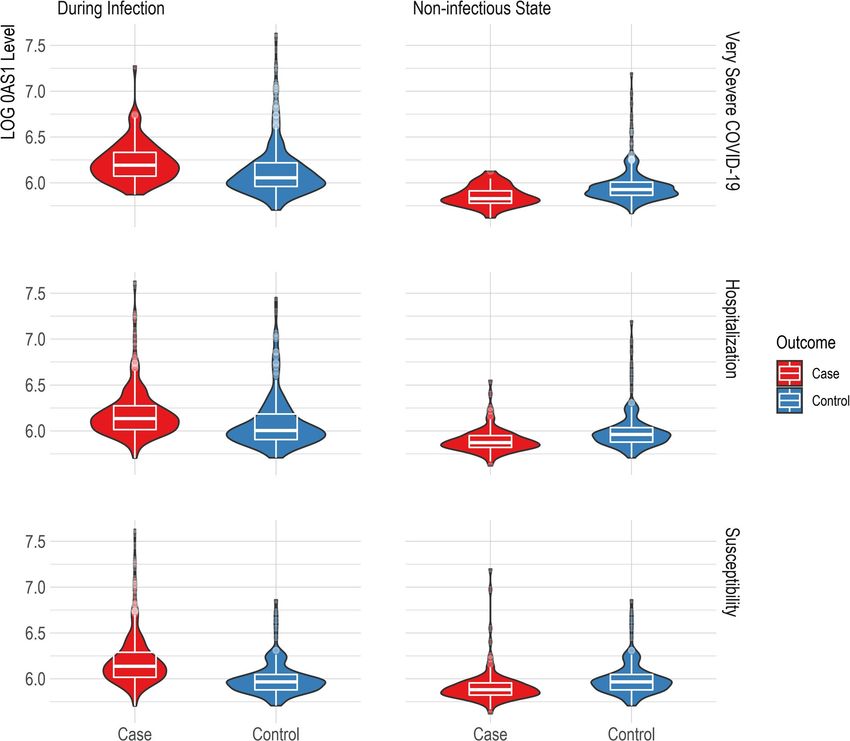
Articles                                                                                                                                              Nature Medicine

genetic determinants of circulating protein levels in large-scale           or protein levels, as catalogued in PhenoScanner23. rs4767027 was
proteomic GWASs and then used MR to assess whether these                    not associated with any other traits or protein levels (P < 5.0 × 10⁻⁵).
cis-pQTLs were associated with COVID-19 outcomes in large                   These findings reduce the possibility that the MR estimate of the
COVID-19 GWASs. Next, we investigated expression QTL (eQTL)
                                                                                                                                                        0.0 0.2 0.4 0.6 0.8 1.0
and splice QTL (sQTL) effects of lead proteins. We then measured
                                                                                                                                         2
the most promising protein, OAS1, in individuals ascertained for                                                                        r to rs4767027
SARS-CoV-2 infection, followed for longitudinal sampling during
and after their infection.                                                                                                                           rs4767027
                                                                                                                   25

Results
MR using cis-pQTLs and pleiotropy assessment. The study                                                            20
design is illustrated in Fig. 1. We began by obtaining the genetic

                                                                                     OAS1 −log10(p)
determinants of circulating protein levels from six large proteomic                                                15
GWASs of individuals of European ancestry (Sun et al.15 n = 3,301;
Emilsson et al.16 n = 3,200; Pietzner et al.17 n = 10,708; Folkersen
                                                                                                                   10
et al.18 n = 3,394; Yao et al.19 n = 6,861 and Suhre et al.20 n = 997). A
total of 931 proteins from these six studies had genome-wide sig-
nificant cis-pQTLs or highly correlated LD proxies (r2 > 0.8) in the                                                5
meta-analyses of data from the COVID-19 Host Genetics Initiative21,
which included results from the GenOMICC program22. We then                                                         0
undertook MR analyses using 1,425 cis-pQTLs and 39 LD proxies
as genetic instruments for circulating proteins in three COVID-19                                                               PP shared SNP signal: 0.718
outcomes: 1) very severe COVID-19 disease (defined as individuals                                                  10
experiencing death, mechanical ventilation, non-invasive ventila-
tion, high-flow oxygen or use of extra-corporeal membrane oxygen-
ation; 99.7% of these individuals were of European ancestry) using                                                  8

4,336 cases and 623,902 controls; 2) COVID-19 disease requiring                      Severe −log10(p)
hospitalization using 6,406 cases and 902,088 controls of European                                                  6
ancestry; and 3) COVID-19 susceptibility using 14,134 cases and
1,284,876 controls of European ancestry. In all outcomes, cases
                                                                                                                    4
required evidence of SARS-CoV-2 infection. For the very severe
COVID-19 and hospitalization outcomes, COVID-19 cases were
defined as laboratory-confirmed SARS-CoV-2 infection based on                                                       2
nucleic acid amplification or serology tests. For the COVID-19 sus-
ceptibility outcome, cases were also identified by review of health                                                 0
records (using International Classification of Disease (ICD) codes
or physician notes).
                                                                                                                                PP shared SNP signal: 0.821
    MR analyses revealed that the levels of three circulating pro-
teins—2′–5′ oligoadenylate synthetase 1 (OAS1), interleukin-10                                                      8
receptor beta subunit (IL10RB) and ABO—were associated with
at least two COVID-19 outcomes after Benjamini–Hochberg false
                                                                                          Hospitalized −log10(p)

discovery rate correction (Table 1 and Supplementary Tables 1–6).                                                   6
Notably, increased OAS1 levels were strongly associated with pro-
tection from all three COVID-19 outcomes. Furthermore, these
effect sizes were more pronounced with more severe outcomes, such                                                   4
that each s.d. increase in OAS1 levels was associated with decreased
odds of very severe COVID-19 (OR = 0.54, 95% confidence inter-
val (CI) 0.44–0.68, P = 7.0 × 10⁻⁸), hospitalization (OR = 0.61, 95%                                                2
CI 0.51–0.73, P = 8.3 × 10⁻⁸) and susceptibility (OR = 0.78, 95%
CI 0.69–0.87, P = 7.6 × 10⁻⁶) (Fig. 2a). We also identified OAS1
                                                                                                                    0
cis-pQTLs in Emilsson et al.16 and Pietzner et al.17, which were not
included in the initial MR due to lack of genome-wide significance
                                                                                                                                PP shared SNP signal: 0.885
for their association with OAS1 levels16 or not included in their
COVID-19 discovery panel17. MR analyses of using these additional
cis-pQTLs yielded concordant results (Supplementary Table 7).                                                       5
    We next assessed whether the cis-pQTL for OAS1 levels
(rs4767027) was associated with over 5,000 other diseases, traits                                                   4
                                                                                         Susceptible −log10(p)

                                                                                                                    3

Fig. 3 | Co-localization of the genetic determinants of OAS1 plasma                                                 2
protein levels and COVID-19 outcomes. Co-localization of genetic signal
for OAS1 levels (top plot) and COVID-19 outcomes (three bottom plots)                                               1
in the 1-Mb region around OAS1 pQTL rs4767027; color shows SNPs in the
                                                                                                                    0
region in LD (r2) with rs4767027 (purple). The posterior probability (PP)
of a shared single signal between OAS1 levels and the three COVID-19                                                    112.9   113.1        113.3    113.5      113.7
outcomes was estimated by coloc.                                                                                                 Chromosome 12 (Mbp)

662                                                                         Nature Medicine | VOL 27 | April 2021 | 659–667 | www.nature.com/naturemedicine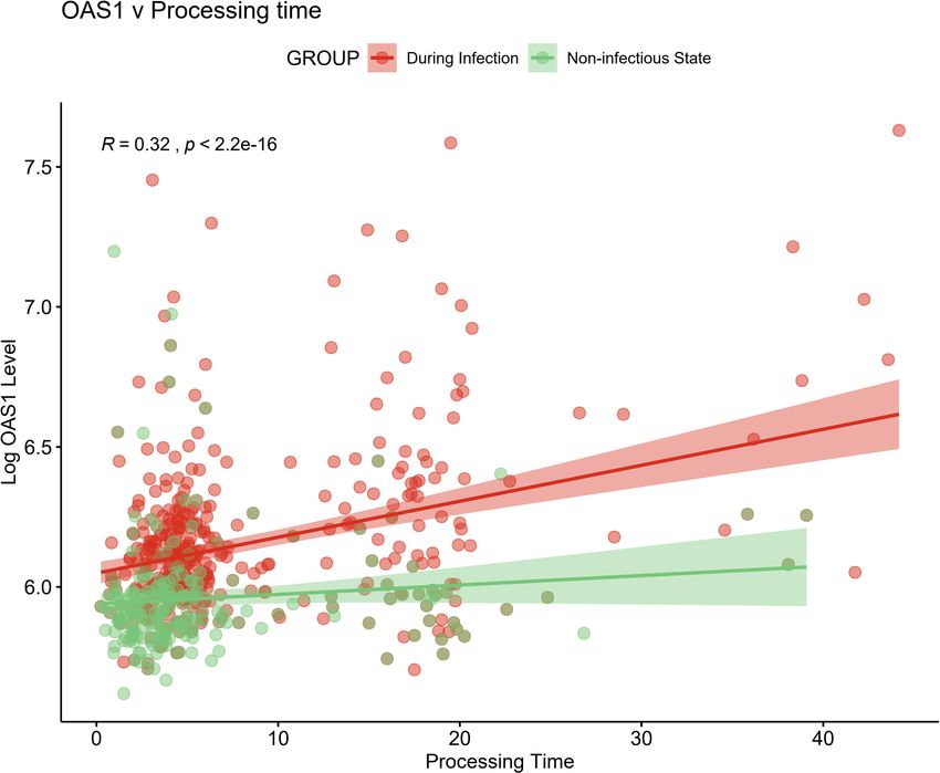
Nature Medicine                                                                                                                                    Articles
                                                                                                   was also replicated using OAS1 cis-pQTL identified by Pietzner
Table 2 | Participant demographics of the BQC19 cohort
                                                                                                   et al.17 (Supplementary Table 7). This suggests that there is likely a
included in this study
                                                                                                   single shared causal signal for OAS1 circulating protein levels and
Sample demographics                                   Number of individuals (%)                    COVID-19 outcomes.
                                                      (total n = 504)                                 Co-localization of ABO levels and different COVID-19 out-
Sex                                                                                                comes also showed co-localization between ABO level and differ-
                                                                                                   ent COVID-19 outcomes (posterior probability of single shared
    Female                                            250 (49.6%)                                  signal = 0.90, 0.98 and 1 for ABO level and very severe COVID-19,
    Male                                              254 (50.4%)                                  hospitalization due to COVID-19 and susceptibility, respectively)
    Age (years) a                                     65.4 (18.0)                                  (Extended Data Fig. 1). We were unable to perform co-localization
    Body mass indexa                                  28.6 (6.18)
                                                                                                   analyses for IL10RB due to a lack of genome-wide summary-level
                                                                                                   data from the original proteomic GWAS16.
    Missing                                           225 (44.6%)
SARS-CoV-2 PCR test                                                                                Aptamer-binding effects. Protein-altering variants (PAVs)15 might
    Positive                                          399 (79.2%)                                  influence binding of affinity agents, such as aptamers or antibod-
                                                                                                   ies, that are used to quantify protein levels. We, thus, assessed if
    Negative                                          105 (20.8%)
                                                                                                   the cis-pQTLs for the MR-prioritized proteins were PAVs or in LD
Hospitalization                                                                                    (r2 > 0.8) with PAVs. rs2834167 (IL10RB) is a nonsense variant and
    Hospitalized                                      406 (80.6%)                                  could, therefore, be subject to potential binding effects. rs505922
    Outpatient treatment only                         98 (19.4%)                                   (ABO) is not in LD with known missense variants. rs4767027
                                                                                                   (OAS1) is an intronic variant, which is in LD with a missense variant
    Hospitalization duration (d)b                     14.0 (6.00, 27.0)
                                                                                                   rs2660 (r2 = 1) in European ancestry. However, because expression
Death                                                                                              studies derived from RNA sequencing are not subject to potential
    Deceased                                          43 (8.5%)                                    effects of missense variants that could influence aptamer binding,
    Survived                                          461 (91.5%)                                  we next explored whether rs4767027 also influences OAS1 expres-
                                                                                                   sion and/or splicing.
Respiratory support
    No oxygen                                         233 (46.2%)                                  sQTL and eQTL studies for OAS genes. sQTLs are genetic vari-
    Oxygen supplementation                            143 (28.4%)                                  ants that influence the transcription of different isoforms of a
    Mechanical ventilation                            128 (25.4%)
                                                                                                   protein. The aptamer that targets OAS1 was developed against a
                                                                                                   synthetic protein comprising the amino acid sequence 1–364 of
    Days on ventilator   b
                                                      14.0 (6.75, 23.5)                            NP002525.2, which is common to the two major OAS1 isoforms:
a
 Mean (s.d.) bMedian (25% interquartile range and 75% interquartile range), which was calculated   p46 and p42. Hence, the aptamer might identify both or either iso-
among individuals who were hospitalized and individuals on a ventilator, respectively.             forms. rs10774671 is a known sQTL for OAS1 that induces alter-
                                                                                                   nate splicing and creates p46 and p42 isoforms. Most present-day
                                                                                                   individuals of European ancestry carry the alternative variant
effect of OAS1 on COVID-19 outcomes is due to horizontal plei-                                     (rs10774671-A). The ancestral variant (rs10774671-G) is the major
otropy. Finally, except for COVID-19 susceptibility, the effect of                                 allele in African populations and became fixed in Neanderthal and
rs4767027 did not demonstrate evidence of heterogeneity across                                     Denisovan genomes24,25. However, the ancestral variant, with its
COVID-19 Host Genetics Initiative GWAS meta-analyses (Table 1).                                    increased expression of the p46 isoform, was reintroduced into the
   Using a cis-pQTL for IL10RB (rs2834167), we found that                                          European population via gene flow from Neanderthals26. Previous
a 1-s.d. increase in circulating IL10RB level was associated                                       analyses suggest that individuals with either the GG or GA genotype
with decreased odds of very severe COVID-19 (OR = 0.47,                                            at rs10774671 express higher amounts of p46 (ref. 26), which is also
95% CI 0.32–0.68, P = 7.1 × 10⁻⁵) and hospitalization (OR = 0.53,                                  the predominant isoform found in circulating blood27. Differences
95% CI 0.39–0.73, P = 8.8 × 10⁻⁵) but not susceptibility (Fig. 2a).                                in antiviral activity have been observed between isoforms, with p46
Using PhenoScanner, we did not find evidence of pleiotropic                                        being more active in certain viral infections28. Interestingly, the
effects of the cis-pQTL for IL10RB. A 1-s.d. increase in circulating                               OAS1 pQTL rs4767027 is in high LD (r2 = 0.97) with rs10774671
ABO level was associated with increased odds of adverse COVID-                                     (ref. 26) in European populations. Functional studies support that
19 outcomes (Table 1); however, we found that the cis-pQTL for                                     the G allele at rs10774671 increases expression of the p46 isoform
ABO (rs505922) was strongly associated with the levels of several                                  but decreases expression of the p42 isoform27. This G allele at the
other proteins, suggesting potential horizontal pleiotropic effects                                sQTL rs10774671 reflects the T allele at pQTL rs4767027, which
(Supplementary Table 8). Given ABO’s known involvement in                                          itself is associated with higher measured OAS1 levels and reduced
multiple physiological processes, these results were expected but                                  odds of COVID-19 severity and susceptibility. These separate lines
highlight that MR analyses might suffer from significant bias from                                 of evidence suggest that OAS1 levels, as measured by the SomaScan
horizontal pleiotropy.                                                                             platform, predominantly identify the p46 isoform, which might
                                                                                                   protect against COVID-19 outcomes.
Co-localization studies. To test whether confounding due to LD                                         Undertaking MR studies of OAS1 splicing, we found that
might have influenced the estimated effect of circulating OAS1 on                                  increased expression of the p46 isoform (as defined by normal-
COVID-19 outcomes, we tested the probability that the genetic                                      ized read counts of the intron cluster defined by LeafCutter29,30) was
determinants of OAS1 circulating protein level were shared with                                    associated with reduced odds of COVID-19 outcomes (OR = 0.29,
the three COVID-19 outcomes using co-localization analyses, as                                     95% CI 0.17–0.49, P = 4.1 × 10⁻⁶ for susceptibility, OR = 0.09, 95%
implemented in coloc12. The posterior probability that OAS1 levels                                 CI 0.04–0.21, P = 2.0 × 10⁻⁸ for hospitalization and OR = 0.05, 95%
and COVID-19 outcomes shared a single causal signal in the 1-Mb                                    CI 0.02–0.13, P = 3.1 × 10⁻⁹ for very severe COVID-19) (Fig. 2b).
locus around the cis-pQTL, rs4767027, was 0.72 for very severe                                     Co-localization analyses also supported a shared causal signal
COVID-19, 0.82 for hospitalization due to COVID-19 and 0.89                                        among the sQTL for OAS1, the pQTL and COVID-19 outcomes
for COVID-19 susceptibility (Fig. 3). This co-localization result                                  (Extended Data Fig. 2). Interestingly, the co-localization analyses

Nature Medicine | VOL 27 | April 2021 | 659–667 | www.nature.com/naturemedicine                                                                                      663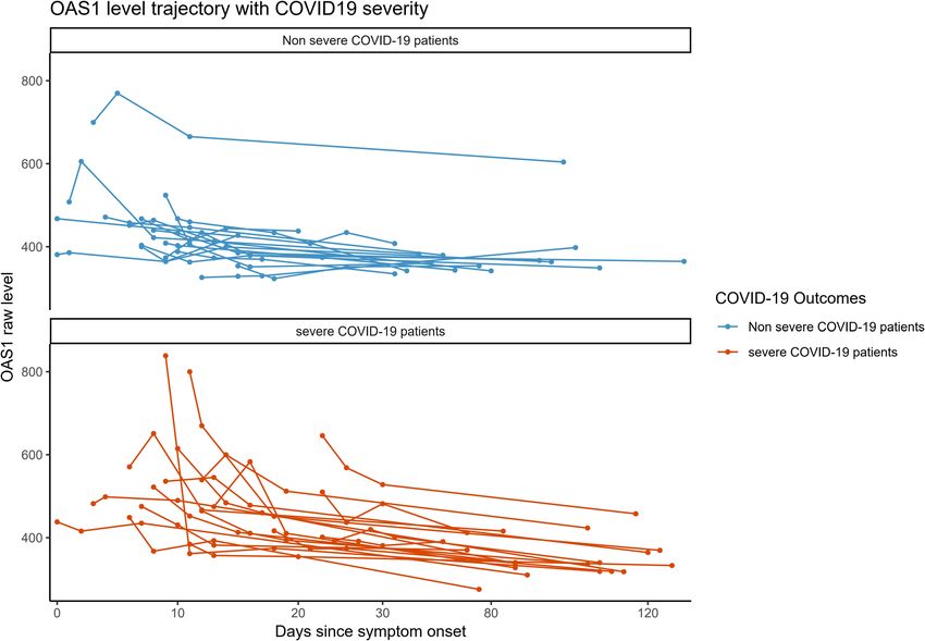
Articles                                                                                                                                                   Nature Medicine

                                                                           OR = 4.39 (n case/control = 308/103)
                                                                           P = 1.1 × 10⁻¹¹

                      During infection
                                                                            OR = 1.93 (n case/control = 260/151)
                                                                            P = 4.8 × 10⁻⁶

                                                                           OR = 1.50 (n case/control = 90/321)
                                                                           P = 0.0007

                                                               OR = 0.69 (n case/control = 113/103)
                                                               P = 0.04
                                                                                                                           COVID-19 outcomes
                      Non-infectious state

                                                                                                                                   Susceptibility
                                                               OR = 0.46 (n case/control = 91/125)
                                                               P = 0.002                                                           Hospitalization
                                                                                                                                   Very severe

                                                               OR = 0.20 (n case/control = 31/185)
                                                               P = 0.001

                                             0.0   0.5   1.0       1.5      2.0     2.5     3.0       3.5    4.0     4.5     5.0        5.5         6.0   6.5   7.0
                                                                Odds ratio (95% confidence interval) per 1-s.d. increase of log OAS1 level

Fig. 4 | Association of OAS1 levels with COVID-19 outcomes from the case–control study in BQC19. Forest plot showing ORs and 95% CIs from logistic
regression analyses (two sided). P values are unadjusted. During infection: patient samples that were collected within 14 d from the date of symptom
onset. For individuals with two or more samples collected within 14 d of symptom onset, the earliest time point was used. Non-infectious state: patient
samples that were collected at least 31 d from the date of symptom onset. For individuals with two or more samples collected at different time points at
least 31 d from symptom onset, the latest time point was used. Additional information is also described in Supplementary Table 10.

supported a stronger probability of a shared signal with the sQTL                                     individuals with samples from patients positive for SARS-CoV-2
than the pQTL, suggesting that the p46 isoform might be the driver
Nature Medicine                                                                                                                     Articles
Discussion                                                                            OAS1, OAS2 and OAS3 share considerable homology. As an
Disease-specific therapies are needed to reduce the morbidity and                 interferon-stimulated gene39, OAS1 polymorphisms have been asso-
mortality associated with COVID-19 outcomes. In this large-scale,                 ciated with the host immune response to several classes of viral
two-sample MR study of 931 proteins assessed for three COVID-                     infection40–44. Given that OAS1 is an intracellular enzyme-activating
19 outcomes in up to 14,134 cases and 1.2 million controls of                     RNase L leading to viral RNA degradation, it is probable that the
European ancestry, we provide evidence that increased OAS1 lev-                   circulating levels of this enzyme reflect intracellular levels of this
els in the non-infectious state are strongly associated with reduced              protein. However, there is experimental evidence that extracellular
risks of very severe COVID-19, hospitalization and susceptibility.                OAS1 might also be important in the viral immune response33.
The protective effect size was particularly large, such that a 50%                    Molecules currently exist that can influence OAS1 expression.
decrease in the odds of very severe COVID-19 was observed per                     Interferon beta-1b, which activates a cytokine cascade leading to
s.d. increase in OAS1 circulating levels. OAS proteins are part of the            increased OAS1 expression45, is currently used to treat multiple
innate immune response against RNA viruses. They are induced by                   sclerosis and has been shown to induce OAS1 expression in blood
interferons and activate latent RNase L, resulting in direct viral and            cells46. Interferon-based therapy has also been used in other viral
endogenous RNA destruction, as demonstrated in in vitro studies33.                infections47. However, recent randomized trials have shown incon-
Thus, OAS1 has a plausible biological activity against SARS-CoV-2.                sistent results. Although intravenous interferon beta-1b combined
Because therapies exist that activate OAS1, repositioning them as                 with lopinavir–ritonavir reduced mortality due to MERS-CoV
potential COVID-19 treatments should be prioritized.                              infections48, in the unblinded SOLIDARITY trial49 there was
    In populations outside of Sub-Saharan Africa, the protective                  no demonstrated benefit of intravenous interferon-beta-1b. On
alleles at both rs4767027-T (the OAS1 pQTL) and rs10774671-G                      the other hand, a recent phase 2 trial testing the effect of inhaled
(the OAS1 sQTL) are found on a Neanderthal haplotype34, which                     nebulized interferon beta-1a (which is closely related to interferon
was passed on to modern humans ~50,000–60,000 years ago35.                        beta-1b) showed improved COVID-19 symptoms in the treatment
The correspondence between the previously described gene flow35                   arm50. Although this study was not powered to show a difference in
from Neandertals at this locus and the haplotype associated with                  mortality, all deaths occurred in the placebo group. Inhaled nebu-
protection against COVID-19 in the GWAS22 was recently demon-                     lized interferon beta results in a much higher tissue availability in
strated34. Even though these two single-nucleotide polymorphisms                  the lung and might result in improved antiviral activity. Moreover,
(SNPs) share a haplotype, their evolutionary histories differ. The                timing of administration is likely to play a role, as the administra-
rs4767027-T allele is derived from the Neanderthal lineage, whereas,              tion of a pro-inflammatory cytokine might not provide benefit
for the rs10774671-G allele, Neanderthals preserved the ancestral                 during the inflammation-driven phase of the disease. However,
state. OAS1 alternative splicing regulated by the rs10774671-G allele             data on timing of administration are currently unavailable in the
increases the isoform p46, which has a higher enzymatic activity                  SOLIDARITY trial, and conclusions cannot yet be drawn. Lastly,
against viruses than the p42 isoform36 and is the only OAS1 isoform               the effect of interferon supplement might vary across ancestral pop-
robustly upregulated during infection26. Although further studies                 ulations, as different ancestries have different amounts of the more
are needed to fully elucidate the functional relevance of the pQTL                active p46 isoform of OAS1. Our study was limited to individuals
and sQTL for OAS1, the antiviral activity of the gene products is                 of European ancestry, a population with higher expression of the
higher for the Neandertal haplotype than the common haplotype                     p46 isoform. Interestingly, the SOLIDARITY trial enrolled 78% of
in Europeans28. In Europeans, the Neandertal haplotype has under-                 its patients in South Asia, the Middle East, North Africa and Latin
gone positive selection26, and the rs4767027-T allele reaches an                  America, populations that might have higher expression of the p42
allele frequency of 0.32. Using MR and measurements of circulat-                  OAS1 isoform, whereas the study on inhaled interferon beta com-
ing proteins, we demonstrated here that increased OAS1 levels of                  prised 80% White patients from the United Kingdom. It is possible
the Neandertal haplotype in modern-day individuals of European                    that interferon beta-1b might have different effects in populations
ancestry confer this protective effect.                                           of different ancestry due to different frequency of genetic variants
    Our MR evidence indicated that higher p46 isoform levels of                   in different populations.
OAS1 and higher OAS1 total protein levels, as measured by the                         There is in vitro evidence that pharmacological inhibition of
SomaScan assay, had protective effects on COVID-19 outcomes.                      phosphodiesterase-12, which degrades 2′–5′ oligoadenylate syn-
These results were strongly supported by co-localization analysis.                thesized by OAS1, potentiates OAS-mediated antiviral activity51,52.
Given the consistent co-localization between the sQTL and pQTL                    Interestingly, coronaviruses in the same family as SARS-CoV-2
for OAS1, the lack of co-localization between the eQTL and pQTL                   have been shown to produce viral proteins that degrade 2′–5′ oli-
for OAS1 and the evidence that the SomaScan assay likely measures                 goadenylate and reduce RNase-L activity, leading to evasion of the
p46 isoforms, it seems probable that the protective effect of OAS1                host immune response53,54. Our findings are also consistent with
is derived from the p46 isoform. However, further investigations                  recent experimental work55 showing that there are situations where
are required to specifically measure each isoform in circulation,                 SARS-CoV-2 is sensitive to OAS1-related antiviral defenses. Our
and isoform activity assays will be required to better understand if              findings motivate pharmacologic strategies to increase OAS1 lev-
the p46 isoform, rather than total OAS1 levels, is most protective                els or activity, as well as further evaluation of the possible antivi-
against COVID-19 outcomes.                                                        ral activity of extracellular OAS1 (ref. 33). Thus, existing preclinical
    The ancestral OAS1 splice variant encoding the more active p46                molecules that lead to increased OAS1 levels51 could be optimized
isoform was lost in the modern human population that left Africa.                 and tested for their effect on COVID-19 outcomes.
Several scenarios might explain this loss of function—for example,                    Our MR analyses found that higher levels of OAS3 expression
loss of purifying selection during the out-of-Africa exodus, which                is associated with worse COVID-19 outcomes, which is an oppo-
might be due to changes in environmental pathogens or potential                   site direction of effect compared to OAS1. The discordant effects of
harm induced by OAS1 antiviral activity37. Unfortunately, we do                   the p46 isoform for OAS1 and OAS3 were also reported by a previ-
not have sufficient data to test if the OAS1 p46 ancestral allele in              ous study26, which might reflect complex biology of OAS genes for
Sub-Saharan Africans also offers protection against COVID-19.                     innate immune response. In a recent transcription-wide association
Nevertheless, these findings further emphasize the importance of                  study from the GenOMICC program22, genetically predicted high
the Neanderthal genome in COVID-19 risk modulation, because a                     expression of OAS3 in lungs and whole blood was associated with
risk locus on chromosome 3 has also been reported to be inherited                 a higher risk of patients with COVID-19 becoming critically ill.
from Neanderthals38.                                                              Although further studies to assess the roles of OAS genes specific

Nature Medicine | VOL 27 | April 2021 | 659–667 | www.nature.com/naturemedicine                                                                       665
Articles                                                                                                                            Nature Medicine

to SARS-CoV-2 are needed, it is likely that OAS1 is the main driver              4. Sterne, J. A. C. et al. Association between administration of systemic
of the protective effect of the p46 isoform for COVID-19 outcomes                    corticosteroids and mortality among critically ill patients with COVID-19: a
                                                                                     meta-analysis. JAMA 324, 1330–1341 (2020).
given previous functional studies demonstrating the antiviral effect             5. Beigel, J. H., Tomashek, K. M. & Dodd, L. E. Remdesivir for the treatment of
of OAS genes26.                                                                      Covid-19—preliminary report. N. Engl. J. Med. 383, 994 (2020).
    This study had limitations. First, we used MR to test the effect of          6. Cavalcanti, A. B. et al. Hydroxychloroquine with or without azithromycin in
circulating protein levels measured in a non-infected state because                  mild-to-moderate Covid-19. N. Engl. J. Med. 383, 2041–2052 (2020).
                                                                                 7. Nelson, M. R. et al. The support of human genetic evidence for approved
the effect of the cis-pQTLs on circulating proteins was estimated
                                                                                     drug indications. Nat. Genet. 47, 856–860 (2015).
in individuals who had not been exposed to SARS-CoV-2. Once a                    8. Cook, D. et al. Lessons learned from the fate of AstraZeneca’s drug pipeline: a
person contracts SARS-CoV-2 infection, levels of circulating pro-                    five-dimensional framework. Nat. Rev. Drug Discov. 13, 419–431 (2014).
teins could be altered, and this might be especially relevant for                9. Zheng, J. et al. Phenome-wide Mendelian randomization mapping the
cytokines such as IL10 (which binds to IL10RB) and OAS1. Thus,                       influence of the plasma proteome on complex diseases. Nat. Genet. 52,
                                                                                     1122–1131 (2020).
the MR results presented in this paper should be interpreted as an               10. Filbin, M. R. et al. Plasma proteomics reveals tissue-specific cell death and
estimation of the effect of circulating protein levels when measured                 mediators of cell–cell interactions in severe COVID-19 patients. Preprint at
in the non-infected state. Ongoing studies will help to clarify if the               bioRxiv https://doi.org/10.1101/2020.11.02.365536 (2020).
same cis-pQTLs influence circulating protein levels during infec-                11. Davey Smith, G., Ebrahim, S., Smith, G. D. & Ebrahim, S. ‘Mendelian
tion. Second, this type of study suffers a high false-negative rate.                 randomization’: can genetic epidemiology contribute to understanding
                                                                                     environmental determinants of disease? Int. J. Epidemiol. 32, 1–22 (2003).
Our goal was not to identify every circulating protein influenc-                 12. Giambartolomei, C. et al. Bayesian test for colocalisation between pairs of
ing COVID-19 outcomes but, rather, to provide evidence for a                         genetic association studies using summary statistics. PLoS Genet. 10,
few proteins with strong cis-pQTLs, because these proteins are                       e1004383 (2014).
more likely to be robust to the assumptions of MR studies. Future                13. Lawlor, D. A., Tilling, K. & Smith, G. D. Triangulation in aetiological
large-scale proteomic studies with more circulating proteins                         epidemiology. Int. J. Epidemiol. 45, 1866–1886 (2016).
                                                                                 14. COVID-19 Host Genetics Initiative. The COVID-19 Host Genetics Initiative,
properly assayed should help to overcome these limitations. Third,                   a global initiative to elucidate the role of host genetic factors in susceptibility
most MR studies assume a linear relationship between the exposure                    and severity of the SARS-CoV-2 virus pandemic. Eur. J. Hum. Genet. 28,
and the outcome. Thus, our findings would not identify proteins                      715–718 (2020).
whose effect on COVID-19 outcomes has a clear threshold effect.                  15. Sun, B. B. et al. Genomic atlas of the human plasma proteome. Nature 558,
Fourth, the overall OAS1 levels measured by RNA sequencing (not                      73–79 (2018).
                                                                                 16. Emilsson, V. et al. Co-regulatory networks of human serum proteins link
only p46) might be biased by the effect of alternative splicing, and                 genetics to disease. Science 361, 769–773 (2018).
the role of overall OAS1 and OAS3 levels indicated by the asso-                  17. Pietzner, M. et al. Genetic architecture of host proteins involved in
ciation of the cis-pQTL of OAS1 in protection against COVID-19                       SARS-CoV-2 infection. Nat. Commun. 11, 6397 (2020).
are possible and not yet explored. We also could not completely                  18. Folkersen, L. et al. Mapping of 79 loci for 83 plasma protein biomarkers in
exclude the possibility that measurement of OAS1 levels might be                     cardiovascular disease. PLoS Genet. 13, e1006706 (2017).
                                                                                 19. Yao, C. et al. Genome-wide mapping of plasma protein QTLs identifies
influenced by aptamer-binding effects. Last, all data presented in                   putatively causal genes and pathways for cardiovascular disease. Nat.
this paper pertain to individuals of European ancestry only—once                     Commun. 9, 3268 (2018).
again underlining the importance of genotyping efforts in other                  20. Suhre, K. et al. Connecting genetic risk to disease end points through the
populations.                                                                         human blood plasma proteome. Nat. Commun. 8, 14357 (2017).
                                                                                 21. COVID-19 Host Genetics Initiative. https://www.covid19hg.org/results/
    In conclusion, we used genetic determinants of circulating pro-
                                                                                     (2021).
tein levels and COVID-19 outcomes obtained from large-scale                      22. Pairo-Castineira, E. et al. Genetic mechanisms of critical illness in Covid-19.
studies and found compelling evidence that OAS1 has a protective                     Nature https://doi.org/10.1038/s41586-020-03065-y (2020).
effect on COVID-19 susceptibility and severity. Measuring plasma                 23. Staley, J. R. et al. PhenoScanner: a database of human genotype–phenotype
OAS1 levels in a case–control study demonstrated that higher cir-                    associations. Bioinformatics 32, 3207–3209 (2016).
                                                                                 24. Prüfer, K. et al. A high-coverage Neandertal genome from Vindija Cave in
culating levels of this protein in a non-infectious state are strongly               Croatia. Science 358, 655–658 (2017).
associated with reduced risk of adverse COVID-19 outcomes.                       25. Meyer, M. et al. A high-coverage genome sequence from an archaic
Interestingly, the available evidence suggests that the protective                   Denisovan individual. Science 338, 222–226 (2012).
effect from OAS1 in individuals of European ancestry is likely due               26. Sams, A. J. et al. Adaptively introgressed Neandertal haplotype at the OAS
to the Neanderthal-introgressed p46 OAS1 isoform. Known phar-                        locus functionally impacts innate immune responses in humans. Genome
                                                                                     Biol. 17, 246 (2016).
macological agents that increase OAS1 levels51 could be explored for             27. Li, H. et al. Identification of a Sjögren’s syndrome susceptibility locus at OAS1
their effect on COVID-19 outcomes.                                                   that influences isoform switching, protein expression, and responsiveness to
                                                                                     type I interferons. PLoS Genet. 13, e1006820 (2017).
Online content                                                                   28. Liu, X. et al. A functional variant in the OAS1 gene is associated with
Any methods, additional references, Nature Research report-                          Sjögren’s syndrome complicated with HBV infection. Sci. Rep. 7,
                                                                                     17571 (2017).
ing summaries, source data, extended data, supplementary infor-                  29. Li, Y. I. et al. Annotation-free quantification of RNA splicing using
mation, acknowledgements, peer review information; details of                        LeafCutter. Nat. Genet. 50, 151–158 (2018).
author contributions and competing interests; and statements of                  30. Aguet, F. et al. The GTEx Consortium atlas of genetic regulatory effects
data and code availability are available at https://doi.org/10.1038/                 across human tissues. Science 369, 1318–1330 (2020).
s41591-021-01281-1.                                                              31. Aguet, F. et al. Genetic effects on gene expression across human tissues.
                                                                                     Nature 550, 204–213 (2017).
                                                                                 32. Hornung, V., Hartmann, R., Ablasser, A. & Hopfner, K. P. OAS proteins and
Received: 11 October 2020; Accepted: 5 February 2021;                                cGAS: unifying concepts in sensing and responding to cytosolic nucleic acids.
Published online: 25 February 2021                                                   Nat. Rev. Immunol. 14, 521–528 (2014).
                                                                                 33. Kristiansen, H. et al. Extracellular 2′–5′ oligoadenylate synthetase stimulates
References                                                                           RNase L-independent antiviral activity: a novel mechanism of virus-induced
1. Johns Hopkins University of Medicine. Coronavirus Resource Center. https://       innate immunity. J. Virol. 84, 11898–11904 (2010).
   coronavirus.jhu.edu/ (2020).                                                  34. Zeberg, H. & Pääbo, S. A genomic region associated with protection against
2. Weinreich, D. M. et al. REGN-COV2, a neutralizing antibody cocktail, in           severe COVID-19 is inherited from Neandertals. Proc. Natl Acad. Sci. USA
   outpatients with Covid-19. N. Engl. J. Med. 384, 238–251 (2020).                  118, e2026309118 (2021).
3. Horby, P. et al. Dexamethasone in hospitalized patients with Covid-           35. Mendez, F. L., Watkins, J. C. & Hammer, M. F. Neandertal origin of
   19—preliminary report. N. Engl. J. Med. https://doi.org/10.1056/                  genetic variation at the cluster of OAS immunity genes. Mol. Biol. Evol. 30,
   NEJMoa2021436 (2020).                                                             798–801 (2013).

666                                                                              Nature Medicine | VOL 27 | April 2021 | 659–667 | www.nature.com/naturemedicine
Nature Medicine                                                                                                                                        Articles
36. Bonnevie-Nielsen, V. et al. Variation in antiviral 2′–5′-oligoadenylate              47. Lin, F. & Young, H. A. Interferons: success in anti-viral immunotherapy.
    synthetase (2′5′AS) enzyme activity is controlled by a single-nucleotide                 Cytokine Growth Factor Rev. 25, 369–376 (2014).
    polymorphism at a splice-acceptor site in the OAS1 gene. Am. J. Hum. Genet.          48. Arabi, Y. M. et al. Interferon beta-1b and lopinavir–ritonavir for Middle East
    76, 623–633 (2005).                                                                      respiratory syndrome. N. Engl. J. Med. 383, 1645–1656 (2020).
37. Carey, C. M. et al. Recurrent loss-of-function mutations reveal costs to OAS1        49. WHO Solidarity Trial Consortium. Repurposed antiviral drugs for
    antiviral activity in primates. Cell Host Microbe 25, 336–343 (2019).                    COVID-19—interim WHO SOLIDARITY trial results. N. Engl. J. Med. 384,
38. Zeberg, H. & Pääbo, S. The major genetic risk factor for severe COVID-19 is              497–511 (2021).
    inherited from Neanderthals. Nature 587, 610–612 (2020).                             50. Monk, P. D. et al. Safety and efficacy of inhaled nebulised interferon beta-1a
39. Schneider, W. M., Chevillotte, M. D. & Rice, C. M. Interferon-stimulated genes:          (SNG001) for treatment of SARS-CoV-2 infection: a randomised,
    a complex web of host defenses. Annu. Rev. Immunol. 32, 513–545 (2014).                  double-blind, placebo-controlled, phase 2 trial. Lancet Respir. Med. 9,
40. Min, J.-Y. & Krug, R. M. The primary function of RNA binding by the                      196–206 (2020).
    influenza A virus NS1 protein in infected cells: inhibiting the 2′–5′                51. Wood, E. R. et al. The role of phosphodiesterase 12 (PDE12) as a negative
    oligo (A) synthetase/RNase L pathway. Proc. Natl Acad. Sci. USA 103,                     regulator of the innate immune response and the discovery of antiviral
    7100–7105 (2006).                                                                        inhibitors. J. Biol. Chem. 290, 19681–19696 (2015).
41. Hu, B. et al. Cellular responses to HSV-1 infection are linked to specific types     52. Silverman, R. H. & Weiss, S. R. Viral phosphodiesterases that antagonize
    of alterations in the host transcriptome. Sci. Rep. 6, 28075 (2016).                     double-stranded RNA signaling to RNase L by degrading 2-5A. J. Interferon
42. Lim, J. K. et al. Genetic variation in OAS1 is a risk factor for initial infection       Cytokine Res. 34, 455–463 (2014).
    with West Nile virus in man. PLoS Pathog. 5, e1000321 (2009).                        53. Zhao, L. et al. Antagonism of the interferon-induced OAS-RNase L pathway
43. Simon-Loriere, E. et al. High anti-dengue virus activity of the OAS gene                 by murine coronavirus ns2 protein is required for virus replication and liver
    family is associated with increased severity of dengue. J. Infect. Dis. 212,             pathology. Cell Host Microbe 11, 607–616 (2012).
    2011–2020 (2015).                                                                    54. Zhang, R. et al. Homologous 2′,5′-phosphodiesterases from disparate RNA
44. Hamano, E. et al. Polymorphisms of interferon-inducible genes OAS-1 and                  viruses antagonize antiviral innate immunity. Proc. Natl Acad. Sci. USA 110,
    MxA associated with SARS in the Vietnamese population. Biochem. Biophys.                 13114–13119 (2013).
    Res. Commun. 329, 1234–1239 (2005).                                                  55. Li, Y. et al. SARS-CoV-2 induces double-stranded RNA-mediated innate
45. Cheng, G. et al. Pharmacologic activation of the innate immune system to                 immune responses in respiratory epithelial derived cells and cardiomyocytes.
    prevent respiratory viral infections. Am. J. Respir. Cell Mol. Biol. 45,                 Preprint at bioRxiv https://doi.org/10.1101/2020.09.24.312553 (2020).
    480–488 (2011).
46. Harari, D., Orr, I., Rotkopf, R., Baranzini, S. E. & Schreiber, G. A robust type
    I interferon gene signature from blood RNA defines quantitative but not              Publisher’s note Springer Nature remains neutral with regard to jurisdictional claims in
    qualitative differences between three major IFNβ drugs in the treatment of           published maps and institutional affiliations.
    multiple sclerosis. Hum. Mol. Genet. 24, 3192–3205 (2014).                           © The Author(s), under exclusive licence to Springer Nature America, Inc. 2021

Nature Medicine | VOL 27 | April 2021 | 659–667 | www.nature.com/naturemedicine                                                                                               667
Articles Nature Medicine
Methods with other circulating proteins (that is, if they were trans-pQTLs to other proteins
pQTL GWAS. We systematically identified pQTL associations from six large or significantly associated with other unrelated diseases or traits). For cis-pQTLs
proteomic GWASs15–20. Each of these studies undertook proteomic profiling using of MR-prioritized proteins measured on the SomaLogic platform, we assessed the
either SomaLogic SomaScans or O-link proximal extension assays. possibility of potential aptamer-binding effects (where the presence of PAVs might
affect protein measurements). We also checked if cis-pQTLs of MR-prioritized
COVID GWAS and COVID-19 outcomes. To assess the association of cis-pQTLs proteins had significantly heterogeneous associations across COVID-19
with COVID-19 outcomes, we used COVID-19 meta-analytic GWASs (data freeze populations in each COVID-19 outcome GWAS.
4) from the COVID-19 Host Genetics Initiative21. For our study, we used three of
these GWAS meta-analyses, which included 25 cohorts of European ancestry and Co-localization analysis. Next, we tested co-localization of the genetic signal
one cohort of admixed American ancestry. The outcomes tested were very severe for the circulating protein and each of the three COVID-19 outcomes using
COVID-19, hospitalization due to COVID-19 and susceptibility to COVID-19 co-localization analyses, which assess potential confounding by LD. Specifically, for
(named A2, B2 and C2, respectively, by the COVID-19 Host Genetics Initiative). each of these MR-significant proteins with genome-wide summary data available,
Very severe COVID-19 cases were defined as hospitalized individuals for the proteomic GWASs a stringent Bayesian analysis was implemented in coloc12
with COVID-19 as the primary reason for hospital admission with R package to analyze all variants in the 1-Mb genomic locus centered on the
laboratory-confirmed SARS-CoV-2 infection (nucleic acid amplification tests or cis-pQTL. Co-localizations with posterior probability for hypothesis 4 (PP4, that
serology based) and death or respiratory support (invasive ventilation, continuous there is an association for both protein level and COVID-19 outcomes, and they
positive airway pressure, bilevel positive airway pressure or continuous external are driven by the same causal variant) > 0.5 were considered likely to co-localize
negative pressure, high-flow nasal or face mask oxygen). Simple supplementary (which means the highest posterior probability for all five coloc hypotheses), and
oxygen (for example, 2 L min−1 via nasal cannula) did not qualify for case status. PP4 > 0.8 was considered to be highly likely to co-localize.
Controls were all individuals in the participating cohorts who did not meet this
case definition. sQTL and eQTL MR and co-localization studies for OAS genes. We performed
Hospitalized COVID-19 cases were defined as individuals hospitalized with MR and co-localization analysis using GTEx project v8 (ref. 31) GWAS summary
laboratory-confirmed SARS-CoV-2 infection (using the same microbiology data to understand the effects of expression and alternative splicing of OAS
methods as for the very severe phenotype), where hospitalization was due to genes in whole blood. The genetic instruments were conditionally independent
COVID-19-related symptoms. Controls were all individuals in the participating (r2 < 0.001) sQTLs and eQTLs for OAS1 and eQTLs for OAS2 and OAS3 identified
cohorts who did not meet this case definition. by using stepwise regression in GTEx31. The sQTL SNP for OAS1 (rs10774671)
Susceptibility to COVID-19 cases was defined as individuals with was originally identified for the normalized read counts of LeafCutter29 cluster of
laboratory-confirmed SARS-CoV-2 infection, health record evidence of COVID- the last intron of the p46 isoform (chr12:112,917,700–112,919,389, GRCh38) in
19 (ICD coding or physician confirmation) or with self-reported infections (for GTEx30 and was used to estimate the effect of the p46 isoform. Co-localization
example, by questionnaire). Controls were all individuals who did not meet this analysis was performed using GWAS summary statistics from GTEx by restricting
case definition. to the regions within 1 Mb of each QTL.
Two-sample MR. We used two-sample MR analyses to screen and test potential Measurement of plasma OAS1 protein levels associated with COVID-19
circulating proteins for their role in influencing COVID-19 outcomes. In outcomes in BQC19. BQC19 is a Québec-wide initiative to enable research into
two-sample MR, the effect of SNPs on the exposure and outcome are taken from the causes and consequences of COVID-19 disease. The patients included in
separate GWASs. This method often improves statistical power because it allows this study were recruited at the Jewish General Hospital (JGH) and the Centre
for larger sample sizes for the exposure and outcome GWAS56. Hospitalier de l’Université de Montréal (CHUM) in Montréal, Québec, Canada.
Exposure definitions: We conducted MR using six large proteomic GWAS COVID-19 case–control status was defined to be consistent with the GWAS
studies15–20. Circulating proteins from Sun et al., Emilsson et al. and Pietzner et al. study from the COVID-19 Host Genetics Initiative, from which the MR results
were measured on the SomaLogic platform; Suhre et al., Yao et al. and Folkersen were derived. Namely, we tested the association of OAS1 protein levels with
et al. used protein measurements on the O-link platform. We selected proteins the three different COVID-19 outcome definitions both in samples procured
with only cis-pQTLs to test their effects on COVID-19 outcomes because they from non-infected stages and samples procured during the acute phase of the
are less likely to be affected by potential horizontal pleiotropy. The cis-pQTLs infection. The three outcomes were as follows. 1) Very severe COVID-19—defined
were defined as the genome-wide significant SNPs (P < 5 × 10⁻⁸) with the lowest P as hospitalized individuals with laboratory-confirmed SARS-CoV-2 infection
value within 1 Mb of the transcription start site of the gene encoding the measured (nucleic acid amplification tests or serology based) and death or respiratory
protein9. For proteins from Emilsson et al., Pietzner et al., Suhre et al., Yao et al. support (invasive ventilation, continuous positive airway pressure, bilevel positive
and Folkersen et al., we used the sentinel cis-pQTL per protein per study as these airway pressure or continuous external negative pressure, high-flow nasal or
were the data available. For proteins from Sun et al., we used PLINK 1.9 (ref. 57) face mask oxygen). Controls were all individuals who did not meet this case
and the 1000 Genome58 European population reference panels to clump and select definition. 2) Hospitalized COVID-19 cases—defined as individuals hospitalized
LD-independent cis-pQTL (r2 < 0.001, distance 1,000 kb) with the lowest P value with laboratory-confirmed SARS-CoV-2 infection. Controls were all individuals
from reported summary statistics for each SOMAmer-bound protein. We included who did not meet this case definition. 3) Susceptibility to COVID-19—cases
the same proteins represented by different cis-pQTLs from different studies to were defined as individuals with laboratory-confirmed SARS-CoV-2 infection,
cross-examine the findings. For cis-pQTLs that were not present in the COVID- and controls were all individuals who underwent PCR testing for SARS-CoV-2
19 GWAS, SNPs with LD r2 > 0.8 and with minor allele frequency (MAF) < 0.42 but were negative. The date of symptom onset for patients with COVID-19 was
were selected as proxies; MAF > 0.3 was used for allelic alignment for proxy SNPs. collected from patient charts or estimated from their first positive COVID-19
cis-pQTLs with palindromic effects and with MAF > 0.42 were removed before MR tests if missing. Case inclusion criteria were not exclusive, which means that some
to prevent allele mismatches. Benjamini–Hochberg correction was used to control individuals who were cases in the susceptibility analyses were also included in the
for the total number of proteins tested using MR. MR analyses were performed hospitalization and very severe COVID-19 cohorts if they met case definitions.
using the TwoSampleMR package in R59. For proteins with a single (sentinel) A total of 125 individuals were recruited from CHUM, and 379 individuals
cis-pQTL, we used the Wald ratio to estimate the effect of each circulating protein were recruited from the JGH. Individuals had blood sampling done at up to five
on each of the three COVID-19 outcomes. For any proteins/SOMAmer reagents different time points (200 individuals had one measurement, 113 individuals
with multiple independent cis-pQTL, an inverse variance-weighted method was had two measurements, 152 individuals had three measurements, 38 individuals
used to meta-analyze their combined effects. After harmonizing the cis-pQTLs of had four measurements and one individual had five measurements). Days from
proteins with COVID-19 GWAS, a total of 566 SOMAmer reagents (529 proteins, symptom onset (T1) were calculated for each sample based on the date of symptom
565 directly matched cis-pQTL and 26 proxies) from Sun et al., 760 proteins (747 onset and blood draw date. For individuals who were negative for COVID-19, T1
directly matched cis-pQTL and 11 proxies) from Emilsson et al., 91 proteins (90 was set to 0. Sample processing time (in hours) for each sample was also calculated
directly matched cis-pQTLs and two proxies) from Pietzner et al., 74 proteins (72 to measure the duration of time from sample collection to processing to account
directly matched cis-pQTL) from Suhre et al., 24 proteins (24 directly matched for the increase in the amount of protein released from cell lysis due to extended
cis-pQTLs) from Yao et al. and 13 proteins (13 directly matched cis-pQTLs) from sample handling time.
Folkersen et al. were used as instruments for the MR analyses across the three Protein levels in citrated (ACD) plasma samples were measured using the
COVID-19 outcomes (Supplementary Tables 11 and 12)15–20. SomaScan assay. In total, 1,039 samples from 399 patients who were positive for
SARS-CoV-2 and 105 patients who were negative for SARS-CoV-2 of mainly
Pleiotropy assessments. A common pitfall of MR is horizontal pleiotropy, which European descent underwent SomaScan assays, which included 5,284 SOMAmer
occurs when the genetic variant affects the outcome via pathways independent reagents targeting 4,742 proteins. The SomaScan assay uses single-stranded
of the exposure. The use of circulating protein cis-pQTLs greatly reduces the DNA aptamers (‘SOMAmers’), which are designed to selectively bind to a
possibility of pleiotropy, for reasons described above. We also searched in the particular protein target60. SOMAmer reagent binding is quantified by microarray,
PhenoScanner23 database, a large catalog of observed SNP–outcome relationships measuring abundance in relative fluorescent units (RFUs). The RFUs for each
involving >5,000 GWASs done to date to assess potentially pleiotropic effects of protein underwent four normalization processes, including hybridization control,
the cis-pQTLs of MR-prioritized proteins by testing the association of cis-pQTLs intraplate median signal normalization, plate scaling and calibration and median
Nature Medicine | www.nature.com/naturemedicineYou can also read