Identification of a Shrimp E3 Ubiquitin Ligase TRIM50-Like Involved in Restricting White Spot Syndrome Virus Proliferation by Its Mediated ...
←
→
Page content transcription
If your browser does not render page correctly, please read the page content below
ORIGINAL RESEARCH
                                                                                                                                                 published: 11 May 2021
                                                                                                                                        doi: 10.3389/fimmu.2021.682562

                                           Identification of a Shrimp E3
                                           Ubiquitin Ligase TRIM50-Like
                                           Involved in Restricting White
                                           Spot Syndrome Virus Proliferation
                                           by Its Mediated Autophagy
                                           and Ubiquitination
                                           Chao Zhao 1,2, Chao Peng 1, Pengfei Wang 1,2, Lulu Yan 1,2, Sigang Fan 1,2
                                           and Lihua Qiu 1,2,3*
                                           1 Key Laboratory of South China Sea Fishery Resources Exploitation and Utilization, Ministry of Agriculture and Rural Affairs,
                                           South China Sea Fisheries Research Institute, Chinese Academy of Fishery Sciences, Guangzhou, China, 2 Sanya Tropical
                                           Fisheries Research Institute, Sanya, China, 3 Key Laboratory of Aquatic Genomics, Ministry of Agriculture and Rural Affairs,
                                           Chinese Academy of Fishery Science, Beijing, China
                            Edited by:
           Humberto Lanz-Mendoza,
    National Institute of Public Health,   Most tripartite motif (TRIM) family proteins are critical components of the autophagy
                                 Mexico
                                           machinery and play important roles in host defense against viral pathogens in mammals.
                      Reviewed by:
                                           However, the roles of TRIM proteins in autophagy and viral infection have not been studied
                            Jing Xing,
     Ocean University of China, China      in lower invertebrates, especially crustaceans. In this study, we first identified a TRIM50-
                    Sarah J. Poynter,      like gene from Penaeus monodon (designated PmTRIM50-like), which, after a white spot
      University of Waterloo, Canada
                                           syndrome virus (WSSV) challenge, was significantly upregulated at the mRNA and protein
                  *Correspondence:
                           Lihua Qiu
                                           levels in the intestine and hemocytes. Knockdown of PmTRIM50-like led to an increase in
           qiugroup_bio@outlook.com        the WSSV quantity in shrimp, while its overexpression led to a decrease compared with
                                           the controls. Autophagy can be induced by WSSV or rapamycin challenge and has been
                   Specialty section:
         This article was submitted to
                                           shown to play a positive role in restricting WSSV replication in P. monodon. The mRNA
           Comparative Immunology,         and protein expression levels of PmTRIM50-like significantly increased with the
               a section of the journal    enhancement of rapamycin-induced autophagy. The autophagy activity induced by
              Frontiers in Immunology
                                           WSSV or rapamycin challenge could be inhibited by silencing PmTRIM50-like in shrimp.
           Received: 18 March 2021
            Accepted: 26 April 2021        Further studies showed that rapamycin failed to induce autophagy or inhibit WSSV
            Published: 11 May 2021         replication after knockdown of PmTRIM50-like. Moreover, pull-down and in vitro
                               Citation:   ubiquitination assays demonstrated that PmTRIM50-like could interact with WSSV
Zhao C, Peng C, Wang P, Yan L, Fan S
   and Qiu L (2021) Identification of a
                                           envelope proteins and target them for ubiquitination in vitro. Collectively, this study
 Shrimp E3 Ubiquitin Ligase TRIM50-        demonstrated that PmTRIM50-like is required for autophagy and is involved in
            Like Involved in Restricting   restricting the proliferation of WSSV through its ubiquitination. This is the first study to
          White Spot Syndrome Virus
          Proliferation by Its Mediated    report the role of a TRIM family protein in virus infection and host autophagy
        Autophagy and Ubiquitination.      in crustaceans.
          Front. Immunol. 12:682562.
    doi: 10.3389/fimmu.2021.682562         Keywords: Penaeus monodon, PmTRIM50-like, WSSV, autophagy, ubiquitination

Frontiers in Immunology | www.frontiersin.org                                       1                                            May 2021 | Volume 12 | Article 682562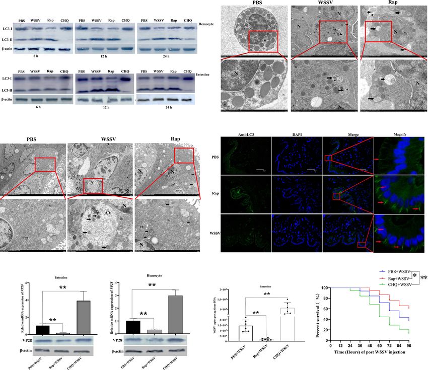
Zhao et al.                                                                                                   PmTRIM50 Restricting WSSV by Autophagy

INTRODUCTION                                                                     components for degradation (15, 16). Additionally, autophagy
                                                                                 can activate innate and adaptive immunity by capturing and
Protein post-translational modifications, such as phosphorylation,                exposing viral components to specific pattern recognition
acetylation, and ubiquitination, are important biological                        receptors or antigen-presenting cells (17). For example,
processes that regulate many intracellular signaling pathways                    autophagy might be involved in delivering viral nucleic acids
(1). Ubiquitination is a post-translational modification                          to Toll-like receptor 7 (TLR7), which mediates the induction of
pathway that regulates many host cellular processes,                             type 1 interferon (IFN) production. Moreover, autophagy might
including DNA repair, differentiation, and regulation of                         be involved in delivering endogenously synthesized microbial
immune responses (2). Ubiquitination involves the covalent                       antigens and self-antigens to late endosomes, which are loaded
attachment of ubiquitin to a lysine residue on the substrate                     onto MHC class II molecules for presentation to CD4+ T cells
protein (3). The process of ubiquitination requires three                        (16). However, autophagy could also dampen the antiviral
enzymes: E1 ubiquitin-activating enzyme, E2 ubiquitin-                           defense pathway by degrading the signaling components of the
conjugating enzyme, and E3 ubiquitin ligase to accomplish                        cytokine response, thereby inhibiting antiviral gene expression
three distinct activities including activation, conjugation, and                 (18). Recent studies have shown that TRIM proteins play an
ligation, respectively (4). E3 ubiquitin ligases are mainly                      important role in regulating autophagy and viral infections (18).
responsible for determining the substrate specificity (5).                        Using siRNA screening, over 30 TRIM proteins have been found
These enzymes operate in a concerted manner to poly-                             to trigger autophagy induction (19). TRIM20 and TRIM21 are
ubiquitinate a substrate protein and define its subsequent                        essential for IFNg-induced autophagy (20). TRIM5a plays an
fate (6). The chain of ubiquitin molecules regulates                             important role in pp242 (an mTOR inhibitor)-induced
molecular signaling depending on the lysine linkage type of                      autophagy (21). TRIM50 is a p62/SQSTM1 interacting protein
the poly-ubiquitin moiety, its length, and additional post-                      that promotes the formation and autophagy clearance of
translational modifications. Poly-ubiquitin chains can be                         aggresome-associated polyubiquitinated proteins through
formed via seven distinct lysine residues (K6, K11, K27, K29,                    HDAC6 interaction (22, 23). TRIM23 mediates virus-induced
K33, K48, and K63). The type of lysine linkage generally                         autophagy via the activation of TANK-binding kinase 1 (TBK1)
determines the fate of the substrate protein. For example,                       (24). The role of TRIM proteins in antiviral autophagy involves
K48-linked chain-directed ubiquitination of substrate protein                    at least two different mechanisms: acting as specific cargo
leads to its proteasomal degradation, whereas a substrate                        receptors that directly recognize viral components and target
protein conjugated with K63-linked chain is targeted for                         them for degradation (20, 25), or interacting with cargo/target-
kinase-mediated cell signaling (7).                                              recognizing proteins such as p62 and core regulators of
    Over 600 putative E3 ubiquitin ligases are encoded in the                    autophagy and forming protein complexes called
human genome (8). Based on domain structures, E3 ubiquitin                       ‘TRIMosomes’ (11). Although the role of TRIM proteins in
ligases have been classified into two major families: homologous                  regulating innate immunity and autophagy has been well
to the E6-AP COOH terminus (HECT) family and the RING-                           established in mammals, reports on the role of TRIM proteins
finger-containing protein family (9, 10). The tripartite motif                    in aquatic invertebrates are very limited, except the report
(TRIM) proteins, which contain a typical RING-finger-                             showing that TRIM9 homologs from Litopenaeus vannamei
containing domain, are a group of highly conserved proteins                      and the oyster Crassostrea hongkongensis can act as negative
that participate in a variety of biological processes such as                    regulators of the NF-kB pathway (26, 27). There are still no
regulation of development, autophagy, apoptosis,                                 reports about the role of TRIM proteins in autophagy and viral
carcinogenesis, and innate immunity (11). Because they                           infection in crustaceans.
contain a RING-finger domain, most TRIM proteins have been                           Black tiger shrimp (Penaeus monodon), which is one of the
defined as E3 ubiquitin ligases and hence contain E3 ubiquitin                    three major cultured shrimp species in the world, has important
ligase activity (11). TRIM proteins play an integral role in                     economic value (28). However, with the rapid development of
mammalian defense against pathogens. In vertebrates, TRIM                        intensive aquaculture and severe pollution of the marine
proteins exert antiviral activity by at least three major                        ecological environment, the outbreak of diseases poses a
mechanisms: directly antagonizing specific viral components,                      serious threat to the shrimp farming industry, which greatly
regulating transcription-dependent antiviral responses such as                   limits the sustainable development of shrimp aquaculture (29).
proinflammatory cytokine induction, and modulating other                          White spot syndrome virus (WSSV) is one of the most virulent
im p o rt a nt c el l-i nt ri ns ic d e fe ns e p a t hw a y s s u c h a s       pathogens in shrimp leading to huge economic losses in the
autophagy (12).                                                                  shrimp industry (30). Great progress has been made in the
    Autophagy is an evolutionarily conserved process that can                    research on molecular immune mechanisms of shrimp against
lead to the formation of autophagic lysosomes to wrap and                        WSSV infection. For example, scavenger receptor C of
degrade damaged proteins or organelles and pathogen-derived                      Marsupenaeus japonicus interacted with WSSV VP19 via its
components (13). Autophagy is induced by many different                          extracellular domain and invoked hemocyte phagocytosis to
viruses, yet the impact of autophagy on viral replication is                     restrict WSSV systemic infection (31). The polymeric
highly virus- and cell type-specific (14). Autophagy can serve                    immunoglobulin receptor of Marsupenaeus japonicas interacts
as an antiviral defense pathway by directly targeting virus or its               with VP24 and mediates WSSV internalization via the pIgR-

Frontiers in Immunology | www.frontiersin.org                                2                                   May 2021 | Volume 12 | Article 682562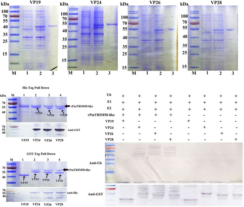
Zhao et al.                                                                                              PmTRIM50 Restricting WSSV by Autophagy

CaM-Clathrin endocytosis pathway to facilitate virus                      of rapamycin or chloroquine on shrimp, we found that 4 mM
proliferation (32). However, no effective method has been                 rapamycin was toxic to shrimp (data not shown), whereas 1 mM
established to restrict the uncontrolled occurrence and rapid             rapamycin was not toxic but was effective in inducing autophagy.
spread of WSSV disease. Understanding the molecular immune                Although 8 mM chloroquine did not show toxicity to shrimp
mechanism against WSSV infection might help identify new                  (data not shown), 4 mM chloroquine was sufficient to inhibit
strategies to prevent WSSV infection.                                     autophagy. Therefore, 1 mM rapamycin and 4 mM chloroquine
    In this study, an E3 ubiquitin ligase, PmTRIM50-like, was             were chosen to activate or inhibit shrimp autophagy activity in
identified in P. monodon, and the anti-WSSV function of                    further experiments.
PmTRIM50-like was analyzed during WSSV infection.                             After treatment with PBS, WSSV, rapamycin, or chloroquine,
                                                                          shrimp hemocytes and intestines were collected at various time
                                                                          points after injection (0, 6, 12, 24, 48, and 72 h) to detect
MATERIALS AND METHODS                                                     PmTRIM50-like expression or autophagy. To investigate the
                                                                          relationship between autophagy and WSSV infection, shrimp
Experimental Animals and Sample                                           were first treated with PBS, rapamycin, or chloroquine, and then
Preparation                                                               12 h later, challenged with WSSV. The survival rate was recorded
Healthy shrimp (P. monodon; mass 15 ± 2 g) were collected from the        within 96 h of WSSV injection. Previous studies have shown that
Zhuhai Experimental Base of the South China Sea Fisheries                 the expression level of VP28 acts as a marker of WSSV
Research Institute, China Academy of Fisheries Sciences                   replication, which indicates the number of virus replications
(Guangdong, China). The shrimp were acclimated for two days in            (31). In this study, shrimp intestines and hemocytes were
aerated seawater (5% salinity) at 24 ± 1°C. Healthy tissues from          collected after 72 h of WSSV challenge to detect the expression
three randomly selected shrimp were examined to determine the             levels of VP28 and virus copies.
distribution of PmTRIM50-like in the muscle, hepatopancreas,
intestine, heart, hemocytes, stomach, brain, and gills. The samples       Quantification of WSSV Copies
were snap frozen in liquid nitrogen and stored at 80°C until use.         The open reading frame (ORF) of the WSSV VP28 fragment was
                                                                          amplified and inserted into the pMD18-Tvector (TaKaRa,
WSSV Challenge and Tissue Collection                                      Dalian, China). The recombinant plasmid was quantified using
The WSSV inoculum was extracted from WSSV-infected shrimp                 a NanoDrop 2000/2000C (Thermo Scientific, Beijing, China).
and viral quantification was detected by quantitative real-time PCR        The copy number of the plasmid was calculated using the known
(qPCR) as described in a previous study (33). Each shrimp was             molecular weight of the plasmid. Subsequently, the plasmid was
injected with 100 ml of WSSV virions (1×10⁶ copies/mL). One               diluted gradually (109, 108, 107, 106, 105, 104, 103) and the diluted
hundred microliters of sterile phosphate-buffered saline (PBS) (140       samples were used as templates for qPCR with primers VP28-
mM NaCl, 2.7 mM KCl, 10 mM Na2HPO4, and 1.8 mM KH2PO4,                    RT-F and VP28-RT-R (Table 1). The cycle threshold (CT) values
pH 7.4) was injected into shrimp used as control. Intestines and          and the quantity of the template were used to generate a standard
hemocytes were collected from the shrimp at different time points         curve for WSSV quantification. Genomic DNA extracted from
(0, 3, 6, 12, 24, 48, and 72 h post injection) for RNA or protein         the viral inoculum or WSSV-infected tissue, along with the
extraction. For hemocyte collection, shrimp hemolymph was                 gradient diluted plasmid samples, was analyzed by qPCR to
extracted using a 1 mL syringe preloaded with anticoagulant (0.45         obtain the absolute copies of WSSV from the inocula or
M NaCl, 10 mM KCl, 10 mM EDTA, and 10 mM HEPES; pH 7.45).                 infected tissue.
After centrifugation at 800 × g for 6 min at 4°C, hemocytes were
collected and used to extract RNA or protein. At least three shrimp       RNA Extraction, cDNA Synthesis, and DNA
were used for tissue collection in each group.                            and Protein Extraction
                                                                          Total RNA was extracted from approximately 50 mg of tissue
Rapamycin and Chloroquine Injection                                       obtained from shrimp using TRIzol reagent (Invitrogen,
Rapamycin (Rap) is widely used to induce autophagy by directly            Shanghai, China) according to the manufacturer’s instructions.
targeting rapamycin receptor (TOR) kinase (34, 35). Rapamycin             After detecting the concentration and quality of total RNA, the
(5 mg/kg) was used to induce autophagy in the Chinese mitten              PrimeScript reverse transcriptase kit (TaKaRa, Dalian, China)
crab, Eriocheir sinensis (36). Chloroquine (CHQ) is a well-               was used for reverse transcription with 1 mg of total RNA
established inhibitor of autophagic proteolysis, which acts by            according to the manufacturer’s instructions. The RNA extract
inhibiting acidification of lysosomes and endosomes (37).                  and cDNA were stored at −80°C until use. Genomic DNA was
Chloroquine (10 mg/kg) was used to inhibit lysosomal activity             extracted using a genomic DNA extraction kit (Tiangen, Beijing,
in shrimp (31). In this study, rapamycin was diluted to different         China). Protein samples from different organs were
concentrations (0.1, 0.5, 1, 2, and 4 mM) with PBS. Chloroquine           homogenized separately in radioimmunoprecipitation assay
was diluted to different concentrations (0.5, 1, 2, 4, and 8 mM)          (RIPA) buffer (50 mM Tris-HCl, 150 mM NaCl, 0.1% SDS,
with PBS. Different concentrations of rapamycin or chloroquine            0.5% Nonidet P-40, 1 mM EDTA, 0.5 mM PMSF, pH 7.5). The
(100 ml) were injected intramuscularly into virus-free shrimp.            tissue homogenate was centrifuged at 12000 × g for 10 min at 4°C
By evaluating the toxic effects of different concentrations               to collect the supernatant for further analysis.

Frontiers in Immunology | www.frontiersin.org                         3                                     May 2021 | Volume 12 | Article 682562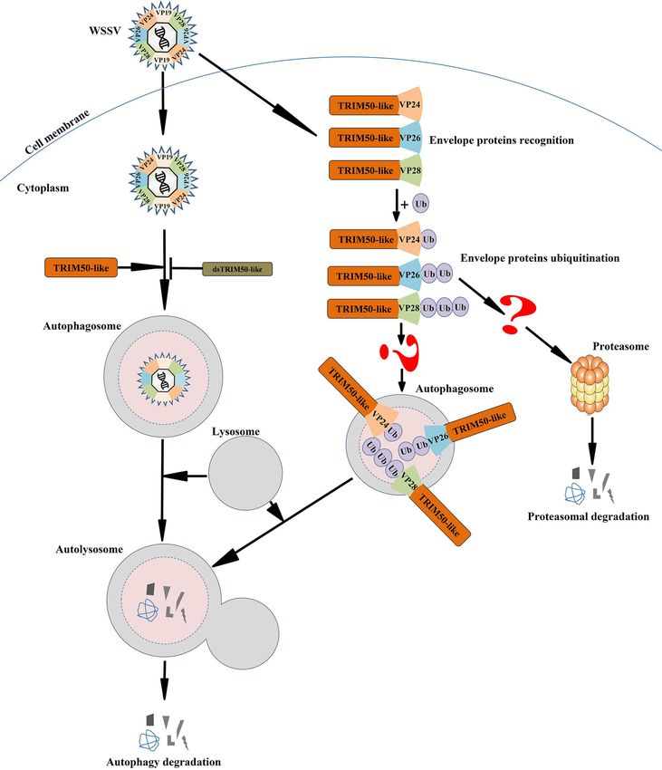
Zhao et al.                                                                                              PmTRIM50 Restricting WSSV by Autophagy

TABLE 1 | Primers used in this study.

Primers                                                     Sequence (5’!3’)                                                     Usage

TRIM50-like-F                               ATGCTGCCTGAGTGCGGTC                                                          ORF cloning
TRIM50-like-R                               TCAATTCGTCGCAAAGTCGC                                                         ORF cloning
rTRIM50-like-F                              TACTCAGAATTCATGCTGCCTGAGTGCGGTC                                              Recombinant expression
rTRIM50-like-R                              TACTCACTCGAGTCAATTCGTCGCAAAGTCGC                                             Recombinant expression
rVP19-F                                     GAATTCATGGCCACCACGACTAACAC                                                   Recombinant expression
rVP19-R                                     CTCGAGTTAATCCCTGGTCCTGTTCTTAT                                                Recombinant expression
rVP24-F                                     GAATTCATGCACATGTGGGGGGTTTA                                                   Recombinant expression
rVP24-R                                     CTCGAGTTATTTTTCCCCAACCTTAA                                                   Recombinant expression
rVP26-F                                     GGATCCACACGTGTTGGAAGAAGCGT                                                   Recombinant expression
rVP26-R                                     GAATTCTTACTTCTTCTTGATTTCGTCCTTG                                              Recombinant expression
rVP28-F                                     GAATTCATGGATCTTTCTTTCACTCTTTCGG                                              Recombinant expression
rVP28-R                                     CTCGAGTTACTCGGTCTCAGTGCCAGAGTAG                                              Recombinant expression
VP28-RT-F                                   CTCCGCAATGGAAAGTCTGA                                                         qRT-PCR
VP28-RT-R                                   GGGTGAAGGAGGAGGTGTT                                                          qRT-PCR
b-actin-F                                   CCCTGTTCCAGCCCTCATT                                                           qRT-PCR
b-actin-R                                   GGATGTCCACGTCGCACTT                                                           qRT-PCR
qTRIM50-like-F                              AGCGCTAGGGGAGTGTCATA                                                          qRT-PCR
qTRIM50-like-R                              CACAATGGTCACGTCCCTCA                                                          qRT-PCR
dsTRIM50-like-F                             GATCACTAATACGACTCACTATAGGGCCTTCTGCAGGGAGTGTCTC                               RNA interference
dsTRIM50-like-R                             GATCACTAATACGACTCACTATAGGGCACACCGCAGCTTCATCTTA                               RNA interference
dsGFP-F                                     CGAGCTCTGGAGTGGTCCCAGTTCTTGTTGA                                              RNA interference
dsGFP-R                                     ACGCGTCGACGCCATTCTTTGGTTTGTCTCCCAT                                           RNA interference

cDNA Cloning and Sequence Analysis                                          [Nomascus leucogenys]; XP_017949750.1, E3 ubiquitin-protein
The ORF sequence was amplified using PmTRIM50-like-F/R                       ligase TRIM50 [Xenopus tropicalis]; XP_020844498.1, tripartite
primers (Table 1). The PCR program for amplification was one                 motif-containing protein 59 [Phascolarctos cinereus];
cycle of 94°C for 3 min; 40 cycles of 94°C for 30 s, 60.5°C for 30 s,       XP_017265762.1, E3 ubiquitin-protein ligase TRIM13
and 72°C for 1.5 min, followed by one cycle of 72°C for 10 min.             [Kryptolebias marmoratus]; EMP32723.1, Tripartite motif-
The resulting PCR product was further verified by                            containing protein 59, partial [Chelonia mydas]; XP_032071003.1,
electrophoresis on a 1.2% agarose gel and purified by PCR.                   E3 ubiquitin-protein ligase TRIM50 [Thamnophis elegans];
The product was ligated into the pMD18-Tvector (TaKaRa,                     KGL84188.1, Tripartite motif-containing protein 59, partial
Dalian, China) and transformed into DH5a cells, and the                     [Tinamus guttatus]; XP_037362732.1, E3 ubiquitin-protein ligase
positive clones were picked for sequencing (Ruibiotech, China).             TRIM50 [Talpa occidentalis]; and NP_775107.1, tripartite motif-
    The PmTRIM50-like sequence was analyzed using the BLAST                 containing protein 59 [Homo sapiens]).
program (http://blast.ncbi.nlm.nih.gov/Blast.cgi) from the NCBI
server. Protein domain analysis of PmTRIM50-like was                        Recombinant Expression, Purification, and
performed using the conserved domain search service (https://               Antiserum Production of PmTRIM50-Like
www.ncbi.nlm.nih.gov/Structure/cdd/wrpsb.cgi). The molecular                The ORF of PmTRIM50-like was amplified using the PCR
size and theoretical isoelectric point of PmTRIM50-like were                primers PmTRIM50-like-F and PmTRIM50-like-R (Table 1)
analyzed online using ExPASy software (http://www.expasy.org/).             and cloned into the pET32a plasmid. The recombinant
Phylogenic analysis was performed using the neighbor-joining (NJ)           plasmid was transformed into E. coli BL21 cells, and the
method using ClustalW and MEGA 6. All PmTRIM50-like                         protein was induced with 0.6 m M isopropyl-b-D-
alignment sequences were from the GenBank database                          thiogalactopyranoside when the OD 600 of the bacterial
(XP_037788838.1, E3 ubiquitin-protein ligase TRIM11-like                    concentration reached 0.6. Recombinant rPmTRIM50-like was
[Penaeus monodon]; MPC09098.1, Tripartite motif-containing                  purified by affinity chromatography using a His-bind
protein 10 [Portunus trituberculatus]; MPC27339.1, Tripartite               purification kit (Beyotime, Shanghai, China). The protein was
motif-containing protein 10 [Portunus trituberculatus];                     separated by 12% sodium dodecyl sulfate-polyacrylamide gel
MPC09100.1, Tripartite motif-containing protein 72 [Portunus                electrophoresis (SDS-PAGE) and visualized using a GE Image
trituberculatus]; KAB7494620.1, E3 ubiquitin-protein ligase                 scanner III. Rabbit antiserum against PmTRIM50-like was
TRIM50 [Armadillidium nasatum]; ACU46018.1,                                 prepared following a previously reported method (38).
TRIM5/cyclophilin A fusion protein [Macaca fascicularis];
KFU97184.1, Tripartite motif-containing protein 59, partial                 Western Blotting Analysis
[Pterocles gutturalis]; XP_006123500.1, tripartite motif-containing         The purified protein or total protein extracted from shrimp
protein 59 [Pelodiscus sinensis]; XP_007441599.1, E3 ubiquitin-             tissue was separated by SDS-PAGE and transferred onto
protein ligase TRIM50 [Python bivittatus]; XP_012302766.1,                  nitrocellulose membranes. The membranes were blocked in 5%
tripartite motif-containing protein 59 [Aotus nancymaae];                   skim milk and resuspended in 0.1 M phosphate buffer solution-
XP_003256425.1, tripartite motif-containing protein 59                      Tween (PBST) at room temperature for 1 h. The membranes

Frontiers in Immunology | www.frontiersin.org                           4                                  May 2021 | Volume 12 | Article 682562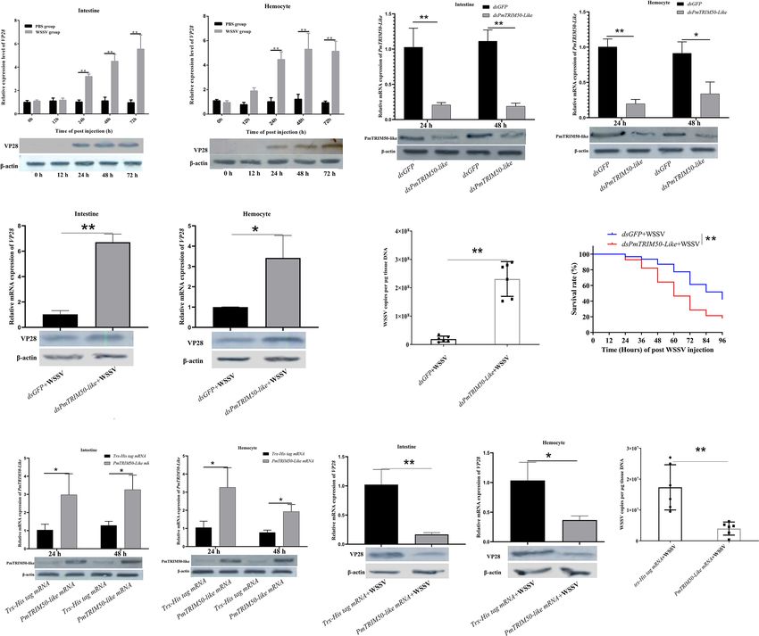
Zhao et al.                                                                                              PmTRIM50 Restricting WSSV by Autophagy

were washed with 0.1 M PBST three times for five min each. To               (50 mg) of dsPmTRIM50-like was injected into the abdominal
detect the purified PmTRIM50-like protein, the membranes were               segment of each shrimp. dsGFP was used as a control. The
incubated with 6×His-Tag HRP Antibody (SAB, Guangzhou,                     intestine and hemocytes were collected at 24 h and 48 h post
China; 1:3000 in PBS) or HRP-conjugated GST tag mouse                      injection, and RNA interference efficiency at the mRNA and
monoclonal antibody (Proteintech, Guangzhou, China; 1:20000                protein levels was detected by qRT-PCR and western blotting,
in PBS) for 1–2 h at 37°C per the manufacturer’s instructions,             respectively. b-Actin was used as an internal reference.
and washed with 0.1 M PBST three times for ten min each. For                   To investigate the role of PmTRIM50-like in autophagy or
detecting the endogenous proteins, the membranes were                      WSSV infection, the shrimp were first injected with dsRNA and
incubated with antiserum against PmTRIM50-like (1:500                      then challenged with WSSV or Rap 12 h later. Shrimp intestines
dilution in 0.1 M PBST), anti-LC3 antibody (Abcam, Shanghai,               were collected for detecting autophagy after 12 and 24 h of
China; 1:2000 dilution in 0.1 M PBS), anti-VP28 antibody                   WSSV or Rap challenge. Shrimp intestines and hemocytes were
(Abcam, Shanghai, China; 1:500 dilution in 0.1 M PBS), or                  collected to detect virus replication after 72 h of WSSV
anti-b-actin antibody (SAB, Guangzhou, China; 1:500 dilution               challenge. The survival rates in the dsPmTRIM50-like+WSSV
in 0.1 M PBS) for 2 h at room temperature. The membranes were              and dsGFP+WSSV groups were recorded within 96 h of
washed three times for ten min with 0.1 M PBST and then                    WSSV injection. To investigate the role of PmTRIM50-like in
incubated with horseradish peroxidase (HRP)-conjugated goat                autophagy and WSSV infection, the shrimp were first co-injected
anti-rabbit antibodies (SAB, Guangzhou, China; 1:20000 dilution            with dsRNA and Rap, and 12 h later, shrimp were challenged
in 0.1 M PBS). Immune active bands were visualized by staining             with WSSV. Shrimp intestines were collected at 12 h and 72 h
with horseradish peroxidase (HRP)- diaminobenzidine                        post WSSV injection to detect autophagy or viral replication.
(Tiangen, Beijing, China) and visualized with a GE Image                   The survival rates in the dsPmTRIM50-like+Rap+WSSV and
scanner III. b-actin expression was used to normalize the                  dsGFP+Rap+WSSV groups were recorded within 96 h of
amount of loaded proteins.                                                 WSSV injection.
    To detect the occurrence of autophagy in shrimp, the protein               Overexpression of the target gene was carried out by injecting
expression level of LC3-II/LC3-I was adopted as the reference              mature body mRNA (31, 32). The recombinant pET32a-
standard in the present study. The ratio of LC3-II/LC3-I has been          PmTRIM50-like plasmid was used as a template to transcribe
proven to be a hallmark of autophagy. LC3 was transformed                  single-stranded and capped PmTRIM50-like mRNAs using an
from LC3-I to LC3-II upon induction of autophagy, and the                  mMESSAGE mMACHINE™ T7 Transcription Kit (Ambion
electrophoresis rate of lipid-acylated LC3-II in polyacrylamide            Europe LTD, Cambridgeshire, UK), according to the
gel was faster than that of LC3-I (39).                                    manufacturer’s instructions. The empty pET32a vector was
                                                                           used as a template to synthesize Trx (thioredoxin)-His tag
Quantitative Real Time PCR                                                 mRNA as a control. Each shrimp was injected with 100 mg
The mRNA expressions of PmTRIM50-like and VP28 were                        PmTRIM50-like (or Trx-His tag) mRNA, and at least three
detected by qRT-PCR with b-actin (GenBank accession No.                    shrimp were used in each group. After 24 h and 48 h of
JN808449.1) used as an internal control. The primers used for              mRNA injection, the efficiency of overexpression at the mRNA
qRT-PCR are listed in Table 1. qRT-PCR was performed in                    and protein levels was detected by qRT-PCR and western
triplicate for each concentration using 384-well plates with a total       blotting, respectively. b-Actin was used as an internal reference.
volume of 12.5 mL, including 6.25 mL of 2×SYBR Preix Ex Taq II                 To investigate the effect of PmTRIM50-like overexpression
(TaKaRa, Dalian, China), 0.5 mL of each specific primer, 1 mL of            on WSSV infection, the shrimp were first injected with
the cDNA template, and 4.25 mL of DEPC water. The program                  PmTRIM50-like mRNA or Trx-His tag mRNA, and then
was as follows: denaturation at 94°C for 2 min; 40 cycles of 94°C          challenged with WSSV 12 h later. Shrimp intestines and
for 15 s, 58°C for 20 s, and 72°C for 20 s. The PCR product was            hemocytes were collected to detect virus replication after 72 h
denatured to produce a melting curve to determine its specificity.          of WSSV challenge.
Each reaction was carried out in three separate tubes, and the test
was repeated three times. PCR data were calculated using the               Transmission Electron Microscopy
2−DDCT method and expressed as the mean ± SD. Student’s t-test             The intestine and hemocytes of WSSV-treated, Rap-treated, and
was used to analyze the significant differences among PCR data,             PBS-treated shrimp were harvested at 12 h post injection. Tissues
and a significant difference was accepted at p < 0.05.                      were fixed in 2.5% glutaraldehyde overnight at 4°C. The samples
                                                                           were post-fixed in 1% osmium tetroxide for 1 h, dehydrated with
RNA Interference and Overexpression                                        graded ethanol, and embedded in epoxy resin. Ultrathin sections
Primers dsPmTRIM50-like-F and dsPmTRIM50-like-R                            were double-stained with uranyl acetate and lead citrate. Images
(Table 1), incorporating a T7 promoter, were used to amplify               were collected using JEM1400.
the template to produce dsRNA with T7 RNA polymerase.
Primers of dsGFP-F and dsGFP-R with the T7 promoter                        Immunocytochemistry Assay
sequences (Table 1) were used to clone a 289 bp DNA                        Intestines from the WSSV, Rap, PBS, dsPmTRIM50-like+WSSV,
fragment of the green fluorescent protein (GFP) gene. dsRNA                 dsGFP+WSSV, dsPmTRIM50-like+Rap, and dsGFP+Rap
was synthesized in vitro using the Transcription T7 Kit (TaKaRa,           groups were dissected after 12 h of WSSV or Rap challenge.
Dalian, China) following the manufacturer’s protocol. dsRNA                Tissues were fixed with 4% paraformaldehyde diluted in PBS.

Frontiers in Immunology | www.frontiersin.org                          5                                   May 2021 | Volume 12 | Article 682562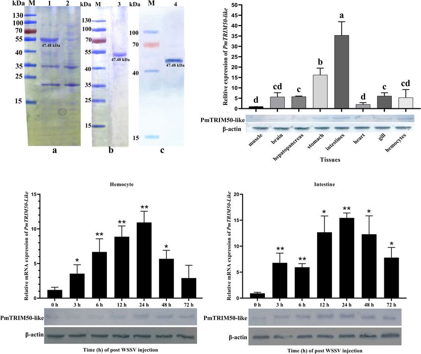
Zhao et al.                                                                                                      PmTRIM50 Restricting WSSV by Autophagy

Immunocytochemistry assays were performed following the                 RESULTS
method described by Zhang et al. (40). Briefly, after
dehydration with ethanol, the tissues were embedded in                  PmTRIM50-Like Was Upregulated by
paraffin, sectioned at a thickness of 5 mm, and rehydrated in            WSSV Challenge in Shrimp
water. The sections were blocked with 5% skim milk in PBS and           The transcript of PmTRIM50-like obtained from the
then incubated with Anti-LC3 antibody (1:200 in 3% BSA;                 transcriptome database of P. monodon was validated by PCR
Abcam, Shanghai, China) for 1–2 h at 37°C. Antibody binding             and confirmed by sequencing. The full-length ORF of
was visualized by fluorescence microscopy using FITC-                    PmTRIM50-Like was 1245 bp, encoding a protein of 414 aa
conjugated goat anti-rabbit IgG (1:10,000 in PBS). DAPI                 length (GenBank accession No. MW208625). The deduced
(1:10,000 in PBS) was used to dye the nuclei for 10 min.                amino acid sequence of PmTRIM50-like contained a typical
                                                                        RING domain of the RING-finger-containing protein family
Pull-Down Assay                                                         (positions 2–28 aa), a B-box domain (positions 64–106
Pull-down assays were performed to explore whether                      aa), and a cyclophilin domain (positions 362–404 aa)
recombinant rPmTRIM50-like could interact with the main                 (Figure S1A). To identify the phylogenetic relationships of
envelope proteins of WSSV (VP19, VP24, VP26, and VP28).                 PmTRIM50-like, a phylogenetic tree was constructed using the
The primers used for the recombinant expression of VPs (VP-F            neighbor-joining method. The results showed that PmTRIM50-
and VP-R) are shown in Table 1. The amplified sequences of               like was most closely related to the E3 ubiquitin-protein ligase
VP19 (GenBank accession No. DQ681071.1), VP24 (GenBank                  TRIM50 of Talpa occidentalis, which formed a clade
accession No. DQ196431.1), VP26 (GenBank accession No.                  (Figure S1B).
AY220746.1), and VP28 (GenBank accession No. DQ681069.1)                     The recombinant PmTRIM50-like protein was then
were ligated separately into the vector PGEX-4T-1 and                   expressed and purified (Figure 1A). The theoretical molecular
transformed into E. coli BL21 cells for expression. The                 weight of the recombinant protein was 46.64 kDa, which
recombinant proteins were purified by affinity chromatography             consisted of a His tag (0.84 kDa) at the carboxyl terminus. The
using a GST-resin (Beyotime, Shanghai, China). For the pull-            target protein (approximately 47.48 kDa) was detected in the
down assay, purified His-tagged rPmTRIM50-like was incubated             supernatant of the resuspended bacteria that were destroyed by
with Ni-NTA beads, to which purified VP19, VP24, VP26, or                ultrasonication (Figure 1Aa); it was further confirmed by
VP28 was added and incubated at 4°C overnight with slight               western blot analysis (Figure 1Ac). The recombinant protein
rotation. The mixture was washed thoroughly with wash buffer            was purified and stored at −80°C for use in subsequent
(20 nM imidazole, 50 mM Tris-HCl, pH 8.0), eluted in elution            experiments (Figure 1Ab). Anti-PmTRIM50-like sera were
buffer (250 nM imidazole, 50 mM Tris-HCl, pH 8.0), and then             prepared using the purified recombinant protein
analyzed using 12.5% SDS-PAGE, followed by western blotting             PmTRIM50-like.
with anti-GST antibody. For the GST pull-down assay, purified                 The spatial expression pattern of PmTRIM50-Like mRNA
GST-tagged VP19, VP24, VP26, or VP28 and purified His-                   was determined by quantitative real-time PCR (qRT-PCR) using
tagged rPmTRIM50-like were incubated with glutathione beads             b-actin as an internal control. The results showed that
at 4°C overnight with slight rotation. The beads were washed            PmTRIM50-Like mRNA was ubiquitously expressed in the
with TBS thoroughly, the proteins were eluted with elution buffer       i n t e s ti n e , s t o m a c h , h e m o c y t e s , g i l l s , h e a r t , b r a i n ,
(10 mM reduced glutathione and 50 mM Tris-HCl, pH 8.0), and             hepatopancreas, and muscle (Figure 1B, upper panel).
then analyzed using 12.5% SDS-PAGE, followed by western                 Western blotting analysis showed that PmTRIM50-Like
blotting with anti-His antibody.                                        protein was also distributed in the intestine and hemocytes
                                                                        (Figure 1B, lower panel).
In Vivo Ubiquitination Assay                                                 After challenge with WSSV, the expression of PmTRIM50-
The ubiquitination assay was performed as described by Choo             like mRNA in hemocytes increased significantly during 3–48 h
et al. (41). The reaction mixture (40 ml) contained 8 ml of 5X          post-challenge and then recovered to the same level as that in the
ubiquitination buffer (100 mM Tris-HCl, pH 7.5, 25 mM MgCl2,            PBS group. The peak value reached at 24 h post WSSV challenge,
2.5 mM DTT, 10 mM ATP), 250 ng of ubiquitination E1 (E-305,             which was 10.6-fold higher than that of the control group
Boston Biochem, Inc), 500 ng of ubiquitination E2 (E2-656,              (p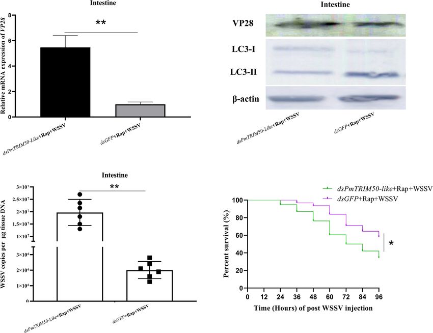
Zhao et al.                                                                                                                 PmTRIM50 Restricting WSSV by Autophagy

                A                                                                   B

                C                                                                   D

 FIGURE 1 | PmTRIM50-like was upregulated in shrimp by WSSV infection. (A) Expression of recombinant PmTRIM50-like in E. coli and protein purification:
 (a) SDS-PAGE of induced PmTRIM50-like; Lane M, molecular weight marker; 1, recombinant protein of PmTRIM50-like at 12-h-induced; 2, recombinant
 PmTRIM50-like protein of none-induced. (b) SDS-PAGE analysis of purified PmTRIM50-like; Lanes M, molecular weight marker; 3, purified PmTRIM50-like protein.
 (c) Western blotting of PmTRIM50-like; Lane M, molecular weight marker; Lane 4, 12-h-induced PmTRIM50-like protein. (B) Tissue distribution of PmTRIM50-like
 was analyzed by qRT-PCR (upper panel) and western blotting (lower panel). Different lowercase letters indicate statistically significant differences (P < 0.05). (C, D)
 Expression patterns of PmTRIM50-like mRNA and protein were detected by qRT-PCR (upper panel) and western blotting (lower panel) in hemocyte (C) and intestine
 (D) after WSSV challenge. b-actin was used as an internal reference. Asterisks indicate significant differences (*P< 0.05 and **P
Zhao et al. PmTRIM50 Restricting WSSV by Autophagy A B C D E F G H I J K L M FIGURE 2 | PmTRIM50-like restricted WSSV infection in shrimp. (A, B) Expression patterns of VP28 mRNA and protein were detected by qRT-PCR (upper panel) and western blotting (lower panel) in intestine (A) and hemocyte (B) after WSSV challenge. (C) Efficiency of dsPmTRIM50-like was detected by qRT-PCR (upper panel) and western blotting (lower panel) in the intestine. (D) Efficiency of dsPmTRIM50-like was detected by qRT-PCR (upper panel) and western blotting (lower panel) in hemocytes. (E) Expression of VP28 in the intestine of dsPmTRIM50-like+WSSV group and dsGFP+WSSV group was determined by qRT-PCR (upper panel) and western blotting (lower panel). (F) Expression of VP28 in hemocytes of dsPmTRIM50-like+WSSV group and dsGFP+WSSV group was determined by qRT-PCR (upper panel) and western blotting (lower panel). (G) The copy number of WSSV in the intestine of dsPmTRIM50-like+WSSV group and dsGFP+WSSV group was detected by qPCR. (H) The survival rate in dsPmTRIM50-like+WSSV group and dsGFP+WSSV group was detected after challenged by WSSV. Survival rates were analyzed statistically using the Kaplan-Meier plots (log-rank X2). (I) Efficiency of PmTRIM50-like overexpression was detected by qRT-PCR (upper panel) and western blotting (lower panel) in the intestine. (J) Efficiency of PmTRIM50-like overexpression was detected by qRT-PCR (upper panel) and western blotting (lower panel) in hemocyte. (K) Expression of VP28 in the intestine of PmTRIM50-like mRNA+WSSV group and Trx-His tag mRNA+WSSV group was determined by qRT-PCR (upper panel) and western blotting (lower panel). (L) Expression of VP28 in hemocytes of PmTRIM50-like mRNA+WSSV group and Trx-His tag mRNA+ WSSV group was determined by qRT-PCR (upper panel) and western blotting (lower panel). (M) The copy number of WSSV in the intestine of PmTRIM50-like mRNA+WSSV group and Trx-His tag mRNA+WSSV group was detected by qPCR. The experiments were repeated three times. b-actin was used as an internal reference. Asterisks indicate significant differences *P< 0.05 and **P
Zhao et al.                                                                                         PmTRIM50 Restricting WSSV by Autophagy

permg genomic DNA) was significantly higher than in the                and Rap-challenged shrimp was stronger and more concentrated
dsGFP+WSSV group (1.9 × 10⁷ copies per mg genomic DNA)                (marked by red arrows) than in the PBS control group
(p
Zhao et al.                                                                                                            PmTRIM50 Restricting WSSV by Autophagy

   A                                                                         B

   C                                                                         D

   E                                    F                                    G                                     H

 FIGURE 3 | Autophagy played a positive role in restricting WSSV infection in shrimp. (A) The expression of LC3-II/LC3-I in hemocytes (upper panel) and the
 intestine (lower panel) was detected by Western blotting. (B) The formation of autophagosome-like vesicles in hemocytes of PBS, WSSV, or Rap challenged shrimp
 was observed by TEM. Vesicles with the characteristics of autophagosomes are indicated by black arrows. N, nucleus; M, mitochondrion. (C) The formation of
 autophagosome-like vesicles in the intestine of PBS, WSSV, or Rap challenged shrimp was observed by TEM. Vesicles with the characteristics of autophagosomes
 are indicated by black arrows. N, nucleus; M, mitochondrion; ER, endoplasmic reticulum; AV, autophagic vacuole. (D) The distribution of the autophagy marker LC3
 in the intestine of PBS, WSSV, or Rap challenged shrimp was detected by immunocytochemistry. Positive immunoreactivities of LC3 are indicated by red arrows.
 (E) The VP28 expression in the intestine of PBS+WSSV group, Rap+WSSV group, and CHQ+WSSV group was determined by using qRT-PCR (upper panel) and
 western blotting (lower panel). (F) VP28 expression in the hemocytes of PBS+WSSV, Rap+WSSV, and CHQ+WSSV groups was determined by using qRT-PCR
 (upper panel) and western blotting (lower panel). (G) Virus copy number in the intestines of PBS+WSSV, Rap+WSSV, and CHQ+WSSV groups was detected by
 qPCR. (H) The survival rates in PBS+WSSV, Rap+WSSV, and CHQ+WSSV groups were recorded. The survival rates were calculated and survival curves were
 presented as Kaplan-Meier plots. Differences between the two groups were analyzed with log-rank test using the software of GraphPad Prism 8.02. The experiments
 were repeated at least three times. Asterisks indicate significant differences *P< 0.05 and **P
Zhao et al. PmTRIM50 Restricting WSSV by Autophagy
A B
C
D
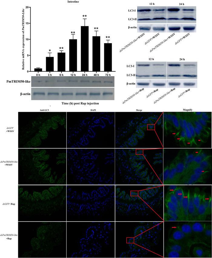
FIGURE 4 | PmTRIM50-like was required for autophagy in shrimp. (A) Expression patterns of PmTRIM50-like mRNA and protein were detected by qRT-PCR
(upper panel) and western blotting (lower panel) in the intestine after rapamycin challenge. b-actin was used as an internal reference. (B) The expression of LC3-II/
LC3-I in dsPmTRIM50-like+WSSV and dsGFP+WSSV groups was detected by western blotting. (C) The expression of LC3-II/LC3-I in dsPmTRIM50-like+Rap
and dsGFP+Rap groups was detected by western blotting. (D) The distribution of the autophagy marker LC3 in dsPmTRIM50-like+WSSV, dsGFP+WSSV,
dsPmTRIM50-like+Rap, and dsGFP+Rap groups was detected by immunocytochemistry. Positive immunoreactivities of LC3 are indicated by red arrows. Asterisks
indicate significant differences (*PZhao et al. PmTRIM50 Restricting WSSV by Autophagy
inhibited when the expression of PmTRIM50-like was silenced in the dsGFP+Rap+WSSV group (Figure 5B, middle panel). The
shrimp. This strongly suggests that PmTRIM50-like might be replication of WSSV in the dsPmTRIM50-like+Rap+WSSV
required for autophagy induction in shrimp. group increased significantly compared with the dsGFP+Rap+
WSSV group, as a higher VP28 expression (Figures 5A, B, upper
PmTRIM50-Like Restricted WSSV panel), higher WSSV copies (Figure 5C), and lower survival
Infection via Mediating Autophagy rate (Figure 5D) were detected in the dsPmTRIM50-like+
in Shrimp Rap+WSSV group. These results further showed that
One mechanism of action of TRIM proteins in antiviral PmTRIM50-like is indispensable for autophagy induction and
autophagy is to regulate autophagy activity during viral thus, a possible mechanism for PmTRIM50-like-mediated
infection (18). In this study, autophagy could restrict WSSV inhibition of WSSV replication may be via mediating
replication, while PmTRIM50-like could both promote autophagy in shrimp.
autophagy and restrict WSSV replication in shrimp. This
indicated that PmTRIM50-like might restrict WSSV replication PmTRIM50-Like Recognized Components
by mediating autophagy. To investigate whether PmTRIM50- of WSSV and Targeted Them for
like regulated anti-WSSV autophagy, shrimp were co-injected Ubiquitination In Vitro
with dsPmTRIM50-like and Rap, followed by WSSV challenge. Another mechanism of action of TRIM proteins acting in
Host autophagy and WSSV replication were also detected. The antiviral autophagy is to act as specific cargo receptors that
results showed that the expression of LC3-II/LC3-I in the directly recognize viral components and target them for
dsPmTRIM50-like+Rap+WSSV group was lower than that in degradation by autophagy (18). To investigate the possibility of
A B
C D
FIGURE 5 | PmTRIM50-like restricted WSSV infection via mediating autophagy in shrimp. (A) The expression of VP28 in dsPmTRIM50-like+Rap+WSSV and
dsGFP+Rap+WSSV groups was detected by qRT-PCR. (B) The expressions of VP28 protein (upper panel) and LC3-II/LC3-I (middle panel) in dsPmTRIM50-like+
Rap+WSSV and dsGFP+Rap+WSSV groups were detected by western blotting. b-actin was used as control (lower panel). (C) Virus copy number of WSSV in the
intestines of dsPmTRIM50-like+Rap+WSSV and dsGFP+Rap+WSSV groups were detected by qPCR. (D) The survival rates in dsPmTRIM50-like+Rap+WSSV and
dsGFP+Rap+WSSV groups were recorded. The survival rates were calculated and survival curves were presented as Kaplan-Meier plots. Differences between the
two groups were analyzed with log-rank test using the software of GraphPad Prism 8.02. The experiments were repeated three times. Asterisks indicate significant
differences *P< 0.05 and **PZhao et al. PmTRIM50 Restricting WSSV by Autophagy
A B C D
E G
F
FIGURE 6 | PmTRIM50-like recognized components of WSSV and targeted them for ubiquitination in vitro. (A–D) Purified GST tagged WSSV structural proteins of
VP19 (A), VP24 (B), VP26 (C), and VP28 (D). (E) His tagged PmTRIM50-like that interacted with GST-VP19, VP24, VP26, and VP28 was obtained in His pull-down
assay and visualized by coomassie blue staining (upper panel) and confirmed by western blotting with anti-GST antibody (lower panel). (F) GST tagged VP19, VP24,
VP26, and VP28 that interacted with His-PmTRIM50-like was obtained in GST pull-down assay and visualized by coomassie blue staining (upper panel) and
confirmed by western blotting with anti-His antibody (lower panel). (G) In vitro ubiquitination of VP19, VP24, VP26, and VP28 by rPmTRIM50-like plus E1, E2, ATP
and ubiquitin (Ub) as detected by anti-Ub antibody (upper panel) and anti-GST antibody (lower panel).
PmTRIM50-like to recognize WSSV components, we conducted no ubiquitination of VP19 was observed in the presence of
GST pulldown and in vitro ubiquitination assays. First, the GST- rPmTRIM50-like. These results indicated that PmTRIM50-like
tagged proteins VP19, VP24, VP26, and VP28 were expressed protein could interact with the viral envelope proteins of WSSV
and purified in E. coli BL21 cells (Figures 6A–D). The pulldown (VP24, VP26, and VP28) and target them for ubiquitination in
assay showed that interaction occurred between rPmTRIM50- vitro. PmTRIM50-like might act as a cargo receptor that directly
like and three viral envelope proteins VP24, VP26, and VP28. recognizes envelope proteins of WSSV and target them for
However, no interaction was detected between rPmTRIM50-Like degradation by autophagy in shrimp. Therefore, we concluded
and VP19 (Figures 6E, F). Many TRIM proteins contain a that another possible mechanism for PmTRIM50-like-mediated
typical RING-finger domain and have E3 ubiquitin ligase inhibition of WSSV replication might be by recognizing WSSV
activity, and thus, can directly couple ubiquitin to a specific components and targeting them for degradation.
substrate. The in vitro ubiquitination assay showed that
PmTRIM50-like could ubiquitinate WSSV components as an
obvious polyubiquitination smear of VP24, VP26, and VP28 DISCUSSION
were detected by anti-Ub antibody in the presence of
rPmTRIM50-Like protein (Figure 6G, upper panel). VP24, TRIM proteins are widely recognized as important antiviral
VP26, and VP28 were also detected by anti-GST antibody with restriction factors or modulators of signaling pathways that
increased molecular weight (Figure 6G, lower panel). However, lead to the induction of antiviral or proinflammatory
Frontiers in Immunology | www.frontiersin.org 13 May 2021 | Volume 12 | Article 682562Zhao et al. PmTRIM50 Restricting WSSV by Autophagy
cytokines. In mammals, TRIM proteins function as autophagy autophagy can be induced by a few viruses (14). On the other
receptors and regulators of autophagosome formation. Many hand, autophagy can be inhibited and subverted by a few viruses
mammalian TRIM proteins play essential roles in viral-induced to promote their replication (52). Previous studies have shown
autophagy. However, there has been no report on the that autophagy can be induced by WSSV infection in
involvement of TRIM proteins in viral infection and host Marsupenaeus japonicus and Cherax quadricarinatus (53, 54).
autophagy in crustaceans. In this study, we identified a In this study, the ratio of LC3-II/LC3-I protein was upregulated
member of the tripartite motif family proteins (PmTRIM50- by WSSV challenge. In the TEM observation of WSSV-infected
like) from P. monodon, which could restrict WSSV replication shrimp, we observed an increased number of autophagosome-
by positively regulating autophagy in shrimp. To our knowledge, like vesicles and autophagic vesicles in hemocytes and intestines.
this is the first report of a TRIM protein being involved in viral The LC3 positive signal in WSSV-challenged shrimp was
infection and host autophagy in crustaceans. stronger and more concentrated than that in the PBS control
The TRIM family of proteins participate in a variety of group. These results indicate that WSSV infection promotes
biological processes in vertebrates, including playing an autophagy in P. monodon. As mentioned earlier, the impact of
important role in the innate immune response and virus- host autophagy on viral replication is highly virus-and cell-type-
induced autophagy (18). Research has shown that roughly half specific. Autophagy can regulate innate and acquired immunity
of the 75 human TRIM family proteins can enhance the innate to protect against viral infection (12). However, autophagy can
immune response (42). Due to their importance in regulating the also facilitate viral replication by serving as a site for viral
innate immune response, TRIMs have become an increasingly replication (55). In our study, the amount of WSSV decreased
studied protein family. Typically, TRIM proteins are composed significantly in shrimp after the induction of autophagy by Rap,
of a RING finger domain, one or two B-BOX domains, a coiled- compared with the control. The amount of WSSV increased
coil region in the N-terminus, and various specific domains in significantly in shrimp after the inhibition of autophagy by
the C-terminus, although eight TRIM proteins in humans lack chloroquine. These results were similar to those of previous
the RING finger domain (43). Many proteins containing the studies in which Rap-induced autophagy played a role in
RING domain have E3 ubiquitin ligase activity and can activate restricting WSSV replication in hematopoietic tissue stem cells
specific signaling pathways by targeting specific proteins for of Cherax quadricarinatus (54). Chloroquine inhibits autophagic
degradation, thereby regulating the antiviral immune response flux by decreasing autophagosome-lysosome fusion. In
(44). For example, the RING finger protein CqRNF152-like in Marsupenaeus japonicus, the amount of WSSV increased
Cherax quadricarinatus has self-ubiquitination activity and significantly when kuruma shrimp were challenged with
interacts with WSSV VP28 (45). Porcine RING finger protein chloroquine, indicating that inhibiting autophagy increased the
114 inhibits classical swine fever virus replication via K27-linked amount of WSSV in shrimp (31). Our results indicate that host
polyubiquitination of viral NS4B (46). The PmTRIM50-like autophagy is probably involved in the clearance of WSSV in
protein contains a typical RING domain, a B-box domain, and P. monodon.
a cyclophilin domain in the C terminus, indicating that Some TRIM proteins are critical components or regulators of
PmTRIM50-like might contain E3 ubiquitin ligase activity and autophagy machinery. For example, TRIM5a can interact with
have antiviral function. The phylogenetic tree showed that LC3s/GABARAPs, p62, and Beclin1 to form a core complex of
PmTRIM50-like was most closely related to Talpa occidentalis the autophagy machinery (21). TRIM13 localizes to the ER and
E3 ubiquitin-protein ligase TRIM50 and hence was named induces autophagy during ER stress via its coiled-coil domain
PmTRIM50-like in this study. Upon WSSV challenge, the and interacts with p62 (56). TRIM59 regulates autophagy by
expression of PmTRIM50-like was upregulated in both modulating the transcription and ubiquitination of Beclin1 (57).
hemocytes and intestines, suggesting that PmTRIM50-like was In this study, host autophagy was found to be induced by WSSV
responsive to WSSV infection. Among TRIM family proteins, and rapamycin challenge in shrimp. The expression of
many TRIM proteins are widely recognized as important PmTRIM50-like was upregulated by WSSV and rapamycin
antiviral restriction factors or modulators of signaling challenge. With the increase in WSSV- or rapamycin-induced
pathways (47). In this study, shrimps with knockdown of autophagy, the expression level of PmTRIM50-like also
PmTRIM50-like were more susceptible to WSSV infection as increased, indicating that PmTRIM50-like might be involved
higher virus copies and higher mortality rates were detected. in the process of autophagy in shrimp. However, WSSV- or
However, shrimps injected with PmTRIM50-like mRNA were rapamycin-induced autophagy could be clearly damaged by
more resistant to WSSV infection and lower virus copies were silencing of PmTRIM50-like in shrimp, indicating that
detected, suggesting that PmTRIM50-like restricted WSSV PmTRIM50-like might be required for autophagy induction
replication in P. monodon. in shrimp.
Autophagy is a homeostatic process that not only sustains cell TRIM family proteins can act as antiviral autophagy inducers
survival under stress but is also induced during a viral infection in two different ways. A few TRIM proteins are critical
(48). Considering the important role of autophagy in antiviral components of the autophagy machinery that mediate viral
processes, some viruses are equipped with sophisticated clearance. TRIM23 is a core component of the autophagy
mechanisms to modulate autophagy in order to facilitate the machinery and is required for the induction of antiviral
stability and replication of the virus (49–51). On one hand, autophagy by several viruses (24). TRIM16 was found to
Frontiers in Immunology | www.frontiersin.org 14 May 2021 | Volume 12 | Article 682562Zhao et al. PmTRIM50 Restricting WSSV by Autophagy FIGURE 7 | Predicted schematic diagram for PmTRIM50-like-mediated anti-WSSV autophagy in shrimp. After WSSV infection, the expression of PmTRIM50-like was upregulated and autophagy was induced in shrimp. Subsequently, autophagy degraded WSSV in a PmTRIM50-like-dependent manner. In shrimp cells, PmTRIM50-like might act as a cargo receptor to recognize and ubiquitinate WSSV envelope proteins of VP24, VP26, and VP28. The recognition and ubiquitination might mark and target WSSV for degradation by autophagy. Frontiers in Immunology | www.frontiersin.org 15 May 2021 | Volume 12 | Article 682562
Zhao et al.                                                                                                             PmTRIM50 Restricting WSSV by Autophagy

promote antiviral autophagy by facilitating activation of the p62-                   that PmTRIM50-like could interact with WSSV envelope
NRF2 axis (58). In this study, rapamycin-induced autophagy                           proteins and target them for ubiquitination in vitro.
significantly restricted the replication of WSSV, but when                                In conclusion, PmTRIM50-like mediates anti-WSSV
PmTRIM50-like was silenced, rapamycin-induced autophagy                              autophagy in P. monodon. After WSSV infection, the
was inhibited, resulting in an increase in WSSV replication.                         expression of PmTRIM50-like was upregulated, and autophagy
This demonstrated that PmTRIM50-like might restrict WSSV                             was induced in shrimp. Subsequently, autophagy degraded
replication by mediating autophagy.                                                  WSSV in a PmTRIM50-like-dependent manner. In shrimp
    Certain TRIMs can act as specific cargo receptors that directly                   cells, PmTRIM50-like may act as a cargo receptor to recognize
recognize viral components and target them for degradation by                        and ubiquitinate the WSSV envelope proteins VP24, VP26, and
autophagy. For example, TRIM5a directly interacts with the                           VP28. Moreover, recognition and ubiquitination might mark
capsid of human immunodeficiency virus-1 (HIV-1), leading                             and target WSSV for degradation by autophagy (Figure 7). This
to premature disassembly of the capsid (59). The recognition of                      is the first report of a TRIM family protein mediating antiviral
HIV-1 capsid protein (p24) by TRIM5a leads to proteasomal and                        autophagy in shrimp. However, the molecular mechanism by
autophagosome degradation. For autophagic clearance, the p24-                        which PmTRIM50-Like regulates antiviral autophagy in shrimp
TRIM5a complex is recruited to autophagosomes, where                                 needs further investigation.
TRIM5a induces autophagy in a Beclin-1- and ULK1-
dependent manner (59). The recognition of viral components
by other TRIM proteins may also destroy virus replication in
different ways. TRIM11 directly interacts with the capsid of
                                                                                     DATA AVAILABILITY STATEMENT
human immunodeficiency virus-1 (HIV-1) to affect the release                          The original contributions presented in the study are included in
of HIV-1 particles (60). TRIM25 targets CpG-rich sites in the                        the article/Supplementary Material. Further inquiries can be
genomes of Sindbis virus (SINV) and HIV-1, and inhibits                              directed to the corresponding author.
translation of the incoming virus genome (61, 62). TRIM21
suppresses hepatitis B virus (HBV) DNA replication by
promoting the ubiquitination of HBV DNA polymerase (63).
TRIM41 targets the viral nucleoprotein of vesicular stomatitis                       AUTHOR CONTRIBUTIONS
virus (VSV) for ubiquitination and subsequent protein
                                                                                     CZ and CP performed the experiments and written original draft
degradation (64). The recognition of viral components can
                                                                                     preparation. CZ: reviewed and edited the paper. PW, LY, and SF:
directly lead to premature capsid disassembly and degradation,
                                                                                     provided resources. LQ: acquired the funding. All authors
block the lifecycle of the virus, or act as “autophagy receptors” to
                                                                                     contributed to the article and approved the submitted version.
initiate autophagy. In this study, PmTRIM50-like could interact
with VP24, VP26, and VP28, and target them for ubiquitination
in vitro. VP19, VP24, VP26, and VP28 are the four major
envelope proteins of WSSV and function in virus entry and                            FUNDING
systemic infection (65–67). VP24, VP26, and VP28 share high
sequence homology with each other; however, VP19 has a                               This study was supported by National Key R&D Program of China
unique structure and biological character (68, 69). Therefore,                       (2018YFD0900103); Central Public-interest Scientific Institution
we concluded that one possible mechanism for PmTRIM50-like                           Basal Research Fund, CAFS (2020TD21); National Natural
to restrict WSSV replication was by recognizing envelope                             Science Foundation of China (42006113); and the Central Public-
proteins of WSSV and targeting them for ubiquitination. The                          interest Scientific Institution Basal Research Fund, South China Sea
recognition and ubiquitination of WSSV might lead to the                             Fisheries Research Institute, CAFS (2019TS11).
disassembly and degradation of WSSV by autophagy or
proteasomes in shrimp.
    In summary, knockdown and overexpression analyses                                SUPPLEMENTARY MATERIAL
revealed that PmTRIM50-like possessed antiviral functions in
shrimp. Further, our study found that PmTRIM50-like was                              The Supplementary Material for this article can be found online
required for autophagy induction and could restrict WSSV                             at: https://www.frontiersin.org/articles/10.3389/fimmu.2021.
replication by mediating autophagy. Moreover, we discovered                          682562/full#supplementary-material

REFERENCES                                                                            3. Zhu J, Tsai N-P. Ubiquitination and E3 Ubiquitin Ligases in Rare
                                                                                         Neurological Diseases With Comorbid Epilepsy. Neuroscience (2020)
 1. Heaton SM, Borg NA, Dixit VM. Ubiquitin in the Activation and Attenuation            428:90–9. doi: 10.1016/j.neuroscience.2019.12.030
    of Innate Antiviral Immunity. J Exp Med (2016) 213(1):1–13. doi: 10.1084/         4. Callis J. The Ubiquitination Machinery of the Ubiquitin System. Arabidopsis
    jem.20151531                                                                         Book (2014) 12:e0174. doi: 10.1199/tab.0174
 2. Ciechanover A, Orian A, Schwartz AL. Ubiquitin-Mediated Proteolysis:              5. HJ M, Martin S, Sylvie B, HP M. A Family of Proteins Structurally and
    Biological Regulation via destruction. Bioessays (2000) 22(5):442–51.                Functionally Related to the E6-AP Ubiquitin-Protein Ligase. PloS One (1995)
    doi: 10.1002/(SICI)1521-1878(200005)22:53.0.CO;2-Q                   92(7):2563–7. doi: 10.1073/pnas.92.7.2563

Frontiers in Immunology | www.frontiersin.org                                   16                                         May 2021 | Volume 12 | Article 682562
You can also read