Staphylococcus saprophyticus From Clinical and Environmental Origins Have Distinct Biofilm Composition - Frontiers
←
→
Page content transcription
If your browser does not render page correctly, please read the page content below
ORIGINAL RESEARCH
                                                                                                                                                  published: 07 June 2021
                                                                                                                                         doi: 10.3389/fmicb.2021.663768

                                             Staphylococcus saprophyticus From
                                             Clinical and Environmental Origins
                                             Have Distinct Biofilm Composition
                                             Opeyemi U. Lawal 1,2 , Marta Barata 1 , Maria J. Fraqueza 3 , Peder Worning 4 ,
                                             Mette D. Bartels 4 , Luisa Goncalves 5 , Paulo Paixão 6 , Elsa Goncalves 7 , Cristina Toscano 7 ,
                                             Joanna Empel 8 , Malgorzata Urbaś 8 , Maria A. Domiìnguez 9 , Henrik Westh 4,10 ,
                                             Hermínia de Lencastre 2,11 and Maria Miragaia 1*
                                             1
                                               Laboratory of Bacterial Evolution and Molecular Epidemiology, Instituto de Tecnologia Química e Biológica António Xavier,
                                             Universidade NOVA de Lisboa, Oeiras, Portugal, 2 Laboratory of Molecular Genetics, Instituto de Tecnologia Química e
                                             Biológica António Xavier, Universidade NOVA de Lisboa, Oeiras, Portugal, 3 Centre for Interdisciplinary Research in Animal
                                             Health, Faculdade de Medicina Veterinária, Universidade de Lisboa, Lisbon, Portugal, 4 Department of Clinical Microbiology,
                                             Hvidovre University Hospital, Hvidovre, Denmark, 5 SAMS Hospital, Lisbon, Portugal, 6 Hospital da Luz, Lisbon, Portugal,
                                             7
                           Edited by:          Hospital Egas Moniz, Lisbon, Portugal, 8 Department of Epidemiology and Clinical Microbiology, Narodowy Instytut Leków,
            Santiago Castillo Ramírez,       Warszawa, Poland, 9 Hospital Universitari de Bellvitge, Barcelona, Spain, 10 Institute of Clinical Medicine, Faculty of Health
      National Autonomous University         Sciences, University of Copenhagen, Copenhagen, Denmark, 11 The Laboratory of Microbiology and Infectious Diseases,
                   of Mexico, Mexico         The Rockefeller University, New York, NY, United States
                        Reviewed by:
                     Timothy J. Foster,
                                             Biofilm formation has been shown to be critical to the success of uropathogens.
        Trinity College Dublin, Ireland
                       Jaime Esteban,        Although Staphylococcus saprophyticus is a common cause of urinary tract infections,
        University Hospital Fundación        its biofilm production capacity, composition, genetic basis, and origin are poorly
                  Jiménez Díaz, Spain
                                             understood. We investigated biofilm formation in a large and diverse collection of
                  *Correspondence:
                       Maria Miragaia
                                             S. saprophyticus (n = 422). Biofilm matrix composition was assessed in representative
                  miragaia@itqb.unl.pt       strains (n = 63) belonging to two main S. saprophyticus lineages (G and S)
                                             recovered from human infection, colonization, and food-related environment using
                   Specialty section:
         This article was submitted to       biofilm detachment approach. To identify factors that could be associated with biofilm
           Evolutionary and Genomic          formation and structure variation, we used a pangenome-wide association study
                         Microbiology,
                                             approach. Almost all the isolates (91%; n = 384/422) produced biofilm. Among
               a section of the journal
             Frontiers in Microbiology       the 63 representative strains, we identified eight biofilm matrix phenotypes, but
        Received: 03 February 2021           the most common were composed of protein or protein–extracellular DNA (eDNA)–
           Accepted: 27 April 2021           polysaccharides (38%, 24/63 each). Biofilms containing protein–eDNA–polysaccharides
          Published: 07 June 2021
                                             were linked to lineage G and environmental isolates, whereas protein-based biofilms
                              Citation:
                  Lawal OU, Barata M,        were produced by lineage S and infection isolates (p < 0.05). Putative biofilm-associated
 Fraqueza MJ, Worning P, Bartels MD,         genes, namely, aas, atl, ebpS, uafA, sasF, sasD, sdrH, splE, sdrE, sdrC, sraP, and ica
  Goncalves L, Paixão P, Goncalves E,
                                             genes, were found with different frequencies (3–100%), but there was no correlation
       Toscano C, Empel J, Urbaś M,
            Domiìnguez MA, Westh H,          between their presence and biofilm production or matrix types. Notably, icaC_1 was
      de Lencastre H and Miragaia M          ubiquitous in the collection, while icaR was lineage G-associated, and only four strains
(2021) Staphylococcus saprophyticus
      From Clinical and Environmental        carried a complete ica gene cluster (icaADBCR) except one that was without icaR.
           Origins Have Distinct Biofilm     We provided evidence, using a comparative genomic approach, that the complete
                         Composition.
                                             icaADBCR cluster was acquired multiple times by S. saprophyticus and originated from
          Front. Microbiol. 12:663768.
     doi: 10.3389/fmicb.2021.663768          other coagulase-negative staphylococci. Overall, the composition of S. saprophyticus

Frontiers in Microbiology | www.frontiersin.org                                      1                                             June 2021 | Volume 12 | Article 663768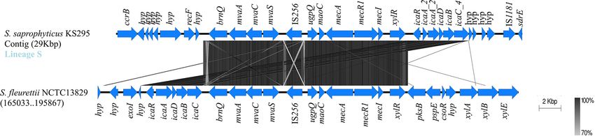
Lawal et al.                                                                                                            Biofilm Composition in S. saprophyticus

                                             biofilms was distinct in environmental and clinical isolates, suggesting that modulation
                                             of biofilm structure could be a key step in the pathogenicity of these bacteria. Moreover,
                                             biofilm production in S. saprophyticus is ica-independent, and the complete icaADBCR
                                             was acquired from other staphylococci.
                                             Keywords: Staphylococcus saprophyticus, evolution, pan-GWAS, WGS, biofilm structure, ica cluster, urinary tract
                                             infection

INTRODUCTION                                                                        Bacterial cells within the biofilm matrix exhibit phenotypic
                                                                                    characteristics different from those of planktonic or free-living
Staphylococcus saprophyticus is a uropathogen associated with                       cells (Martins et al., 2019). For instance, free-living bacteria cells
10–20% of urinary tract infection (UTI) in sexually active young                    that are susceptible to antibiotics sometimes become resistant or
women worldwide (Raz et al., 2005; Kline and Lewis, 2016).                          tolerant to such antibiotics or other antimicrobial agents in the
Possible complications such as acute pyelonephritis, urethritis                     matrix (Martins et al., 2019). In fact, S. saprophyticus biofilms
(Hovelius et al., 1984), and endocarditis (Garduño et al., 2005;                    were reported to be resistant to antibiotics used in the empirical
Choi et al., 2006), especially in immunocompromised individuals,                    treatment of UTI and to biocides used for decontamination
have been documented. S. saprophyticus is a frequent colonizer                      because of the protective function of the biofilm against the action
of the human gastrointestinal tract, cervix, urethra, vagina,                       of these agents (Fagerlund et al., 2016; Martins et al., 2019).
perineum, and rectum (Latham et al., 1983; Rupp et al., 1992).                      Additionally, biofilms constitute effective barriers against host-
Also, it colonizes the gut and skin of food-producing animals                       immune evasion and low urine pH (Becker et al., 2014; Heilmann
(Hedman et al., 1993), which could serve as a source of                             et al., 2019). Biofilm could also be a hotspot for horizontal
contamination of food-related environments.                                         gene transfer and a risk for development and dissemination of
   The success of S. saprophyticus as a uropathogen is due to its                   multidrug-resistant strains (Baker-Austin et al., 2006; Fagerlund
ability to survive in harsh and toxic conditions, which is provided                 et al., 2016; Martins et al., 2019).
by the accumulation of genetic determinants encoding high                               The composition of the biofilm matrix could be different
resistance to heavy metals (Lawal et al., 2021b) and detoxification                 between species and from strain to strain (Fagerlund et al.,
of uric acid and D-serine (Gatermann and Marre, 1989; Korte-                        2016), but the biofilm matrix is essentially composed of
Berwanger et al., 2013). Moreover, S. saprophyticus pathogenicity                   bacterial cells embedded in polysaccharides, extracellular DNA
has been described to be associated with its capacity to adhere                     (eDNA), and proteins. In staphylococcal biofilms, polysaccharide
to uroepithelial cells promoted by adhesins, surface proteins, and                  intercellular adhesin (PIA) is one of the main components.
biofilm production (Kuroda et al., 2005; Martins et al., 2019).                     PIA is a homoglycan with beta-1,6-linked N-acetylglucosamine
   Previous studies mainly done on uropathogenic Escherichia                        residues and de-N-acetylated amino groups in its composition
coli have shown that biofilm is an important pathogenicity factor                   (Mack et al., 2016). The synthesis of PIA in biofilm formation
in either medical device-associated UTI (Jacobsen et al., 2008)                     is mediated by an operon (icaADBCR). This comprises the
or cystitis (Anderson et al., 2003; Rosen et al., 2007; Blango                      N-acetylglucosamine transferase icaA that synthetizes PIA
and Mulvey, 2010). In particular, it was demonstrated in a                          oligomers from UDP-N-acetylglucosamine and the product of
mouse model of cystitis that E. coli and Klebsiella pneumoniae                      icaD, which gives optimal efficiency to icaA (Arciola et al.,
can exist in biofilm-like large aggregates of bacteria (pods or                     2015). The icaB encodes N-deacetylase and is involved in the
intracellular bacterial communities) in the bladder epithelial                      partial deacetylation of PIA. The product of icaC is involved
cells, a phenomenon that was suggested to be responsible for                        in the export of the polysaccharide, while icaR is the negative
recurrent cystitis (Anderson et al., 2003; Rosen et al., 2007; Blango               transcriptional regulator of the operon (Rohde et al., 2010;
and Mulvey, 2010). The importance of biofilm for UTI was                            Arciola et al., 2015).
additionally shown during occurrence of urinary stones/calculi                          Extracellular DNA has been described in staphylococcal
by urease-producing bacteria (Norman and Stamey, 1971;                              biofilms from Staphylococcus aureus (Eckhart et al., 2007; Izano
McLean and Nickel, 1994). Regarding S. saprophyticus, the role of                   et al., 2008) and Staphylococcus epidermidis (Qin et al., 2007;
biofilm on pathogenesis was mainly evidenced by the occurrence                      Izano et al., 2008) and was described to be part of the
of medical device-associated UTI (Hovelius et al., 1984; Choi                       biofilm of two clinical strains of S. saprophyticus (Soumya
et al., 2006; Magarifuchi et al., 2015), but it remains to be                       et al., 2017). Staphylococcal biofilms are often additionally
demonstrated whether biofilms formed by these bacteria are also                     composed of surface-associated and cell wall-anchored proteins
implicated in cystitis or urinary stones.                                           such as microbial surface components recognizing adhesive
   Biofilms are organized bacterial cell communities contained                      matrix molecules, which are essential for different stages of
in an extracellular matrix that mediate adherence to abiotic and                    attachment to surfaces and biofilm accumulation (Jönsson et al.,
biotic surfaces (Heilmann et al., 2003; Izano et al., 2008; Tojo                    1991; Patti et al., 1994). Other commonly found proteins
et al., 2009; Fagerlund et al., 2016). Biofilms play a significant                  associated with proteinaceous biofilm in staphylococci are
role in an array of infections, namely, medical device associated,                  autolysins/adhesins such as AtlE, Aap in S. epidermidis, and
valve endocarditis, and UTI (O’Gara, 2007; Becker et al., 2014).                    Bap in S. aureus (Heilmann et al., 1997; Cucarella et al., 2001;

Frontiers in Microbiology | www.frontiersin.org                                 2                                       June 2021 | Volume 12 | Article 663768
Lawal et al.                                                                                                        Biofilm Composition in S. saprophyticus

Hirschhausen et al., 2012). In spite of the clinical relevance of                 60◦ C for 60 min and stained with 0.06% crystal violet; and excess
S. saprophyticus biofilms, its composition remains unclear.                       dye was removed by washing (4×) with sterile deionized water.
    A recent study showed that S. saprophyticus causing UTI                       The plates were air-dried at room temperature. Biofilm produced
in humans belonged to two major clonal lineages (G and                            by each isolate was quantified by adding 30% acetic acid to each
S) that originated in food/production animals and humans,                         well, measured for absorbance at OD595 nm , and classified as
respectively, (Lawal et al., 2021a). However, it is still unknown if              described below (Stephanovic et al., 2007). The assay was done in
the mechanisms of disease caused by the two lineages are related.                 triplicates. S. epidermidis RP62A and TSB were used as positive
    In this study, we aimed to explore the heterogeneity in matrix                and negative controls, respectively.
composition of biofilms produced by S. saprophyticus and to
explore how biofilm phenotypes are distributed in the population
and how they correlate with genetic content.
                                                                                  Classification of Biofilm Production
                                                                                  The method of Stephanovic et al. (2007) was employed to
                                                                                  classify the biofilm production. Briefly, the average OD595 nm
MATERIALS AND METHODS                                                             of the four blank wells (TSB only) was calculated, and
                                                                                  the ODc was obtained by applying the following formula:
                                                                                  ODc = AverageOD595 nm blank + 4 ∗ Standard Deviationblank . The
Ethical Considerations
                                                                                  final optical density (OD) value of an isolate was expressed as
The human isolates were recovered as part of the routine
                                                                                  average OD value of the strain less ODc. Biofilm formation for
clinical diagnostic testing; ethical approval and informed consent
                                                                                  each test strain was classified as follows: OD ≤ ODc = no biofilm
were not required. All data were handled anonymously. Sample
                                                                                  produced; ODc < OD ≤ 2 × ODc = weak biofilm producer;
collection was in accordance with the European Parliament
                                                                                  2 × ODc < OD ≤ 4 × ODc = moderate biofilm producer; and
and Council decision for the epidemiological surveillance and
                                                                                  4 × ODc < OD = strong biofilm producer.
control of communicable disease in the European community1 .
Slaughterhouse samples were part of the routine control practices
for evaluation of good hygiene practices and programs to assure                   Biofilm Detachment Assay
meat safety (CE No. 853/2004).                                                    We used three biofilm-detaching agents previously described
                                                                                  (Fagerlund et al., 2016; Sugimoto et al., 2018; Tasse et al., 2018),
Bacterial Collection                                                              namely, Proteinase K (100 µg/ml, Sigma-Aldrich, St. Louis,
We assembled a large collection of 422 Staphylococcus                             United States), DNAse I (100 µg/ml, Sigma-Aldrich, St. Louis,
saprophyticus isolates recovered in seven countries from                          United States), and sodium periodate (50 mM, Sigma-Aldrich,
human infection and colonization as well as food-related                          St. Louis, United States; prepared in sodium acetate buffer),
environment between 1997 and 2017 (Supplementary Table 1).                        which disperse biofilm matrix composed of protein, eDNA and
Out of a total number of biofilm producers (n = 384), we selected                 polysaccharide, respectively. Biofilms were grown in microtiter
63 strains with high biofilm production representing the different                plates in TSBsG for 18 h, as described above. The planktonic cells
phylogenetic clusters identified when isolates were studied by                    were removed, and the plates were washed with distilled water
single-nucleotide polymorphism (SNP) analysis (Lawal et al.,                      (4×). The disruptor was added to each well and incubated for
2021a). The selected isolates comprised isolates from both clonal                 2 h at 37◦ C. For the control wells, only buffer was added instead.
lineages G (n = 42) and S (n = 21; Supplementary Figure 1).                       The suspensions were removed; the plates were washed, heat
Selected strains were recovered from human infection and                          fixed, and stained with crystal violet as described above. Biofilm
colonization (n = 47) and food-related environment (n = 16;                       remaining after treatment with disruptors was determined by
Supplementary Table 2).                                                           comparing the test assays with their respective control. The
                                                                                  main component of the biofilms produced by each representative
Biofilm Formation Assay                                                           isolate was assessed and classified as described below. All assays
We assessed the biofilm formation capacity in 422                                 were done in triplicates.
S. saprophyticus using the modified polystyrene microtiter
plates in a static condition as previously described (Stepanović                 Biofilm Composition and Definition of
et al., 2000; Stephanovic et al., 2007). Briefly, a colony from an
overnight culture was suspended in tryptic soy broth (TSB) and
                                                                                  Biofilm Types
grown overnight at 37◦ C with aeration. The culture was adjusted                  To determine the relative biofilm composition, we compared
to 0.5 McFarland standards with TSB supplemented with 1%                          the biofilm biomass of each strain after disruption with
glucose (w/v; BDH, England; TSBsG); and each suspension                           its corresponding control (without disruptors) expressed in
was inoculated onto 96-well microtiter plates (Corning Inc.,                      percentage (%), as previously described (Kogan et al., 2006;
United States) and incubated at 37◦ C for 18 h. The free-floating                 Fagerlund et al., 2016). Isolates with >70% reduction in biofilm
planktonic bacteria from each well were removed and washed                        after treatment with specific biofilm detaching agents were
(4×) with sterile distilled water. Attached cells were heat fixed at              interpreted to be composed of the component targeted by
                                                                                  the disruptor (Fagerlund et al., 2016); isolates with 30–70%
1
 http://www.ecdc.europa.eu/en/activities/surveillance/EARS-Net/Pages/index.       reduction in biofilm after disruption were classified as partially
aspx                                                                              composed of these components; and isolates with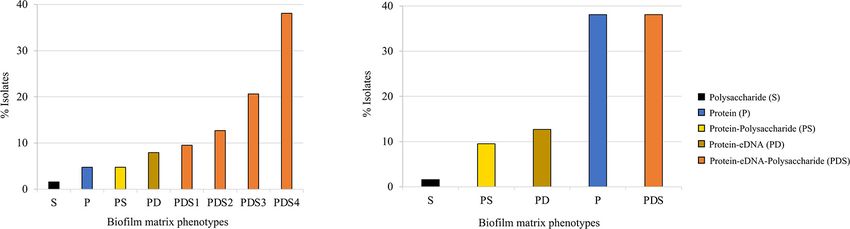
Lawal et al.                                                                                                Biofilm Composition in S. saprophyticus

reduction after disruption were considered as not containing the         Student’s t-test. Chi square was used to test the significance of the
component (Kogan et al., 2006; Fagerlund et al., 2016).                  link between S. saprophyticus biofilm phenotypes of strains from
                                                                         different lineages and origin. Statistical analyses were performed
Whole-Genome Sequencing and                                              with GraphPad Prism v6.0 (GraphPad Software, Inc., San Diego,
Assembly                                                                 CA, United States).
Paired-end sequence reads produced on an Illumina MiSeq with
an average coverage of 103 per genome reported in Lawal et al.
(2021a) with the accession number PRJNA604222 were retrieved             RESULTS
from sequence read archives. Low Q-score ends (Q < 20)
were trimmed of the Illumina reads using Trimmomatic v0.36               The Great Majority of Staphylococcus
(Bolger et al., 2014). Reads were assembled using SPAdes v3.11.1         saprophyticus Isolates Produced Biofilm
(Bankevich et al., 2012). QUAST v5.1 (Gurevich et al., 2013)             All S. saprophyticus isolates (n = 422) recovered from human
was used to evaluate the quality of assemblies. All contigs with         colonization and infection including UTI and food-related
 0.2) produced
Phylogeny Reconstruction and                                             biofilm, among which 91% (n = 349/384) were strong biofilm
Comparison                                                               producers, and 5% each (n = 18/384) were moderate and weak
Single-nucleotide polymorphism-based phylogeny of all                    biofilm producers. Among the weak (n = 18) and non-biofilm
S. saprophyticus isolates in the collection and the representative       producers (n = 36) in this collection, 83% (n = 15/18) and 64%
strains was done separately using CSIPhylogeny v1.4 (Kaas                (23/36), respectively, were recovered from human infection and
et al., 2014) with the default parameters. Maximum likelihood            dispersed in clonal lineage G.
trees were reconstructed using RAxML v8.2.12 (Stamatakis,
2014). The general time reversible model was performed with              Biofilm Composition Is Highly Diverse in
100 bootstrap resampling for node support. Phylogenetic trees            Staphylococcus saprophyticus
were re-rooted midpoint and visualized using web-based tool
                                                                         Biofilm matrix phenotype in 63 representative S. saprophyticus
microreact (Argimón et al., 2016).
                                                                         isolates (Supplementary Figure 1) was determined using biofilm
                                                                         detachment assay. Our results showed that proteinase K, when
Genome Annotation and Pangenome                                          compared with the controls, detached > 75% of the biofilm
Construction                                                             matrix formed in almost all the isolates (98%; 62/63) in this
Genomes were annotated using Prokka v1.14.6 (Seemann, 2014).             collection. The treatment of isolates’ biofilm matrix with DNAse
The pangenomes of the representative strains (n = 63) and                and sodium periodate showed that in 54% (34/63) and 46%
those of the entire collection (n = 422) were defined using              (29/63) of the isolates, respectively, >70% of the matrix was
Roary v3.13.0 (Page et al., 2015) with 85% BLASTp homologues             detached (Supplementary Figure 2), while a partial detachment
clustering with split paralogues. The accessory genomes were             (30–70% biofilm detached) was observed in 35% (22/63) and 41%
defined as the pan minus the core genome. Pangenome-wide                 (26/63), respectively, (Supplementary Figure 2). Considering
association study (pan-GWAS) approach was used to determine              that the amount of biofilm detached is directly correlated to
the association between genomic, demographic, and phenotypic             the amount of the targeted biofilm component, almost all
data using Scoary v1.6.16 (Brynildsrud et al., 2016). Genes with a       the S. saprophyticus tested produced biofilms composed of
Benjamini–Hochberg p value < 0.05 and odds ratio > 1, with no            similar amounts of proteins, while the content in eDNA and
duplicated function in the pangenome, were considered.                   polysaccharide varied from strain to strain.
                                                                            We classified the observed phenotypes into groups based
Comparative Genomic Analysis                                             on the % of reduction in biofilm biomass after detachment
The nucleotide sequences of genes of interest found in the               in comparison with the respective control. Based on this
collection were compared using blast (blastn) analysis (Altschul         classification, we found five major different biofilm matrix
et al., 1997, 2005). Contigs were reordered with MAUVE (Darling          types in S. saprophyticus population. The great majority (81%;
et al., 2004) using S. saprophyticus American Type Culture               51/63) of the isolates produced biofilm composed of protein–
Collection (ATCC) 15305 (AP008934.1) as reference. Gene                  eDNA–polysaccharide (PDS). The remaining isolates (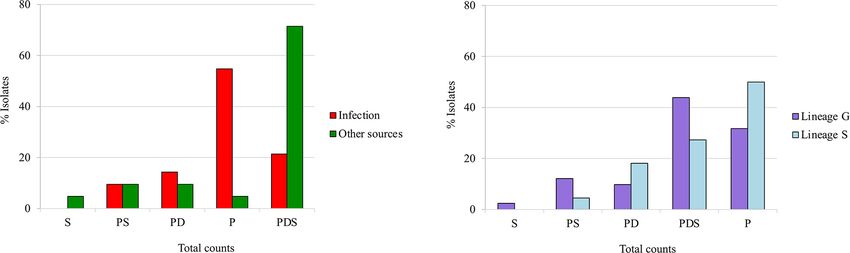
Lawal et al.                                                                                                                    Biofilm Composition in S. saprophyticus

  FIGURE 1 | Quantitative classification of preformed biofilm in 63 Staphylococcus saprophyticus strains based on matrix phenotypes. (A) Activity of biofilm-degrading
  agents, namely, proteinase K, DNase, and sodium periodate, was assessed on biofilm produced. Isolates with >70% biofilm reduction after treatment with specific
  biofilm detaching agents were interpreted to be composed of the component targeted by the disruptor, while 30–70% or
Lawal et al.                                                                                                                    Biofilm Composition in S. saprophyticus

  FIGURE 2 | Classification of the biofilm matrix phenotypes produced by Staphylococcus saprophyticus based on the (A) source and (B) genetic lineages of isolates.
  Associations of biofilm matrix phenotypes with S. saprophyticus isolates belonging to two different genetic lineages and recovered from different sources were tested
  using chi square at p < 0.05. The proportions of isolates of infection and colonization (A) and lineage G and S (B) that have a specific biofilm phenotype are shown.

  FIGURE 3 | Maximum likelihood tree of 63 Staphylococcus saprophyticus strains showing the source, genetic lineages, matrix components of biofilm, and
  distribution of virulence genes. Distribution of five main biofilm matrix phenotypes produced by S. saprophyticus including S, polysaccharide; PS,
  protein–polysaccharide; PD, protein–eDNA; P, protein; and PDS, protein–eDNA–polysaccharide is shown. Isolates marked with asterisks carried additional ica genes
  (icaA, icaD, and/or icaB). Each node represents different strains, and nodes with the same color belonged to the same lineage. The tree was constructed from the
  core-genome single-nucleotide polymorphism (SNP) alignment without recombination by RAxML using general time reversible model and 100 bootstrap values for
  node support. A comparison figure was generated using microreact.

the pangenome was constructed, and the prevalence of ica                               and reordered the contigs against the reference genome
genes was assessed.                                                                    S. saprophyticus 15305 and annotated the contigs containing ica
   The icaR gene encoding a negative transcriptional regulator                         genes. The ubiquitous icaC (icaC_1) and icaR genes were in
of the ica operon (Conlon et al., 2002) was exclusively found                          different chromosomal regions; however, both were located in the
in isolates belonging to clonal lineage G, whereas an allele                           first quarter of the genome (ATCC 15305 AP008934.1; icaC_1:
of icaC (icaC_1), a gene encoding an acetyltransferase that                            336965. . .338035; icaR: 134063...134569; Kuroda et al., 2005).
exports polysaccharide (Arciola et al., 2015), was ubiquitous in                           The icaADB was located in three different chromosomal
S. saprophyticus. The remaining ica genes, which are part of                           regions, always accompanied by an additional copy of icaC
the cluster, namely, icaA, icaD, and icaB, were found in a very                        (icaC_2, icaC_3) different from the ubiquitous allele (icaC_1).
small fraction of the population (4/422;
Lawal et al.                                                                                                                  Biofilm Composition in S. saprophyticus

  FIGURE 4 | Evidence for probable loss of ica gene cluster in Staphylococcus saprophyticus. Blocks of identical color represent nucleotide sequence homology
  between regions found in the two species. The darker-shade region depicts the highest homology, and genes are represented by arrows facing the direction of
  transcription. The icaR gene was ubiquitous in lineage G; and within this lineage, additional ica genes that were found in some of the isolates were located
  downstream. saeS gene was located in place of icaR in lineage S. Comparison figure was generated using EasyFig.

different insertion sequences (IS256 and IS1181). Although this                      (Naushad et al., 2019) and also contains icaC_1 ubiquitously and
ica-containing genomic region in S. saprophyticus resembles a                        icaR in low frequency (n = 16/57) in their genome, suggesting
SCCmec-like structure, due to the presence of the two central                        that these genes could have been transmitted during vertical
structural elements of SCCmec (mec complex A and ccrB gene;                          evolution through speciation. However, the extremely low GC
Figure 5), we could not find the inverted repeat regions and ISs                     content observed in these two genes in both S. saprophyticus
defining the boundaries of the element. These elements appear                        and S. xylosus (icaC: 28.85, 30.81%; icaR: 25.9, 27.84%) when
to be inserted in a location far apart from the characteristic                       compared with the remaining genome implies that they were
orfX chromosomal integration site (>1.5 Mb apart; see Figure 5;                      acquired from other genus into Staphylococcus.
Rolo et al., 2017). Our results suggest that the complete ica                            Among the four ica-positive strains from lineage G, two
cluster was acquired multiple times by S. saprophyticus in diverse                   carried icaADBC (icaA, icaD, icaB, and icaC_2; KS11 and KS98,
chromosomal locations.                                                               Figure 3) located downstream the ubiquitous icaR (Figure 4).
                                                                                     Like with ubiquitous icaR, the remaining ica genes (icaADBC)
                                                                                     had the closest homology with those of S. xylosus (nt id = 73–78%;
The ica Cluster From Staphylococcus                                                  Table 1). The gene organization within the clusters was similar
saprophyticus Originated From Other                                                  to the ica cluster from Staphylococcus aureus or S. epidermidis
Coagulase-Negative Staphylococci                                                     (Lerch et al., 2019). The only exception was that the icaR
The diverse ica cluster genetic environment in S. saprophyticus                      had the same transcriptional direction as the remaining ica
suggests a complex evolutionary history for this group of genes.                     genes (Figure 4).
To better understand the origin and evolution of the cluster in                          The other two ica-positive strains from lineage G that carried
S. saprophyticus, we used blast analysis (Altschul et al., 1997,                     icaADBC downstream fmhA gene appear to have a completely
2005) to find homologs of these genes and compared the gene                          different origin. They contained no icaR (icaADBC1R); and all
environment with those of related species.                                           the other ica genes (icaA_1, icaB_1, icaD_1, and icaC_3) were
   The ubiquitous icaC_1 and icaR from lineage G in                                  highly similar (nt id ≥ 98%) to those in Staphylococcus cohnii
S. saprophyticus was closely related homologous to the one                           BKAW01 (Table 1 and Supplementary Figure 4). The high
found in Staphylococcus xylosus and Staphylococcus equorum (nt                       similarity of ica genes detected in these strains with those of
id: icaC_1—87% and 84%; icaR—72% and 62%, respectively).                             S. cohnii suggests a probable recent acquisition from this species.
Furthermore, the genetic environment of ubiquitous icaC_1 in                         In fact, genes encoding a tRNA [tRNA-Asn (att)], a putative
both lineages (G and S) was very similar in terms of nucleotide                      recombinase (bin), and a plasmid replication protein (rep), all
sequence (nt id ∼ 80%) and gene synteny to the icaC region                           genes associated with mobilization and recombination, were
of S. xylosus and S. equorum, although it was inverted in these                      located downstream of the ica cluster, suggesting that these genes
species (Supplementary Figure 3 and Table 1). The fact that                          might have been exogenously acquired.
S. xylosus and S. equorum are close phylogenetic relatives of S.                         In the single isolate from lineage S that contained an entire
saprophyticus and icaC genes are within a similar chromosomal                        ica cluster (icaA_2, icaD_2, icaB_2, icaC_4, and icaR_1) in a
region implies that they could have been inherited via vertical                      SCCmec-like structure (KS295; Figures 3, 5), the ica cluster
evolution during speciation. Actually, the average nucleotide                        genes were almost identical (nt. Id ≥ 99.7%) to those found
identity between these species was identical (∼80%) to the                           in Staphylococcus fleurettii having a much lower identity with
homology observed for the genes understudy, which further                            ica genes than other staphylococcal species (S. aureus, 75–
supports the hypothesis of vertical inheritance.                                     83%; S. epidermidis, ≤67%; Staphylococcus sciuri, 73–83%; and
   Staphylococcus xylosus is one of the species most closely                         S. xylosus, ≤70%; Table 1). This is the only case in which direction
related to S. saprophyticus in the Staphylococcus phylogenetic tree                  of transcription of icaR is opposite to the remaining ica genes,

Frontiers in Microbiology | www.frontiersin.org                                  7                                            June 2021 | Volume 12 | Article 663768
Lawal et al. Biofilm Composition in S. saprophyticus
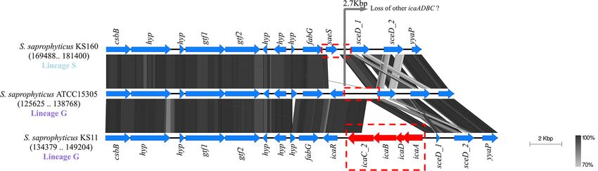
FIGURE 5 | Evidence for the probable acquisition of ica gene cluster and SCCmec-associated genes in Staphylococcus saprophyticus. Structure of ica operon
located downstream of SCCmec elements that were found in one S. saprophyticus isolate in lineage S. Blocks of identical color represent nucleotide sequence
homology between regions found in the two species. The darker-shade region depicts the highest homology, and genes are represented by arrows facing the
direction of transcription. The ica genes and the SCCmec-associated genes and the vicinity found in S. saprophyticus KS295 were compared with the closed
genome of Staphylococcus fleurettii NCTC13829. Comparison figure was generated using EasyFig.
TABLE 1 | ica genes carried by four Staphylococcus saprophyticus strains in lineage G and S and their nucleotide homology with those found in other
staphylococcal species.
Gene allele Strains (lineage) % Nucleotide identity
Staphylococcus Staphylococcus Staphylococcus Staphylococcus Staphylococcus Staphylococcus
aureus xylosus cohnii fleurettii sciuri epidermidis
icaC_1 G (326); S (n = 95) 56.0 87.0 55.0 53.0 54.0 56.0
icaR All (G) 61.0 75.0 – 56.0 56.0 61.0
icaA (group_4834) KS11, KS98 (G) 71.0 78.0 41.0 46.0 70.0 71.0
icaD 61.0 74.0 – 60.0 59.0 61.0
icaB (group_4832) 64.0 73.0 68.0 63.0 48.0 64.0
icaC_2 69.0 73.0 63.0 54.0 56.0 50.0
(group_3085)
icaA_1 KS313 (G) 71.0 78.0 99.6 67.0 70.0 71.0
icaD_1(group_4346) 60.0 64.0 98.0 59.0 58.0 59.0
icaB_1 70.0 73.0 99.0 63.0 62.0 66.0
icaC_3 67.0 71.0 99.0 63.0 61.0 68.0
(group_2023)
icaR_1 KS295 (S) 75.0 56.0 – 99.8 73.0 56.0
icaA_2 83.0 70.0 65.0 99.8 83.0 65.0
icaD_2 80.0 60.0 – 99.7 77.0 56.0
icaB_2 77.0 65.0 64.0 100.0 78.0 64.0
icaC_4 79.0 59.0 58.0 99.8 73.0 52.0
depicts genes that either were completely absent or had ≤60% coverage. Values highlighted in bold had the highest identities for each gene.
as it is described for S. aureus and S. epidermidis (Lerch et al., DISCUSSION
2019). Interestingly, in addition to ica cluster, the entire region
spanning from brnQ to xylR, including the mec complex, was In this study, we confirmed that almost all (91%) the 422 included
highly identical to the mecA region in S. fleurettii NCTC13829 S. saprophyticus isolates produced biofilm irrespective of the
(99–100% nt id; Table 2 and Figure 5). Furthermore, the gene source or genetic lineage of the isolates. This rate was higher
synteny of this region in both S. saprophyticus and S. fleurettii than that found in a previous study wherein 70% (119/169)
NCTC13829 was similar. The only exception was the position of the S. saprophyticus recovered from infection and food-
of ica gene cluster that was found upstream of the mec complex produced biofilm (Martins et al., 2019). These high rates of
and SCCmec elements in S. saprophyticus and downstream these biofilm formation in the population suggest that biofilm is
elements in S. fleurettii (Figure 5). The chromosomal location probably the main mode of living of these bacteria. Biofilm
of the mec complex in S. fleurettii, so-called the native location formation is an important step in bacterial colonization and
(approximately 200 kb apart from orfX; Rolo et al., 2017), adaptation in a variety of environments and contributes to disease
differed with the location of mec complex and ica genes in this development in the host (Speziale et al., 2014; Jeong et al.,
S. saprophyticus strain. Overall, our data suggest that ica genes 2016). Biofilm formation in this study was assessed using the
in S. saprophyticus were probably acquired from different CoNS in vitro microtiter plate assay in rich standard medium. This
species and were inserted in different chromosomal locations. does not completely mimic the in vivo scenario; and, hence
Frontiers in Microbiology | www.frontiersin.org 8 June 2021 | Volume 12 | Article 663768Lawal et al. Biofilm Composition in S. saprophyticus
TABLE 2 | SCCmec-associated genes carried by a Staphylococcus saprophyticus strain (KS295) in lineage S and their homology with those found in other
staphylococcal species.
Genes % Nucleotide identity
Staphylococcus aureus Staphylococcus fleurettii Staphylococcus sciuri Staphylococcus vitulinus
ccrB (ccrB3) 86.0 – 91.0 –
brnQ 74.0 99.9 83.0 86.0
mvaA 70.0 100.0 83.0 87.0
paaJ – 99.9 85.0 85.0
mvaS 99.7 99.9 88.0 91.0
IS256 – 99.7 79.0 –
ugpQ 100.0 – – –
maoC 100.0 – – –
mecA 99.9 100 99.9 99.9
mecR1 99.5 99.7 99.5 –
mecI 100.0 100.0 – –
xylR 99.0 99.8 99.0 –
depicts genes that either were completely absent or had ≤60% coverage. Values highlighted in bold had the highest identities for each gene.
there is the possibility for false negatives. Studies on comparison Genes that encode cell wall-anchored proteins, surface
of different techniques would be essential to complement the proteins, autolysins, and ica operon that are linked to biofilm
results obtained here. formation and matrix phenotypes were found in our collection.
The structure and composition of biofilms produced by Adhesin-encoding genes such as aas (Hell et al., 1998), ebpS
bacteria are generally maintained by various macromolecules, (Downer et al., 2002), and uafA (Kuroda et al., 2005) had been
which can vary in type and quantity among species and speculatively linked to biofilm formation in S. saprophyticus with
strains (Arciola et al., 2015; Sugimoto et al., 2018). The scarce experimental proof (Fagerlund et al., 2016). In this study,
S. saprophyticus biofilm matrix composition determined by the presence of these genes alone was not correlated with biofilm
detachment assays in this study was highly heterogeneous, formation ability or composition, suggesting that mutations with
showing at least five different phenotypes. However, the most impact in gene expression level might have an important role
common biofilm types found in this study were composed on biofilm phenotype produced, as previously described (Harris
of only protein or PDSs. Noteworthy, we found significant et al., 2016; Tasse et al., 2018). Further studies would be required
differences in biofilm matrix composition in S. saprophyticus to confirm this hypothesis. Another possibility is that the low
of the two genetic lineages as well as between strains of number of isolates included in the GWAS analysis could have
infection and colonization/environmental origin (lineage hindered the identification of statistically significant associations.
S/infection: protein; lineage G/colonization: PDS). The Among the genes associated with biofilm formation in
heterogeneity in the biofilm matrix phenotypes found in staphylococci, the ica operon, responsible for the production
S. saprophyticus population was previously described for of polysaccharide, is the most important (Rohde et al., 2010).
Staphylococcus aureus (Sugimoto et al., 2018) and S. epidermidis However, in our S. saprophyticus collection, this operon was
(Rohde et al., 2007), among others. Moreover, Tasse and rarely found (∼1%), suggesting that biofilm in this species is
colleagues have previously described the association between ica-independent. Although the complete cluster was scarce, an
biofilm matrix phenotypes and clonal lineages in S. aureus. allele of icaC gene, involved in polysaccharide export, was found
In particular, they reported that S. aureus CC5, CC15, and to be ubiquitous (Arciola et al., 2015), and icaR, a ica negative
CC30 strains produced highly eDNA-dependent biofilm, transcriptional regulator (Conlon et al., 2002), was present in
whereas that S. aureus CC45 was protein-dependent (Tasse all isolates of lineage G. In spite of their high homology with
et al., 2018). This observation might be a long-term adaptive genes within the ica cluster, these two genes, when together in
response to different environmental signals that may vary the same strain, were located far apart in the chromosome. These
between different settings, such as the presence of antibiotics, genes had high similarity (nt id ∼ 80%) with those found in
immune system, acidity, humidity, changes in temperature, and S. xylosus, a close phylogenetic relative of S. saprophyticus, within
other imbalances in the environment that may induce stress a similar chromosomal region, implying that they could have
(Rachid et al., 2000; López et al., 2010; Di Ciccio et al., 2015). been inherited via vertical evolution during speciation.
Results suggest that eradication of biofilms in infection and Besides the ubiquitous icaC/icaR genes, we additionally found
colonization should be done with different approaches. More in the genome of a few strains the complete ica cluster located
knowledge on the composition and genetic basis of biofilm in three different regions of the chromosome: downstream,
formation is important to help develop anti-biofilm strategies the ubiquitous icaR or nearby genes usually associated with
against this pathogen. mobile genetic elements like SCCmec or plasmids. While
Frontiers in Microbiology | www.frontiersin.org 9 June 2021 | Volume 12 | Article 663768Lawal et al. Biofilm Composition in S. saprophyticus
ica cluster genes located nearby the ubiquitous icaR had a low AUTHOR CONTRIBUTIONS
identity with ica genes from the closely related species S. xylosus
(∼70% nt id), ica cluster genes inserted nearby mobile genetic OL and MB performed the phenotypic experiments. OL
elements showed a high identity (>98%) with ica genes from performed the bioinformatics analysis. OL and MM carried out
other coagulase-negative staphylococci such as S. cohnii and the data analysis and interpretation and wrote the manuscript.
S. fleurettii. These results suggest that the ica cluster located MF, LG, PP, EG, CT, JE, MU, HL, HW, and MDB provided
downstream icaR was either acquired exogenously, a long time the isolates. MF, LG, PP, EG, CT, JE, MU, HL, HW, PW, and
ago, or inherited via vertical evolution during speciation and MDB were involved in manuscript revision. All authors read and
further lost from the majority of the population to avoid approved the final manuscript.
the “fitness cost” associated with polysaccharide production.
On the other hand, the other ica clusters identified appear
to have been acquired by horizontal gene transfer from
staphylococcal species. FUNDING
OL was supported by Ph.D. grant PD/BD/113992/2015
CONCLUSION from the Fundação para a Ciência e Tecnologia (FCT).
This work was partially supported by project PTDC/FIS-
In this study, we showed that there was a high variability in NAN/0117/2014, project PTDC/CVT-CVT/29510/2017, and
the composition of the biofilm formed by S. saprophyticus. project PTDC/BIAMIC/ 31645/2017; Projects LISBOA-01-0145-
The most common type of biofilm produced by this bacterium FEDER-007660 (Microbiologia Molecular, Estrutural e Celular)
contained protein or PDSs. The biofilm components appear and UID/Multi/04378/2019 funded by FEDER funds through
to differ between food-related and human infection isolates COMPETE2020—Programa Operacional Competitividade e
and between clones, suggesting that modulation of biofilm Internacionalização (POCI); ONEIDA project (LISBOA-01-
composition could be a key step in S. saprophyticus virulence 0145-FEDER-016417) co-funded by FEEI—“Fundos Europeus
and niche adaptation. Our data further showed the possible Estruturais e de Investimento” from “Programa Operacional
origin and multiple acquisition of the ica gene cluster in Regional Lisboa2020”; and national funds through FCT;
S. saprophyticus. Operacional Competitividade e Internacionalização, Programa
Operacional Regional de Lisboa (FEDER) and Fundação para a
Ciência e a Tecnologia.
DATA AVAILABILITY STATEMENT
The datasets presented in this study can be found
in online repositories that can be found in the SUPPLEMENTARY MATERIAL
article/Supplementary Material. Sequence data generated from
this study had earlier been deposited to the Sequence Reads The Supplementary Material for this article can be found
Archives under the project accession number PRJNA604222. online at: https://www.frontiersin.org/articles/10.3389/fmicb.
2021.663768/full#supplementary-material
Supplementary Figure 1 | Maximum likelihood tree of 422 S. saprophyticus
ETHICS STATEMENT constructed with core-genome single nucleotide polymorphism (SNP) highlighting
the selected strains in the population. Each node represents different strains and
The studies involving human participants were reviewed and node of the same color belonged to the same lineage. The core genome
approved by Comissão de Ética para a Investigação e Ensino alignment was constructed using CSI-Phylogeny. Recombination regions were
(CEIE) da Faculdade de Medicina Veterinária, Universidade removed using Gubbins and the phylogenetic tree reconstructed using RAxML
de Lisboa. Written informed consent for participation was with general time reversible model and 100 bootstrap value for node support.
not required for this study in accordance with the national Supplementary Figure 2 | (A) Biofilm present after treatment with biofilm
legislation and the institutional requirements. The animal degrading agents in 63 S. saprophyticus strains. Data presented are means and
study was reviewed and approved by Comissão de Ética para standard errors of the present biofilm after treatment compared to control (sodium
acetate) expressed in percentage. Assays were carried out in triplicates. (B)
a Investigação e Ensino (CEIE) da Faculdade de Medicina Biofilm present after treatment with biofilm degrading agents for 63
Veterinária, Universidade de Lisboa. Written informed consent S. saprophyticus strains. Data presented are means and standard errors after
for participation was not obtained from the owners because treatment compared to control (sodium acetate) measured at OD595 nm. Assays
Oral informed consent was obained for animal participation. were carried out in triplicates.
Slaughterhouse samples were part of the routine control practices Supplementary Figure 3 | Comparison of icaC environment in S. saprophyticus,
for evaluation of good hygiene practices and programs to assure S. xylosus, and S. equorum. Blocks of identical color represent nucleotide
meat safety (CE No. 853/2004). The sampling was done as sequence homology between regions found in the two species. Darker shade
part of the routine infection control program and no additional colored region depicts highest homology and genes are represented by arrows
facing the direction of transcription. IcaC and its vicinity found in S. saprophyticus
sampling was perfomed. The samples were non-invasive or lineages were compared with each other and with that of S. saprophyticus ATCC
minimal invasive and were the animal commensal bacteria and 15305, as well as with the closed genome of the phylogenetic relative species of
not animals that were studied. this bacterium. Comparison figure was generated using EasyFig.
Frontiers in Microbiology | www.frontiersin.org 10 June 2021 | Volume 12 | Article 663768Lawal et al. Biofilm Composition in S. saprophyticus
Supplementary Figure 4 | Structure and comparison of ica gene cluster found in the ica genes was compared with the draft genome of S. cohnii BKAW01.
a S. saprophyticus recovered from food-related environment and belonging to Comparison figure was generated using EasyFig.
lineage G. Blocks of identical color represent nucleotide sequence homology
between regions found in the two species. Darker shade colored region depicts Supplementary Table 1 | Characteristics of S. saprophyticus used in this study.
highest homology and genes are represented by arrows facing the direction of
transcription. All the ica genes including some of the genes in its vicinity are highly Supplementary Table 2 | Characteristics of S. saprophyticus isolates used for
similar (≥98% nucleotide sequence) with those found in S. cohnii. The vicinity of the biofilm detachment assay.
REFERENCES Darling, A. C. E., Mau, B., Blattner, F. R., and Perna, N. T. (2004). Mauve: Multiple
alignment of conserved genomic sequence with rearrangements. Genome Res.
Altschul, S., Madden, T., Schäffer, A., Zhang, J., Zhang, Z., Miller, W., et al. 14, 1394–1403. doi: 10.1101/gr.2289704.tion
(1997). Gapped BLAST and PSI-BLAST: a new generation of protein database Di Ciccio, P., Vergara, A., Festino, A. R., Paludi, D., Zanardi, E., Ghidini, S., et al.
search programs. Nucleic Acids Res. 25, 3389–3402. doi: 10.1371/journal.pone. (2015). Biofilm formation by Staphylococcus aureus on food contact surfaces:
0026263 Relationship with temperature and cell surface hydrophobicity. Food Control.
Altschul, S., Wootton, J., Gertz, M., Agarwala, R., Morgulis, A., Schäffer, A., et al. 50, 930–936. doi: 10.1016/j.foodcont.2014.10.048
(2005). Protein database searches using compositionally adjusted substitution Downer, R., Roche, F., Park, P. W., Mecham, R. P., and Foster, T. J. (2002).
matrices. FEBS J. 272, 5101–5109. The elastin-binding protein of Staphylococcus aureus (EbpS) is expressed
Anderson, G. G., Palermo, J. J., Schilling, J. D., Roth, R., Heuser, J., and Hultgren, at the cell surface as an integral membrane protein and not as a cell
S. J. (2003). Intracellular bacterial biofilm-like pods in urinary tract infections. wall-associated protein. J. Biol. Chem. 277, 243–250. doi: 10.1074/jbc.M1076
Science 301, 105–107. doi: 10.1126/science.1084550 21200
Arciola, C. R., Campoccia, D., Ravaioli, S., and Montanaro, L. (2015). Eckhart, L., Fischer, H., Barken, K. B., Tolker-Nielsen, T., and Tschachler, E.
Polysaccharide intercellular adhesin in biofilm: Structural and regulatory (2007). DNase1L2 suppresses biofilm formation by Pseudomonas aeruginosa
aspects. Front. Cell. Infect. Microbiol. 5:7. doi: 10.3389/fcimb.2015.00007 and Staphylococcus aureus. Br. J. Dermatol. 156, 1342–1345. doi: 10.1111/j.
Argimón, S., Abudahab, K., Goater, R. J. E., Fedosejev, A., Bhai, J., Glasner, C., 1365-2133.2007.07886.x
et al. (2016). Microreact: visualizing and sharing data for genomic epidemiology Fagerlund, A., Langsrud, S., Heir, E., Mikkelsen, M. I., and Møretrø, T. (2016).
and phylogeography. Microb. Genomics 2, 1–11. doi: 10.1099/mgen.0.00 Biofilm matrix composition affects the susceptibility of food associated
0093 staphylococci to cleaning and disinfection agents. Front. Microbiol. 7:856. doi:
Baker-Austin, C., Wright, M. S., Stepanauskas, R., and McArthur, J. V. (2006). 10.3389/fmicb.2016.00856
Co-selection of antibiotic and metal resistance. Trends Microbiol. 14, 176–182. Garduño, E., Márquez, I., Beteta, A., Said, I., Blanco, J., and Pineda, T. (2005).
doi: 10.1016/j.tim.2006.02.006 Staphylococcus saprophyticus causing native valve endocarditis. Scand. J. Infect.
Bankevich, A., Nurk, S., Antipov, D., Gurevich, A. A., Dvorkin, M., Kulikov, A. S., Dis. 37, 690–691. doi: 10.1080/00365540510027200
et al. (2012). SPAdes: a new genome assembly algorithm and its applications Gatermann, S., and Marre, R. (1989). Cloning and expression of Staphylococcus
to single-cell sequencing. J. Comput. Biol. 19, 455–477. doi: 10.1089/cmb.2012. saprophyticus urease gene sequences in Staphylococcus carnosus and
0021 contribution of the enzyme to virulence. Infect. Immun. 57, 2998–
Barros, E. M., Lemos, M., Souto-Padrón, T., and Giambiagi-deMarval, M. 3002.
(2015). Phenotypic and genotypic characterization of biofilm formation in Gurevich, A., Saveliev, V., Vyahhi, N., and Tesler, G. (2013). QUAST: Quality
Staphylococcus haemolyticus. Curr. Microbiol. 70, 829–834. doi: 10.1007/ assessment tool for genome assemblies. Bioinformatics 29, 1072–1075. doi: 10.
s00284-015-0794-x 1093/bioinformatics/btt086
Becker, K., Heilmann, C., and Peters, G. (2014). Coagulase-negative Harris, L. G., Murray, S., Pascoe, B., Bray, J., Meric, G., Magerios, L., et al.
staphylococci. Clin. Microbiol. Rev. 27, 870–926. doi: 10.1128/CMR.001 (2016). Biofilm morphotypes and population structure among Staphylococcus
09-13 epidermidis from commensal and clinical samples. PLoS One 11:e0151240. doi:
Blango, M. G., and Mulvey, M. A. (2010). Persistence of Uropathogenic Escherichia 10.1371/journal.pone.0151240
coli in the Face of Multiple Antibiotics. Antimicrob. Agents Chemother. 54, Hedman, P., Ringertz, O., Lindström, M., and Olsson, K. (1993). The origin of
1855–1863. doi: 10.1128/AAC.00014-10 Staphylococcus saprophyticus from cattle and pigs. Scand. J. Infect. Dis. 25,
Bolger, A. M., Lohse, M., and Usadel, B. (2014). Trimmomatic: A flexible 57–60. doi: 10.1080/00365549309169670
trimmer for Illumina sequence data. Bioinformatics 30, 2114–2120. doi: 10. Heilmann, C., Hussain, M., Peters, G., and Götz, F. (1997). Evidence for autolysin-
1093/bioinformatics/btu170 mediated primary attachment of Staphylococcus epidermidis to a polystyrene
Brynildsrud, O., Bohlin, J., Scheffer, L., and Eldholm, V. (2016). Erratum to: Rapid surface. Mol. Microbiol. 24, 1013–1024. doi: 10.1046/j.1365-2958.1997.
scoring of genes in microbial pan-genome-wide association studies with Scoary 4101774.x
[Genome Biol (2016), 17, 238]. Genome Biol. 17, 1–9. doi: 10.1186/s13059-016- Heilmann, C., Thumm, G., Chhatwal, G. S., Hartleib, J., Uekötter, A., and Peters,
1132-8 G. (2003). Identification and characterization of a novel autolysin (Aae) with
Carver, T., Harris, S. R., Berriman, M., Parkhill, J., and McQuillan, J. A. (2012). adhesive properties from Staphylococcus epidermidis. Microbiology 149, 2769–
Artemis: An integrated platform for visualization and analysis of high- 2778. doi: 10.1099/mic.0.26527-0
throughput sequence-based experimental data. Bioinformatics 28, 464–469. doi: Heilmann, C., Ziebuhr, W., and Becker, K. (2019). Are coagulase-negative
10.1093/bioinformatics/btr703 staphylococci virulent? Clin. Microbiol. Infect. 25, 1071–1080. doi: 10.1016/j.
Choi, S. H., Woo, J. H., Jeong, J. Y., Kim, N. J., Kim, M. N., Kim, Y. S., et al. cmi.2018.11.012
(2006). Clinical significance of Staphylococcus saprophyticus identified on blood Hell, W., Meyer, H. G. W., and Gatermann, S. G. (1998). Cloning of aas, a gene
culture in a tertiary care hospital. Diagn. Microbiol. Infect. Dis. 56, 337–339. encoding a Staphylococcus saprophyticus surface protein with adhesive and
doi: 10.1016/j.diagmicrobio.2006.08.012 autolytic properties. Mol. Microbiol. 29, 871–881. doi: 10.1046/j.1365-2958.
Conlon, K. M., Humphreys, H., and O’Gara, J. P. (2002). icaR encodes a 1998.00983.x
transcriptional repressor involved in environmental regulation of ica operon Hirschhausen, N., Schlesier, T., Peters, G., and Heilmann, C. (2012).
expression and biofilm formation in Staphylococcus epidermidis. J. Bacteriol. Characterization of the modular design of the autolysin/adhesin aaa from
184, 4400–4408. doi: 10.1128/JB.184.16.4400-4408.2002 Staphylococcus aureus. PLoS One 7:e40353. doi: 10.1371/journal.pone.0040353
Cucarella, C., Solano, C., Valle, J., Amorena, B., Lasa, I., and Penades, J. (2001). Hovelius, B., Colleen, S., and Mardh, P. A. (1984). Urinary tract infections in
Bap, a Staphylococcus aureus surface protein involved in biofilm. J. Bacteriol. men caused by Staphylococcus saprophyticus. Scand. J. Infect. Dis. 16, 37–41.
183, 2888–2896. doi: 10.1128/JB.183.9.2888 doi: 10.3109/00365548409068407
Frontiers in Microbiology | www.frontiersin.org 11 June 2021 | Volume 12 | Article 663768Lawal et al. Biofilm Composition in S. saprophyticus
Izano, E. A., Amarante, M. A., Kher, W. B., and Kaplan, J. B. (2008). Differential McLean, R. J. C., and Nickel, J. C. (1994). Glycosaminoglycans and struvite calculi.
roles of poly-N-acetylglucosamine surface polysaccharide and extracellular World J. Urol. 12, 49–51. doi: 10.1007/BF00182051
DNA in Staphylococcus aureus and Staphylococcus epidermidis biofilms. Appl. Naushad, S., Kanevets, U., Nobrega, D., Carson, D., Dufour, S., Jean-Philippe Roy,
Environ. Microbiol. 74, 470–476. doi: 10.1128/AEM.02073-07 et al. (2019). Staphylococcus debuckii sp. Nov., a coagulase-negative species from
Jacobsen, S. M., Stickler, D. J., Mobley, H. L. T., and Shirtliff, M. E. (2008). bovine milk. Int. J. Syst. Evol. Microbiol. 69, 2239–2249. doi: 10.1099/ijsem.0.
Complicated Catheter-Associated Urinary Tract Infections Due to Escherichia 003457
coli and Proteus mirabilis. Clin. Microbiol. Rev. 21, 26–59. doi: 10.1128/CMR. Norman, J., and Stamey, T. A. (1971). Surgical, bacteriological and biochemical
00019-07 management of " Infection Stones ". JAMA J. Am. Med. Assoc. 215, 1470–1476.
Jefferson, K. K. (2004). What drives bacteria to produce a biofilm? FEMS Microbiol. O’Gara, J. P. (2007). ica and beyond: Biofilm mechanisms and regulation in
Lett. 236, 163–173. doi: 10.1016/j.femsle.2004.06.005 Staphylococcus epidermidis and Staphylococcus aureus. FEMS Microbiol. Lett.
Jeong, D. W., Lee, B., Her, J. Y., Lee, K. G., and Lee, J. H. (2016). Safety and 270, 179–188. doi: 10.1111/j.1574-6968.2007.00688.x
technological characterization of coagulase-negative staphylococci isolates from Page, A. J., Cummins, C. A., Hunt, M., Wong, V. K., Reuter, S., Holden,
traditional Korean fermented soybean foods for starter development. Int. J. M. T. G., et al. (2015). Roary: Rapid large-scale prokaryote pan genome analysis.
Food Microbiol. 236, 9–16. doi: 10.1016/j.ijfoodmicro.2016.07.011 Bioinformatics 31, 3691–3693. doi: 10.1093/bioinformatics/btv421
Jönsson, K., Signäs, C., Müller, H.-P., and Lindberg, M. (1991). Two differente Patti, J. M., Allen, B. L., Mcgavin, M. J., and Hook, M. (1994). MSCRAMM -
genes encode fibronection binding proteins in Staphylococcus aureus. Eur. J. mediated adherence of microorganisms to host tissues. Annu. Rev. Microbiol.
Biochem. 202, 1041–1048. 48, 585–617.
Kaas, R. S., Leekitcharoenphon, P., Aarestrup, F. M., and Lund, O. (2014). Qin, Z., Ou, Y., Yang, L., Zhu, Y., Tolker-Nielsen, T., Molin, S., et al. (2007).
Solving the problem of comparing whole bacterial genomes across different Role of autolysin-mediated DNA release in biofilm formation of Staphylococcus
sequencing platforms. PLoS One 9:e104984. doi: 10.1371/journal.pone.01 epidermidis. Microbiology 153, 2083–2092. doi: 10.1099/mic.0.2007/006031-0
04984 Rachid, S., Ohlsen, K., Witte, W., Hacker, J., and Ziebuhr, W. (2000). Effect of
Kline, K. A., and Lewis, A. L. (2016). Gram-positive uropathogens, polymicrobial subinhibitory antibiotic concentrations on polysaccharide intercellular adhesin
urinary tract infection, and the emerging microbiota of the urinary tract. expression in biofilm-forming Staphylococcus epidermidis. Antimicrob. Agents
Microbiol. Spectr. 4, 1–31. doi: 10.1128/microbiolspec.UTI-0012-2012 Chemother. 44, 3357–3363. doi: 10.1128/AAC.44.12.3357-3363.2000
Kogan, G., Sadovskaya, I., Chaignon, P., Chokr, A., and Jabbouri, S. (2006). Raz, R., Colodner, R., and Kunin, C. M. (2005). Who are you - Staphylococcus
Biofilms of clinical strains of Staphylococcus that do not contain polysaccharide saprophyticus? Clin. Infect. Dis. 40, 896–898. doi: 10.1086/428353
intercellular adhesin. FEMS Microbiol. Lett. 255, 11–16. doi: 10.1111/j.1574- Rohde, H., Burandt, E. C., Siemssen, N., Frommelt, L., Burdelski, C., Wurster, S.,
6968.2005.00043.x et al. (2007). Polysaccharide intercellular adhesin or protein factors in biofilm
Korte-Berwanger, M., Sakinc, T., Kline, K., Nielsen, H. V., Hultgren, S., and accumulation of Staphylococcus epidermidis and Staphylococcus aureus isolated
Gatermann, S. G. (2013). Significance of the d-serine-deaminase and d-serine from prosthetic hip and knee joint infections. Biomaterials 28, 1711–1720.
metabolism of Staphylococcus saprophyticus for virulence. Infect. Immun. 81, doi: 10.1016/j.biomaterials.2006.11.046
4525–4533. doi: 10.1128/IAI.00599-13 Rohde, H., Frankenberger, S., Zähringer, U., and Mack, D. (2010). Structure,
Kuroda, M., Yamashita, A., Hirakawa, H., Kumano, M., Morikawa, K., Higashide, function and contribution of polysaccharide intercellular adhesin (PIA) to
M., et al. (2005). Whole genome sequence of Staphylococcus saprophyticus Staphylococcus epidermidis biofilm formation and pathogenesis of biomaterial-
reveals the pathogenesis of uncomplicated urinary tract infection. PNAS 102, associated infections. Eur. J. Cell Biol. 89, 103–111. doi: 10.1016/j.ejcb.2009.10.
13272–13277. doi: 10.1073/pnas.0502950102 005
Latham, R. H., Running, K., and Stamm, W. E. (1983). Urinary tract infections in Rolo, J., Worning, P., Nielsen, J., Bowden, R., Bouchami, O., Damborg, P., et al.
young adult women caused by Staphylococcus saprophyticus. J. Am. Med. Assoc. (2017). Evolutionary origin of the staphylococcal cassette chromosome mec
250, 3063–3066. doi: 10.1001/jama.1983.03340220031028 (SCCmec). Antimicrob. Agents Chemother. 61, 1–16. doi: 10.1128/AAC.023
Lawal, O. U., Fraqueza, M., Bouchami, O., Worning, P., Bartels, M., Gonçalves, M., 02-16
et al. (2021a). Foodborne Origin and Local and Global Spread of Staphylococcus Rosen, D. A., Hooton, T. M., Stamm, W. E., Humphrey, P. A., and Hultgren, S. J.
saprophyticus Causing Human Urinary Tract Infections. Emerg. Infect. Dis. 27, (2007). Detection of intracellular bacterial communities in human urinary tract
880–893. doi: 10.3201/eid2703.200852 infection. PLoS Med. 4:e329. doi: 10.1371/journal.pmed.0040329
Lawal, O. U., Fraqueza, M., Worning, P., Bouchami, O., Bartels, M., Gonçalves, M., Rupp, M. E., Soper, D. E., and Archer, G. L. (1992). Colonization of the female
et al. (2021b). Staphylococcus saprophyticus causing infections in humans are genital tract with Staphylococcus saprophyticus. J. Clin. Microbiol. 30, 2975–
associated with high resistance to heavy metals. Antimicrob. Agents Chemother 2979.
doi: 10.1128/AAC.02685-20 [Epub Online ahead of print]. Seemann, T. (2014). Prokka: Rapid prokaryotic genome annotation. Bioinformatics
Lerch, M. F., Schoenfelder, S. M. K., Marincola, G., Wencker, F. D. R., Eckart, M., 30, 2068–2069. doi: 10.1093/bioinformatics/btu153
Förstner, K. U., et al. (2019). A non-coding RNA from the intercellular adhesion Soumya, K. R., Philip, S., Sugathan, S., Mathew, J., and Radhakrishnan, E. K. (2017).
(ica) locus of Staphylococcus epidermidis controls polysaccharide intercellular Virulence factors associated with coagulase negative staphylococci isolated from
adhesion (PIA)-mediated biofilm formation. Mol. Microbiol. 111, 1571–1591. human infections. Biotechnology 7, 1–10. doi: 10.1007/s13205-017-0753-2
doi: 10.1111/mmi.14238 Speziale, P., Pietrocola, G., Foster, T. J., and Geoghegan, J. A. (2014). Protein-
López, D., Vlamakis, H., and Kolter, R. (2010). Biofilms. Cold Spring Harb. Perspect based biofilm matrices in staphylococci. Front. Cell. Infect. Microbiol. 4:171.
Biol. 2, 1–11. doi: 10.3389/fcimb.2014.00171
Mack, D., Egge, H., Krokotsch, A., Fischer, W., Leopold, K., Laufs, R., et al. (2016). Stamatakis, A. (2014). RAxML version 8: A tool for phylogenetic analysis and
The intercellular adhesin involved in biofilm accumulation of Staphylococcus post-analysis of large phylogenies. Bioinformatics 30, 1312–1313. doi: 10.1093/
epidermidis is a linear beta-1,6-linked glucosaminoglycan: purification and bioinformatics/btu033
structural analysis. J. Bacteriol. 178, 175–183. doi: 10.1128/jb.178.1.175-183. Stepanović, S., Vuković, D., Dakić, I., Savić, B., and Švabić-Vlahović, M. (2000).
1996 A modified microtiter-plate test for quantification of staphylococcal biofilm
Magarifuchi, H., Kusaba, K., Yamakuchi, H., Hamada, Y., Urakami, T., and Aoki, formation. J. Microbiol. Methods 40, 175–179. doi: 10.1016/S0167-7012(00)
Y. (2015). Staphylococcus saprophyticus native valve endocarditis in a diabetic 00122-6
patient with neurogenic bladder: A case report. J. Infect. Chemother. 21, 695– Stephanovic, S., Vukovic, D., Hola, V., Bonaventura, G. D., Djukic, S., Cirkovic,
699. doi: 10.1016/j.jiac.2015.05.008 I., et al. (2007). Quantification of biofilm in microtiter plates: Overview of
Martins, K. B., Ferreira, A. M., Pereira, V. C., Pinheiro, L., De Oliveira, A., testing conditions and practical recommendations for assessment of biofilm
De Lourdes Ribeiro, et al. (2019). In vitro effects of antimicrobial agents on production by staphylococci. APMIS 115, 891–899. doi: 10.1111/j.1600-0463.
planktonic and biofilm forms of Staphylococcus saprophyticus isolated from 2007.apm_630.x
patients with urinary tract infections. Front. Microbiol. 10:40. doi: 10.3389/ Sugimoto, S., Sato, F., Miyakawa, R., Chiba, A., Onodera, S., Hori, S., et al. (2018).
fmicb.2019.00040 Broad impact of extracellular DNA on biofilm formation by clinically isolated
Frontiers in Microbiology | www.frontiersin.org 12 June 2021 | Volume 12 | Article 663768You can also read