Aspergillus niger Spores Are Highly Resistant to Space Radiation - DLR
←
→
Page content transcription
If your browser does not render page correctly, please read the page content below
ORIGINAL RESEARCH
                                                                                                                                             published: 03 April 2020
                                                                                                                                     doi: 10.3389/fmicb.2020.00560

                                             Aspergillus niger Spores Are Highly
                                             Resistant to Space Radiation
                                             Marta Cortesão 1* , Aram de Haas 1 , Rebecca Unterbusch 1 , Akira Fujimori 2 ,
                                             Tabea Schütze 3 , Vera Meyer 3 and Ralf Moeller 1
                                             1
                                              Space Microbiology Research Group, Radiation Biology Department, Institute of Aerospace Medicine, German Aerospace
                                             Center, Cologne, Germany, 2 Department of Basic Medical Sciences for Radiation Damages, National Institutes for Quantum
                                             and Radiological Science and Technology, Chiba, Japan, 3 Chair of Applied and Molecular Microbiology, Institute
                                             of Biotechnology, Technische Universität Berlin, Berlin, Germany

                                             The filamentous fungus Aspergillus niger is one of the main contaminants of the
                                             International Space Station (ISS). It forms highly pigmented, airborne spores that have
                                             thick cell walls and low metabolic activity, enabling them to withstand harsh conditions
                                             and colonize spacecraft surfaces. Whether A. niger spores are resistant to space
                          Edited by:         radiation, and to what extent, is not yet known. In this study, spore suspensions of
                     André Antunes,
          Macau University of Science
                                             a wild-type and three mutant strains (with defects in pigmentation, DNA repair, and
              and Technology, China          polar growth control) were exposed to X-rays, cosmic radiation (helium- and iron-ions)
                       Reviewed by:          and UV-C (254 nm). To assess the level of resistance and survival limits of fungal
                    Douglas Galante,
                                             spores in a long-term interplanetary mission scenario, we tested radiation doses up to
         National Center for Research
    in Energy and Materials (CNPEM),         1000 Gy and 4000 J/m2 . For comparison, a 360-day round-trip to Mars yields a dose of
                                 Brazil      0.66 ± 0.12 Gy. Overall, wild-type spores of A. niger were able to withstand high doses
                  Marta Filipa Simões,
          Macau University of Science
                                             of X-ray (LD90 = 360 Gy) and cosmic radiation (helium-ion LD90 = 500 Gy; and iron-
               and Technology, China         ion LD90 = 100 Gy). Drying the spores before irradiation made them more susceptible
                 *Correspondence:            toward X-ray radiation. Notably, A. niger spores are highly resistant to UV-C radiation
                      Marta Cortesão
                                             (LD90 = 1038 J/m2 ), which is significantly higher than that of other radiation-resistant
                marta.cortesao@dlr.de
                                             microorganisms (e.g., Deinococcus radiodurans). In all strains, UV-C treated spores
                   Specialty section:        (1000 J/m2 ) were shown to have decreased biofilm formation (81% reduction in wild-
         This article was submitted to
                                             type spores). This study suggests that A. niger spores might not be easily inactivated
                Extreme Microbiology,
               a section of the journal      by exposure to space radiation alone and that current planetary protection guidelines
             Frontiers in Microbiology       should be revisited, considering the high resistance of fungal spores.
         Received: 21 January 2020
          Accepted: 16 March 2020            Keywords: Aspergillus niger, A. niger spores, spore survival, space, radiation, X-ray, UV, international
                                             space station
           Published: 03 April 2020
                            Citation:
             Cortesão M, de Haas A,
 Unterbusch R, Fujimori A, Schütze T,
                                            INTRODUCTION
       Meyer V and Moeller R (2020)
  Aspergillus niger Spores Are Highly
                                            Radiation is the most challenging factor for life in the space environment (Horneck et al., 2010;
       Resistant to Space Radiation.        Chancellor et al., 2014). On the one hand, the Sun emits UV radiation (non-ionizing), X-rays
             Front. Microbiol. 11:560.      (ionizing electromagnetic waves) and solar flares (intense bursts of high-energy ionizing radiation)
     doi: 10.3389/fmicb.2020.00560          (Sliney, 2007). On the other hand, cosmic events such as supernova explosions or pulsars, emit

Frontiers in Microbiology | www.frontiersin.org                                   1                                               April 2020 | Volume 11 | Article 560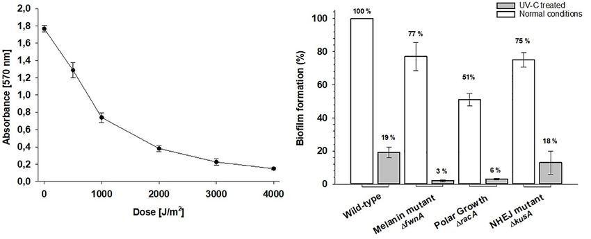
Cortesão et al.                                                                                                           Aspergillus niger Radiation

galactic cosmic radiation (GCR) (Chancellor et al., 2018). GCR               The spore cell wall is composed of polysaccharides (mainly
particle spectrum spans from light particles, such as hydrogen-              chitin and glucans), and is covered by an outer layer of
ions (85%) and helium-ions (He, 14%), to high charge Z and                   rodlets (hydrophobins) and pigments (e.g., melanin). These
energy particles (HZE) like iron-ions (Fe, 0.03%) (Horneck et al.,           outer layers of the cell wall make spores highly hydrophobic
2010). Radiation shielding on the International Space Station                and highly pigmented (Beauvais et al., 2014). Pigments, such
(ISS) is provided by both the Earth’s magnetosphere and the                  as melanin, are known to be involved in different cellular
walls of the space station. However, not all types of radiation              processes, from adhesion to virulence, as well as to protect
are easily shielded. For instance, HZE particles are still capable           cells from radiation-induced stress and ROS (Cockell and
of penetrating current space vehicles (Chancellor et al., 2018).             Knowland, 1999; Eisenman and Casadevall, 2012; Cordero
Protecting living systems from radiation becomes particularly                and Casadevall, 2017). Melanized fungi have been reported in
challenging beyond low Earth orbit (LEO). Due to the absence                 Chernobyl sites (Zhdanova et al., 2000; Casadevall et al., 2017),
of Earth’s magnetosphere, space missions toward the Moon or                  and some were even found displaying increased growth after
Mars will be exposed to substantially higher radiation doses than            X-ray irradiation (Dadachova et al., 2007). Previous studies
those currently experienced on the ISS (Cucinotta et al., 2013;              have reported the presence of melanin in A. niger spores as
Chancellor et al., 2014, 2018; Narici et al., 2017). Studies on how          an adaptive trait conferring resistance toward UV-A (315–
radiation affects cells have identified two main types of damage:            400) (Singaravelan et al., 2008). Moreover, studies on clinical
direct and indirect. Direct damage targets DNA (e.g., single-                isolates of Aspergillus fumigatus reported the involvement of
or double-strand breaks), proteins, or lipids. Whereas indirect              DHN-melanin in UV-C protection. Here, loss of a polyketide
damage is induced by the generation of reactive oxygen species               synthase from the DHN-melanin pathway (1pksP) resulted
(ROS) – which are produced by the interaction of radiation with              in decreased survival, when exposed to 100 J/m2 . This was
cellular water molecules in a process called radiolysis (Cadet et al.,       not the case for an A. fumigatus strain isolated from the ISS,
2015; Moeller et al., 2017).                                                 where loss of pksP did neither reduce nor increase viability
    Regardless of the damage, many microorganisms, especially                (Blachowicz et al., 2020). However, a recent review emphasizes
spore formers, are able to withstand high radiation doses                    that pigment biosynthesis in Aspergillus species is not yet fully
(Horneck et al., 2010; Moeller et al., 2010). Spores of the                  understood (Chang et al., 2020). Pigmentation in A. niger
bacterium Bacillus subtilis are known to be highly resistant                 is particularly puzzling. The pigment spectrum of A. niger
to extreme space conditions and therefore are currently being                spores was shown to have two main absorbance peaks, which
used as indicators for decontamination protocols and planetary               together absorb light in the entire VIS-spectrum and thus
protection policies (Kminek et al., 2019). However, while survival           result in the black color. These are thought to be two distinct
of bacterial spores has been extensively studied in both Earth and           components – one green (peak at ∼575 nm), and one brown
spaceflight contexts (Moeller et al., 2014; Setlow, 2014; Khodadad           component (∼425 nm) – and were both shown to be FwnA
et al., 2017), survival of fungal spores has not. Samples from               dependent (Jorgensen et al., 2011). FwnA is an ortholog of
the ISS indoor microbiome identified Aspergillus niger as one                pksP, and deletion of the fwnA gene (1fwnA) results in fawn-
of the most common fungal contaminants (Novikova et al.,                     colored (not white as for A. fumigatus) spores. Knowing if
2006; Checinska et al., 2015). Contrary to B. subtilis spores,               and how pigmentation is involved in spore resistance will be
which are formed as a response to stressful conditions, asexual              crucial to understand the limits of spore survival, which will,
spores of A. niger (i.e., conidia) are produced as a natural part            in turn, help develop adequate decontamination approaches.
of its life cycle (Krijgsheld et al., 2013). A. niger spores are             Another important component in fungal spore resistance is
highly pigmented and can be easily dispersed through the air                 DNA repair. When damage occurs, several pathways can be
which facilitates habitat colonization. Also, as an opportunistic            activated: nucleotide excision repair (NER), mismatch repair
human pathogen, inhalation of A. niger spores can lead to                    (MMR), homologous recombination (HR), or non-homologous
human respiratory infections (Silverman et al., 1967; Latgé, 1999;           end-joining (NHEJ) recombination (Sinha and Hader, 2002).
Esbelin et al., 2013). Thus, the ability of A. niger to survive              A. niger strains deficient in NHEJ (1kusA) are widely used
and grow in the spaceflight environment is a potential threat                to generate mutant strains, but it was also shown that
to both astronaut health and spacecraft safety. Nonetheless,                 these strains are more sensitive to UV and X-ray irradiation
A. niger is also a well-established cell factory used in modern-             (Meyer et al., 2007).
day biotechnology to produce various compounds such as                          When considering fungal contamination in indoor habitats,
proteins, enzymes, and pharmaceuticals (Meyer et al., 2015;                  the ability to colonize is not only dependent on spore survival,
Cairns et al., 2018). This makes A. niger a potential asset in               but also on the ability for spores to adhere to a surface,
long-term space missions, where astronauts will have to produce              germinate, and grow. Germination and hyphal growth are
their own compounds of interest such as vitamins or antibiotics              established through polarized growth (Kwon et al., 2011, 2013).
(Cortesão et al., 2020).                                                     In A. niger, the stabilization of polarity axes during germination
    Despite the relevance of A. niger spores in the space context,           is dependent on the Rho GTPase RacA. A racA deletion
it is not yet known whether they are able to withstand extreme               displays a hyperbranching phenotype which results in compact
space radiation conditions. Fungal spore survival generally                  colonies (Kwon et al., 2011). Spore adhesion to surfaces is
depends on two main components. One is the spore cell                        facilitated by proteins in the cell wall that help fungi to grow
wall, which helps to prevent radiation damage on the DNA.                    on a wide-range of substrates (e.g., from quartz used on

Frontiers in Microbiology | www.frontiersin.org                          2                                        April 2020 | Volume 11 | Article 560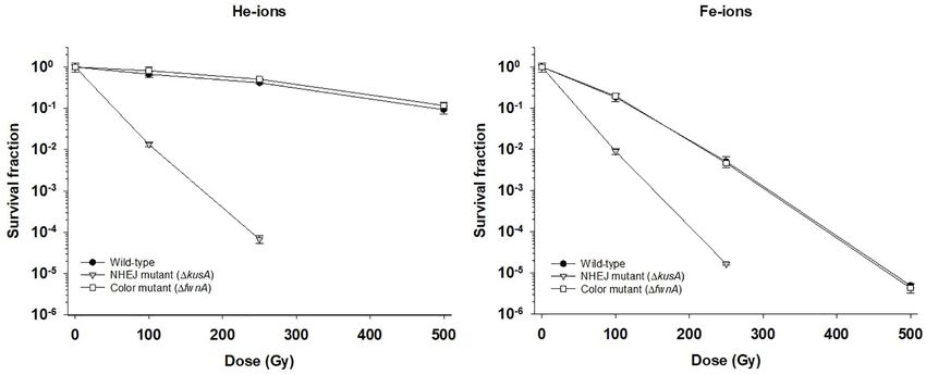
Cortesão et al.                                                                                                          Aspergillus niger Radiation

windows to silicone and polycarbonate used in medical/scientific           Figure 1). Radiation exposure was performed using the RS225
instruments) (Makimura et al., 2001; Mora et al., 2016, 2019).             X-ray machine (Gulmay Medical Systems, Camberley, Surrey,
Furthermore, fungal growth is surface-associated, which can                United Kingdom) operated without filter at 200 kV and 15 mA
induce biocorrosion. In fact, fungal-induced biocorrosion has              which allowed exposure of high doses in a short amount of
led to major problems in spacecraft safety such as those in                time. Dose rate, in Gy/min, was determined using the UNIDOS
the Mir space station (Klintworth and Reher, 1999; Novikova                webline with an ionization chamber type TM30013 (PTW,
et al., 2006). Understanding whether polar growth impacts                  Freiburg, Germany). For each desired dose, the sample exposure
spore revival and subsequent surface-associated growth is                  time was adjusted given that distance and dose rate were kept
important to better control fungal contamination in the                    constant. X-ray exposure time was calculated as follows:
spaceflight context.
   For these reasons, understanding whether A. niger spores                                                    R (Gy)
                                                                                                t (min) =
resist to space radiation, and to what extent, will be crucial to                                            d (Gy/min)
assess both the risks and opportunities of fungal spore survival
during space travel. This study has assessed A. niger spore survival       where t = time (in minutes); R = desired radiation dose (in Gy),
to different types of space radiation (X-rays, cosmic radiation,           d (dosimeter value in Gy/min). Samples were exposed to 50,
and UV-C). Three mutant strains were included to elucidate                 100, 250, 500, and 1000 Gy. Given that the average dose rate
the role of pigmentation (1fwnA) and DNA repair (1kusA) on                 was ∼20 Gy/min, the maximum time a sample was exposed to
spore resistance to radiation. In addition, high radiation doses           X-ray radiation was 50 min (corresponding to 1000 Gy). To test
were tested to assess the limits of resistance of fungal spores            irradiation of dried spores, 25 µl of a spore suspension (to a total
and their survival potential during long-term space travel. In             of 107 spores per PCR tube) either in water (H2 O) or in saline
addition, a fourth strain deficient in polar growth (1racA) was            solution (0.9% NaCl) was placed in PCR tubes, which were left
tested to assess the impact of UV-C treatment in spore revival             to air-dry overnight on the bench (22◦ C) before irradiation. After
and consequent ability for surface colonization.                           irradiation, the spores were suspended in 100 µl of water (H2 O)
                                                                           or saline solution (0.9% NaCl). Radiation exposure included at
                                                                           least three biological replicates per strain and was performed two
MATERIALS AND METHODS                                                      independent times (n = 6). Viability was determined by colony
                                                                           forming units (CFUs) (see section “Viability Assay”).
Strains and Media
Aspergillus niger wild-type (N402) and three mutant strains                Cosmic Radiation Exposure
with defects in pigmentation (1fnwA), DNA repair (1kusA),                  Spores were exposed to helium- and iron-ions (two components
and polar growth control (1racA), were used in this study                  of cosmic radiation) in PCR tubes (Brand) each filled with
and are listed in Table 1. A. niger spores were harvested from             100 µl of saline solution (0.9% NaCl) at a concentration
3-day-old cultures incubated on complete medium (CM) at                    of 107 spores/ml. PCR tubes were placed inside Petri dishes
30◦ C [55 mM glucose, 11 mM KH2 PO4 , 7 mM KCl, 178 nM                     stacked together inside plastic bags and placed directly facing
H3 BO3 , 2 mM MgSO4 , 76 nM ZnSO4 , 70 mM NaNO3 ,                          the ion beam. Samples were exposed to 10, 100, 250,
6.2 nM Na2 MoO4 , 18 nM FeSO4 , 7.1 nM CoCl2 , 6.4 nM                      and 500 Gy. Non-irradiated controls were left at room
CuSO4 , 25 nM MnCl2 , 174 nM EDTA, 0.5% (w/v) yeast extract                temperature. Viability was calculated by CFU (see section
and 0.1% (w/v) casamino acids] by flooding the agar plates                 “Viability Assay”). Radiation exposure included three biological
with saline solution (0.9% NaCl) and harvesting the spores                 replicates per strain (n = 3). Cosmic radiation exposure was
using a cotton stick. Spore suspensions were filtered using                performed at the Heavy Ion Medical Accelerator (HIMAC)
Miracloth (Millipore) to remove hyphal fragments and were                  facility at the National Institute of Radiological Sciences
kept at 4◦ C. Counting was done using a Neubauer chamber.                  (NIRS) in Chiba, Japan. The helium-ion beam had an
All experiments were performed with spore suspensions not                  energy of 150 MeV/n and linear energy transfer (LET)
older than 2 weeks. Viability assays were done using minimal               of 2.2 keV/µm. The iron-ion beam had an energy of
medium (MM) [55 mM glucose, 11 mM KH2 PO4 , 7 mM                           500 MeV/n and LET of 200 keV/µm. Each tested dose
KCl, 178 nM H3 BO3 , 2 mM MgSO4 , 76 nM ZnSO4 , 70 mM                      was adjusted by exposure time, since beam energy and LET
NaNO3 , 6.2 nM Na2 MoO4 , 18 nM FeSO4 , 7.1 nM CoCl2 ,                     was kept constant.
6.4 nM CuSO4 , 25 nM MnCl2 , 174 nM EDTA]. For MM or
CM agar plates, 15 g agar was added per liter (adapted from                UV-C Radiation Exposure
Carvalho et al., 2010).                                                    Spores of A. niger were exposed to UV radiation in Petri dishes
                                                                           with an initial concentration of 106 spores/ml in 15 ml of
X-Ray Radiation Exposure                                                   saline solution (0.9% NaCl). The concentration of 106 spores/ml
Spores of A. niger strains were exposed to X-ray radiation in PCR          ensures a spore monolayer and prevents survival due to shielding
tubes (Brand), each filled with 100 µl of saline solution (0.9%            by the spores themselves. The UV lamp (MagneTel, Menomonee
NaCl) at a concentration of 107 spores/ml. This concentration              Falls, WI, United States) was used a UV-C monochromatic
was chosen after testing the effect of initial spore concentration         wavelength of 254 nm. During irradiation, magnetic stirrers
(inoculum) on survivability toward X-rays (Supplementary                   continuously mixed the spore suspension in order to avoid

Frontiers in Microbiology | www.frontiersin.org                        3                                         April 2020 | Volume 11 | Article 560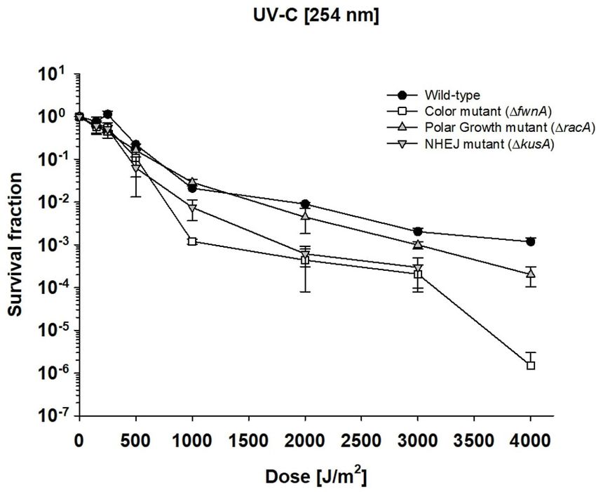
Cortesão et al.                                                                                                                               Aspergillus niger Radiation

TABLE 1 | Aspergillus niger strains used in the study.

Name                      Strain        Relevant         Description                                                                              References
                                        genotype

Wild-type                 N402                           Wild-type strain capable of DNA repair and pigment formation which give                  Bos et al., 1988
                                                         black-colored spores
Color mutant              MA93.1        1fwnA            Loss of pigment, due to lack of polyketide synthase results in fawn-colored spores       Jorgensen et al., 2011
NHEJ mutant               MA78.6        1kusA            Inactive in NHEJ pathway and thus impaired in DNA repair                                 Meyer et al., 2007
Polar growth mutant       MA80.1        1kusA,1racA      Inactive in NHEJ pathway and polar growth control                                        Kwon et al., 2011

mutual shielding of the spores. The radiation dose [J/m2 ] was                         samples and N0 that of the controls). To analyze the microscopic
adjusted through exposure time, since the height of the UV lamp                        morphology, previously irradiated samples were diluted 100-fold
was kept constant. For that, UV fluence was determined using                           before plating 10 µl on a MM agar plate. These were incubated for
the dosimeter (UVP UVX radiometer), and exposure time was                              1 day at 30◦ C, after which a Zeiss microscope (Axio Imager.M2)
adjusted for each sample to reach each desired UV dose (i.e., 150,                     was used to take images of representative areas on the agar plate
250, 500, 1000, 2000, 3000, and 4000 J/m2 ). UV-C exposure time                        for further analysis (Supplementary Figure 2). Viability assays
was calculated as follows:                                                             included three biological replicates per tested strain and were
                                                                                       performed at two independent times (n = 6).
                                     R (J/m2 ) × 100
                         t (s) =
                                       d (µW/cm2 )                                     Crystal Violet Assay
where t = time (in seconds); R = desired radiation dose (in                            To assess fungal biofilm formation, i.e., surface-associated growth
J/m2 ), d (dosimeter value for UV fluence, in µW/cm2 ). After                          with production of extracellular matrix, a crystal violet assay
each time point, corresponding to a certain dose, 100 µl of                            was performed, adapted from Mowat et al. (2007). In a 96-
sample were taken in triplicate from the spore suspension and                          well plate each well contained 100 µl spore suspension (to a
transferred into PCR tubes. Viability was calculated by CFU (see                       total concentration of 105 spores/ml per well), 100 µl MM,
section “Viability Assay”). Radiation exposure included at least                       and 100 µl of distilled H2 O (dH2 O) (the controls contained an
three biological replicates per strain and was performed two                           additional 100 µl MM instead of 100 µl spore suspension) and
independent times (n = 6).                                                             were incubated for 48 h at 30◦ C. After incubation, the wells were
                                                                                       washed three times with dH2 O, and 300 µl of crystal violet (0.5%)
Oxidative Stress Assay                                                                 were added to each well to stain the surface-associated biomass
Oxidative stress resistance toward hydrogen peroxide (H2 O2 )                          (crystal violet stains hyphae and extracellular matrix that do not
was measured using a protocol adapted from Riesenman and                               detach after washing). Excess staining was removed by washing
Nicholson (2000). Spores of A. niger were diluted to a final                           with dH2 O. De-staining was carried out by adding 300 µl of 95%
concentration of 108 spores/ml in saline solution (0.9% NaCl),                         ethanol. The absorbance of the ethanol-crystal violet solution
833 µl of which were placed in a 5 ml tube (Eppendorf).                                was measured at 570 nm. The higher the absorbance value the
Afterward 167 µl of 30% H2 O2 were added (Sigma-Aldrich).                              greater the quantity of biological material. The absorbance was
The spore-H2 O2 suspension was gently mixed at RT (∼22◦ C)                             evaluated using the VICTOR Nivo Multimode Microplate Reader
and incubated for up to 15 min in a final concentration of 5%                          (PerkinElmer, Waltham, MA, United States). The assay included
H2 O2 . 30 µl of the suspension were taken at different time                           eight biological replicates per tested strain and was performed
points and diluted 1:10 in saline solution (0.9% NaCl) with                            twice (n = 16).
bovine catalase (100 µg/ml) to stop the oxidation reaction.
Samples were serially diluted and used to determine viability (see                     Data Analysis
section “Viability Assay”). This assay included three biological                       Student’s t-test was performed to analyze the significance between
replicates per tested strain, and was performed two independent                        individual data points where a two-tailed p-value ≤ 0.05, was
times (n = 6).                                                                         considered significant. Error bars as standard error. Linear
                                                                                       regression on survival fraction data was used to calculate the
Viability Assay                                                                        lethal dose for 90% of the population (LD90 values) – which is
Viability of A. niger spores was determined by their ability to                        the same as D10 values (10% survivability).
form colonies after exposure to the tested environments. Samples
were serially diluted up to 10−8 using a 96-well plate, each well
with a total volume of 300 µl. To count the CFUs, 20 µl of each                        RESULTS
dilution was plated out in triplicate on 1/8 of a Petri dish with
MM agar, containing Triton X-100 (0.05%) to facilitate counting.                       Aspergillus niger Spore Resistance
The plates were incubated for 2 days at 30◦ C before the colonies                      Toward X-Ray Radiation
were counted. This allowed calculation of the survival fraction                        Spores from the wild-type, color mutant (1fwnA) and NHEJ
ratio (N/N0, in which N is the number of CFU of the treated                            mutant (1kusA) strains were exposed to different X-ray doses

Frontiers in Microbiology | www.frontiersin.org                                    4                                                 April 2020 | Volume 11 | Article 560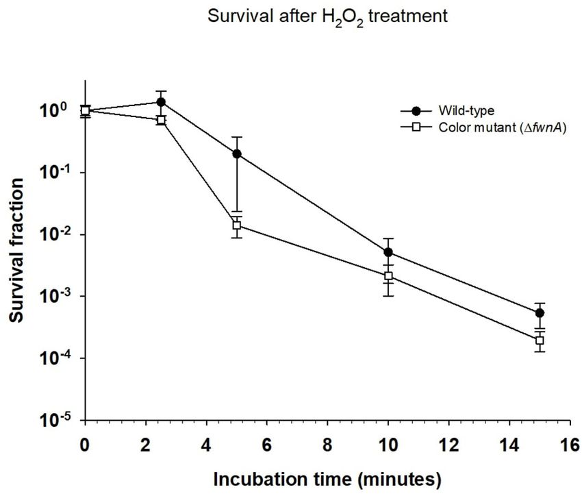
Cortesão et al. Aspergillus niger Radiation
TABLE 2 | Lethal dose (LD90 ) values for Aspergillus niger spores irradiated with X-rays under different space-relevant conditions.
Strain 0.9% NaCl H2 O Air-dried (0.9% NaCl) Air-dried (H2 O)
Wild-type 366 (R2 = 0.97) 362 (R2 = 0.96) 187 (R2 = 0.99) 204 (R2 = 0.99)
Color mutant (1fwnA) 353 (R2 = 0.98) 306 (R2 = 0.99) 175 (R2 = 0.99) 185 (R2 = 0.99)
NHEJ mutant (1kusA) 57 (R2 = 0.93) 55 (R2 = 0.92) 35 (R2 = 0.99) 45 (R2 = 0.95)
Data reported as LD90 values – dose of radiation treatment leading to a 90% inactivation of the initial CFU (same as D10 values). Values in Gray (Gy).
TABLE 3 | Lethal dose (LD90 ) values for Aspergillus niger spores irradiated with different types of ionizing radiation.
Strain X-rays (Gy) Helium-ion (Gy) Iron-ion (Gy)
Wild-type 366 (R2 = 0. 97) 506 (R2 = 0.98) 112 (R2 = 0.99)
Color mutant (1fwnA) 353 (R2 = 0.98) 567 (R2 = 0.96) 112 (R2 = 0.99)
NHEJ mutant (1kusA) 57 (R2 = 0.93) 55 (R2 = 0.99) 50 (R2 = 0.99)
Data reported as LD90 values – dose of radiation treatment leading to a 90% inactivation of the initial CFU (same as D10 values).
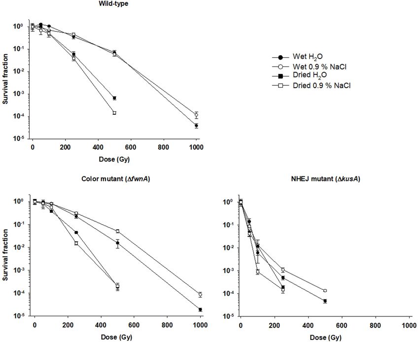
(up to 1000 Gy) in saline solution. The lethal dose required significantly more sensitive (5.2 × 10³ ± 1.2 × 10³ CFU/ml) than
to inactivate 90% of the spores was similar for both wild-type wild-type spores (4.6 × 10⁴ ± 7.2 × 10³ CFU/ml) (p = 0.002).
(LD90 = 366 Gy) and 1fwnA strains (LD90 = 353 Gy). After
the maximum tested radiation dose of 1000 Gy, the 1fwnA Aspergillus niger Spores Resistance
strain demonstrated no significant differences in survival when Toward Cosmic Radiation
compared with the wild-type (5.3 × 10³ ± 1.3 × 10³ CFU/ml, The effect of cosmic radiation on the survival of A. niger
versus 1 × 10⁴ ± 3.2 × 10³ CFU/ml). In contrast, at 500 Gy, was tested by irradiating spores with helium- and iron-
1kusA was shown to be significantly more sensitive to X-ray ions. Spores from the wild-type, color mutant (1fwnA)
radiation (1.25 × 10⁴ ± 1.1 × 10³ CFU/ml with a LD90 of 57 Gy), and NHEJ mutant (1kusA) strains were exposed to up
when compared to the wild-type (5 × 10⁶ ± 8.7 × 10⁵ CFU/ml) to 500 Gy of helium- and iron-ions. At 500 Gy, wild-
(p = 0.000). No CFU were detected above 500 Gy for 1kusA. type spores survived less when irradiated with iron-ions
Survivability and LD90 values of the strains toward X-ray (4.7 × 10² ± 6.01 × 101 CFU/ml) than when irradiated with
radiation, in all tested conditions, are shown summarized X-rays (5 × 10⁶ ± 8.7 × 10⁵ CFU/ml) (p = 0.06), or helium-
in Table 2. ions (8.7 × 10⁶ ± 1.9 × 10⁶ CFU/ml) (p = 0.01) (Table 3). The
same trend holds for 1fwnA spores at 500 Gy, where survival
toward iron-ions (4.3 × 10² ± 1.9 × 10² CFU/ml) was decreased
Air-Drying of A. niger Spores Reduces in comparison to both X-rays (3.4 × 10⁶ ± 5.4 × 10⁵ CFU/ml)
Their Resistance Toward X-Ray (p = 0.004) and helium-ions (1.2 × 10⁷ ± 2.0 × 10⁶ CFU/ml)
Radiation (p = 0.004) (Figures 1, 2 and Table 3). This is consistent
Survivability of air-dried A. niger spores was compared with with the fact that helium-ions are lighter elements in cosmic
survivability of wet spores (both in water and saline solution). radiation whereas iron-ions are heavier particles, which cause
The highest X-ray dose at which there were detectable colonies greater damage to cells (Cucinotta and Durante, 2006). At
was 500 Gy for both wild-type and 1fwnA strains, and 250 Gy 250 Gy, survival of 1kusA spores was significantly reduced when
for 1kusA. Results show that survivability of dried spores was irradiated with iron-ions (1.9 × 10³ ± 9.9 × 101 CFU/ml) than
decreased in comparison to wet spores (both in water and saline when irradiated with X-rays (4.7 × 10² ± 6.01 × 101 CFU/ml)
solution) in all tested strains (Figure 1). At 500 Gy, wild-type (p = 0.02), with no colony formation being observed above
spores in water (4.9 × 10⁶ ± 1.1 × 10⁶ CFU/ml) survive better 250 Gy of cosmic radiation (Figure 2). Exposure to helium-
than dried spores from water (4.6 × 10⁴ ± 7.2 × 10³ CFU/ml) and iron-ions (cosmic radiation) showed that both wild-type
(p = 0.001). The same is seen for spores in saline solution where and 1kusA spores were able to germinate after exposure to 250
wet spores (5 × 10⁶ ± 8.7 × 10⁵ CFU/ml) survived better than and 500 Gy, respectively (Supplementary Figure 2), suggesting
dried spores from saline solution (8.2 × 10³ ± 1.2 × 10³ CFU/m) that there is a greater level of resistance if germination would
(p = 0.000) (Table 2). When comparing water- versus saline- be considered instead of colony forming ability. Since 1fwnA
dried spores, wild-type spores dried from saline solution spores do not show reduced resistance toward X-ray radiation
(8.2 × 10³ ± 1.2 × 10³ CFU/ml) survive significantly less than (and subsequent ROS) in comparison to the wild-type strain,
spores dried from water (4.6 × 10⁴ ± 7.2 × 10³ CFU/ml) we tested whether pigmentation is involved in protecting the
(p = 0.000). Loss of pigmentation did not affect resistance to spore from H2 O2 -induced oxidative-stress by incubating the
desiccation (0 Gy) (p = 0.6); or radiation resistance (at 500 Gy) spores up to 15 min in 5% H2 O2 (Figure 3). Both the wild-type
when spores were air-dried from saline solution (p = 0.4). and color mutant strains decreased in survival when incubated
However, loss of pigmentation decreased spore survival in with H2 O2 . The survival of 1fwnA spores after 15 min was
irradiated spores dried in water: at 500 Gy, 1fwnA spores were lower than that of the wild-type, with the LD90 value for the
Frontiers in Microbiology | www.frontiersin.org 5 April 2020 | Volume 11 | Article 560Cortesão et al. Aspergillus niger Radiation
FIGURE 1 | Effect of desiccation in survival to X-ray radiation of spores of different Aspergillus niger mutant strains. Spores were irradiated in either liquid
suspensions (water or saline solution) or air-dried (from water or from saline solution). Survival fraction was calculated relatively to the non-irradiated controls.
wild-type strain (5 min) being higher than that of the 1fwnA wild-type spores (LD90 ) was 1038 J/m2 . LD90 of 1racA spores
(3.1 min) (Figure 3). was 826 J/m2 , of 1kusA spores was 580 J/m2 , and for 1fwnA
spores it was 512 J/m2 . The data also shows that deletion of racA
Aspergillus niger Spores Are Highly increases survival toward UV-C (Figure 4), in all tested doses.
Resistant Toward UV-C Radiation
To investigate the impact of UV radiation on A. niger spore Defect in Both NHEJ and Polar Growth
survivability, spore suspensions with 106 spores/ml (spore Decreases A. niger Biofilm Formation
monolayer) were exposed to 0–4000 J/m2 UV-C radiation When assessing A. niger biofilm formation (quantified as amount
(254 nm). At the highest tested dose of 4000 J/m2 , both of surface-adhered biomass detected in the well after washing), a
the wild-type (7.1 × 10² ± 4.3 × 10² CFU/ml) and polar defect in pigmentation (1fwnA) decreased biofilm formation by
growth mutant (4.2 × 101 ± 2.5 × 101 CFU/ml) demonstrated 23% (p = 0.02); a defect in the NHEJ pathway (1kusA) decreased
high survival (Figure 4). No CFU were detected for 1kusA biofilm formation by 25% (p = 0.01); and a defect in both NHEJ
at 4000 J/m2 . All tested strains were able to cope with and polar growth (1kusA, 1racA) showed a decrease of 49%
3000 J/m2 of UV-C exposure. At 3000 J/m2 wild-type spores (p = 0.007) (Figure 5B). When assessing the effect of UV-C
(1.3 × 10³ ± 6.8 × 10² CFU/ml) and 1racA spores radiation in wild-type biofilm formation, we found that biomass
(2.3 × 10² ± 1.3 × 10² CFU/ml) displayed high survivability; decreased as doses increased up to 4000 J/m2 (Figure 5A). Thus,
whereas 1fwnA spores (6.3 × 101 ± 3.7 × 101 CFU/ml) and because the UV-C LD90 value for wild-type spores was 1000 J/m2 ,
1kusA spores (1.2 × 10² ± 0.1 × 101 CFU/ml) displayed low we tested biofilm formation after treatment with 1000 J/m2 UV-
survivability. The UV-C dose required to eliminate 90% of the C for all strains. UV-C treatment led to 81% reduction in biofilm
Frontiers in Microbiology | www.frontiersin.org 6 April 2020 | Volume 11 | Article 560Cortesão et al. Aspergillus niger Radiation
FIGURE 2 | Effect of cosmic radiation (helium-ions – left; and iron-ions – right) on survival of spores of different Aspergillus niger strains. Survival fraction was
calculated relatively to the non-irradiated controls.
FIGURE 4 | Effect of UV-C radiation in survival of Aspergillus niger spores.
FIGURE 3 | Effect of incubation with H2 O2 on Aspergillus niger spore survival.
Survival fraction was calculated relative to the non-irradiated control samples.
Survival fraction was calculated relative to the non-treated (0 min) samples.
Error bars as standard error.
formation for wild-type spores (p = 0.04), 97% reduction in
of water is known to decrease radiation resistance via ROS
1fnwA spores, (p = 0.001), 82% in 1kusA spores (p = 0.001), and
formation. Nevertheless, A. niger spores have previously been
94% in 1racA1kusA spores, (p = 0.005) (Figure 5B).
shown to have increased radiation sensitivity when vacuum-dried
and irradiated in air compared to wet spores irradiated in air
(Silverman et al., 1967). However, the same study reported that
DISCUSSION vacuum-dried spores irradiated in vacuum were found to be
more resistant to radiation, which implies that space vacuum
Air Drying A. niger Spores Reduces Their desiccation might increase A. niger spore resilience (Silverman
Resistance Toward X-Ray Radiation et al., 1967). Interestingly, a study assessing the impact of water in
Spore survival toward X-rays was tested in both air-dried and radiation resistance of yeast reports that small amounts of water
liquid conditions (a comparison is provided in Table 4). Results substantially increases radiation sensitivity (Hutchinson et al.,
show that air-dried spores have lower survival rates than spores 1957). Thus, we consider the possibility that drying the spores
irradiated in liquid suspension (either in water, or in saline overnight might have not been enough to retrieve all water from
solution) (Figure 1). This result is unexpected, as the presence the spore suspension.
Frontiers in Microbiology | www.frontiersin.org 7 April 2020 | Volume 11 | Article 560Cortesão et al. Aspergillus niger Radiation
FIGURE 5 | (A) Biofilm formation of Aspergillus niger wild-type after 0 to 4000 J/m2 UV-C (254 nm). Absorbance values are proportional to the amount of detected
biomass. (B) Effect of UV-C treatment on A. niger biofilm formation. The biofilm formation is shown for normal conditions (white bars) and after treatment with
1000 J/m2 UV-C (gray bars).
TABLE 4 | Lethal dose (LD90 ) values for Aspergillus niger spores in comparison to different organisms in response to UV-C radiation (254 nm) and X-ray radiation. Values
for cells/spores in suspension (wet), irradiated in air.
Strain UV-C (J/m2 ) References X-rays (Gy) References
Aspergillus niger (spores) 1038 This study 366 This study
Bacillus subtilis (spores) 100 Newcombe et al., 2005 857 Moeller et al., 2014
Deinococcus radiodurans (cells) 656 Bauermeister et al., 2009 ca. 8000 Moseley and Laser, 1965; Driedger et al., 1970
Data reported as LD90 values – dose of radiation treatment leading to a 90% inactivation of the initial CFU (same as D10 values).
Additionally, pigmentation did not have an effect on low-LET X-ray radiation (Figure 1). This implies that radiation
desiccation resistance (at 0 Gy), which is consistent with previous with iron-ions promotes additional cellular damages besides
findings concerning the role of pigmentation in fungal survival to double-strand breaks, possibly as indirect damage in the form of
low water activity (Segers et al., 2018). When irradiating air-dried ROS generation. Multiple experiments have shown similar results
spores with 500 Gy, there was no difference between wild-type in yeasts exposed to ion radiation (Ikpeme et al., 1995; Kiefer
and color mutant for spores dried in saline. However, the color et al., 2002). Conversely, a study with the filamentous fungus
mutant had increased sensitivity when spores were dried in Neurospora crassa, showed that an NHEJ-deficient strain had
water. Thus, to fully understand the effect of desiccation (air- or ∼80% survival after 100 Gy of high-LET carbon-ion irradiation
vacuum-induced) on fungal spore resistance to extreme radiation (Ma et al., 2013), whereas the NHEJ-deficient A. niger strain
further studies are required. tested in the current study demonstrated survival of only ∼17%
after 100 Gy of low-LET helium-ion radiation (a lighter element
High-Versus Low-LET Response in A. than carbon). Nevertheless, this discrepancy in survival might be
niger Spores attributed to the fact that hyphal compartments of N. crassa can
The biological effectiveness of radiation in a given biological contain up to 100 nuclei, which in turn can lower the hit rate of
sample is dependent on the LET. This means that equal doses of radiation induced DNA damage (Roper et al., 2011).
different types of radiation can induce different types of damage,
and thus different cellular responses (Moeller et al., 2017). With High Resistance of A. niger Spores
this, previous studies with animals suggest that indirect damage Toward UV-C
is the main biological effect of low-LET radiations (such as X-rays UV-derived decontamination methods are commonly used in
or helium-ions), whereas direct damage is the main biological modern laboratories and healthcare systems (Yang et al., 2019),
effect of high-LET radiation (such as iron-ions) (Kennedy, 2014). with UV-C lamps used to sterilize biosafety cabinets reaching
In contrast, the current study suggests that direct damage, in around 300 J/m2 in 12.5 min (Meechan and Wilson, 2006). Yet, in
the form of DNA double-strand breaks, is induced by low-LET this study A. niger wild-type spores demonstrated high resistance
radiation exposure (X-rays and helium-ions), given that A. niger toward UV-C (Figure 4, and Supplementary Figure 3), where
spore survivability was highly dependent on the NHEJ pathway the lethal dose required to eliminate 90% of wild-type spores
(1kusA). Additionally, 1kusA spores showed similar survival was 1038 J/m2 . This is significantly higher than the LD90 of
rates toward both high-LET iron-ion radiation (Figure 2) and other microorganisms (Table 4), and becomes particularly clear
Frontiers in Microbiology | www.frontiersin.org 8 April 2020 | Volume 11 | Article 560Cortesão et al. Aspergillus niger Radiation
in comparison with previously characterized radiation-resistant or cosmic radiation-induced oxidative stress. Previous studies
organisms such as Deinococcus radiodurans (LD90 = 660 J/m2 ) analyzing the effect of H2 O2 on A. niger spores were able
or B. subtilis spores (LD90 = 100 J/m2 ). In a study using UV- to show that ROS scavenging was facilitated by increased
C to treat drinking water, A. niger was found to be completely catalase expression resistance (Angelova et al., 2005). From the
inactivated after exposure to 1920 J/m2 UV-C (Sisti et al., 2017), results presented here, contrary to what has been suggested,
which is in agreement with our study (LD90 = 1038 J/m2 ). pigmentation does not influence A. niger survival to space-like
The NHEJ pathway was shown to be an important DNA ionizing radiation.
repair mechanism for survival of A. niger spores after exposure
to UV-C and X-ray radiation. Interestingly, previous studies UV-C Radiation Decreases Biofilm
with NHEJ mutants of N. crassa and Cryptococcus neoformans
did not show differences in survival after UV-C irradiation Formation Effectively
when compared to the wild-type (Ninomiya et al., 2004; To address the contamination risks and possible
Goins et al., 2006). It is possible that point mutations and single- decontamination procedures concerning A. niger growth in
strand breaks caused by UV radiation can be repaired by KusA the spaceflight environment, wild-type spores were exposed
independent repair mechanisms (Eckardt-Schupp and Klaus, to up to 4000 J/m2 of UV-C and we assessed the impact of
1999; Goldman et al., 2002). However, the higher nucleus number UV-C on surface-associated growth, i.e., biofilm formation
per hyphal compartment in N. crassa, and the capsule formation (Figure 5A). Because wild-type spores were 90% inactivated
of C. neoformans spores (McFadden and Casadevall, 2001) at a dose of 1000 J/m2 , the ability for biofilm formation
may contribute to their increased radiation tolerance despite was assessed, for all strains, before and after treatment with
mutations in this repair pathway. 1000 J/m2 UV-C. Here, one additional strain, deficient in polar
growth, was included. This strain lacks the Rho GTPase RacA
involved in establishing polarized tip extension via regulation
Pigmentation as Key-Protection Against of the actin filaments, which is important for proper cell wall
UV-C but Not X-Rays or Cosmic formation in A. niger hyphae (Kwon et al., 2011, 2013). Results
Radiation show that 1racA spores ability for biofilm formation was
Pigments, such as melanin are involved in different cellular reduced by 49% before UV-C treatment, and 94% after UV-C
processes from adhesion to virulence (Eisenman and Casadevall, treatment, when compared with the wild-type (Figure 5B). The
2012), and are known to help cells against radiation-induced underlying molecular mechanism for reduced surface-associated
stress and ROS (Cockell and Knowland, 1999). Studies growth (before UV-C) might be due reduced adhesion to
concerning the role of pigmentation in radiation resistance hydrophobic surfaces, and/or results in less spore aggregation
of fungal spores have been performed with monochromatic during spore outgrowth – a hypothesis worth studying further.
UV-C (254 nm) and pulsed light up to 1770 J/m2 on inoculated The reduction of surface-associated growth after exposure to
agar with concentrations up to 107 –108 spores/ml (Esbelin et al., radiation suggests that the function of RacA plays a role (direct
2013). The current study irradiated spore suspensions in liquid, or indirect) in UV-C resistance of A. niger. Moreover, the tested
using a concentration of 106 spores/ml. This concentration color mutant (1fwnA) strain also demonstrated decreased
was chosen to guarantee a spore monolayer and prevent self- ability of surface-associated growth, both before and after
shielding (Figure 4). As expected, pigment-deficient spores UV-C treatment, which suggests the involvement of pigments
(1fwnA) demonstrated lower survivability after exposure to in spore adhesion.
UV-C radiation. Interestingly, deficiency in pigmentation did
not alter survivability after exposure to ionizing radiation
(X-rays or cosmic radiation) (Figures 1, 2). This seems CONCLUSION
to contradict previous studies where melanin was shown
to have a protective effect against X-ray irradiation in the This study shows that spores of A. niger are extremely resistant
fungi C. neoformans and Cryomyces antarcticus (Pacelli to space radiation. Spores were able to withstand high doses
et al., 2017). However, it is to note that the strain tested of X-ray (LD90 = 360 Gy), cosmic radiation (helium-ion
in the current study lacks a putative polyketide synthase LD90 = 500 Gy; and iron-ion LD90 = 100 Gy), and UV-C
which results in fawn-colored (not white) spores, which radiation (LD90 = 1038 J/m2 ). A. niger spore resistance to UV-C
might provide sufficient amount of pigmentation to display is particularly interesting, given that it is even higher than that of
protective properties. other radiation-resistant microorganisms (e.g., D. radiodurans).
Both ionizing radiation and hydrogen peroxide are known Air-drying the spores made them more susceptible to X-ray
to affect cell survival through the generation of ROS. To radiation. Moreover, wild-type spores treated with 1000 J/m2 UV-
better understand how pigment-deficient spores resist to C were shown to have decreased biofilm formation ability (81%
ionizing radiation, these were incubated in hydrogen peroxide. reduction). It is important to note that the ionizing radiation
Results show that 1fwnA spores were more sensitive to doses used in this study (up to 1000 Gy) are multiple times higher
hydrogen peroxide than wild-type spores (Figure 3). This than doses expected from traveling in interplanetary space. For
indicates that pigmentation is involved in protecting the example, a 360-day round-trip to Mars would yield a dose of
spore from H2 O2 -induced oxidative-stress, but not in X-ray- 0.66 ± 0.12 Gy (Zeitlin et al., 2013). It is therefore unlikely that
Frontiers in Microbiology | www.frontiersin.org 9 April 2020 | Volume 11 | Article 560Cortesão et al. Aspergillus niger Radiation
A. niger spores become easily inactivated due to space radiation FUNDING
alone. We thus recommend that current planetary protection
guidelines are revisited to address the high resistance of fungal RM was supported by the DLR grant FuE-Projekt “ISS LIFE”
spores in space travel scenarios. In addition, further studies are (Programm RF-FuW, Teilprogramm 475). MC was supported
needed in order to address fungal spore resistance to other space by the DLR/DAAD Research Fellowship Doctoral Studies in
environmental factors such as vacuum, changes in pressure, and Germany, 2017 (57370122). AF and RM – MEXT Grant-in-Aid
extreme temperature fluctuations. for Scientific Research on Innovative Areas “Living in Space”
(Grant Numbers: 15H05935, 15K21745).
DATA AVAILABILITY STATEMENT
ACKNOWLEDGMENTS
All datasets generated for this study are included in the
article/Supplementary Material. We thank Andrea Schröder and the HIMAC operators for their
excellent technical assistance during parts of this research.
AUTHOR CONTRIBUTIONS
SUPPLEMENTARY MATERIAL
MC, AH, and RU performed the experiments, analyzed the
data, and wrote the manuscript. VM, TS, RM, and AF The Supplementary Material for this article can be found
contributed to the conception and design of the study, and online at: https://www.frontiersin.org/articles/10.3389/fmicb.
manuscript preparation. 2020.00560/full#supplementary-material
REFERENCES health risk for exploration astronauts. NPJ Micrograv. 4:8. doi: 10.1038/s41526-
018-0043-2
Angelova, M. B., Pashova, S. B., Spasova, B. K., Vassilev, S. V., and Slokoska, Chancellor, J. C., Scott, G. B. I., and Sutton, J. P. (2014). Space radiation: the
L. S. (2005). Oxidative stress response of filamentous fungi induced by number one risk to astronaut health beyond low earth orbit. Life 4, 491–510.
hydrogen peroxide and paraquat. Mycol. Res. 109, 150–158. doi: 10.1017/ doi: 10.3390/life4030491
s0953756204001352 Chang, P.-K., Cary, J. W., and Lebar, M. D. (2020). Biosynthesis of conidial
Bauermeister, A., Bentchikou, E., Moeller, R., and Rettberg, P. (2009). Roles of and sclerotial pigments in Aspergillus species. Appl. Microbiol. Biotechnol. 104,
PprA, IrrE, and RecA in the resistance of Deinococcus radiodurans to germicidal 2277–2286. doi: 10.1007/s00253-020-10347-y
and environmentally relevant UV radiation. Arch. Microbiol. 191, 913–918. Checinska, A., Probst, A. J., Vaishampayan, P., White, J. R., Kumar, D., Stepanov,
doi: 10.1007/s00203-009-0522-7 V. G., et al. (2015). Microbiomes of the dust particles collected from the
Beauvais, A., Fontaine, T., Aimanianda, V., and Latge, J. P. (2014). Aspergillus international space station and spacecraft assembly facilities. Microbiome 3:50.
cell wall and biofilm. Mycopathologia 178, 371–377. doi: 10.1007/s11046-014- doi: 10.1186/s40168-015-0116-3
9766-0 Cockell, C. S., and Knowland, J. (1999). Ultraviolet radiation screening compounds.
Blachowicz, A., Raffa, N., Bok, J. W., Choera, T., Knox, B., Lim, F. Y., et al. Biol. Rev. Camb. Philos. Soc. 74, 311–345. doi: 10.1111/j.1469-185x.1999.
(2020). Contributions of spore secondary metabolites to UV-C protection and tb00189.x
virulence vary in different Aspergillus fumigatus Strains. mBio 11:e03415-19. Cordero, R. J., and Casadevall, A. (2017). Functions of fungal melanin beyond
doi: 10.1128/mBio.03415-19 virulence. Fungal Biol. Rev. 31, 99–112. doi: 10.1016/j.fbr.2016.12.003
Bos, C. J., Debets, A. J., Swart, K., Huybers, A., Kobus, G., and Slakhorst, S. M. Cortesão, M., Schütze, T., Marx, R., Moeller, R., and Meyer, V. (2020). “Fungal
(1988). Genetic analysis and the construction of master strains for assignment biotechnology in space: why and how?,” in Grand Challenges in Fungal
of genes to six linkage groups in Aspergillus niger. Curr. Genet. 14, 437–443. Biotechnology, ed. H. Nevalainen (Cham: Springer International Publishing),
doi: 10.1007/bf00521266 501–535. doi: 10.1007/978-3-030-29541-7_18
Cadet, J., Grand, A., and Douki, T. (2015). “Solar UV radiation-induced Cucinotta, F. A., and Durante, M. (2006). Cancer risk from exposure to galactic
DNA bipyrimidine photoproducts: formation and mechanistic insights,” cosmic rays: implications for space exploration by human beings. Lancet. Oncol.
in Photoinduced Phenomena in Nucleic Acids II: DNA Fragments and 7, 431–435. doi: 10.1016/s1470-2045(06)70695-7
Phenomenological Aspects, eds M. Barbatti, A. C. Borin, and S. Ullrich Cucinotta, F. A., Kim, M.-H. Y., Chappell, L. J., and Huff, J. L. (2013). How safe is
(Cham: Springer International Publishing), 249–275. doi: 10.1007/128_ safe enough? Radiation risk for a human mission to Mars. PLoS One 8:e74988.
2014_553 doi: 10.1371/journal.pone.0074988
Cairns, T. C., Nai, C., and Meyer, V. (2018). How a fungus shapes biotechnology: Dadachova, E., Bryan, R. A., Huang, X., Moadel, T., Schweitzer, A. D., et al. (2007).
100 years of Aspergillus niger research. Fungal. Biol. Biotechnol. 5:13. doi: 10. Ionizing radiation changes the electronic properties of melanin and enhances
1186/s40694-018-0054-5 the growth of melanized fungi. PLoS One 2:e457. doi: 10.1371/journal.pone.
Carvalho, N. D., Arentshorst, M., Jin Kwon, M., Meyer, V., and Ram, A. F. 0000457
(2010). Expanding the ku70 toolbox for filamentous fungi: establishment Driedger, A. A., James, A. P., and Grayston, M. J. (1970). Cell survival and
of complementation vectors and recipient strains for advanced gene X-ray-induced DNA degradation in Micrococcus radiodurans. Radiat. Res. 44,
analyses. Appl. Microbiol. Biotechnol. 87, 1463–1473. doi: 10.1007/s00253-010- 835–845.
2588-1 Eckardt-Schupp, F., and Klaus, C. (1999). Radiation inducible DNA repair
Casadevall, A., Cordero, R. J. B., Bryan, R., Nosanchuk, J., and Dadachova, processes in eukaryotes. Biochimie 81, 161–171. doi: 10.1016/s0300-9084(99)
E. (2017). Melanin, radiation, and energy transduction in fungi. Microbiol. 80049-2
Spectr 5, 1–6. Eisenman, H. C., and Casadevall, A. (2012). Synthesis and assembly of fungal
Chancellor, J. C., Blue, R. S., Cengel, K. A., Auñón-Chancellor, S. M., Rubins, K. H., melanin. Appl. Microbiol. Biotechnol. 93, 931–940. doi: 10.1007/s00253-011-
Katzgraber, H. G., et al. (2018). Limitations in predicting the space radiation 3777-2
Frontiers in Microbiology | www.frontiersin.org 10 April 2020 | Volume 11 | Article 560Cortesão et al. Aspergillus niger Radiation Esbelin, J., Mallea, S., Ram, A. F., and Carlin, F. (2013). Role of pigmentation niger kusA mutant. J. Biotechnol. 128, 770–775. doi: 10.1016/j.jbiotec.2006. in protecting Aspergillus niger conidiospores against pulsed light radiation. 12.021 Photochem. Photobiol. 89, 758–761. doi: 10.1111/php.12037 Meyer, V., Fiedler, M., Nitsche, B., and King, R. (2015). “The cell factory Aspergillus Goins, C. L., Gerik, K. J., and Lodge, J. K. (2006). Improvements to gene deletion enters the big data era: opportunities and challenges for optimising product in the fungal pathogen Cryptococcus neoformans: absence of Ku proteins formation,” in Filaments in Bioprocesses, eds R. Krull and T. Bley (Cham: increases homologous recombination, and co-transformation of independent Springer), 91–132. doi: 10.1007/10_2014_297 DNA molecules allows rapid complementation of deletion phenotypes. Fungal. Moeller, R., Raguse, M., Leuko, S., Berger, T., Hellweg, C. E., Fujimori, A., et al. Genet. Biol. 43, 531–544. doi: 10.1016/j.fgb.2006.02.007 (2017). STARLIFE-An international campaign to study the role of galactic Goldman, G. H., Mcguire, S. L., and Harris, S. D. (2002). The DNA damage cosmic radiation in astrobiological model systems. Astrobiology 17, 101–109. response in filamentous fungi. Fungal Genet. Biol. 35, 183–195. doi: 10.1006/ doi: 10.1089/ast.2016.1571 fgbi.2002.1344 Moeller, R., Raguse, M., Reitz, G., Okayasu, R., Li, Z., Klein, S., et al. (2014). Horneck, G., Klaus, D. M., and Mancinelli, R. L. (2010). Space microbiology. Resistance of Bacillus subtilis spore DNA to lethal ionizing radiation damage Microbiol. Mol. Biol. Rev. 74, 121–156. doi: 10.1128/MMBR.000 relies primarily on spore core components and DNA repair, with minor effects 16-09 of oxygen radical detoxification. Appl. Environ. Microbiol. 80, 104–109. doi: Hutchinson, F., Preston, A., and Vogel, B. (1957). Radiation sensitivity of enzymes 10.1128/AEM.03136-13 in wet and in dry yeast cells. Radiat. Res. 7, 465–472. Moeller, R., Reitz, G., Berger, T., Okayasu, R., Nicholson, W. L., and Horneck, Ikpeme, S., Lobrich, M., Akpa, T., Schneider, E., and Kiefer, J. (1995). Heavy G. (2010). Astrobiological aspects of the mutagenesis of cosmic radiation on ion-induced DNA double-strand breaks with yeast as a model system. Radiat. bacterial spores. Astrobiology 10, 509–521. doi: 10.1089/ast.2009.0429 Environ. Biophys. 34, 95–99. doi: 10.1007/bf01275213 Mora, M., Mahnert, A., Koskinen, K., Pausan, M. R., Oberauner-Wappis, L., Jorgensen, T. R., Park, J., Arentshorst, M., Van Welzen, A. M., Lamers, G., Vankuyk, Krause, R., et al. (2016). Microorganisms in confined habitats: microbial P. A., et al. (2011). The molecular and genetic basis of conidial pigmentation monitoring and control of intensive care units, operating rooms, cleanrooms in Aspergillus niger. Fungal Genet Biol. 48, 544–553. doi: 10.1016/j.fgb.2011. and the international space station. Front. Microbiol. 7:1573. doi: 10.3389/fmicb. 01.005 2016.01573 Kennedy, A. R. (2014). Biological effects of space radiation and development of Mora, M., Wink, L., Kögler, I., Mahnert, A., Rettberg, P., Schwendner, effective countermeasures. Life Sci. Space Res. 1, 10–43. doi: 10.1016/j.lssr.2014. P., et al. (2019). Space Station conditions are selective but do not 02.004 alter microbial characteristics relevant to human health. Nat. Commun. Khodadad, C. L., Wong, G. M., James, L. M., Thakrar, P. J., Lane, M. A., Catechis, 10:3990. J. A., et al. (2017). Stratosphere conditions inactivate bacterial endospores from Moseley, B. E., and Laser, H. (1965). Repair of X-ray in Micrococcus radiodurans. a Mars spacecraft assembly facility. Astrobiology 17, 337–350. doi: 10.1089/ast. Proc. R. Soc. Lond. B. Biol. Sci. 162, 210–222. doi: 10.1098/rspb.1965.0035 2016.1549 Mowat, E., Butcher, J., Lang, S., Williams, C., and Ramage, G. (2007). Development Kiefer, J., Egenolf, R., and Ikpeme, S. (2002). Heavy ion-induced DNA double- of a simple model for studying the effects of antifungal agents on multicellular strand breaks in yeast. Radiat. Res. 157, 141–148. doi: 10.1667/0033-7587(2002) communities of Aspergillus fumigatus. J. Med. Microbiol. 56, 1205–1212. doi: 157[0141:hiidds]2.0.co;2 10.1099/jmm.0.47247-0 Klintworth, R., and Reher, H. J. (1999). Biological induced corrosion of materials Narici, L., Casolino, M., Di Fino, L., Larosa, M., Picozza, P., Rizzo, A., et al. (2017). II: new test methods and experiences from mir station. Acta Astronaut. 44, Performances of Kevlar and Polyethylene as radiation shielding on-board the 569–578. doi: 10.1016/s0094-5765(99)00069-7 International Space Station in high latitude radiation environment. Sci. Rep. 7, Kminek, G., Fellous, J., Rettberg, P., Moissl-Eichinger, C., Sephton, M. A., Royle, 1644–1644. doi: 10.1038/s41598-017-01707-2 S. H., et al. (2019). The international planetary protection handbook. Space Res. Newcombe, D. A., Schuerger, A. C., Benardini, J. N., Dickinson, D., Tanner, R., and Today 205, e1–e120. doi: 10.1016/j.srt.2019.09.001 Venkateswaran, K. (2005). Survival of spacecraft-associated microorganisms Krijgsheld, P., Bleichrodt, R., Van Veluw, G. J., Wang, F., Müller, W. H., under simulated martian UV irradiation. Appl. Environ. Microbiol. 71, 8147– Dijksterhuis, J., et al. (2013). Development in Aspergillus. Stud. Mycol. 74, 1–29. 8156. doi: 10.1128/aem.71.12.8147-8156.2005 doi: 10.3114/sim0006 Ninomiya, Y., Suzuki, K., Ishii, C., and Inoue, H. (2004). Highly efficient gene Kwon, M. J., Arentshorst, M., Roos, E. D., Van Den Hondel, C. A., Meyer, V., and replacements in Neurospora strains deficient for nonhomologous end-joining. Ram, A. F. (2011). Functional characterization of Rho GTPases in Aspergillus Proc. Natl. Acad. Sci. U.S.A. 101:12248. doi: 10.1073/pnas.0402780101 niger uncovers conserved and diverged roles of Rho proteins within filamentous Novikova, N., De Boever, P., Poddubko, S., Deshevaya, E., Polikarpov, N., Rakova, fungi. Mol. Microbiol. 79, 1151–1167. doi: 10.1111/j.1365-2958.2010.07524.x N., et al. (2006). Survey of environmental biocontamination on board the Kwon, M. J., Nitsche, B. M., Arentshorst, M., Jorgensen, T. R., Ram, A. F., International Space Station. Res. Microbiol. 157, 5–12. doi: 10.1016/j.resmic. and Meyer, V. (2013). The transcriptomic signature of RacA activation 2005.07.010 and inactivation provides new insights into the morphogenetic network of Pacelli, C., Bryan, R. A., Onofri, S., Selbmann, L., Shuryak, I., and Dadachova, E. Aspergillus niger. PLoS One 8:e68946. doi: 10.1371/journal.pone.0068946 (2017). Melanin is effective in protecting fast and slow growing fungi from Latgé, J. P. (1999). Aspergillus fumigatus and aspergillosis. Clin. Microbiol. Rev. 12, various types of ionizing radiation. Environ. Microbiol. 19, 1612–1624. doi: 310–350. doi: 10.1128/CMR.12.2.310 10.1111/1462-2920.13681 Ma, L., Kazama, Y., Inoue, H., Abe, T., Hatakeyama, S., and Tanaka, S. (2013). The Riesenman, P. J., and Nicholson, W. L. (2000). Role of the spore coat layers in type of mutations induced by carbon-ion-beam irradiation of the filamentous Bacillus subtilis spore resistance to hydrogen peroxide, artificial UV-C. UV-B, fungus Neurospora crassa. Fungal. Biol. Biotechnol. 117, 227–238. doi: 10.1016/ and solar UV radiation. Appl. Environ. Microbiol. 66, 620–626. doi: 10.1128/ j.funbio.2013.01.002 aem.66.2.620-626.2000 Makimura, K., Hanazawa, R., Takatorl, K., Tamura, Y., Fujisaki, R., Nishiyama, Roper, M., Ellison, C., Taylor, J. W., and Glass, N. L. (2011). Nuclear and genome Y., et al. (2001). Fungal flora on board the Mir-space station, identification by dynamics in multinucleate ascomycete fungi. Curr. Biol. 21, R786–R793. doi: morphological features and ribosomal DNA sequences. Microbiol. Immunol. 45, 10.1016/j.cub.2011.06.042 357–363. doi: 10.1111/j.1348-0421.2001.tb02631.x Segers, F. J. J., Wosten, H. A. B., and Dijksterhuis, J. (2018). Aspergillus McFadden, D. C., and Casadevall, A. (2001). Capsule and melanin synthesis in niger mutants affected in conidial pigmentation do not have an increased Cryptococcus neoformans. Med. Mycol. 39(Suppl. 1), 19–30. doi: 10.1080/ susceptibility to water stress during growth at low water activity. Lett. Appl. 744118883 Microbiol. 66, 238–243. doi: 10.1111/lam.12846 Meechan, P. J., and Wilson, C. (2006). Use of ultraviolet lights in biological Setlow, P. (2014). Spore resistance properties. Microbiol. Spectr. 2, 1–14. safety cabinets: a contrarian view. Appl. Biosaf. 11, 222–227. doi: 10.1177/ Silverman, G. J., Davis, N. S., and Beecher, N. (1967). Resistivity of spores to 153567600601100412 ultraviolet and gamma radiation while exposed to ultrahigh vacuum or at Meyer, V., Arentshorst, M., El-Ghezal, A., Drews, A. C., Kooistra, R., Van Den atmospheric pressure. Appl. Microbiol. 15, 510–515. doi: 10.1128/aem.15.3.510- Hondel, C. A., et al. (2007). Highly efficient gene targeting in the Aspergillus 515.1967 Frontiers in Microbiology | www.frontiersin.org 11 April 2020 | Volume 11 | Article 560
You can also read