Substrate Stiffness Modulates the Growth, Phenotype, and Chemoresistance of Ovarian Cancer Cells
←
→
Page content transcription
If your browser does not render page correctly, please read the page content below
ORIGINAL RESEARCH
                                                                                                                                           published: 24 August 2021
                                                                                                                                      doi: 10.3389/fcell.2021.718834

                                            Substrate Stiffness Modulates the
                                            Growth, Phenotype, and
                                            Chemoresistance of Ovarian Cancer
                                            Cells
                                            Yali Fan 1† , Quanmei Sun 2* † , Xia Li 1,3 , Jiantao Feng 4 , Zhuo Ao 2 , Xiang Li 2 and
                                            Jiandong Wang 1*
                                            1
                                              Department of Gynecologic Oncology, Beijing Obstetrics and Gynecology Hospital, Capital Medical University, Beijing,
                                            China, 2 Chinese Academy of Sciences (CAS) Center for Excellence in Nanoscience, National Centre for Nanoscience
                                            and Technology, Beijing, China, 3 Hospital of Beijing Forestry University, Beijing Forestry University, Beijing, China,
                                            4
                                              Artemisinin Research Center, Institute of Chinese Materia Medica, China Academy of Chinese Medical Sciences, Beijing,
                                            China

                                            Mechanical factors in the tumor microenvironment play an important role in response
                            Edited by:
                            Jianyu Rao,     to a variety of cellular activities in cancer cells. Here, we utilized polyacrylamide
 University of California, Los Angeles,     hydrogels with varying physical parameters simulating tumor and metastatic target
                          United States
                                            tissues to investigate the effect of substrate stiffness on the growth, phenotype, and
                         Reviewed by:
                             Weibo Yu,
                                            chemotherapeutic response of ovarian cancer cells (OCCs). We found that increasing
 University of California, Los Angeles,     the substrate stiffness promoted the proliferation of SKOV-3 cells, an OCC cell line. This
                          United States     proliferation coincided with the nuclear translocation of the oncogene Yes-associated
                              Mk Chen,
    University of Texas Southwestern        protein. Additionally, we found that substrate softening promoted elements of epithelial-
       Medical Center, United States        mesenchymal transition (EMT), including mesenchymal cell shape changes, increase
                 *Correspondence:           in vimentin expression, and decrease in E-cadherin and β-catenin expression. Growing
                       Quanmei Sun
                  sunqm@nanoctr.cn
                                            evidence demonstrates that apart from contributing to cancer initiation and progression,
                     Jiandong Wang          EMT can promote chemotherapy resistance in ovarian cancer cells. Furthermore, we
       wangjiandongxy@ccmu.edu.cn           evaluated tumor response to standard chemotherapeutic drugs (cisplatin and paclitaxel)
     † These   authors have contributed
                                            and found antiproliferation effects to be directly proportional to the stiffness of the
                    equally to this work
                                            substrate. Nanomechanical studies based on atomic force microscopy (AFM) have
                    Specialty section:      revealed that chemosensitivity and chemoresistance are related to cellular mechanical
         This article was submitted to
                                            properties. The results of cellular elastic modulus measurements determined by AFM
         Cell Adhesion and Migration,
                a section of the journal    demonstrated that Young’s modulus of SKOV-3 cells grown on soft substrates was less
  Frontiers in Cell and Developmental       than that of cells grown on stiff substrates. Gene expression analysis of SKOV-3 cells
                                Biology
                                            showed that mRNA expression can be greatly affected by substrate stiffness. Finally,
           Received: 01 June 2021
           Accepted: 27 July 2021
                                            immunocytochemistry analyses revealed an increase in multidrug resistance proteins,
         Published: 24 August 2021          namely, ATP binding cassette subfamily B member 1 and member 4 (ABCB1 and
                               Citation:    ABCB4), in the cells grown on the soft gel resulting in resistance to chemotherapeutic
     Fan Y, Sun Q, Li X, Feng J, Ao Z,
                                            drugs. In conclusion, our study may help in identification of effective targets for cancer
    Li X and Wang J (2021) Substrate
      Stiffness Modulates the Growth,       therapy and improve our understanding of the mechanisms of cancer progression
    Phenotype, and Chemoresistance          and chemoresistance.
               of Ovarian Cancer Cells.
       Front. Cell Dev. Biol. 9:718834.     Keywords: ovarian cancer, substrate stiffness, Yes-associated protein, epithelial-mesenchymal transition,
       doi: 10.3389/fcell.2021.718834       chemoresistance

Frontiers in Cell and Developmental Biology | www.frontiersin.org                 1                                           August 2021 | Volume 9 | Article 718834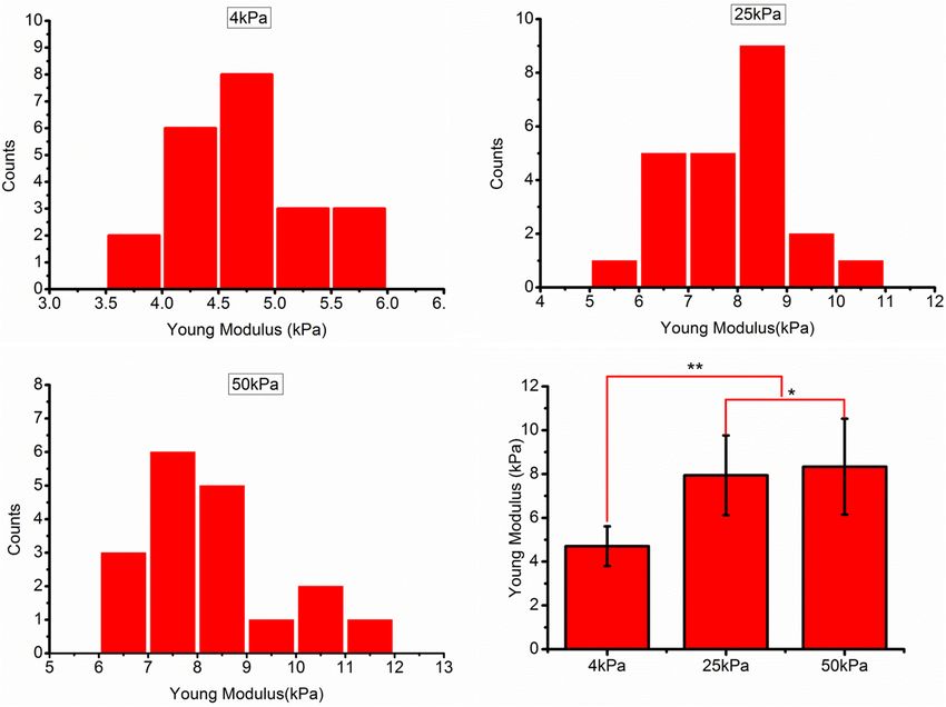
Fan et al.                                                                                             Substrate Stiffness Modulates Ovarian Cancer

INTRODUCTION                                                              (Hazlehurst et al., 2000, 2007). Mounting evidence suggests that
                                                                          not only the composition of the ECM but also its stiffness can
Epithelial ovarian cancer (EOC) is the leading cause of death             significantly affect chemoresistance (Schrader et al., 2011; Liu
among gynecological malignancies (Walker et al., 2015). This              et al., 2015). Drug resistance arises from multiple mechanisms,
poor prognosis is mainly because most patients are diagnosed              such as drug target mutations, drug metabolism, drug efflux, etc.
at a late stage and have drug resistance. The standard treatment          Recently, epithelial-mesenchymal transition (EMT) has received
of EOC is the surgical removal of the tumor tissue followed               increasing attention for its role in cancer drug resistance. Several
by chemotherapy. Cisplatin and paclitaxel represent the two               studies show that cancer cells resistant to cisplatin and/or
most widely used first-line agents for EOC. Most EOC patients             paclitaxel had acquired a mesenchymal phenotype (Kajiyama
are chemotherapy-sensitive; however, 15% experience primary               et al., 2007; Yang et al., 2014), which points to EMT as a possible
platinum resistance (Jemal et al., 2010). Many patients also              driver of resistance.
experience cancer recurrence within 2 years of initial treatment              Instead of the classic patterns of metastasis via the
due to acquired resistance to platinum-based chemotherapy.                hematogenous route and extravasation at a distal site, ovarian
Five-year survival is only 30% in advanced EOC (Mor and Alvero,           cancer cells (OCCs) often metastasize through the intraperitoneal
2013). Chemotherapy resistance is considered to be a major                fluid to the omentum, retroperitoneal lymph nodes, and even to
obstacle to the successful treatment of EOC. This highlights              the parenchyma of the liver or lungs. The prognosis of patients
the need to elucidate mechanisms that drive cancer progression            with EOC is most likely related to the degree of peritoneal
and resistance and to develop therapeutic strategies for drug-            dissemination. The omentum, which is one of the most frequent
resistant relapses.                                                       sites of ovarian cancer metastasis, is predominantly composed
   The role of tissue stiffness has been explored in various              of adipose tissue. Adipocytes are the key components of ovarian
human cancers. Many factors promote tumor tissue stiffening,              cancer TME and have been shown to stimulate the migration
including extracellular matrix (ECM) remodeling and an                    of OCCs toward omentum through secreted cytokines such as
increase in the interstitial pressure due to tumor growth and             IL-8 (Nieman et al., 2011). Omental adipocytes also have been
chaotic microvasculature (Erkan et al., 2012). ECM is primarily           shown to produce fatty acids, which can be used as an energy
composed of collagen and fibrous proteins, proteoglycans.                 source, to support OCCs proliferation. Adipocytes also influence
ECM stiffening in tumors is caused by the reorganization of               OCC chemoresistance. Activated Akt enhances the survival of
the stroma by the excessive activation of ECM enzymes and                 OCCs and promotes the chemoresistance through attenuating
proteins that covalently cross-link collagen fibers and other ECM         p53 proapoptotic signaling (Yang et al., 2006; Fraser et al., 2008).
components (Levental et al., 2009). Cells sense and respond to            Studies have demonstrated that adipocyte-secreted arachidonic
the mechanical properties of the ECM through mechanocellular              acid (AA) acts directly on OCCs to activate AKT and inhibit
systems, including focal adhesion complexes, integrins, the actin         cisplatin-induced apoptosis (Yang et al., 2019). Currently, there
cytoskeleton, and associated molecular motors. Recent reports             is a gap in our understanding of the effects of substrate stiffness
indicate that mechanical forces are also directly transmitted             on OCC chemoresistance and the driving mechanisms that
from the ECM to the nucleus by the physical anchoring of the              underlie it. In the present study, we utilized a collagen-coated
cytoskeleton to the nuclear lamina (Tajik et al., 2016). The Hippo        polyacrylamide hydrogel system with elastic properties that
pathway effector protein Yes-associated protein (YAP) is a well-          mimic those of tumor, and metastatic target tissues for OCC
known intracellular transducer of mechanical stimuli (Dupont              dissemination, to investigate the role of substrate stiffness in
et al., 2011). Studies have shown that the Hippo pathway and              OCC growth and chemotherapeutic response.
YAP not only respond to mechanical cues but are also important
mediators of cellular responses to these stimuli (Dupont et al.,
2011; Aragona et al., 2013). In addition to providing structural          MATERIALS AND METHODS
support, the ECM can regulate cellular behavior. Recently, studies
have reported that the stiffness of the ECM plays a pivotal role in       Cell Culture
tumor initiation, progression, metastasis, and therapeutic efficacy       The EOC cell line, SKOV-3 (American Type Culture Collection
(Sethi et al., 1999; Feng et al., 2013; Liu et al., 2016).                [ATCC]) was cultured in RPMI-1640 media (Gibco, Waltham,
   Cancer cells gain chemoresistance through a variety                    MA, United States) supplemented with 10% fetal bovine serum
of mechanisms. Apart from intrinsic resistance factors                    (Corning, New York, NY, United States), 100 U mL−1 penicillin,
(Galluzzi et al., 2012), tumor chemoresistance is also affected           and 100 mg mL−1 streptomycin (all from Hyclone, Logan, UT,
by the biochemical and physical properties of the tumor                   United States) in a humidified incubator at 37◦ C with 5% CO2 .
microenvironment (TME) (Ostman, 2012; Kharaishvili et al.,
2014; Wu et al., 2021). The physical components of the TME,               Proliferation and Cytotoxicity Analysis
such as high interstitial fluid pressure and densely packed               For proliferation analysis, the OCCs were seeded in 24-well
cells, hinder drug delivery (Correia and Bissell, 2012). Cancer           culture plates, coated with hydrogel substrates of different
cells can acquire chemoresistance via cell-cell and cell-ECM              stiffness (Matrigen, United States), at a concentration of 2,500
interactions (Landowski et al., 2003). When a cancer cell comes           cells/cm2 , and cultured for 6 days. Cell viability was determined
into close contact with the stromal cells or ECM, adhesion                once every 24 h. For cytotoxicity analysis, SKOV-3 cells were
induces the production of pro- and anti-apoptotic molecules               seeded in 96-well culture plates, coated with hydrogel substrates

Frontiers in Cell and Developmental Biology | www.frontiersin.org     2                                     August 2021 | Volume 9 | Article 718834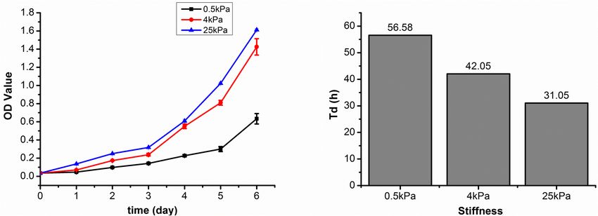
Fan et al.                                                                                              Substrate Stiffness Modulates Ovarian Cancer

of different stiffness (Matrigen, United States), at a concentration       in radioimmunoprecipitation assay (RIPA) buffer plus
of 4 × 10³ cells/well for 24 h. The cells were then treated                PhosStopTM (Biorigin, BN25015). Equal amounts of protein
with varying concentrations of paclitaxel (Sigma, United States)           were separated by gel electrophoresis and transferred onto
or cisplatin (Tokyo Chemical Industry, Japan) for 48 h. The                a polyvinylidene fluoride (PVDF) (Merck Millipore Ltd.,
concentrations of paclitaxel were 0, 10−4 , 10−3 , 10−2 , 10−1 , 1         Tullagreen, Carrigtwohill) membrane. The membrane was
µg/mL. The concentration of cisplatin were 0, 10−2 , 10−1 , 1, 5,          blocked with 5% non-fat dry milk and then incubated with
and 10 µM. After discarding the supernatant, Cell Counting Kit-            a 1:1,500 dilution of primary antibody against α-tubulin
8TM (CCK-8) (DOJINDO, Japan) working solution was added                    (DM1A-3873s, Cell Signaling Technology), paxillin (D9G12-
to each well and incubated for 3 h at 37◦ C. The absorbances at            12065, Cell Signaling Technology), vimentin (D21H3-5741,
450 nm were read using a spectrophotometer (NanoDrop, ND-                  Cell Signaling Technology), E-cadherin (24E10-3195, Cell
100, United States). Each experiment was repeated three times to           Signaling Technology), β-catenin (D10A8-8480, Cell Signaling
assess for the consistency of the results.                                 Technology), YAP (sc-101199, Santa Cruz), ABCB1 (ABP59326,
                                                                           Abbkine), or ABCB4 (ABP59247, Abbkine) overnight at
Environmental Scanning Electron                                            4◦ C. The membrane was then washed and incubated with
                                                                           a secondary peroxidase-conjugated antibody (ab-150077 or
Microscopy
                                                                           ab150113, Abcam) for 1 h after washing. Antibody binding
The SKOV-3 cells were cultured on small circular glass sheets
                                                                           was detected using an enhanced chemiluminescence detection
coated with hydrogel substrates of different stiffness (Matrigen,
                                                                           buffer from Alpha Innotech Imaging System (San Leandro, CA,
United States). The cells were cultured in this environment for
                                                                           United States). ImageJ software (Fiji-win64) was used to analyze
three days. Next, the cells were washed with phosphate-buffered
                                                                           the data. Each experiment was repeated three times to assess for
saline (PBS) three times and fixed using 2.5% glutaraldehyde for
                                                                           consistency of results.
30 min at 25◦ C. The cells were then again washed with PBS three
times. Next, the cells were dehydrated using 30, 50, 70, 85, 95,
and 100% ethanol for 15 min each time. A carbon dioxide critical
                                                                           Atomic Force Microscopy
                                                                           The SKOV-3 cells were seeded in Petri dishes, coated with
point dryer was used to replace the ethanol in the sample for
                                                                           hydrogel substrates of different stiffness (Matrigen, United States)
drying. Finally, the cells were imaged using the low vacuum mode
                                                                           for 3 days. To probe the nanomechanical properties of the
of the environmental scanning electron microscope (Quanta 200
                                                                           cells, we employed an AFM instrument (5,500; Keysight, Santa
FEG, FEI, United States).
                                                                           Rosa, CA, United States) combined with an inverted microscope
                                                                           (TE2000U; Nikon, Tokyo, Japan). As the 0.5 kPa hydrogel
Immunofluorescence Staining                                                was too soft for AFM experiments, we instead used hydrogels
The SKOV-3 cells were seeded on small circular glass sheets,               with stiffness values of 4, 25, and 50 kPa for this test. Each
coated with hydrogel substrates of different stiffness (Matrigen,          cell was probed by recording the approach part of the force-
United States) and were cultured for 48 h. The cells were                  distance curve at the central region of the cytoplasm with a
washed with PBS three times and then were fixed using 4%                   frequency of 1 Hz. According to our previous method (Shi
paraformaldehyde solution for 30 min. After washing with PBS               et al., 2010), silica microspheres (Thermo Fisher Scientific,
three times, the cells were treated with the 2.5% Triton-XTM               United States) with a diameter of approximately 10 µm were
for 30 min. The cells were blocked with 3% bovine serum                    attached to the tipless cantilever with a typical spring constant
albumin (BSA) (Solarbio, China) and were incubated with a                  (k) of about 0.2 Nm−1 (TL-CONT, NANOSENSORS, Neuchatel,
1:100 dilution of primary antibody against α-tubulin (DM1A-                Switzerland). The force-distance curves were converted into
3873s, Cell Signaling Technology, United States), paxillin                 force-indentation curves and fitted to the spherical Hertz model
(sc-365379, Santa Cruz, United States), vimentin (sc-6260,                 to calculate Young’s modulus of the cells. Before AFM indention
Santa Cruz), E-cadherin (sc-21791, Santa Cruz), β-catenin (sc-             testing, the spring constant of the cantilever was determined
7963, Santa Cruz), YAP (sc-101199, Santa Cruz) overnight                   using the thermal tune method. According to the slope of
at 4◦ C. The cells were then washed with PBS three times                   the force-distance curves acquired on the glass substrate, the
and incubated with a secondary antibody (ab150113, Abcam,                  deflection sensitivity of the cantilever was measured in the fluid.
United Kingdom) for 1 h. 40 ,6-diamidino-2-phenylindole (DAPI)
(Cell Signaling Technology), which was diluted with PBS                    mRNA Expression Analysis
to 1 µg/mL, was added and incubate for 5 min, then                         High throughput sequencing was used to identify significant
washed with PBS three times. Finally, images were acquired                 differences in gene expression in the cells grown on different
using a laser scanning confocal microscope (UltraVIEW VoX;                 substrates. The total RNA from each sample was isolated using
PerkinElmer, United States).                                               TRIzol reagent (Merk, Darmstadt, Germany). The triplicate
                                                                                  R

                                                                           samples of all assays were constructed an independent library.
Western Immunoblotting                                                     TruSeq RNA Sample Prep Kit (Illumina, FC-122-1001) was used
The SKOV-3 cells were seeded in 6-well culture plates, coated              with 1 µg of total RNA for the construction of sequencing
with hydrogel substrates of different stiffness (Matrigen,                 libraries. NEBNext Poly(A) mRNA Magnetic Isolation Module
                                                                                               R

United States), at a concentration of 2.5 × 10⁵ cells per                  kit was used to enrich the poly (A) tailed mRNA molecules from
well and were cultured for 72 h. Cell lysates were prepared                1 µg total RNA. The gene expression analysis were performed to

Frontiers in Cell and Developmental Biology | www.frontiersin.org      3                                     August 2021 | Volume 9 | Article 718834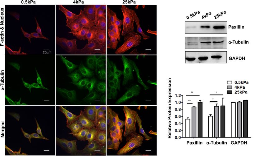
Fan et al. Substrate Stiffness Modulates Ovarian Cancer
analyze the differentia expression genes (DEGs) between samples. Substrate Stiffness Affects Cell Stiffness
Next, pathway analysis was applied to determine the significant Mechanical analysis of the force-distance curves was performed
pathways of the differential genes using the Kyoto Encyclopaedia to determine the relative cell elasticities indicated by Young’s
of Genes and Genomes (KEGG) database. The P-values for the modulus. The curves were then fitted to the spherical Hertz
pathways of all the differential genes were calculated. P-value model. AFM measurements demonstrated that Young’s modulus
were used to carry out significance analysis. Parameters for of SKOV-3 cells grown on soft substrates was less than that of
classifying significantly DEGs are ≥ 2-fold differences (|log2FC| cells grown on stiff substrates (p < 0.05; Figure 3). Values are
≥ 1, FC: the fold change of expressions) in the transcript expressed at mean ± standard deviation. The values of Young’s
abundance and P < 0.05. modulus in the 4, 25, and 50 kPa groups were 4.70 ± 0.91,
7.94 ± 1.82, and 8.34 ± 2.19 kPa, respectively.
Statistics Analysis
Origin Pro (version 8.5, OriginLab Corporation, Northampton, Substrate Stiffness Regulates the
MA, United States) was used to perform statistical analysis with Phenotypes of OCCs
a one-way analysis of variance (ANOVA). A two-sample t-test
Characteristic phenotypic markers of mesenchymal and epithelial
was used to determine statistical significance. Differences with a
cells were assessed to demonstrate EMT across the cell
p-value below 0.05 were considered statistically significant for all
population. These included an increase in vimentin expression,
experiments unless otherwise specified.
decreases in E-cadherin and β-catenin expression, and a
more elongated cell shape. Immunofluorescence staining and
Western blotting showed that SKOV-3 cells grown on stiffer
RESULTS substrates had increased vimentin expression and decreased
E-cadherin and β-catenin expression (Figures 4Aa–f,B). Cell
Substrate Stiffness Influences the OCC shape was also observed to change with stiffness. Images
Proliferation were acquired using an environmental scanning electron
The most common sites of metastasis for OCCs are the microscopy in low vacuum mode to observe the morphological
peritoneum, lymph nodes, lungs, and liver. According to previous response to changes in substrate stiffness (Figures 4Ag–i).
reports, hydrogels with stiffness of 0.5, 4, and 25 kPa are The shapes of cells on the soft substrates (0.5 kPa) were
equivalent to the stiffness of lymph nodes, peritoneum, and mostly spindle-like and the cells were well spread out and
tumor tissues (Levental et al., 2007; McKenzie et al., 2018). flattened on stiffer substrates. The shape of cells grown on
Growth profiles of SKOV-3 cells cultured on different substrates the softer substrates showed characteristics of epithelial cells.
were plotted by calculating cell numbers in 24 h intervals for These results indicated that EMT occurred in OCCs on the
6 days. The SKOV-3 cells in the 0.5, 4, and 25 kPa hydrogels soft substrates.
entered the logarithmic growth phase at the 5th, 4th, and 4th
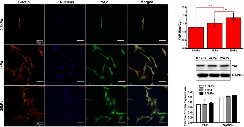
day, respectively (Figure 1A). The doubling time (Td) results Substrate Stiffness Promotes YAP
based on 72 h cultures showed that the SKOV-3 cells in the 0.5, Nuclear Localization
4, and 25 kPa hydrogel substrates had a Td of 56.58, 42.05, and
YAP is the main effector of the Hippo signaling pathway and is
31.05 h, respectively (Figure 1B). These results indicated that the
involved in signal transduction and transcriptional activation of
proliferation rate of SKOV-3 cells was increased when on a more
downstream target factors (Piccolo et al., 2014). YAP was noted
rigid substrate.
as an oncogene in previous studies. High YAP activity drives
proliferation, differentiation, invasion, and metastases of cancer
Substrate Stiffness Influences the cells. Studies have revealed that, apart from the Hippo pathway,
Skeleton of Ovarian Cancer Cell SKOV-3 YAP is regulated by mechanical forces (Benham-Pyle et al., 2015).
Cells adhere to the ECM through focal adhesions that link Therefore, we investigated the effect of substrate stiffness on YAP
the actin cytoskeleton to ECM. The cytoskeleton is a dynamic nuclear localization in OCCs. The immunofluorescence studies
network composed mainly of three kinds of fiber structures, showed that the nuclear distribution of YAP in SKOV-3 cells
namely F-actin, microtubules, and intermediate filaments. The grown on stiff substrates was elevated compared to cells grown on
actin cytoskeleton is known to be highly responsive to mechanical soft substrates (p < 0.01; Figures 5A,B). Western blotting showed
stresses (Fletcher and Mullins, 2010). The cytoskeleton structures that there were no significant differences in the expression of YAP
of SKOV-3 cells on the tested substrates were investigated using among the cells in the three substrate rigidity groups (Figure 5C).
laser scanning confocal microscopy. The results showed that These results demonstrated that substrate stiffness promoted
the fluorescence of F-actin and tubulin were weak in cells on nuclear translocation of YAP. Substrate stiffness regulates the
soft substrates, whereas the cells on rigid substrates exhibited chemosensitivity of OCCs.
prominent stress fibers (Figure 2A). Additionally, the western The chemotherapeutic drugs cisplatin and paclitaxel work
blot assays showed that the expression of tubulin and focal based on distinct molecular mechanisms. Cisplatin forms DNA
adhesion-paxillin increased as substrate stiffness also increased cross-links and platinum adducts between DNA and proteins,
(Figure 2B). These results indicated that the cytoskeleton was which causes DNA damage and subsequent cell death (Florea
remodeled and reinforced to match the force applied by the and Büsselberg, 2011). Paclitaxel induces cell death by binding to
different substrates. microtubulin, thus causing microtubule dysfunction, induction
Frontiers in Cell and Developmental Biology | www.frontiersin.org 4 August 2021 | Volume 9 | Article 718834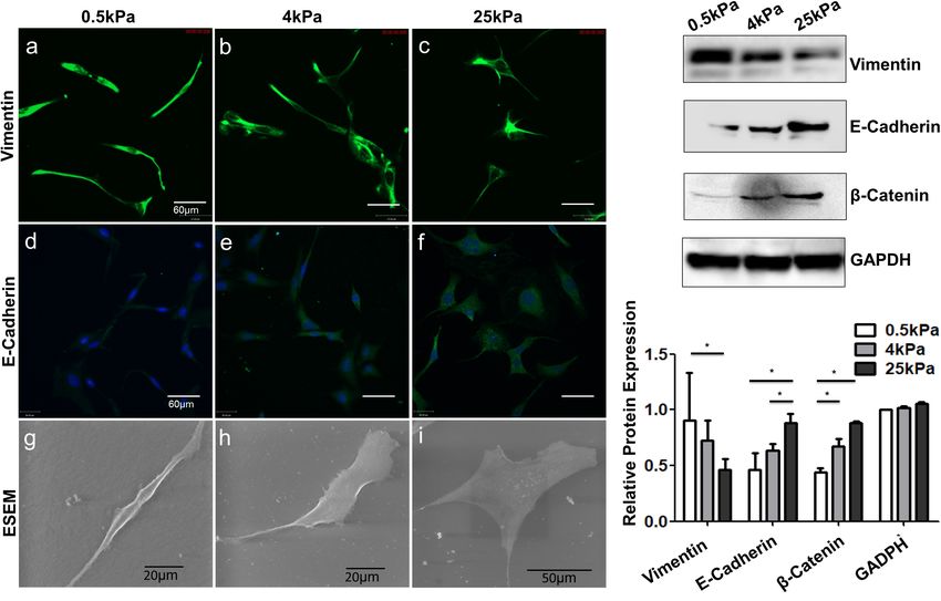
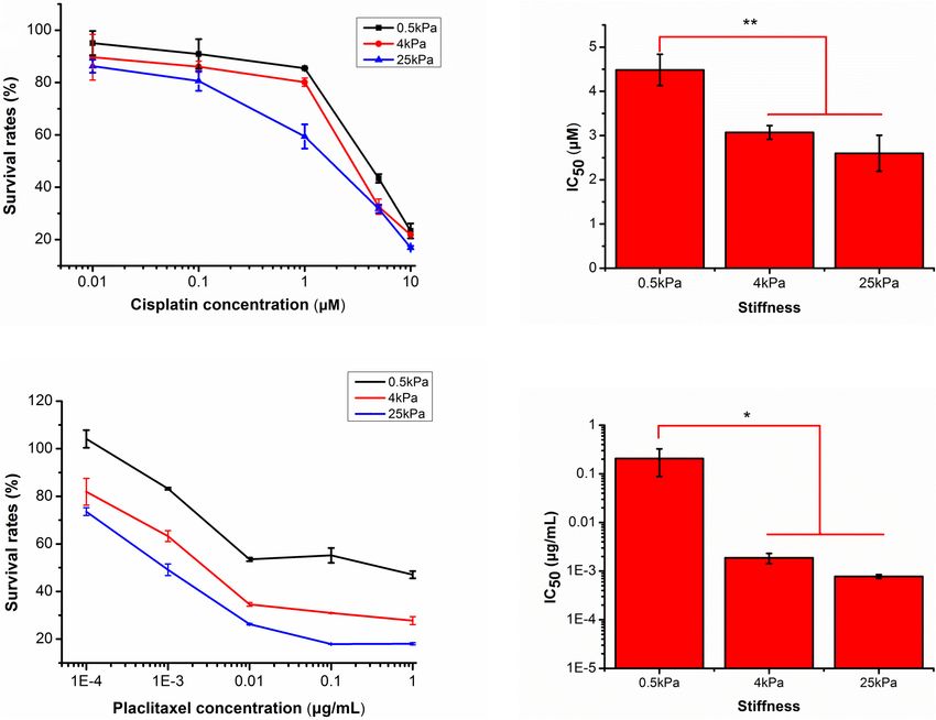
Fan et al.                                                                                                                  Substrate Stiffness Modulates Ovarian Cancer

  FIGURE 1 | (A) SKOV-3 cell growth profiles on substrates of varying stiffness; (B) doubling time (Td) of SKOV-3 cells on different substrates after 72 h.

  FIGURE 2 | (A) Structures of F-actin and microtubulin in SKOV-3 cells grown on substrates of different stiffness. (B) Expression patterns of microtubulin and paxillin
  in MCF-7 cultured on different substrates for 48 h. *P < 0.05 and **P < 0.01.

of cell cycle arrest in G2-M, and activation of apoptosis (Horwitz,                      between the latter two and 0.5 kPa (p < 0.05). At 10 µM, there
1992). The pharmacological responses of the SKOV-3 cells on                              were no significant differences in survival rates of SKOV-3 cells
different substrates to the antitumor drugs cisplatin and paclitaxel                     on different substrates (Figure 6A). Subsequently, the mean IC50
were evaluated. Values are expressed at mean ± standard                                  values for cisplatin were 4.48 ± 0.35, 3.07 ± 0.15, and 2.60 ± 0.41
deviation. For cisplatin at the lowest concentration (less than                          µM for the 0.5, 4, and 25 kPa substrates, respectively (Figure 6B).
1 µM), there were no significant differences in survival rates                           There was a significant difference between 0.5 kPa and the latter
of SKOV-3 cells on different substrates. At 1 µM of cisplatin,                           two (p < 0.01), but there was no significant difference between 4
survival rates of SKOV-3 cells on the 0.5, 4, and 25 kPa                                 and 25 kPa substrate (p < 0.05).
substrates were 79.18 ± 10.84, 81.77 ± 3.06, and 57.99 ± 4.10%,                             For paclitaxel, the effects of substrate stiffness on
respectively. There was no significant difference between 0.5 and                        pharmacological response were similar to that of cisplatin.
4 kPa, but there was a significant difference between the former                         As the concentration of paclitaxel increased, the cell survival
two and the 25 kPa substrate (p < 0.05). However, at 5 µM,                               rates on all substrates gradually decreased. At a concentration
the cell survival rates were 46.48 ± 5.54, 31.08 ± 3.30, and                             of 10−4 µg mL−1 , the cell survival rates on the 0.5, 4, and
33.63 ± 3.43%, respectively. There was no significant difference                         25 kPa hydrogels were 101.47 ± 11.98, 78.81 ± 6.69, and
between 4 and 25 kPa, but there was a significant difference                             71.55 ± 3.72%, respectively. There was a significant difference

Frontiers in Cell and Developmental Biology | www.frontiersin.org                    5                                            August 2021 | Volume 9 | Article 718834
Fan et al.                                                                                                               Substrate Stiffness Modulates Ovarian Cancer

  FIGURE 3 | (A–C) Frequency distribution of the Young’s modulus of SKOV-3 cells cultured on hydrogels with different stiffness for 3 days. (D) Medians of the
  Young’s modulus. *P < 0.05 and **P < 0.01.

  FIGURE 4 | Role of substrate stiffness in epithelial-mesenchymal transition (EMT) induction. (A) Laser confocal microscopy and environmental scanning electron
  microscopy (ESEM) of SKOV-3 cells on substrates of varying stiffness. (B) Western blotting showing expression levels of vimentin, E-cadherin, and β-catenin.
  *P < 0.05.

Frontiers in Cell and Developmental Biology | www.frontiersin.org                  6                                           August 2021 | Volume 9 | Article 718834
Fan et al. Substrate Stiffness Modulates Ovarian Cancer
FIGURE 5 | (A) Characterization of YAP fluorescence and stress fibers in SKOV-3 cells grown on substrates with different stiffness. (B) Quantification of the
nuclear/cytoplasmic ratio of YAP. (C) Western blotting showing the expression of YAP. **P < 0.01.
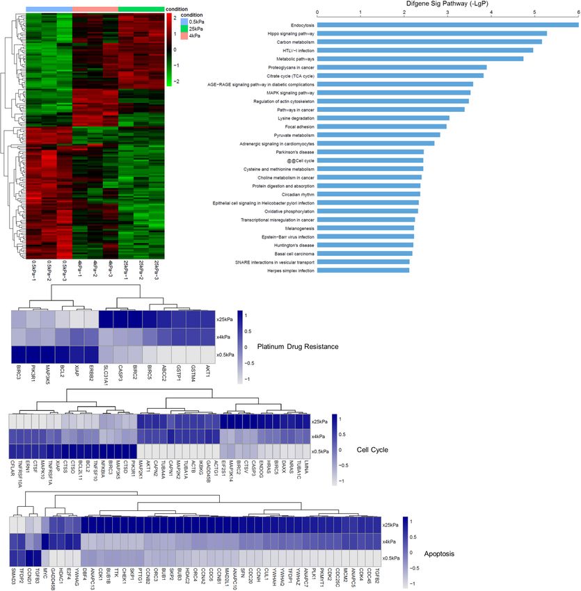
between 0.5 kPa and the latter two (p < 0.05), but there was in the SKOV-3 cells grown on the different substrates via
no significant difference between 4 kPa and 25 kPa substrate microarray analysis. Changes in gene expression were measured
(p < 0.05). When increased to 10−3 µg mL−1 , the cell survival after 3 days of growth on the gels. The hierarchical clustering
rates were 81.99 ± 2.15, 61.25 ± 3.87, and 50.89 ± 3.51%, analysis of these changes in mRNA expression is shown in
respectively. There was a significant difference between 0.5 and Figure 7A. We found that the general direction of changes
4 kPa (p < 0.01), between 4 and 25 kPa (p < 0.05). At 10−2 µg in gene expression (up or downregulated) was more similar
mL−1 , the cell survival rates were 52.18 ± 2.31, 34.2 ± 0.84, and between cells cultured on 25 and 4 kPa substrates. We performed
28.24 ± 3.40%, respectively. There was a significant difference the pathways analyses of the differentially expressed mRNA
between 0.5 and 4 kPa (p < 0.01), and there was no significant according to the KEGG database. Figure 7B lists the 30 most
difference between 4 and 25 kPa. At the highest dose of 10−1 significant signaling pathways. Among these, endocytosis, the
µg mL−1 , the cell survival rates of the cells grown on the 0.5, Hippo signaling pathway, metabolic pathways, proteoglycans
4, and 25 kPa substrates was 51.27 ± 7.11, 29.97 ± 1.72, and in cancer, the MAPK signaling pathway, regulation of actin
18.06 ± 0.3%, respectively. There was a significant difference cytoskeleton, focal adhesion, and cell cycle were ranked the
between 0.5 and 4 kPa (p < 0.01), between 4 and 25 kPa highest. Figure 7C expression levels of genes involved in
(p < 0.05). At the highest dose of 1 µg mL−1 , the cell survival platinum drug resistance, apoptosis and cell cycle. Many genes
rates of the cells grown on the 0.5, 4, and 25 kPa substrates was whose molecular action imparts in apoptosis were downregulated
51.48 ± 7.14, 29.02 ± 2.49, and 16.97 ± 1.85%, respectively in the cells grown on 0.5 kPa substrate. Platinum drug-resistance
(Figure 6C). There was a significant difference between 0.5 and genes including ERBB2, BCL2, MAP3K5, PIK3R1, and BIRC3
4 kPa (p < 0.01), but there was no significant difference between were significantly upregulated in SKOV-3 cells on soft substrates.
4 and 25 kPa substrate (p < 0.05). The mean IC50 values of
paclitaxel for cells grown on the 0.5, 4, and 25 kPa substrates were Substrate Stiffness Affects the
0.21 ± 0.12, 0.0018 ± 0.0004, and 0.0007 ± 0.00005 µg mL−1 , Expression of Multidrug Resistance
respectively (Figure 6D). There was a significant difference
Proteins
between 0.5 kPa and the latter two, but there was no significant
The ABC transporter (ATP-binding cassette transporter)
difference between 4 kPa and 25 kPa substrate (p < 0.05).
superfamily is a group of transmembrane proteins and
transporters that cause drug resistance by using ATP to
Analysis of SKOV-3 Cell Gene Expression excrete multiple anticancer drugs. After tumor cells develop
When Grown on Different Substrates resistance to a drug, they also develop resistance to drugs with
In order to identify the effect of substrate stiffness on gene different structures and mechanisms of action that they have
expression, we evaluated for differentially expressed mRNAs not been exposed to, which is a phenomenon called multidrug
Frontiers in Cell and Developmental Biology | www.frontiersin.org 7 August 2021 | Volume 9 | Article 718834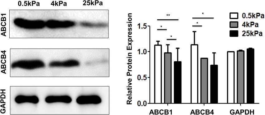
Fan et al.                                                                                                                      Substrate Stiffness Modulates Ovarian Cancer

  FIGURE 6 | Responses of SKOV-3 cells grown on different substrates to cisplatin and paclitaxel. (A) Survival rates of SKOV-3 cells on different substrates after
  treatment with different concentrations of cisplatin. (B) IC50 values of cisplatin on different substrates. (C) Survival rates of SKOV-3 cells on different substrates after
  treatment with different concentrations of paclitaxel. (D) IC50 values of paclitaxel on different substrates. *P < 0.05 and **P < 0.01.

resistance (MDR). Overexpression of ABC transporters is an                                 previous studies. The canonical mechanism of YAP regulation
important cause of MDR. Among the various ABC transporters,                                is a phosphorylation cascade in the Hippo pathway (Huang
ABCB1 (MDR1) and ABCB4 (MDR3) are thought to play                                          et al., 2005). YAP is inhibited through direct phosphorylation
important roles in ovarian cancer. The results showed that the                             of the central components of the Hippo pathway including
expression of ABCB1 and ABCB4 on the soft substrates was                                   the Mst1/2 and Lats1/2 kinase cascades (Piccolo et al., 2014).
higher than that on rigid substrates (Figure 8). These results                             Phosphorylated YAP localizes to the cytoplasm in an inactive
indicated that substrate stiffness affected the expression of genes                        state and dephosphorylated YAP enters the nucleus from the
related to multidrug resistance.                                                           cytoplasm. In the nucleus, YAP mainly induces cell proliferation
                                                                                           and the expression of anti-apoptotic genes by interacting with
                                                                                           various transcription factors such as the TEA region (TEAD)
DISCUSSION                                                                                 family. Besides the canonical Hippo pathway, investigators
                                                                                           have recently uncovered that YAP is regulated by different
In this study, we demonstrated that the growth and the                                     types of mechanical stimuli including physical stretching
chemotherapeutic response of OCCs in vitro are markedly                                    (Benham-Pyle et al., 2015), ECM topography (Aragona et al.,
affected by substrate stiffness. It has previously been                                    2013), and stiffness (Dupont et al., 2011) via Hippo-independent
demonstrated that substrate stiffness can regulate proliferation                           mechanisms. Here, we used confocal microscopy to visualize
in a variety of other cancer cells (Tilghman et al., 2010). In                             and quantify YAP localization in SKOV-3 cells grown on
our study, the growth profiles of the human OCC line SKOV-3                                hydrogels of differing stiffness. The results showed that the
were analyzed and the results showed increased proliferation                               levels of nuclear localization were positively correlated with
and rapid cell cycle progression when cells were cultured on                               the stiffness of the hydrogel substrates. Our data agree with
rigid substrates. In previous studies, it has been demonstrated                            similar findings from other studies linking YAP nuclear
that the expression of cyclin-D was upregulated and critical                               localization to greater proliferation on stiffer substrates. In
mitogenic signaling was promoted on rigid substrates (Schrader                             the literature, cells cultured on rigid substrates showed high
et al., 2011; Hui et al., 2017). YAP was noted as an oncogene in                           nuclear localization and elevated transcriptional activities of

Frontiers in Cell and Developmental Biology | www.frontiersin.org                      8                                              August 2021 | Volume 9 | Article 718834
Fan et al. Substrate Stiffness Modulates Ovarian Cancer FIGURE 7 | Substrate stiffness alters the expression of genes. (A) Changes in the SKOV-3 cell gene expression when cultured on substrates with different stiffness. (B) Significant pathways of differentially expressed genes in SKOV-3 cells on different substrates. (C) Expression levels of selected genes whose human orthologs are involved in platinum-based drug resistance, cell cycle, and apoptosis. YAP, whereas, in cells that were gown on softer substrates, Liu-Chittenden et al. (2012) showed that VP could disrupt YAP translocated to the cytoplasm and was inactivated (Zhao the TEAD-YAP association and inhibit YAP-induced liver et al., 2012; Totaro et al., 2017). Studies suggest that the overgrowth. It was also observed that VP could effectively inhibition of YAP interaction with its transcriptional partners reduce proliferation and inhibit the growth of OCCs in vivo is a potential strategy for cancer therapy. Verteporfin (VP), and in vitro (Feng et al., 2016). Our findings combined with used clinically for the treatment of macular degeneration, those of the previously mentioned studies indicate that targeting has been proven to inhibit the interaction between YAP and YAP signaling may be a promising therapy strategy against TEAD and has shown potential as an anticancer treatment. stiffness-induced proliferation. Frontiers in Cell and Developmental Biology | www.frontiersin.org 9 August 2021 | Volume 9 | Article 718834
Fan et al.                                                                                                            Substrate Stiffness Modulates Ovarian Cancer

  FIGURE 8 | Substrate stiffness affects the expression of multidrug resistance proteins.

    AFM-based nanomechanical studies have revealed that                                 (Wu et al., 1998; Beil et al., 2003). Our study demonstrated
cellular mechanical properties are closely related to certain                           that the organization and expression of various cytoskeleton
disease states. Many studies have reported that cancer cells                            components, including F-actin, microtubulin, and vimentin,
are physically softer than normal cells (Lekka, 2016; Alibert                           varied with changes in substrate stiffness. Our results showed that
et al., 2017) and metastatic cancer cells are more mechanically                         OCCs grown on softer substrates with a lower elastic modulus
compliant than their non-metastatic counterparts (Li et al.,                            and were less sensitive to chemotherapeutic agents. The literature
2008; Xu et al., 2012; Park, 2016). Recent AFM experiments                              regarding the response to mechanical changes of cancer cells and
investigating the mechanical properties of chemo-sensitive                              their normal counterparts to different gel substrate stiffnesses
and resistant cancer cells have contributed to unraveling the                           reported that cancer cells were less susceptible to changes in
multifaceted nature of chemotherapeutic resistance. Sharma et al.                       substrate when substrate stiffness was increased compared to
(2014) demonstrated that cisplatin-resistant OCCs (OVCAR5-                              their normal counterparts. Our study, combined with those
CisR and SKOV3-CisR) were much stiffer than their cisplatin-                            previously mentioned, indicates that research into the effect of
sensitive counterparts (OVCAR5 and SKOV-3, respectively).                               different substrate stiffness on the mechanical properties of drug-
Moreover, their results indicated that cisplatin resistance                             sensitive and drug-resistant cancer cells is of great significance
correlates with the dynamic reorganization of actin filaments                           in understanding the underlying mechanisms of chemoresistance
and nano-mechanical compliance (Sharma et al., 2014). Studies                           from a nanomechanical perspective.
have shown that paclitaxel and cisplatin induce an increase                                 The EMT is a process by which epithelial cancer cells lose
in the stiffness of cancer cells (Ren et al., 2015). In addition,                       cell-cell adhesion, apical-basal polarity, and acquire a spindle-like
cisplatin-sensitive OCCs (A2780) showed a dose-dependent                                morphology. Epithelial markers such as E-cadherin, cytokeratins,
increase in cell stiffness after cisplatin treatment, while resistant                   and occludin are downregulated. Meanwhile, mesenchymal
cells (A7890cis) were unaffected. However, one study showed                             markers such as N-cadherin and vimentin are upregulated
that cisplatin- and paclitaxel-resistant OCCs were softer than                          (Thompson et al., 2005; Taube et al., 2010). EMT can be
drug-sensitive cells (Kapoor et al., 2018). Seo et al. (2015)                           activated not only by a variety of signaling pathways such as
found that there was a bimodal distribution in the histogram                            TGF-β, EGF, miRNA, AKT, and PI3K but also by changes in
of mechanical stiffness. This bimodal distribution implies the                          ECM stiffness and endogenous mechanical stress (De Craene
existence of two different subpopulations. The peak of the                              and Berx, 2013). It has been previously demonstrated that
lower stiffness almost overlapped with the average mechanical                           substrate stiffness can promote EMT in multiple cancer cell
stiffness of sensitive cells. All these findings demonstrated that                      lines (Schrader et al., 2011; Rice et al., 2017). The stiffness
cellular nano-mechanical studies help to reveal how cancer cells                        of metastatic microenvironments is significantly lower than
acquire drug resistance. However, most of these experiments                             that of the primary tumor tissue. In this study, we utilized
were performed on plastic Petri dishes, whose mechanical                                polyacrylamide hydrogels with elastic properties that mimic
properties are very different from those of natural tissues.                            tumor tissue and tissues commonly metastasized by OCCs
The elastic properties of cells are mainly affected by their                            to investigate the role of ECM stiffness in promoting OCC
intracellular actin cytoskeleton. Cytoskeletal reorganization is                        malignancy. We have demonstrated that changes in cellular
responsible for changes in cellular elastic modulus. In addition,                       morphology and the expression of a variety of molecules were
microtubules and intermediate filaments such as vimentin and                            all indicative of EMT following culture on gels of lower rigidity.
keratin play crucial roles in the elastic properties of cells                           The cellular morphology changed to a spindle-like shape, while

Frontiers in Cell and Developmental Biology | www.frontiersin.org                  10                                      August 2021 | Volume 9 | Article 718834
Fan et al. Substrate Stiffness Modulates Ovarian Cancer
epithelial markers including E-cadherin and β-catenin were 1976; Bourhis et al., 1989; Veneroni et al., 1994). Recent studies
downregulated, and mesenchymal markers such as vimentin were indicate that expression of ABCB1 is a useful predictor of
upregulated in softer environments. Our findings suggest that paclitaxel resistant for patients with ovarian cancer (Kamazawa
a reduction in the stiffness of the cancer cell niche, as would et al., 2002; Haque et al., 2020). ABCB1 was expressed at higher
be encountered by disseminated or metastatic OCCs, would be levels in more mesenchymal and therapy-resistant OCC lines,
sufficient to promote EMT. than in more epithelial and chemo-sensitive cell lines (Feng et al.,
In the context of cancer, the EMT mechanism is crucial for 2017; Zhang et al., 2018). Furthermore, ABCB4 (MDR3) is also
cancer initiation and metastasis. Growing evidence demonstrates linked with chemotherapy resistance and is increased in recurrent
that apart from contributing to cancer progression, EMT can ovarian cancers (Duan et al., 2004). Paclitaxel-resistant cell
promote chemotherapy resistance in OCCs. The epithelial lines overexpress both ABCB1 and ABCB4 (Januchowski et al.,
marker E-cadherin is downregulated and the mesenchymal 2014). Correlation analyses showed a high correlation between
marker vimentin is upregulated in paclitaxel-resistant epithelial MDR3 expression and resistance to paclitaxel and doxorubicin
OCCs (NOS-PR, TAOV-PR, and SKOV-3) (Kajiyama et al., in vitro. Our data showed that cells cultured on softer gels
2007). It was demonstrated that an increase in the expression expressed more ABCB1 and ABCB4, which coincidentally were
of miR-181a-induced EMT in OCCs mediated resistance to more mesenchymal and therapy-resistant. These results suggest
paclitaxel-based therapies (Li et al., 2016). Twist1 is a highly that EMT promotes chemoresistance in SKOV-3 cells on soft
evolutionally conserved basic Helix-Loop-Helix transcriptional substrates via the upregulation of ABCB1 and ABCB4.
factor (bHLH). Many studies have highlighted the role of in
promoting cancer cell EMT (Watanabe et al., 2004; Yang et al.,
2004). A study showed that miR-186 regulation of Twist1 CONCLUSION
can be seen as a promising strategy to sensitize OCCs that
have undergone EMT and chemotherapy-induced resistance In conclusion, our study showed that increasing substrate
(Zhu et al., 2016). Recent studies have demonstrated that stiffness promotes the proliferation of SKOV-3 cells and the
cancer cells acquire chemoresistance based on the substrate nuclear localization of YAP. Conversely, a soft environment (as
stiffness-induced EMT. Rice et al. (2017) demonstrated that might be encountered by disseminated or metastatic OCCs)
substrate stiffness induced EMT and promoted chemoresistance induces EMT and chemoresistance. Chemoresistance in SKOV-
in pancreatic cancer cells. Our results suggest that a stiff 3 cells on softer substrates was due to the upregulation of
environment promotes epithelial phenotypes in OCCs, whereas platinum drug-resistant genes, ABCB1, and ABCB4. These
low stiffness induces mesenchymal phenotypes. We further findings provide new therapeutic targets for future anti-
investigated whether EMT affected OCC susceptibility cancer drug designs.
to chemotherapy. We found that the chemosensitivity of
SKOV-3 cells to cisplatin and paclitaxel decreased as the
substrate softened. Our findings highlighted that substrate DATA AVAILABILITY STATEMENT
stiffness plays an important role in EMT and subsequent
chemotherapeutic resistance. The data presented in this study are deposited in https://www.
There are a variety of EMT-driven mechanisms that lead to ncbi.nlm.nih.gov/geo/query/acc.cgi?acc=GSE178888.
the acquisition of chemoresistance such as lower drug uptake,
higher drug efflux, higher DNA repair capacity, and decreased
apoptosis (Loret et al., 2019). In our study, the results of mRNA AUTHOR CONTRIBUTIONS
microarray analysis showed that platinum drug-resistance genes
including ERBB2, BCL-2, MAP3K5, PIK3R1, and BIRC3 are YF: cell culture, western immunoblotting, data analysis, and
significantly upregulated in SKOV-3 cells on soft substrates. manuscript writing. QS: research supervision, cell culture,
Avian erythroblastosis oncogene B2 (ERBB2) also known as proliferation and cytotoxicity analysis, AFM experimental
human epidermal growth factor receptor 1 (HER2) signaling is implementation, statistical analysis of data, manuscript
highly correlated with cisplatin-resistance in OCCs and tumors writing, and critical review and revision. XLa: cell culture,
(Harris et al., 2019). BCL-2, anti-apototic protein, can block immunofluorescence staining, cell staining, and ESEM imaging.
p53-mediated apoptosis (Kassim et al., 1999; Dai et al., 2017) JF: technical adviser, data analysis, and statistical analysis of
and involve in AKT-regulated cell survival in cisplatin resistant data. ZA: MATLAB code writing and data analysis. XLn: mRNA
EOC (Dai et al., 2017). It is a potential predictor of cisplatin- microarray analysis. JW: research supervision, and manuscript
resistance in EOC. ABC transporters exclude drugs from the critical review and revision. All the authors read and approved
cytoplasm and move them to the extracellular environment the final manuscript.
using the energy provided by ATP hydrolysis. EMT transcription
factors can induce the expression of ABC transporters. ABCB1
(MDR1), encoding p-glycoprotein (PgP) is the most studied FUNDING
ABC transporter and the first to be identified to selectively
confer MDR by directly pumping out anticancer drugs, including This work was supported by the National Natural Science
paclitaxel, doxorubicin, topotecan, docetaxel (Juliano and Ling, Foundation of China (No. 11672192).
Frontiers in Cell and Developmental Biology | www.frontiersin.org 11 August 2021 | Volume 9 | Article 718834Fan et al. Substrate Stiffness Modulates Ovarian Cancer
REFERENCES resistance in leukaemia cells. Br. J. Haematol. 136, 269–275. doi: 10.1111/j.1365-
2141.2006.06435.x
Alibert, C., Goud, B., and Manneville, J. (2017). Are cancer cells really softer than Hazlehurst, L. A., Damiano, J. S., Buyuksal, I., Pledger, W. J., and Dalton, W. S.
normal cells? Biol. Cell 109, 167–189. doi: 10.1111/boc.201600078 (2000). Adhesion to fibronectin via beta1 integrins regulates p27kip1 levels and
Aragona, M., Panciera, T., Manfrin, A., Giulitti, S., Michielin, F., Elvassore, N., contributes to cell adhesion mediated drug resistance (CAM-DR). Oncogene 19,
et al. (2013). A mechanical checkpoint controls multicellular growth through 4319–4327. doi: 10.1038/sj.onc.1203782
YAP/TAZ regulation by actin-processing factors. Cell 154, 1047–1059. doi: 10. Horwitz, S. B. (1992). Mechanism of action of taxol. Trends Pharmacol. Sci. 13,
1016/j.cell.2013.07.042 134–136. doi: 10.1016/0165-6147(92)90048-b
Beil, M., Micoulet, A., von Wichert, G., Paschke, S., Walther, P., Omary, M. B., et al. Huang, J., Wu, S., Barrera, J., Matthews, K., and Pan, D. (2005). The Hippo
(2003). Sphingosylphosphorylcholine regulates keratin network architecture signaling pathway coordinately regulates cell proliferation and apoptosis by
and visco-elastic properties of human cancer cells. Nat. Cell Biol. 5, 803–811. inactivating Yorkie, the Drosophila Homolog of YAP. Cell 122, 421–434. doi:
doi: 10.1038/ncb1037 10.1016/j.cell.2005.06.007
Benham-Pyle, B. W., Pruitt, B. L., and Nelson, W. J. (2015). Mechanical strain Hui, L., Zhang, J., Ding, X., Guo, X., and Jiang, X. (2017). Matrix stiffness
induces E-cadherin-dependent Yap1 and β-catenin activation to drive cell cycle regulates the proliferation, stemness and chemoresistance of laryngeal
entry. Science 348, 1024–1027. doi: 10.1126/science.aaaa4559 squamous cancer cells. Int. J. Oncol. 50, 1439–1447. doi: 10.3892/ijo.2017.
Bourhis, J., Goldstein, L. J., Riou, G., Pastan, I., Gottesman, M. M., and Bénard, 3877
J. (1989). Expression of a human multidrug resistance gene in ovarian Januchowski, R., Wojtowicz, K., Andrzejewska, M., and Zabel, M. (2014).
carcinomas. Cancer Res. 49, 5062–5065. Expression of MDR1 and MDR3 gene products in paclitaxel-, doxorubicin-
Correia, A. L., and Bissell, M. J. (2012). The tumor microenvironment is a and vincristine-resistant cell lines. Biomed. Pharmacother. 68, 111–117. doi:
dominant force in multidrug resistance. Drug Resist. Updat. 15, 39–49. doi: 10.1016/j.biopha.2013.09.004
10.1016/j.drup.2012.01.006 Jemal, A., Siegel, R., Xu, J., and Ward, E. (2010). Cancer statistics, 2010. CA Cancer
Dai, Y., Jin, S., Li, X., and Wang, D. (2017). The involvement of Bcl-2 family J Clin 60, 277–300. doi: 10.3322/caac.20073
proteins in AKT-regulated cell survival in cisplatin resistant epithelial ovarian Juliano, R. L., and Ling, V. (1976). A surface glycoprotein modulating drug
cancer. Oncotarget 8, 1354–1368. doi: 10.18632/oncotarget.13817 permeability in chinese hamster ovary cell mutants. Biochim. Biophys. Acta. 455,
De Craene, B., and Berx, G. (2013). Regulatory networks defining EMT during 152–162. doi: 10.1016/0005-2736(76)90160-7
cancer initiation and progression. Nat. Rev. Cancer 13, 97–110. doi: 10.1038/ Kajiyama, H., Shibata, K., Terauchi, M., Yamashita, M., Ino, K., Nawa, A.,
nrc3447 et al. (2007). Chemoresistance to paclitaxel induces epithelial-mesenchymal
Duan, Z., Brakora, K. A., and Seiden, M. V. (2004). Inhibition of ABCB1 (MDR1) transition and enhances metastatic potential for epithelial ovarian carcinoma
and ABCB4 (MDR3) expression by small interfering RNA and reversal of cells. Int. J. Oncol. 31, 277–283. doi: 10.3892/ijo.31.2.277
paclitaxel resistance in human ovarian cancer cells. Mol. Cancer Ther. 3, Kamazawa, S., Kigawa, J., Kanamori, Y., Itamochi, H., Sato, S., Iba, T., et al.
833–838. doi: 10.1016/j.lungcan.2004.01.008 (2002). Multidrug resistance gene-1 is a useful predictor of paclitaxel-based
Dupont, S., Morsut, L., Aragona, M., Enzo, E., Giulitti, S., Cordenonsi, M., et al. chemotherapy for patients with ovarian cancer. Gynecol. Oncol. 86, 171–176.
(2011). Role of YAP/TAZ in mechanotransduction. Nature 474, 179–183. doi: doi: 10.1006/gyno.2002.6738
10.1038/nature10137 Kapoor, A., Barai, A., Thakur, B., Das, A., Patwardhan, S. R., and Monteiro,
Erkan, M., Hausmann, S., Michalski, C. W., Fingerle, A. A., Dobritz, M., Kleeff, M. (2018). Soft drugresistant ovarian cancer cells migrate via two distinct
J., et al. (2012). The role of stroma in pancreatic cancer: diagnostic and mechanisms utilizing myosin II based contractility. Biochim. Biophys. Acta.
therapeutic implications. Nat. Rev. Gastroenterol. Hepatol. 9, 454–467. doi: 1865, 392–405. doi: 10.1016/j.bbamcr.2017.11.012
10.1038/nrgastro.2012.115 Kassim, S. K., Ali, H. S., Sallam, M. M., Fayed, S. T., Seada, L. S., Abd-Elkawy, E.,
Feng, J., Gou, J., Jia, J., Yi, T., Cui, T., and Li, Z. (2016). Verteporfin, a suppressor et al. (1999). Increased bcl-2 expression is associated with primary resistance to
of YAP-TEAD complex, presents promising antitumor properties on ovarian chemotherapy in human epithelial ovarian cancer. Clin. Biochem. 32, 333–338.
cancer. Onco. Targets Ther. 9, 5371–5381. doi: 10.2147/ott.S109979 doi: 10.1016/s0009-9120(99)00026-0
Feng, J. T., Tang, Y., Xu, Y. G., Sun, Q. M., Liao, F. L., and Han, D. (2013). Substrate Kharaishvili, G., Simkova, D., Bouchalova, K., Gachechiladze, M., Narsia, N., and
stiffness influences the outcome of antitumor drug screening in vitro. Clin. Bouchal, J. (2014). The role of cancer-associated fibroblasts, solid stress and
Hemorheol. Micro. 55, 121–131. doi: 10.3233/Ch-131696 other microenvironmental factors in tumor progression and therapy resistance.
Feng, T., Wang, Y., Lang, Y., and Zhang, Y. (2017). KDM5A promotes proliferation Cancer Cell Int. 14:41. doi: 10.1186/1475-2867-14-41
and EMT in ovarian cancer and closely correlates with PTX resistance. Mol. Landowski, T. H., Olashaw, N. E., Agrawal, D., and Dalton, W. S. (2003). Cell
Med. Rep. 16, 3573–3580. doi: 10.3892/mmr.2017.6960 adhesion-mediated drug resistance (CAM-DR) is associated with activation
Fletcher, D. A., and Mullins, R. D. (2010). Cell mechanics and the cytoskeleton. of NF-kappa B (RelB/p50) in myeloma cells. Oncogene 22, 2417–2421. doi:
Nature 463, 485–492. doi: 10.1038/nature08908 10.1038/sj.onc.1206315
Florea, A. M., and Büsselberg, D. (2011). Cisplatin as an anti-tumor drug: cellular Lekka, M. (2016). Discrimination between normal and cancerous cells using AFM.
mechanisms of activity, drug resistance and induced side effects. Cancers 3, Bionanoscience 6, 65–80. doi: 10.1007/s12668-016-0191-3
1351–1371. doi: 10.3390/cancers3011351 Levental, I., Georges, P. C., and Janmey, P. A. (2007). Soft biological materials
Fraser, M., Bai, T., and Tsang, B. K. (2008). Akt promotes cisplatin resistance in and their impact on cell function. Soft Matter 3, 299–306. doi: 10.1039/b610
human ovarian cancer cells through inhibition of p53 phosphorylation and 522j
nuclear function. Int. J. Cancer 122, 534–546. doi: 10.1002/ijc.23086 Levental, K. R., Yu, H., Kass, L., Lakins, J. N., Egeblad, M., Erler, J. T., et al. (2009).
Galluzzi, L., Senovilla, L., Vitale, I., Michels, J., Martins, I., Kepp, O., et al. (2012). Matrix crosslinking forces tumor progression by enhancing integrin signaling.
Molecular mechanisms of cisplatin resistance. Oncogene 31, 1869–1883. doi: Cell 139, 891–906. doi: 10.1016/j.cell.2009.10.027
10.1038/onc.2011.384 Li, L., Xu, Q. H., Dong, Y. H., Li, G. X., and Li, H. Y. (2016). MiR-181a upregulation
Haque, A., Sait, K., Alam, Q., Alam, M. Z., and Rasool, M. (2020). MDR1 Gene is associated with epithelial-To-mesenchymal transition (EMT) and multidrug
polymorphisms and its association with expression as a clinical relevance in resistance (MDR) of ovarian cancer cells. Eur. Rev. Med. Pharmacol. Sci. 20,
terms of response to chemotherapy and prognosis in ovarian cancer. Front. 2004–2010.
Genet. 11:516. doi: 10.3389/fgene.2020.00516 Li, Q. S., Lee, G., Ong, C. N., and Lim, C. T. (2008). AFM indentation study of breast
Harris, F. R., Zhang, P., Yang, L., Hou, X., Leventakos, K., Weroha, S. J., et al. (2019). cancer cells. Biochem. Biophys. Res. Commun. 374, 609–613. doi: 10.1016/j.bbrc.
Targeting HER2 in patient-derived xenograft ovarian cancer models sensitizes 2008.07.078
tumors to chemotherapy. Mol. Oncol. 13, 132–152. doi: 10.1002/1878-0261. Liu, C., Liu, Y., Xie, H. G., Zhao, S., Xu, X. X., Fan, L. X., et al. (2015). Role of three-
12414 dimensional matrix stiffness in regulating the chemoresistance of hepatocellular
Hazlehurst, L. A., Argilagos, R. F., and Dalton, W. S. (2007). Beta1 integrin carcinoma cells. Biotechnol. Appl. Biochem. 62, 556–562. doi: 10.1002/bab.
mediated adhesion increases Bim protein degradation and contributes to drug 1302
Frontiers in Cell and Developmental Biology | www.frontiersin.org 12 August 2021 | Volume 9 | Article 718834Fan et al. Substrate Stiffness Modulates Ovarian Cancer Liu, C. Y., Li, X., Hua, W. D., Li, J. J., Han, X. X., Ha, Q., et al. (2016). Porous matrix Totaro, A., Castellan, M., Battilana, G., Zanconato, F., Azzolin, L., Giulitti, S., et al. stiffness modulates response to targeted therapy in breast carcinoma. Small 12, (2017). YAP/TAZ link cell mechanics to notch signalling to control epidermal 4675–4681. doi: 10.1002/smll.201601365 stem cell fate. Nat. Commun. 8:15206. doi: 10.1038/ncomms15206 Liu-Chittenden, Y., Huang, B., Shim, J. S., Chen, Q., Lee, S. J., Anders, R. A., et al. Veneroni, S., Zaffaroni, N., Daidone, M. G., Benini, E., Villa, R., and Silvestrini, (2012). Genetic and pharmacological disruption of the TEAD-YAP complex R. (1994). Expression of p-glycoprotein and in vitro or in vivo resistance to suppresses the oncogenic activity of YAP. Genes Dev. 26, 1300–1305. doi: 10. doxorubicin and cisplatin in breast and ovarian cancers. Eur. J. Cancer 30A, 1101/gad.192856.112 1002–1007. doi: 10.1016/0959-8049(94)90132-5 Loret, N., Denys, H., Tummers, P., and Berx, G. (2019). The role of epithelial-to- Walker, J. L., Powell, C. B., Chen, L. M., Carter, J., Bae Jump, V. L., Parker, L. P., et al. mesenchymal plasticity in ovarian cancer progression and therapy resistance. (2015). Society of gynecologic oncology recommendations for the prevention of Cancers 11:838. doi: 10.3390/cancers11060838 ovarian cancer. Cancer 121, 2108–2120. doi: 10.1002/cncr.29321 McKenzie, A. J., Hicks, S. R., Svec, K. V., Naughton, H., Edmunds, Z. L., and Howe, Watanabe, O., Imamura, H., Shimizu, T., Kinoshita, J., Okabe, T., Hirano, A., et al. A. K. (2018). The mechanical microenvironment regulates ovarian cancer cell (2004). Expression of twist and wnt in human breast cancer. Anticancer Res. 24, morphology, migration, and spheroid disaggregation. Sci. Rep. 8:7228. doi: 3851–3856. 10.1038/s41598-018-25589-0 Wu, H. W., Kuhn, T., and Moy, V. T. (1998). Mechanical properties of L929 Mor, G., and Alvero, A. (2013). The duplicitous origin of ovarian cancer. Rambam cells measured by atomic force microscopy: effects of anticytoskeletal drugs Maimonides Med. J. 4:e0006. doi: 10.5041/RMMJ.10106 and membrane crosslinking. Scanning 20, 389–397. doi: 10.1002/sca.1998. Nieman, K. M., Kenny, H. A., Penicka, C. V., Ladanyi, A., Buell-Gutbrod, R., 4950200504 Zillhardt, M. R., et al. (2011). Adipocytes promote ovarian cancer metastasis Wu, P., Gao, W., Su, M., Nice, E. C., Zhang, W., Lin, J., et al. (2021). Adaptive and provide energy for rapid tumor growth. Nat. Med. 17, 1498–1503. doi: mechanisms of tumor therapy resistance driven by tumor microenvironment. 10.1038/nm.2492 Front Cell Dev. Biol. 9:641469. doi: 10.3389/fcell.2021.641469 Ostman, A. (2012). The tumor microenvironment controls drug sensitivity. Nat. Xu, W., Roman, M., Byungkyu, K., Wang, L., John, M., Todd, S., et al. (2012). Cell Med. 18, 1332–1334. doi: 10.1038/nm.2938 stiffness is a biomarker of the metastatic potential of ovarian cancer cells. PLoS Park, S. (2016). Nano-mechanical phenotype as a promising biomarker to evaluate One 7:e46609. doi: 10.1371/journal.pone.0046609 cancer development, progression, and anti-cancer drug efficacy. J. Cancer Prev. Yang, J., Mani, S. A., Donaher, J. L., Ramaswamy, S., Itzykson, R. A., Come, 21, 73–80. doi: 10.15430/JCP.2016.21.2.73 C., et al. (2004). Twist, a master regulator of morphogenesis, plays an Piccolo, S., Dupont, S., and Cordenonsi, M. (2014). The biology of YAP/TAZ: hippo essential role in tumor metastasis. Cell 117, 927–939. doi: 10.1016/j.cell.2004. signaling and beyond. Physiol. Rev. 94, 1287–1312. doi: 10.1152/physrev.00005. 06.006 2014 Yang, J., Zaman, M. M., Vlasakov, I., Roy, R., Huang, L., Martin, C. R., et al. Ren, J., Huang, H., Liu, Y., Zheng, X., and Zou, Q. (2015). An atomic force (2019). Adipocytes promote ovarian cancer chemoresistance. Sci. Rep. 9:13316. microscope study revealed two mechanisms in the effect of anticancer drugs doi: 10.1038/s41598-019-49649-1 on rate-dependent young’s modulus of human prostate cancer cells. PLoS One Yang, Q., Huang, J., Wu, Q., Cai, Y., Zhu, L., Lu, X., et al. (2014). Acquisition 10:e0126107. doi: 10.1371/journal.pone.0126107 of epithelial-mesenchymal transition is associated with Skp2 expression in Rice, A. J., Cortes, E., Lachowski, D., Cheung, B. C. H., Karim, S. A., Morton, paclitaxel-resistant breast cancer cells. Br. J. Cancer 110, 1958–1967. doi: 10. J. P., et al. (2017). Matrix stiffness induces epithelial–mesenchymal transition 1038/bjc.2014.136 and promotes chemoresistance in pancreatic cancer cells. Oncogenesis 6:e352. Yang, X., Fraser, M., Moll, U. M., Basak, A., and Tsang, B. K. (2006). Akt-mediated doi: 10.1038/oncsis.2017.54 cisplatin resistance in ovarian cancer: modulation of p53 action on caspase- Schrader, J., Gordon-Walker, T. T., Aucott, R. L., van Deemter, M., Quaas, dependent mitochondrial death pathway. Cancer Res. 66, 3126–3136. doi: 10. A., Walsh, S., et al. (2011). Matrix stiffness modulates proliferation, 1158/0008-5472.Can-05-0425 chemotherapeutic response, and dormancy in hepatocellular carcinoma cells. Zhang, Y., Huang, S., Guo, Y., and Li, L. (2018). MiR-1294 confers cisplatin Hepatology 53, 1192–1205. doi: 10.1002/hep.24108 resistance in ovarian cancer cells by targeting IGF1R. Biomed. Pharmacother. Seo, Y. H., Jo, Y. N., Oh, Y. J., and Park, S. (2015). Nano-mechanical reinforcement 106, 1357–1363. doi: 10.1016/j.biopha.2018.07.059 in drug-resistant ovarian cancer cells. Biol. Pharm. Bull. 38, 389–395. doi: 10. Zhao, B., Li, L., Wang, L., Wang, C. Y., Yu, J., and Guan, K. L. (2012). Cell 1248/bpb.b14-00604 detachment activates the Hippo pathway via cytoskeleton reorganization to Sethi, T., Rintoul, R. C., Moore, S. M., MacKinnon, A. C., Salter, D., Choo, C., et al. induce anoikis. Genes Dev. 26, 54–68. doi: 10.1101/gad.173435.111 (1999). Extracellular matrix proteins protect small cell lung cancer cells against Zhu, X., Shen, H., Yin, X., Long, L., Xie, C., Liu, Y., et al. (2016). MiR-186 regulation apoptosis: a mechanism for small cell lung cancer growth and drug resistance of Twist1 and ovarian cancer sensitivity to cisplatin. Oncogene 35, 323–332. in vivo. Nat. Med. 5, 662–668. doi: 10.1038/9511 doi: 10.1038/onc.2015.84 Sharma, S., Santiskulvong, C., Rao, J., Gimzewski, J. K., and Dorigo, O. (2014). The role of Rho GTPase in cell stiffness and cisplatin resistance in ovarian cancer Conflict of Interest: The authors declare that the research was conducted in the cells. Integr. Biol. 6, 611–617. doi: 10.1039/c3ib40246k absence of any commercial or financial relationships that could be construed as a Shi, L., Shi, S., Li, J., Sun, Q., Feng, K., Chen, P., et al. (2010). AFM and fluorescence potential conflict of interest. imaging of nanomechanical response in periodontal ligament cells. Front. Biosci. 2:1028–41. doi: 10.2741/e161 The handling editor declared a past co-authorship with one of the authors JF. Tajik, A., Zhang, Y. J., Wei, F. X., Sun, J., Jia, Q., Zhou, W. W., et al. (2016). Transcription upregulation via force-induced direct stretching of chromatin. Publisher’s Note: All claims expressed in this article are solely those of the authors Nat. Mater. 15, 1287–1296. doi: 10.1038/Nmat4729 and do not necessarily represent those of their affiliated organizations, or those of Taube, J. H., Herschkowitz, J. I., Komurov, K., Zhou, A. Y., Gupta, S., Yang, the publisher, the editors and the reviewers. Any product that may be evaluated in J., et al. (2010). Core epithelial-to-mesenchymal transition interactome gene- this article, or claim that may be made by its manufacturer, is not guaranteed or expression signature is associated with claudin-low and metaplastic breast endorsed by the publisher. cancer subtypes. Proc. Natl. Acad. Sci. U. S. A. 107, 15449–15454. doi: 10.1073/ pnas.1004900107 Copyright © 2021 Fan, Sun, Li, Feng, Ao, Li and Wang. This is an open-access Thompson, E. W., Newgreen, D. F., and Tarin, D. (2005). Carcinoma invasion article distributed under the terms of the Creative Commons Attribution License and metastasis: a role for epithelial-mesenchymal transition? Cancer Res. 65, (CC BY). The use, distribution or reproduction in other forums is permitted, provided 5991–5995. doi: 10.1158/0008-5472.CAN-05-0616 the original author(s) and the copyright owner(s) are credited and that the original Tilghman, R. W., Cowan, C. R., Mih, J. D., Koryakina, Y., Gioeli, D., Slack-Davis, publication in this journal is cited, in accordance with accepted academic practice. J. K., et al. (2010). Matrix rigidity regulates cancer cell growth and cellular No use, distribution or reproduction is permitted which does not comply with phenotype. PLoS One 5:e12905. doi: 10.1371/journal.pone.0012905 these terms. Frontiers in Cell and Developmental Biology | www.frontiersin.org 13 August 2021 | Volume 9 | Article 718834
You can also read