Primary Cilia Formation Does Not Rely on WNT/β-Catenin Signaling - Frontiers
←
→
Page content transcription
If your browser does not render page correctly, please read the page content below
BRIEF RESEARCH REPORT
                                                                                                                                       published: 26 February 2021
                                                                                                                                     doi: 10.3389/fcell.2021.623753

                                            Primary Cilia Formation Does Not
                                            Rely on WNT/β-Catenin Signaling
                                            Ondrej Bernatik 1 , Petra Paclikova 2 , Anna Kotrbova 2 , Vitezslav Bryja 2 and
                                            Lukas Cajanek 1*
                                            1
                                             Department of Histology and Embryology, Faculty of Medicine, Masaryk University, Brno, Czechia, 2 Section of Animal
                                            Physiology and Immunology, Department of Experimental Biology, Faculty of Science, Masaryk University, Brno, Czechia

                                            Primary cilia act as crucial regulators of embryo development and tissue homeostasis.
                                            They are instrumental for modulation of several signaling pathways, including Hedgehog,
                                            WNT, and TGF-β. However, gaps exist in our understanding of how cilia formation and
                                            function is regulated. Recent work has implicated WNT/β-catenin signaling pathway in
                                            the regulation of ciliogenesis, yet the results are conflicting. One model suggests that
                                            WNT/β-catenin signaling negatively regulates cilia formation, possibly via effects on cell
                                            cycle. In contrast, second model proposes a positive role of WNT/β-catenin signaling
                                            on cilia formation, mediated by the re-arrangement of centriolar satellites in response
                         Edited by:
        Francesc R. Garcia-Gonzalo,         to phosphorylation of the key component of WNT/β-catenin pathway, β-catenin. To
    Autonomous University of Madrid,        clarify these discrepancies, we investigated possible regulation of primary cilia by the
                              Spain
                                            WNT/β-catenin pathway in cell lines (RPE-1, NIH3T3, and HEK293) commonly used
                        Reviewed by:
                                            to study ciliogenesis. We used WNT3a to activate or LGK974 to block the pathway,
            Helen Louise May-Simera,
      Johannes Gutenberg University         and examined initiation of ciliogenesis, cilium length, and percentage of ciliated cells.
                      Mainz, Germany        We show that the treatment by WNT3a has no- or lesser inhibitory effect on cilia
               Colin Anfimov Johnson,
 University of Leeds, United Kingdom        formation. Importantly, the inhibition of secretion of endogenous WNT ligands using
                   *Correspondence:         LGK974 blocks WNT signaling but does not affect ciliogenesis. Finally, using knock-out
                        Lukas Cajanek       cells for key WNT pathway components, namely DVL1/2/3, LRP5/6, or AXIN1/2 we
                cajanek@med.muni.cz;
                                            show that neither activation nor deactivation of the WNT/β-catenin pathway affects the
             lukas.cajanek@gmail.com
                                            process of ciliogenesis. These results suggest that WNT/β-catenin-mediated signaling is
                    Specialty section:      not generally required for efficient cilia formation. In fact, activation of the WNT/β-catenin
         This article was submitted to
         Cell Adhesion and Migration,
                                            pathway in some systems seems to moderately suppress ciliogenesis.
                a section of the journal
                                            Keywords: primary cilia, Wnt/β-catenin, ciliogenesis, cell signaling, Wnt3a, RPE-1, HEK293, NIH3T3
  Frontiers in Cell and Developmental
                                Biology
         Received: 30 October 2020          INTRODUCTION
         Accepted: 04 January 2021
        Published: 26 February 2021
                                            Primary cilia are tubulin-based rod-shaped organelles on the surface of most mammalian cells.
                               Citation:    They play a fundamental role in embryo development and tissue homeostasis. Importantly, defects
               Bernatik O, Paclikova P,     in primary cilia structure and function lead to variety of developmental disorders collectively called
    Kotrbova A, Bryja V and Cajanek L
                                            ciliopathies (Hildebrandt et al., 2011; Mitchison and Valente, 2017; Reiter and Leroux, 2017).
  (2021) Primary Cilia Formation Does
          Not Rely on WNT/β-Catenin
                                            Moreover, primary cilia defects have been related to cancer (Han et al., 2009; Wong et al., 2009;
                               Signaling.   Jenks et al., 2018).
       Front. Cell Dev. Biol. 9:623753.         Cilium formation is organized by the mother centriole (MC)-derived basal body, the older
      doi: 10.3389/fcell.2021.623753        centriole of the pair that makes up the centrosome. While centrosome is best known as microtubule

Frontiers in Cell and Developmental Biology | www.frontiersin.org                 1                                        February 2021 | Volume 9 | Article 623753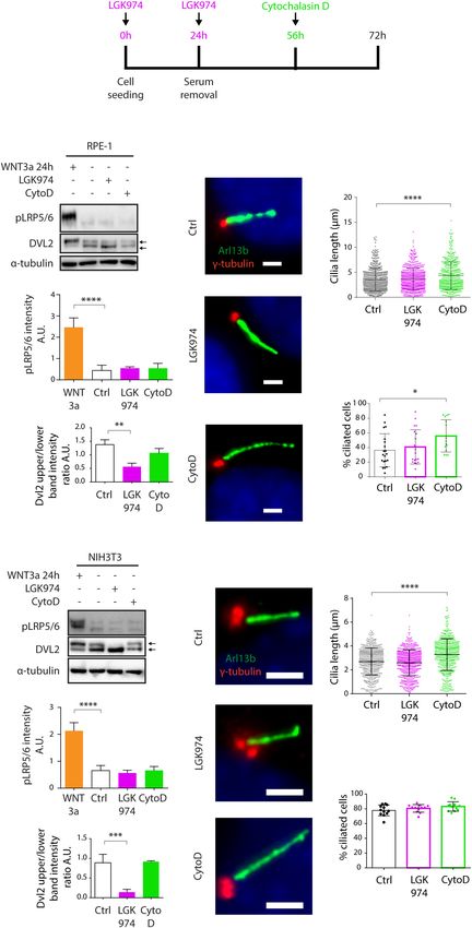
Bernatik et al.                                                                                                  Wnt3a Does Not Induce Ciliogenesis

organizing center coordinating mitosis, primary cilium                     Active β-catenin (ABC) accumulates, translocates to the nucleus
formation is tightly connected with G1/G0 phase (Ford                      where it binds transcription factors of TCF-LEF family to trigger
et al., 2018; Mirvis et al., 2018). The growth of primary cilium           transcription of target genes (Behrens et al., 1996; Molenaar
itself is preceded by the accumulation of vesicles at MC distal            et al., 1996). Not surprisingly, many developmental disorders
appendages (Sorokin, 1962; Westlake et al., 2011; Schmidt et al.,          and cancers are directly caused by WNT pathways deregulation
2012; Lu et al., 2015; Wu et al., 2018) and by the removal of              (Zhan et al., 2017; Humphries and Mlodzik, 2018).
CEP97/CP110 capping complex specifically from MC distal end                    Whilst the connections between primary cilia and hedgehog
(Spektor et al., 2007). Major role in the cilia initiation is linked       signaling are well documented (Huangfu et al., 2003; Corbit
to the Tau tubulin kinase 2 (TTBK2) activity (Goetz et al., 2012).         et al., 2005; Rohatgi et al., 2007), the relationship between cilia
Once recruited to MC by distal appendage protein CEP164                    and WNT signaling is still rather controversial. The exception
(Čajánek and Nigg, 2014; Oda et al., 2014), TTBK2 seems to                here seems to be the WNT/PCP pathway [one of the non-
control both the process of vesicle docking and the CP110/CEP97            canonical WNT pathways (Butler and Wallingford, 2017)], which
removal (Goetz et al., 2012; Lo et al., 2019). In turn, this allows        was described to affect cilia formation and functions via effects
the extension of tubulin-based axoneme sheathed by ciliary                 on cytoskeleton and basal body positioning (Wallingford and
membrane from MC-derived basal body. The formed cilium is                  Mitchell, 2011; May-Simera and Kelley, 2012; Carvajal-Gonzalez
physically separated from the rest of a cell by ciliary transition         et al., 2016; Bryja et al., 2017). As for the WNT/β-catenin
zone, a selective barrier ensuring only specific proteins to enter         pathway, there are reports showing that primary cilia loss or
the cilium (Garcia-Gonzalo and Reiter, 2017; Gonçalves and                 disruption leads to upregulation of the pathway activity (Corbit
Pelletier, 2017; Nachury, 2018). Such compartmentation and                 et al., 2008; McDermott et al., 2010; Wiens et al., 2010; Lancaster
hence specific protein composition of primary cilium is the basis          et al., 2011; Liu et al., 2014; Zingg et al., 2018; Patnaik et al.,
for its instrumental role in the Hedgehog signaling pathway                2019), but also studies that deny any involvement of primary
in vertebrates (Bangs and Anderson, 2017; Nachury and Mick,                cilia in WNT/β-catenin signaling (Huang and Schier, 2009;
2019). In addition, several links between primary cilia and                Ocbina et al., 2009). Some of these discrepancies can perhaps
other signaling pathways such as WNT or TGF-β have recently                be explained by context-specific activity of involved ciliary
emerged (Anvarian et al., 2019).                                           components (Lancaster et al., 2011; Patnaik et al., 2019) or effects
    WNT signaling pathways are developmentally important                   directly on WNT/β-catenin pathway independently of the role
signaling routes regulating cell differentiation, migration, and           in cilia formation (Balmer et al., 2015; Kim et al., 2016), or the
proliferation and their activity controls shaping of the embryo            requirement for intact basal bodies rather than cilia (Vertii et al.,
(Nusse and Clevers, 2017). WNT signaling pathways can be                   2015; Vora et al., 2020).
distinguished based on whether they use β-catenin as an effector               To make the matters even more puzzling, two opposing
protein. The pathway relying on stabilization of β-catenin is              models have recently emerged regarding possible function of
termed the WNT/β-catenin pathway and regulates stemness,                   WNT/β-catenin pathway in cilia formation. Activation of the
cell differentiation and proliferation, while the β-catenin-               WNT/β-catenin pathway in neural progenitors of the developing
independent or non-canonical WNT pathways regulate                         cerebral cortex was reported to hamper cilia formation in
cytoskeleton, cell polarity, and cell movements (Humphries                 mice (Nakagawa et al., 2017), arguing for a negative role
and Mlodzik, 2018; Steinhart and Angers, 2018). These two                  of the excesive WNT/β-catenin signaling in ciliogenesis. In
branches of WNT pathways are activated by a distinct set                   contrast, a recent report described a direct involvement of
of extracellularly secreted WNT ligand proteins (Angers and                WNT/β-catenin signaling pathway in promotion of primary
Moon, 2009). WNTs are posttranslationally palmitoylated                    cilia formation through β-catenin driven stabilization of
by O-Acyl-transferase Porcupine, and only after the lipid                  centriolar satellites in RPE-1 cell line (Kyun et al., 2020). We
modification are the WNT proteins fully active (Willert et al.,            approached this conundrum using cell lines that commonly
2003; Zhai et al., 2004). Following their secretion, WNTs bind             serve as ciliogenesis model systems (RPE-1, NIH3T3, and
to seven-pass transmembrane receptors from Frizzled family                 HEK293). Using either pharmacological or genetic means to
that form heterodimeric complexes with various coreceptors.                manipulate the WNT/β-catenin pathway, we found no evidence
WNT/β-catenin pathway uses LRP5/6 coreceptors (Pinson et al.,              of facilitated ciliogenesis in response to the activation of
2000; Tamai et al., 2000; Wehrli et al., 2000). Signal received by         WNT/β-catenin signaling.
the receptor-coreceptor pair on the cell membrane is then relayed
to Dishevelled (DVL) proteins that, following phosphorylation
by CK1-δ/ε and other kinases (Bernatik et al., 2011; González-             MATERIALS AND METHODS
Sancho et al., 2013; Hanáková et al., 2019), are used both by
the non-canonical and the WNT/β-catenin pathways (Sokol,                   Cell Culture
1996; Wallingford et al., 2000). β-catenin destruction complex,            RPE-1 cells were grown in DMEM/F12 (Thermo Fisher
composed of proteins Adenomatous polyposis coli (APC),                     Scientific, 11320033) supplemented by 10% FBS (Biosera, cat.
AXIN and two kinases; GSK3-β and CK1-α, is then inactivated                No. FB-1101/500), 1% Penicillin/Streptomycin (Biosera, cat. No.
by DVL sequestration of AXIN proteins (Tamai et al., 2004).                XC-A4122/100) and 1% L-glutamine (Biosera, cat. No. XC-
Then β-catenin phosphorylation by GSK3-β and CK1-α on its                  T1715/100), HEK293 T-Rex (referred to as HEK293, cat.no.
N-terminal degron is terminated and the non-phosphorylated                 R71007, Invitrogen) and NIH3T3 cells were grown in DMEM

Frontiers in Cell and Developmental Biology | www.frontiersin.org      2                                    February 2021 | Volume 9 | Article 623753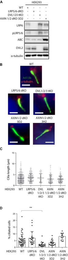
Bernatik et al.                                                                                                  Wnt3a Does Not Induce Ciliogenesis

Glutamax (Thermo Fisher Scientific, 10569069) supplemented
              R
                                                                            (analysis of CP110 and TTBK2 presence on the MC), 3–4 fields
by 10% FBS and 1% Penicillin/Streptomycin. Where indicated,                 of vision (200–400 cells) were analyzed per experimental run,
RPE-1 cells were starved by serum free medium, NIH3T3 cells                 n = 3. Statistical analyses by one-way ANOVA were performed
were starved by 0.1% FBS containing medium, and HEK293 cells                using Graphpad Prism, P < 0.05 (∗ ), P < 0.01 (∗∗ ), P < 0.001
were starved by serum free medium for 24 h. Cells were seeded at            (∗∗∗ ), and P < 0.0001 (∗∗∗∗ ). Results are presented as mean
50,000/well (RPE-1 and NIH3T3) or 120000/well (HEK293) of 24                plus SEM. Primary antibodies used: Arl13b (Proteintech, Cat.no.
well plate. Treatments by small molecules were done for indicated           17711-1-AP), γ-tubulin (Merck, T6557), CP110 (Proteintech,
times: LGK974 (0.4 µM) (Sellcheck, cat. No. S7143) for 72 h                 12780-1-AP), and TTBK2 (Merck, Cat.no. HPA018113).
(LGK974 was re-added to the starvation medium as indicated in
Figure 2A), Cytochalasin D (500nM) (Merck Cat. No. C8273) for               Dual Luciferase (TopFLASH) Assay,
16 h, PF670462 (1 µM) (Merck, SML0795) for 24 h. WNT3a (90                  Transfection of HEK293
ng/ml) (R&D systems, Cat.no. 5036-WN) for 2 h or 24 h.                      Transfection and dual luciferase assay of HEK293 WT and KO
                                                                            cells was carried out as previously described (Paclíková et al.,
Western Blot and Quantification                                             2017). In brief, in 0.1 µg of the pRLtkLuc plasmid and 0.1 µg
Western blot was performed as previously described (Bernatik                of the Super8X TopFlash plasmid per well of 24 well plate were
et al., 2020). Antibodies used: LRP6 (Cell signaling, Cat.no.               cotransfected, on the next day cells were treated by 90ng/ml
#2560), Phospho-LRP5/6 (Ser1493/Ser1490; Cell signaling,                    WNT3a and signal was measured after 24 h treatment.
Cat.no. #2568), AXIN1 (Cell signaling, Cat.no. #3323) DVL2
(Cell signaling, Cat.no. #3216), Active-β-catenin (Merck, Cat.              CRISPR/Cas9 Generation of LRP5/6
no. 05-665-25UG), and α-tubulin (Proteintech, Cat.no. 66031-
                                                                            Double Knock-Out and AXIN1/2 Double
1-Ig). Quantifications were performed using Fiji distribution
of ImageJ. Intensity of pLRP5/6 and ABC band was measured                   Knock-Out HEK293 Cells
and normalized to mean value from all conditions of given                   Used guide RNAs were following: LRP5 gRNA
experiment. Intensity of LRP6 and DVL2 was calculated as the                gagcgggccgacaagactag, LRP6 gRNA ttgccttagatccttcaagt,
ratio of the upper to lower band intensity (the bands are indicated         AXIN1 gRNA cgaacttctgaggctccacg, and AXIN2 gRNA
by arrows in the corresponding Figures) and normalized to mean              tccttattgggcgatcaaga. gRNAs were cloned into pSpCas9
value from all conditions of given experiment. Quantification was           (BB)-2A-GFP (PX458) (Addgene plasmid, 41815) or pU6-
performed on n = 3. Statistical analyses by students t-test or one-         (BbsI)_CBh-Cas9-T2A-mCherry (Addgene plasmid, 64324)
way ANOVA were performed using Graphpad Prism, P < 0.05                     plasmids. Following transfection by Lipofectamine 2000
(∗ ), P < 0.01 (∗∗ ), P < 0.001 (∗∗∗ ), and P < 0.0001 (∗∗∗∗ ).             (Thermo Fisher Scientific) the transfected cells were FACS
                                                                            sorted [FACSAria Fusion (BD Biosciences)] and clonally
Immunocytochemistry                                                         expanded. Genotyping of LRP5 KO and AXIN2 KO mutants
RPE-1, NIH3T3 and HEK293 cells were seeded on glass                         was done following genomic DNA isolation (DirectPCR
coverslips, treated as indicated, washed by PBS and fixed for               Lysis Reagent; 301-C, Viagen Biotech) by PCR using
10 min in −20o C methanol, washed 3× by PBS, blocked (2%                    DreamTaq DNA Polymerase (Thermo Fisher Scientific). Used
BSA in PBS with 0.01% NaN3 ), 3× washed by PBS, incubated                   primers: LRP5 forward: gttcggtctgacgcagtaca, LRP5 reversed:
with primary antibodies for 1 h, 3× washed by PBS, incubated                aggatggcctcaatgactgt, AXIN2 forward: cagtgccaggggaagaag, and
with secondary antibodies (Goat anti-Rabbit IgG Alexa Fluor                 AXIN2 reversed: gtcttggtggcaggcttc. PCR products were cut
488 Secondary Antibody, Cat.no. A11008; Goat anti-Mouse IgG                 by BfaI (R0568S, NEB) in case of LRP5 KO and Hpy188III
Alexa Fluor 568 Secondary Antibody, Cat.no. A11031, all from                (R0622S, NEB) for AXIN2 KO screening, respectively. Successful
Thermo Fisher Scientific) for 2 h in dark, washed 3× by PBS,                disruption of individual ORFs was confirmed by sequencing,
incubated 5 min with DAPI, 2× washed by PBS and mounted to                  Supplementary Figures 2A,D, 3A–E.
glycergel (DAKO #C0563). Microscopy analysis was done using
Zeiss AxioImager.Z2 with Hamamatsu ORCA Flash 4.0 camera,
                                                                            RESULTS
63× Apo oil immersion objective, and ZEN Blue 2.6 acquisition
SW (Zeiss). Image stacks acquired using Zeiss AxioImager.Z2
were projected as maximal intensity images by using ImageJ
                                                                            Treatment by Recombinant WNT3a
distribution FIJI (Schindelin et al., 2012). Where appropriate,             Induces WNT/β-Catenin Pathway
contrast and/or brightness of images were adjusted by using                 Activation but Not Ciliogenesis
Photoshop CS5 (Adobe) or FIJI. To assess effects on ciliogenesis            First, we tested if primary ciliogenesis can be modulated by
or cilia length, at least 4–5 fields of vision (approximately 200–400       activation of WNT/β-catenin pathway in RPE-1 by recombinant
cells per experiment) were analyzed per experimental condition,             WNT3a. Experiment outline is schematized (Figure 1A). We
on at least n = 3. Cilia present on HEK293 cells were counted               initially treated the cells for 2 h. While we observed the expected
manually. Cilia present on RPE-1 or NIH3T3 were counted                     accumulation of active β-catenin (ABC), phosphorylation and
in ACDC software semiautomatic mode, all cilia present were                 shift of LRP5/6 coreceptors (LRP6, pLRP5/6, S1490/S1493),
verified and adjusted manually as recommended (Lauring et al.,              and phosphorylation and upshift of DVL2 (Figures 1B–E and
2019). For the experiments in Supplementary Figures 1D–G                    Supplementary Figure 1A), WNT3a did not alter the length

Frontiers in Cell and Developmental Biology | www.frontiersin.org       3                                   February 2021 | Volume 9 | Article 623753
Bernatik et al. Wnt3a Does Not Induce Ciliogenesis
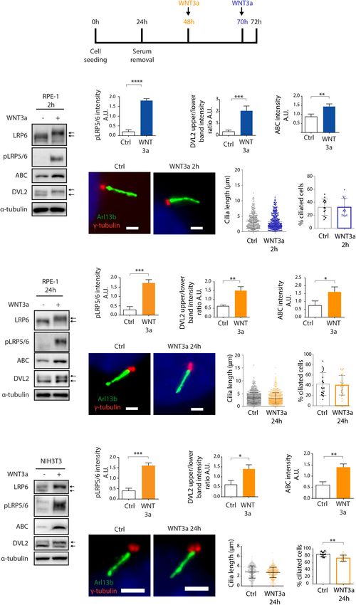
FIGURE 1 | WNT3a does not promote ciliogenesis or cilia length. (A) Experimental scheme of WNT3a treatment experiment. Cells were seeded and grown for 24 h,
then starved for additional 48 h. A 2 h treatment (RPE-1) by WNT3a is indicated in blue, 24 h treatment is indicated in orange (RPE-1 and NIH3T3). (B) Western blot
analysis of 2 h WNT3a treatment of RPE-1. The treatment leads to LRP6 shift and increased LRP5/6 phosphorylation, DVL2 phosphorylation and upshift, and
accumulation of ABC. The quantitation of pLRP5/6 intensity is shown in (C) n = 3, DVL2 band intensities (upper to lower band intensity ratio, the bands are indicated
by arrows) is shown in (D) n = 3, the quantification of relative ABC levels is presented in (E) n = 3. (F) Representative images of RPE-1 cells treated by WNT3a or
vehicle (control) for 2 h and stained for Arl13b (green) and γ-tubulin (red). Scale bar = 2 µm. DAPI (blue) was used to counter stain nuclei. The corresponding
quantification of the cilia length (G) and the percentage of cells with Arl13+ cilium (H). Each dot indicates either length of a single primary cilium (G) or percentage of
ciliated cells in a single image (H). (I) Western blot analysis of 24 h WNT3a treatment of RPE-1. The treatment leads to LRP6 shift, increased LRP5/6
phosphorylation, DVL2 phosphorylation and upshift, and accumulation of ABC. The quantification of pLRP5/6 intensity is shown in (J) n = 3, DVL2 bands (indicated
by arrows) intensity ratio is shown in (K) n = 3, quantification of relative ABC levels is presented in (L) n = 3. (M) Representative images of RPE-1 cells treated by
WNT3a or vehicle (control) for 24 h and stained for Arl13b (green) and γ-tubulin (red). Scale bar = 2 µm. DAPI (blue) was used to counter stain nuclei. The
corresponding quantification of the cilia length and the percentage of cells with Arl13+ cilium is shown in (N,O), respectively. Each dot indicates either length
of a single primary cilium (N) or percentage of ciliated cells in a single image (O) n = 4. (P) Western blot analysis of NIH3T3 cells treated by WNT3a for 24 h shows LRP6
(Continued)
Frontiers in Cell and Developmental Biology | www.frontiersin.org 4 February 2021 | Volume 9 | Article 623753Bernatik et al. Wnt3a Does Not Induce Ciliogenesis
FIGURE 1 | Continued
shift and LRP5/6 phosphorylation, DVL2 phosphorylation and upshift, and accumulation of ABC. The quantification of pLRP5/6 intensity is shown in (Q) n = 3, DVL2
band intensities (upper to lower band intensity ratio, the bands are indicated by arrows) is shown in (R) n = 3, quantification of relative ABC intensity (S) n = 3.
(T) Representative images of NIH3T3 cells treated by WNT3a for 24 h, stained for Arl13b (green), and γ-tubulin (red). Scale bar = 2 µm. DAPI (blue) was used to
counter stain nuclei. The corresponding quantification of the cilia length (U) and the percentage of cells with Arl13+ cilium (V). Each dot indicates either length of a
single primary cilium (U) or percentage of ciliated cells in one image frame (V) n = 3.
or number of Arl13b positive cilia (Figures 1F–H). Next, we but LGK974 treatment failed to show any effect on cilia length,
examined effects of prolonged treatment of RPE-1 cells by in contrast to CytoD treatment (Figures 2K,L). We noted the
WNT3a. Importantly, we were able to detect that WNT/β-catenin CytoD treatment in NIH3T3 did not increase the cilia numbers
pathway is still active after 24 h, as visible from the mobility (Figure 2M), possibly due to high basal ciliation rate of NIH3T3
shift of LRP6 (Figure 1I and Supplementary Figure 1B) or compared to RPE-1. In sum, these data imply that signaling
the elevated levels of ABC, pLRP5/6 or DVL2 phosphorylation mediated by endogenous WNT ligands is not required for
(Figures 1I–L), but the treatment did not show any notable primary ciliogenesis.
effects on cilia length or numbers (Figures 1M–O). In agreement
with these data, WNT3a treatment failed to alter either TTBK2 Genetic Ablation of WNT/β-Catenin
recruitment to MC (Supplementary Figures 1D,E) or MC- Pathway Does Not Alter Primary
specific loss of CP110 (Supplementary Figures 1F,G). To Ciliogenesis
corroborate these findings, we also tested the influence of WNT3a
To corroborate our findings, we established a panel of HEK293
in NIH3T3 cell line. Similarly, to RPE-1, WNT3a treatment for
cells devoid of critical components of WNT signaling pathways.
24 h was able to activate the WNT/β-catenin pathway in NIH3T3
To specifically block the course of WNT/β-catenin pathway
cells (Figures 1P–S and Supplementary Figure 1C), but the
we used LRP5/6 double knock out HEK293 cells, to block
length of cilia was not affected (Figures 1T,U). Intriguingly, we
the course of any WNT signaling pathway we used DVL1/2/3
detected a decrease in the percentage of ciliated cells following
triple knock out HEK293 cells (Paclíková et al., 2017) and to
the WNT3a treatment (Figure 1V).
overactivate WNT/β-catenin pathway we used AXIN1/2 double
knock out HEK293 cells.
Inhibition of WNT Secretion Halts WNT First, we have verified successful disruption of LRP5 gene
Signaling but Not Ciliogenesis by sequencing (Supplementary Figures 2A, 3A), and lack of
Having found WNT3a-activated WNT/β-catenin signaling is not LRP6 and pLRP5/6 signals in LRP5/6 null cells by western blot
sufficient to promote cilia formation, we tested a possibility that (Figure 3A and Supplementary Figure 2B). Furthermore, we
steady state WNT signaling is required for effective ciliogenesis. confirmed these cells cannot activate WNT/β-catenin signaling
WNT/β-catenin pathway is intensively studied as a driver of (Supplementary Figure 2C). Similarly, we confirmed disruption
oncogenic growth, thus there are currently available various of AXIN1 and AXIN2 genes in AXIN1/2 dKO by sequencing
small molecules that inhibit WNT ligand secretion. To this (Supplementary Figures 2D, 3B–E), and lack of AXIN1 by
end, we used a Porcupine inhibitor LGK974 to block the western blot (Supplementary Figure 2E). In addition, we
secretion of endogenous WNT ligands and in turn block the observed that loss of AXIN1/2 function leads to excessive ABC
steady state WNT signaling (Jiang et al., 2013). As a positive accumulation (Figure 3A) and in turn to overactivation of
control in these experiments we used cytochalasin D (CytoD), WNT/β-catenin signaling in AXIN1/2dKO cells (Supplementary
an actin polymerization inhibitor known to facilitate ciliogenesis Figure 2F), as expected.
and promote cilia elongation (Kim et al., 2015). Experiment Having characterized our model system, we examined cilia
outline is schematized (Figure 2A). While we observed no formation in those cells. Consistently with previous work,
visible change in pLRP5/6 levels following the LGK974 treatment HEK293 cells form cilia less frequently than RPE-1 or NIH3T3
(Figures 2B,C), perhaps because the basal levels of pLRP5/6 cells (Lancaster et al., 2011; Bernatik et al., 2020). We were
were at our detection limit, we detected downshift of DVL2 able to detect about 5% of cells with Arl13b+ primary cilium
(Figures 2B,D) confirming the endogenous WNT signaling in WT HEK293. The percentage of ciliated cells, but not the
was successfully ablated. Importantly, however, the LGK974 cilia length, was reduced in DVL1/2/3 tKO cells (Figures 3B–
treatment did not alter primary ciliogenesis, in contrast to D). This observation is in agreement with the role of DVL and
CytoD that facilitated CP110 removal from MC (Supplementary WNT/PCP pathway in the regulation of basal body positioning
Figures 1F,G), cilia elongation (Figures 2E,F), and formation and ciliogenesis (Park et al., 2008; Shnitsar et al., 2015; Sampilo
(Figures 2E,G). In addition, we inhibited WNT signaling at the et al., 2018). Systemic activation of WNT/β-catenin pathway
level of CK1-δ/ε using small molecule PF670462 (Badura et al., by AXIN1/2 removal produced a somewhat mixed result.
2007; Janovska et al., 2018), and found no effect on ciliogenesis Using AXIN1/2 dKO clone 3D2 we initially observed a non-
(Supplementary Figures 1H–J). Next, we applied the approach significant negative trend on the cilia formation. However, this
outlined in Figure 2A also to NIH3T3 cells, with very similar was not confirmed using an independent clone 3H2 (Figure 3D).
results - LGK974 caused no visible change in pLRP5/6 levels but Importantly, the ablation of WNT/β-catenin pathway in LRP5/6
inhibited WNT signaling on the level of DVL2 (Figures 2H–J), dKO cells had no effect on either the percentage of ciliated cells
Frontiers in Cell and Developmental Biology | www.frontiersin.org 5 February 2021 | Volume 9 | Article 623753Bernatik et al. Wnt3a Does Not Induce Ciliogenesis FIGURE 2 | Inhibition of WNT secretion has no effect on ciliogenesis or cilia length. (A) Experimental scheme illustrating the time points of LGK974 (Purple) or CytoD (Green) treatments. (B) Western blot analysis of RPE-1 treated by LGK974 or CytoD. WNT3a was used as positive control to activate WNT/β-catenin pathway. pLRP5/6 intensity is quantified in (C) n = 3, DVL2 shift (upper to lower band intensity ratio) is quantified in (D) n = 3. (E) Representative images of RPE-1 cells following the indicated treatment, stained for Arl13b (green) and γ-tubulin (red). Scale bar = 2 µm. DAPI (blue) was used to counter stain nuclei. Quantification of the cilia length (F) and the percentage of cells with Arl13+ cilium (G). Each dot indicates either length of a single primary cilium (F) or percentage of ciliated cells in one image frame (G) n ≥ 3. (H) Western blot analysis NIH3T3 treated by LGK974 or CytoD. pLRP5/6 intensity is quantified in (I) n = 3, (J) Quantification of DVL2 band intensities (upper to lower band intensity ratio) n = 3. (K) Representative images of NIH3T3 cells following treatment with LGK974 or CytoD, stained for Arl13b (green) and γ-tubulin (red). Scale bar = 2 µm. DAPI (blue) was used to counter stain nuclei. Quantification of the cilia length (L) and the percentage of cells with Arl13+ cilium (M). Each dot indicates either length of a single primary cilium (L) or percentage of ciliated cells in one image frame (M) n = 3. Frontiers in Cell and Developmental Biology | www.frontiersin.org 6 February 2021 | Volume 9 | Article 623753
Bernatik et al.                                                                                                                        Wnt3a Does Not Induce Ciliogenesis

                                                                                          FIGURE 3 | Continued
                                                                                          and DAPI (blue), and analyzed by IF microscopy. Representative images are
                                                                                          shown in (B). Scale bar = 2 µm. Quantification of cilia length and percentage
                                                                                          of ciliated cells is shown in (C,D), respectively Each dot indicates either length
                                                                                          of a single primary cilium (C) or percentage of ciliated cells in one image (D).
                                                                                          n = 4.

                                                                                         or cilia length (Figures 3B–D), in agreement with our earlier
                                                                                         observations based on pharmacological inhibition of endogenous
                                                                                         WNT signaling in RPE-1 or NIH3T3. In sum, from these data
                                                                                         we conclude that WNT/β-catenin signaling is not required for
                                                                                         effective ciliogenesis.

                                                                                         DISCUSSION
                                                                                         Regulation of ciliogenesis is a complex process involving
                                                                                         multiple factors directly or indirectly influencing cilia initiation
                                                                                         and elongation. The regulators of cilium formation encompass
                                                                                         a wide range of molecules such as components of centrioles,
                                                                                         regulators of vesicular trafficking, intraflagellar transport
                                                                                         proteins, membrane proteins, and components of cytoskeleton
                                                                                         (Seeley and Nachury, 2010; Ishikawa and Marshall, 2017; Wang
                                                                                         and Dynlacht, 2018; Conkar and Firat-Karalar, 2020).
                                                                                             WNT3a is considered a prototypical “canonical” WNT
                                                                                         ligand that activates WNT/β-catenin pathway (Willert et al.,
                                                                                         2003). Moreover, WNT3a and hence the WNT/β-catenin
                                                                                         pathway are well known for their mitogenic potential in many
                                                                                         experimental systems (Niehrs and Acebron, 2012). In addition,
                                                                                         WNT/β-catenin pathway has been shown to act mainly during
                                                                                         G2/M phase of the cell cycle (Davidson et al., 2009), while
                                                                                         primary cilia form during G0/G1 and during the G2/M they
                                                                                         disassemble (Rieder et al., 1979; Ford et al., 2018). Furthermore,
                                                                                         mitogenic signals typically promote cilium disassembly (Rieder
                                                                                         et al., 1979; Tucker et al., 1979; Pugacheva et al., 2007). From this
                                                                                         perspective, the recently reported positive role of WNT3a and
                                                                                         WNT/β-catenin signaling on primary cilia formation (Kyun et al.,
                                                                                         2020) is counterintuitive and puzzling.
                                                                                             Principally, there are several important methodological
                                                                                         differences between our work and the previous results (Kyun
                                                                                         et al., 2020) which may account for the different outcomes. (1)
                                                                                         In our experiments we activated the WNT/β-catenin pathway
                                                                                         by recombinant WNT3a, in contrast to WNT3a conditioned
                                                                                         medium often used in the previous study (Kyun et al., 2020).
                                                                                         Thus, some of the reported effects of WNT3a conditioned
                                                                                         medium may be a result of secondary effects. (2) We applied up to
                                                                                         24 h stimulation by WNT3a to activate or 72 h LGK974 to block
                                                                                         the pathway, respectively. We cannot formally exclude that the
                                                                                         longer WNT3a treatments used by Kyun et al., could account
                                                                                         for the observed differences. However, we argue this seems
  FIGURE 3 | Ablation of WNT β-catenin pathway does not alter primary
                                                                                         unlikely, given that full activation of the WNT/β-catenin pathway
  ciliogenesis. (A) Western blot analysis of individual HEK293 KO cell lines using       or cilium formation typically happens within several hours
  the indicated antibodies. Note that LRP5/6 dKO cells lack LRP6 and phospho             following the proper stimuli (Bryja et al., 2007; Naik and Piwnica-
  LRP5/6 (pSer1493/pSer1490), DVL1/2/3 tKO cell do not have detectable                   Worms, 2007; Pitaval et al., 2010; Lu et al., 2015; Wu et al., 2018;
  levels of DVL2. AXIN1/2 dKO cells shown elevated level of ABC. (B–D)
                                                                                         Pejskova et al., 2020). In fact, prolonged WNT/β-catenin pathway
  HEK293 Cells were starved for 48 h, stained for Arl13b (green), γ-tubulin (red),
                                                                       (Continued)
                                                                                         stimulation increases a chance for indirect secondary effects.
                                                                                         Indeed, WNT signaling has been shown to regulate expression of

Frontiers in Cell and Developmental Biology | www.frontiersin.org                    7                                            February 2021 | Volume 9 | Article 623753
Bernatik et al. Wnt3a Does Not Induce Ciliogenesis
a number of ligands from FGF (Kratochwil et al., 2002; Barrow DATA AVAILABILITY STATEMENT
et al., 2003; Shimokawa et al., 2003; Chamorro et al., 2005;
Hendrix et al., 2006) or BMP (Baker et al., 1999; Kim et al., 2002; The original contributions presented in the study are included
Shu et al., 2005) families that might in turn affect ciliogenesis in the article/Supplementary Material, further inquiries can be
(Neugebauer et al., 2009; Komatsu et al., 2011; Cibois et al., directed to the corresponding author.
2015; Bosakova et al., 2018). 3. Finally, we visualized cilia by
staining for Arl13b, a small GTPase from Arf/Arl-family highly
enriched in the ciliary membrane (Caspary et al., 2007; Cantagrel AUTHOR CONTRIBUTIONS
et al., 2008; Hori et al., 2008; Duldulao et al., 2009; Cevik
et al., 2010; Li et al., 2010). In the report by Kyun et al., OB designed and performed the experiments, and wrote and
acetylated α-tubulin antibody staining was used to assess the edited the manuscript. PP and AK performed selection and
cilia length, thickness, and numbers. From this perspective, it verification of CRISPR edited HEK293 cell lines. VB edited
is plausible some of the reported changes in cilia length or the manuscript. LC designed the experiments, and wrote and
thickness in fact reflect changes in the acetylation of ciliary edited the manuscript. All authors contributed to the article and
tubulin rather than changes in cilium size. That being said, there approved the submitted version.
is an evidence that individual cilia differ significantly in the
levels of tubulin post-translation modifications and the levels of
tubulin modifications may dramatically change in response to
the appropriate stimuli (Piperno et al., 1987; Berbari et al., 2013; FUNDING
He et al., 2018).
This work was supported by grants from the Czech Science
Our data show that while WNT3a consistently activates
Foundation (19-05244S) and the Swiss National Science
the WNT/β-catenin pathway, it has no or minor negative
Foundation (IZ11Z0_166533) to LC. OB was supported by funds
effects on ciliogenesis. Elevated β-catenin levels following
from the Faculty of Medicine MU to junior researcher (Ondrej
APC ablation have been related to reduced ciliogenesis
Bernatik, ROZV/28/LF/2020). VB was supported from European
and cell cycle defects in the developing cortex in mice
Structural and Investment Funds, Operational Program
(Nakagawa et al., 2017). Indeed, we detected modest decrease
Research, Development and Education – Preclinical Progression
in the percentage of ciliated NIH3T3 cells following WNT3a
of New Organic Compounds with Targeted Biological Activity”
induced β-catenin accumulation. We speculate we did
(Preclinprogress) – CZ.02.1.01/0.0/0.0/16_025/0007381.
not observe comparable negative effect on cilia following
the WNT/β-catenin pathway activation after AXIN1/2 loss
due to abnormal cell cycle regulation in HEK293, which
hampers detection of relatively subtle deviations in their ACKNOWLEDGMENTS
cell cycle progression (Löber et al., 2002; Stepanenko and
Dmitrenko, 2015). These data are in contrast to Kyun et al., We acknowledge the core facility CELLIM supported by the
where accumulation of β-catenin by WNT3a conditioned Czech-BioImaging large RI project (LM2018129 funded by MEYS
medium treatment or by expression of S45A non-degradable CR) for their support with obtaining scientific data presented in
oncogenic mutant variant of β-catenin (Liu et al., 2002) this article. hCas9 (BB)-2A-GFP was a gift from George Church
facilitates ciliogenesis. (Addgene plasmid #41815) and pU6-(BbsI)_CBh-Cas9-T2A-
In sum, we found no evidence that endogenous mCherry was a gift from Ralf Kuehn (Addgene plasmid # 64324).
WNT/β-catenin signaling, while ablated either pharmacologically
in RPE-1 or NIH3T3 by LGK974, or genetically by removal
of LRP5/6 in HEK293, is required for primary cilia to form. SUPPLEMENTARY MATERIAL
Our findings presented in this article challenge some of
the published evidence and argue against positive role of The Supplementary Material for this article can be found
WNT3a or WNT/β-catenin pathway in ciliogenesis or cilia online at: https://www.frontiersin.org/articles/10.3389/fcell.2021.
length regulation. 623753/full#supplementary-material
REFERENCES rhythms under free-running and entrained conditions. J. Pharmacol. Exp. Ther.
322, 730–738. doi: 10.1124/jpet.107.122846
Angers, S., and Moon, R. T. (2009). Proximal events in Wnt signal transduction. Baker, J. C., Beddington, R. S. P., and Harland, R. M. (1999). Wnt signaling in
Nat. Rev. Mol. Cell Biol. 10, 468–477. doi: 10.1038/nrm2717 Xenopus embryos inhibits Bmp4 expression and activates neural development.
Anvarian, Z., Mykytyn, K., Mukhopadhyay, S., Pedersen, L. B., and Christensen, Genes Dev. 13, 3149–3159. doi: 10.1101/gad.13.23.3149
S. T. (2019). Cellular signalling by primary cilia in development, organ Balmer, S., Dussert, A., Collu, G. M., Benitez, E., Iomini, C., and Mlodzik,
function and disease. Nat. Rev. Nephrol. 15, 199–219. doi: 10.1038/s41581-019- M. (2015). Components of intraflagellar transport complex a function
0116-9 independently of the cilium to regulate canonical Wnt signaling
Badura, L., Swanson, T., Adamowicz, W., Adams, J., Cianfrogna, J., Fisher, K., in Drosophila. Dev. Cell 34, 705–718. doi: 10.1016/j.devcel.2015.
et al. (2007). An inhibitor of casein kinase Iε induces phase delays in circadian 07.016
Frontiers in Cell and Developmental Biology | www.frontiersin.org 8 February 2021 | Volume 9 | Article 623753Bernatik et al. Wnt3a Does Not Induce Ciliogenesis
Bangs, F., and Anderson, K. V. (2017). Primary cilia and mammalian hedgehog ciliary and non-ciliary mechanisms. Nat. Cell Biol. 10, 70–76. doi: 10.1038/
signaling. Cold Spring Harb. Perspect. Biol. 9:a028175. doi: 10.1101/cshperspect. ncb1670
a028175 Davidson, G., Shen, J., Huang, Y. L., Su, Y., Karaulanov, E., Bartscherer, K., et al.
Barrow, J. R., Thomas, K. R., Boussadia-Zahui, O., Moore, R., Kemler, R., Capecchi, (2009). Cell cycle control of Wnt receptor activation. Dev. Cell 17, 788–799.
M. R., et al. (2003). Ectodermal Wnt3β-catenin signaling is required for the doi: 10.1016/j.devcel.2009.11.006
establishment and maintenance of the apical ectodermal ridge. Genes Dev. 17, Duldulao, N. A., Lee, S., and Sun, Z. (2009). Cilia localization is essential for in vivo
394–409. doi: 10.1101/gad.1044903 functions of the Joubert syndrome protein Arl13b/Scorpion. Development 136,
Behrens, J., Von Kries, J. P., Kühl, M., Bruhn, L., Wedlich, D., Grosschedl, R., et al. 4033–4042. doi: 10.1242/dev.036350
(1996). Functional interaction of β-catenin with the transcription factor LEF- 1. Ford, M. J., Yeyati, P. L., Mali, G. R., Keighren, M. A., Waddell, S. H., Mjoseng,
Nature 382, 638–642. doi: 10.1038/382638a0 H. K., et al. (2018). A cell/cilia cycle biosensor for single-cell kinetics reveals
Berbari, N. F., Sharma, N., Malarkey, E. B., Pieczynski, J. N., Boddu, R., Gaertig, persistence of cilia after G1/S transition is a general property in cells and mice.
J., et al. (2013). Microtubule modifications and stability are altered by cilia Dev. Cell 47, 509.e5–523.e5. doi: 10.1016/j.devcel.2018.10.027
perturbation and in cystic kidney disease. Cytoskeleton 70, 24–31. doi: 10.1002/ Garcia-Gonzalo, F. R., and Reiter, J. F. (2017). Open sesame: how transition fibers
cm.21088 and the transition zone control ciliary composition. Cold Spring Harb. Perspect.
Bernatik, O., Pejskova, P., Vyslouzil, D., Hanakova, K., Zdrahal, Z., and Cajanek, Biol. 9:a028134. doi: 10.1101/cshperspect.a028134
L. (2020). Phosphorylation of multiple proteins involved in ciliogenesis by Tau Goetz, S. C., Liem, K. F., and Anderson, K. V. (2012). The spinocerebellar ataxia-
Tubulin kinase 2. Mol. Biol. Cell 31, 1032–1046. doi: 10.1091/MBC.E19-06- associated gene tau tubulin kinase 2 controls the initiation of ciliogenesis. Cell
0334 151, 847–858. doi: 10.1016/j.cell.2012.10.010
Bernatik, O., Sri Ganji, R., Dijksterhuis, J. P., Konik, P., Cervenka, I., Polonio, Gonçalves, J., and Pelletier, L. (2017). The ciliary transition zone: finding the pieces
T., et al. (2011). Sequential activation and inactivation of dishevelled in the and assembling the gate. Mol. Cells 40, 243–253. doi: 10.14348/molcells.2017.
Wnt/β-catenin pathway by casein kinases. J. Biol. Chem. 286, 10396–10410. 0054
doi: 10.1074/jbc.M110.169870 González-Sancho, J. M., Greer, Y. E., Abrahams, C. L., Takigawa, Y., Baljinnyam, B.,
Bosakova, M. K., Varecha, M., Hampl, M., Duran, I., Nita, A., Buchtova, M., Lee, K. H., et al. (2013). Functional consequences of Wnt-induced dishevelled
et al. (2018). Regulation of ciliary function by fibroblast growth factor signaling 2 phosphorylation in canonical and noncanonical Wnt signaling. J. Biol. Chem.
identifies FGFR3-related disorders achondroplasia and thanatophoric dysplasia 288, 9428–9437. doi: 10.1074/jbc.M112.448480
as ciliopathies. Hum. Mol. Genet. 27, 1093–1105. doi: 10.1093/hmg/ddy031 Han, Y. G., Kim, H. J., Dlugosz, A. A., Ellison, D. W., Gilbertson, R. J., and Alvarez-
Bryja, V., Červenka, I., and Čajánek, L. (2017). The connections of Wnt pathway Buylla, A. (2009). Dual and opposing roles of primary cilia in medulloblastoma
components with cell cycle and centrosome: side effects or a hidden logic? development. Nat. Med. 15, 1062–1065. doi: 10.1038/nm.2020
Crit. Rev. Biochem. Mol. Biol. 52, 614–637. doi: 10.1080/10409238.2017.13 Hanáková, K., Bernatík, O., Kravec, M., Micka, M., Kumar, J., Harnoš, J., et al.
50135 (2019). Comparative phosphorylation map of Dishevelled 3 links phospho-
Bryja, V., Schulte, G., and Arenas, E. (2007). Wnt-3a utilizes a novel low dose and signatures to biological outputs. Cell Commun. Signal. 17:170. doi: 10.1186/
rapid pathway that does not require casein kinase 1-mediated phosphorylation s12964-019-0470-z
of Dvl to activate β-catenin. Cell. Signal. 19, 610–616. doi: 10.1016/j.cellsig.2006. He, K., Ma, X., Xu, T., Li, Y., Hodge, A., Zhang, Q., et al. (2018). Axoneme
08.011 polyglutamylation regulated by Joubert syndrome protein ARL13B controls
Butler, M. T., and Wallingford, J. B. (2017). Planar cell polarity in development and ciliary targeting of signaling molecules. Nat. Commun. 9:3310. doi: 10.1038/
disease. Nat. Rev. Mol. Cell Biol. 18, 375–388. doi: 10.1038/nrm.2017.11 s41467-018-05867-1
Čajánek, L., and Nigg, E. A. (2014). Cep164 triggers ciliogenesis by recruiting Hendrix, N. D., Wu, R., Kuick, R., Schwartz, D. R., Fearon, E. R., and Cho, K. R.
Tau tubulin kinase 2 to the mother centriole. Proc. Natl. Acad. Sci. U. S. A. (2006). Fibroblast growth factor 9 has oncogenic activity and is a downstream
111:E2841-50. doi: 10.1073/pnas.1401777111 target of Wnt signaling in ovarian endometrioid adenocarcinomas. Cancer Res.
Cantagrel, V., Silhavy, J. L., Bielas, S. L., Swistun, D., Marsh, S. E., Bertrand, J. Y., 66, 1354–1362. doi: 10.1158/0008-5472.CAN-05-3694
et al. (2008). Mutations in the cilia gene ARL13B lead to the classical form of Hildebrandt, F., Benzing, T., and Katsanis, N. (2011). Ciliopathies. N. Engl. J. Med.
joubert syndrome. Am. J. Hum. Genet. 83, 170–179. doi: 10.1016/j.ajhg.2008.06. 364, 1533–1543. doi: 10.1056/nejmra1010172
023 Hori, Y., Kobayashi, T., Kikko, Y., Kontani, K., and Katada, T. (2008). Domain
Carvajal-Gonzalez, J. M., Mulero-Navarro, S., and Mlodzik, M. (2016). Centriole architecture of the atypical Arf-family GTPase Arl13b involved in cilia
positioning in epithelial cells and its intimate relationship with planar cell formation. Biochem. Biophys. Res. Commun. 373, 119–124. doi: 10.1016/j.bbrc.
polarity. BioEssays 38, 1234–1245. doi: 10.1002/bies.201600154 2008.06.001
Caspary, T., Larkins, C. E., and Anderson, K. V. (2007). The graded response Huang, P., and Schier, A. F. (2009). Dampened Hedgehog signaling but normal
to sonic hedgehog depends on cilia architecture. Dev. Cell 12, 767–778. doi: Wnt signaling in zebrafish without cilia. Development 136, 3089–3098. doi:
10.1016/j.devcel.2007.03.004 10.1242/dev.041343
Cevik, S., Hori, Y., Kaplan, O. I., Kida, K., Toivenon, T., Foley-Fisher, C., et al. Huangfu, D., Liu, A., Rakeman, A. S., Murcia, N. S., Niswander, L., and Anderson,
(2010). Joubert syndrome Arl13b functions at ciliary membranes and stabilizes K. V. (2003). Hedgehog signalling in the mouse requires intraflagellar transport
protein transport in Caenorhabditis elegans. J. Cell Biol. 188, 953–969. doi: proteins. Nature 426, 83–87. doi: 10.1038/nature02061
10.1083/jcb.200908133 Humphries, A. C., and Mlodzik, M. (2018). From instruction to output: Wnt/PCP
Chamorro, M. N., Schwartz, D. R., Vonica, A., Brivanlou, A. H., Cho, K. R., and signaling in development and cancer. Curr. Opin. Cell Biol. 51, 110–116. doi:
Varmus, H. E. (2005). FGF-20 and DKK1 are transcriptional targets of β-catenin 10.1016/j.ceb.2017.12.005
and FGF-20 is implicated in cancer and development. EMBO J. 24, 73–84. Ishikawa, H., and Marshall, W. F. (2017). Intraflagellar transport and ciliary
doi: 10.1038/sj.emboj.7600460 dynamics. Cold Spring Harb. Perspect. Biol. 9:a021998. doi: 10.1101/cshperspect.
Cibois, M., Luxardi, G., Chevalier, B., Thomé, V., Mercey, O., Zaragosi, L. E., a021998
et al. (2015). BMP signalling controls the construction of vertebrate mucociliary Janovska, P., Verner, J., Kohoutek, J., Bryjova, L., Gregorova, M., Dzimkova, M.,
epithelia. Development 142, 2352–2363. doi: 10.1242/dev.118679 et al. (2018). Casein kinase 1 is a therapeutic target in chronic lymphocytic
Conkar, D., and Firat-Karalar, E. N. (2020). Microtubule-associated proteins leukemia. Blood 131, 1206–1218. doi: 10.1182/blood-2017-05-786947
and emerging links to primary cilium structure, assembly, maintenance, and Jenks, A. D., Vyse, S., Wong, J. P., Kostaras, E., Keller, D., Burgoyne, T., et al. (2018).
disassembly. FEBS J. doi: 10.1111/febs.15473 Online ahead of print Primary cilia mediate diverse kinase inhibitor resistance mechanisms in cancer.
Corbit, K. C., Aanstad, P., Singla, V., Norman, A. R., Stainier, D. Y. R., and Reiter, Cell Rep. 23, 3042–3055. doi: 10.1016/j.celrep.2018.05.016
J. F. (2005). Vertebrate smoothened functions at the primary cilium. Nature 437, Jiang, X., Hao, H. X., Growney, J. D., Woolfenden, S., Bottiglio, C., Ng, N., et al.
1018–1021. doi: 10.1038/nature04117 (2013). Inactivating mutations of RNF43 confer Wnt dependency in pancreatic
Corbit, K. C., Shyer, A. E., Dowdle, W. E., Gaulden, J., Singla, V., and Reiter, ductal adenocarcinoma. Proc. Natl. Acad. Sci. U.S.A. 110, 12649–12654. doi:
J. F. (2008). Kif3a constrains β-catenin-dependent Wnt signalling through dual 10.1073/pnas.1307218110
Frontiers in Cell and Developmental Biology | www.frontiersin.org 9 February 2021 | Volume 9 | Article 623753Bernatik et al. Wnt3a Does Not Induce Ciliogenesis
Kim, J., Jo, H., Hong, H., Kim, M. H., Kim, J. M., Lee, J. K., et al. (2015). Actin Naik, S., and Piwnica-Worms, D. (2007). Real-time imaging of β-catenin dynamics
remodelling factors control ciliogenesis by regulating YAP/TAZ activity and in cells and living mice. Proc. Natl. Acad. Sci. U.S.A. 104, 17465–17470. doi:
vesicle trafficking. Nat. Commun. 6:6781. doi: 10.1038/ncomms7781 10.1073/pnas.0704465104
Kim, J. S., Crooks, H., Dracheva, T., Nishanian, T. G., Singh, B., Jen, J., et al. (2002). Nakagawa, N., Li, J., Yabuno-Nakagawa, K., Eom, T. Y., Cowles, M., Mapp,
Oncogenic β-catenin is required for bone morphogenetic protein 4 expression T., et al. (2017). APC sets the Wnt tone necessary for cerebral cortical
in human cancer cells. Cancer Res. 62, 2744–2748. progenitor development. Genes Dev. 31, 1679–1692. doi: 10.1101/gad.3026
Kim, M., Suh, Y. A., Oh, J. H., Lee, B. R., Kim, J., and Jang, S. J. (2016). KIF3A 79.117
binds to β-arrestin for suppressing Wnt/β-catenin signalling independently Neugebauer, J. M., Amack, J. D., Peterson, A. G., Bisgrove, B. W., and Yost, H. J.
of primary cilia in lung cancer. Sci. Rep. 6:32770. doi: 10.1038/srep3 (2009). FGF signalling during embryo development regulates cilia length in
2770 diverse epithelia. Nature 458, 651–654. doi: 10.1038/nature07753
Komatsu, Y., Kaartinen, V., and Mishina, Y. (2011). Cell cycle arrest in node cells Niehrs, C., and Acebron, S. P. (2012). Mitotic and mitogenic Wnt signalling. EMBO
governs ciliogenesis at the node to break left-right symmetry. Development 138, J. 31, 2705–2713. doi: 10.1038/emboj.2012.124
3915–3920. doi: 10.1242/dev.068833 Nusse, R., and Clevers, H. (2017). Wnt/β-catenin signaling, disease, and emerging
Kratochwil, K., Galceran, J., Tontsch, S., Roth, W., and Grosschedl, R. (2002). therapeutic modalities. Cell 169, 985–999. doi: 10.1016/j.cell.2017.05.016
FGF4, a direct target of LEF1 and Wnt signaling, can rescue the arrest of tooth Ocbina, P. J. R., Tuson, M., and Anderson, K. V. (2009). Primary cilia are not
organogenesis in Lef1-/- mice. Genes Dev. 16, 3173–3185. doi: 10.1101/gad. required for normal canonical Wnt signaling in the mouse embryo. PLoS One
1035602 4:e6839. doi: 10.1371/journal.pone.0006839
Kyun, M. L., Kim, S. O., Lee, H. G., Hwang, J. A., Hwang, J., Soung, N. K., et al. Oda, T., Chiba, S., Nagai, T., and Mizuno, K. (2014). Binding to Cep164, but
(2020). Wnt3a stimulation promotes primary ciliogenesis through β-catenin not EB1, is essential for centriolar localization of TTBK2 and its function in
phosphorylation-induced reorganization of centriolar satellites. Cell Rep. 30, ciliogenesis. Genes to Cells 19, 927–940. doi: 10.1111/gtc.12191
1447.e5–1462.e5. doi: 10.1016/j.celrep.2020.01.019 Paclíková, P., Bernatík, O., Radaszkiewicz, T. W., and Bryja, V. (2017). The
Lancaster, M. A., Schroth, J., and Gleeson, J. G. (2011). Subcellular spatial N-terminal part of the dishevelled DEP domain is required for Wnt/β-catenin
regulation of canonical Wnt signalling at the primary cilium. Nat. Cell Biol. 13, signaling in mammalian cells. Mol. Cell. Biol. 37:e145-17. doi: 10.1128/mcb.
700–708. doi: 10.1038/ncb2259 00145-17
Lauring, M. C., Zhu, T., Luo, W., Wu, W., Yu, F., and Toomre, D. (2019). New Park, T. J., Mitchell, B. J., Abitua, P. B., Kintner, C., and Wallingford, J. B.
software for automated cilia detection in cells (ACDC). Cilia 8:1. doi: 10.1186/ (2008). Dishevelled controls apical docking and planar polarization of basal
s13630-019-0061-z bodies in ciliated epithelial cells. Nat. Genet. 40, 871–879. doi: 10.1038/n
Li, Y., Wei, Q., Zhang, Y., Ling, K., and Hu, J. (2010). The small GTPases ARL-13 g.104
and ARL-3 coordinate intraflagellar transport and ciliogenesis. J. Cell Biol. 189, Patnaik, S. R., Kretschmer, V., Brücker, L., Schneider, S., Volz, A. K., Oancea-
1039–1051. doi: 10.1083/jcb.200912001 Castillo, L., et al. (2019). Bardet–Biedl Syndrome proteins regulate cilia
Liu, B., Chen, S., Cheng, D., Jing, W., and Helms, J. A. (2014). Primary cilia disassembly during tissue maturation. Cell. Mol. Life Sci. 76, 757–775. doi:
integrate hedgehog and Wnt signaling during tooth development. J. Dent. Res. 10.1007/s00018-018-2966-x
93, 475–482. doi: 10.1177/0022034514528211 Pejskova, P., Reilly, M. L., Bino, L., Bernatik, O., Dolanska, L., Ganji, R. S.,
Liu, C., Li, Y., Semenov, M., Han, C., Baeg, G. H., Tan, Y., et al. (2002). Control of et al. (2020). KIF14 controls ciliogenesis via regulation of Aurora A and is
β-catenin phosphorylation/degradation by a dual-kinase mechanism. Cell 108, important for Hedgehog signaling. J. Cell Biol. 219:e201904107. doi: 10.1083/
837–847. doi: 10.1016/S0092-8674(02)00685-2 JCB.201904107
Lo, C. H., Lin, I. H., Yang, T. T., Huang, Y. C., Tanos, B. E., Chou, P. C., et al. (2019). Pinson, K. I., Brennan, J., Monkley, S., Avery, B. J., and Skarnes, W. C. (2000).
Phosphorylation of CEP83 by TTBK2 is necessary for cilia initiation. J. Cell Biol. An LDL-receptor-related protein mediates Wnt signalling in mice. Nature 407,
218, 3489–3505. doi: 10.1083/JCB.201811142 535–538. doi: 10.1038/35035124
Löber, C., Lenz-Stöppler, C., and Dobbelstein, M. (2002). Adenovirus E1- Piperno, G., LeDizet, M., and Chang, X. J. (1987). Microtubules containing
transformed cells grow despite the continuous presence of transcriptionally acetylated alpha-tubulin in mammalian cells in culture. J. Cell Biol. 104, 289–
active p53. J. Gen. Virol. 83, 2047–2057. doi: 10.1099/0022-1317-83-8-2047 302. doi: 10.1083/jcb.104.2.289
Lu, Q., Insinna, C., Ott, C., Stauffer, J., Pintado, P. A., Rahajeng, J., et al. (2015). Pitaval, A., Tseng, Q., Bornens, M., and Théry, M. (2010). Cell shape and
Early steps in primary cilium assembly require EHD1/EHD3-dependent ciliary contractility regulate ciliogenesis in cell cycle-arrested cells. J. Cell Biol. 191,
vesicle formation. Nat. Cell Biol. 17, 228–240. doi: 10.1038/ncb3109 303–312. doi: 10.1083/jcb.201004003
May-Simera, H., and Kelley, M. W. (2012). Planar cell polarity in the inner Pugacheva, E. N., Jablonski, S. A., Hartman, T. R., Henske, E. P., and Golemis,
ear. Curr. Top. Dev. Biol. 101, 111–140. doi: 10.1016/B978-0-12-394592-1.00 E. A. (2007). HEF1-dependent aurora a activation induces disassembly of the
006-5 primary cilium. Cell 129, 1351–1363. doi: 10.1016/j.cell.2007.04.035
McDermott, K. M., Liu, B. Y., Tlsty, T. D., and Pazour, G. J. (2010). Primary cilia Reiter, J. F., and Leroux, M. R. (2017). Genes and molecular pathways
regulate branching morphogenesis during mammary gland development. Curr. underpinning ciliopathies. Nat. Rev. Mol. Cell Biol. 18, 533–547. doi: 10.1038/
Biol. 20, 731–737. doi: 10.1016/j.cub.2010.02.048 nrm.2017.60
Mirvis, M., Stearns, T., and Nelson, W. J. (2018). Cilium structure, assembly, Rieder, C. L., Jensen, C. G., and Jensen, L. C. W. (1979). The resorption of primary
and disassembly regulated by the cytoskeleton. Biochem. J. 475, 2329–2353. cilia during mitosis in a vertebrate (PtK1) cell line. J. Ultrasructure Res. 68,
doi: 10.1042/BCJ20170453 173–185. doi: 10.1016/S0022-5320(79)90152-7
Mitchison, H. M., and Valente, E. M. (2017). Motile and non-motile cilia in human Rohatgi, R., Milenkovic, L., and Scott, M. P. (2007). Patched1 regulates hedgehog
pathology: from function to phenotypes. J. Pathol. 241, 294–309. doi: 10.1002/ signaling at the primary cilium. Science 317, 372–376. doi: 10.1126/science.
path.4843 1139740
Molenaar, M., Van De Wetering, M., Oosterwegel, M., Peterson-Maduro, J., Sampilo, N. F., Stepicheva, N. A., Zaidi, S. A. M., Wang, L., Wu, W.,
Godsave, S., Korinek, V., et al. (1996). XTcf-3 transcription factor mediates Wikramanayake, A., et al. (2018). Inhibition of microRNA suppression of
β-catenin-induced axis formation in xenopus embryos. Cell 86, 391–399. doi: dishevelled results in Wnt pathway-associated developmental defects in sea
10.1016/S0092-8674(00)80112-9 urchin. Development 145:dev167130. doi: 10.1242/dev.167130
Nachury, M. V. (2018). The molecular machines that traffic signaling receptors into Schindelin, J., Arganda-Carreras, I., Frise, E., Kaynig, V., Longair, M., Pietzsch, T.,
and out of cilia. Curr. Opin. Cell Biol. 51, 124–131. doi: 10.1016/j.ceb.2018.03. et al. (2012). Fiji: an open-source platform for biological-image analysis. Nat.
004 Methods 9, 676–682. doi: 10.1038/nmeth.2019
Nachury, M. V., and Mick, D. U. (2019). Establishing and regulating the Schmidt, K. N., Kuhns, S., Neuner, A., Hub, B., Zentgraf, H., and Pereira, G. (2012).
composition of cilia for signal transduction. Nat. Rev. Mol. Cell Biol. 20, Cep164 mediates vesicular docking to the mother centriole during early steps of
389–405. doi: 10.1038/s41580-019-0116-4 ciliogenesis. J. Cell Biol. 199, 1083–1101. doi: 10.1083/jcb.201202126
Frontiers in Cell and Developmental Biology | www.frontiersin.org 10 February 2021 | Volume 9 | Article 623753Bernatik et al. Wnt3a Does Not Induce Ciliogenesis Seeley, E. S., and Nachury, M. V. (2010). The perennial organelle: assembly and Wang, L., and Dynlacht, B. D. (2018). The regulation of cilium assembly and disassembly of the primary cilium. J. Cell Sci. 123, 511–518. doi: 10.1242/jcs. disassembly in development and disease. Development 145:dev151407. doi: 10. 061093 1242/dev.151407 Shimokawa, T., Furukawa, Y., Sakai, M., Li, M., Miwa, N., Lin, Y. M., et al. Wehrli, M., Dougan, S. T., Caldwell, K., O’Keefe, L., Schwartz, S., Valzel- (2003). Involvement of the FGF18 gene in colorectal carcinogenesis, as a novel Ohayon, D., et al. (2000). Arrow encodes an LDL-receptor-related protein downstream target of the β-catenin/T-cell factor complex. Cancer Res. 63, essential for Wingless signalling. Nature 407, 527–530. doi: 10.1038/3503 6116–6120. 5110 Shnitsar, I., Bashkurov, M., Masson, G. R., Ogunjimi, A. A., Mosessian, S., Cabeza, Westlake, C. J., Baye, L. M., Nachury, M. V., Wright, K. J., Ervin, K. E., Phu, L., et al. E. A., et al. (2015). PTEN regulates cilia through dishevelled. Nat. Commun. (2011). Primary cilia membrane assembly is initiated by Rab11 and transport 6:8388. doi: 10.1038/ncomms9388 protein particle II (TRAPPII) complex-dependent trafficking of Rabin8 to the Shu, W., Guttentag, S., Wang, Z., Andl, T., Ballard, P., Lu, M. M., et al. (2005). centrosome. Proc. Natl. Acad. Sci. U.S.A. 108, 2759–2764. doi: 10.1073/pnas. Wnt/β-catenin signaling acts upstream of N-myc, BMP4, and FGF signaling 1018823108 to regulate proximal-distal patterning in the lung. Dev. Biol. 283, 226–239. Wiens, C. J., Tong, Y., Esmail, M. A., Oh, E., Gerdes, J. M., Wang, J., et al. (2010). doi: 10.1016/j.ydbio.2005.04.014 Bardet-biedl syndrome-associated small GTPase ARL6 (BBS3) functions at Sokol, S. Y. (1996). Analysis of dishevelled signalling pathways during Xenopus or near the ciliary gate and modulates Wnt signaling. J. Biol. Chem. 285, development. Curr. Biol. 6, 1456–1467. doi: 10.1016/S0960-9822(96)00750-6 16218–16230. doi: 10.1074/jbc.M109.070953 Sorokin, S. (1962). Centrioles and the formation of rudimentary cilia by fibroblasts Willert, K., Brown, J. D., Danenberg, E., Duncan, A. W., Weissman, I. L., Reya, T., and smooth muscle cells. J. Cell Biol. 15, 363–377. doi: 10.1083/jcb.15.2.363 et al. (2003). Wnt proteins are lipid-modified and can act as stem cell growth Spektor, A., Tsang, W. Y., Khoo, D., and Dynlacht, B. D. (2007). Cep97 and CP110 factors. Nature 423, 448–452. doi: 10.1038/nature01611 suppress a cilia assembly program. Cell 130, 678–690. doi: 10.1016/j.cell.2007. Wong, S. Y., Seol, A. D., So, P. L., Ermilov, A. N., Bichakjian, C. K., Epstein, 06.027 E. H., et al. (2009). Primary cilia can both mediate and suppress Hedgehog Steinhart, Z., and Angers, S. (2018). Wnt signaling in development and tissue pathway-dependent tumorigenesis. Nat. Med. 15, 1055–1061. doi: 10.1038/nm. homeostasis. Development 145:dev146589. doi: 10.1242/dev.146589 2011 Stepanenko, A. A., and Dmitrenko, V. V. (2015). HEK293 in cell biology and Wu, C. T., Chen, H. Y., and Tang, T. K. (2018). Myosin-Va is required for preciliary cancer research: phenotype, karyotype, tumorigenicity, and stress-induced vesicle transportation to the mother centriole during ciliogenesis. Nat. Cell Biol. genome-phenotype evolution. Gene 569, 182–190. doi: 10.1016/j.gene.2015.0 20, 175–185. doi: 10.1038/s41556-017-0018-7 5.065 Zhai, L., Chaturvedi, D., and Cumberledge, S. (2004). Drosophila Wnt-1 undergoes Tamai, K., Semenov, M., Kato, Y., Spokony, R., Liu, C., Katsuyama, Y., et al. a hydrophobic modification and is targeted to lipid rafts, a process that (2000). LDL-receptor-related proteins in Wnt signal transduction. Nature 407, requires porcupine. J. Biol. Chem. 279, 33220–33227. doi: 10.1074/jbc.M40340 530–535. doi: 10.1038/35035117 7200 Tamai, K., Zeng, X., Liu, C., Zhang, X., Harada, Y., Chang, Z., et al. (2004). A Zhan, T., Rindtorff, N., and Boutros, M. (2017). Wnt signaling in cancer. Oncogene Mechanism for Wnt coreceptor activation. Mol. Cell 13, 149–156. doi: 10.1016/ 36, 1461–1473. doi: 10.1038/onc.2016.304 S1097-2765(03)00484-2 Zingg, D., Debbache, J., Peña-Hernández, R., Antunes, A. T., Schaefer, S. M., Tucker, R. W., Pardee, A. B., and Fujiwara, K. (1979). Centriole ciliation is related Cheng, P. F., et al. (2018). EZH2-mediated primary cilium deconstruction to quiescence and DNA synthesis in 3T3 cells. Cell 17, 527–535. doi: 10.1016/ drives metastatic melanoma formation. Cancer Cell 34, 69.e14–84.e14. doi: 0092-8674(79)90261-7 10.1016/j.ccell.2018.06.001 Vertii, A., Bright, A., Delaval, B., Hehnly, H., and Doxsey, S. (2015). New frontiers: discovering cilia-independent functions of cilia proteins. EMBO Rep. 16, 1275– Conflict of Interest: The authors declare that the research was conducted in the 1287. doi: 10.15252/embr.201540632 absence of any commercial or financial relationships that could be construed as a Vora, S. M., Fassler, J. S., and Phillips, B. T. (2020). Centrosomes are required for potential conflict of interest. proper β-catenin processing and Wnt response. Mol. Biol. Cell 31, 1951–1961. doi: 10.1091/mbc.E20-02-0139 Copyright © 2021 Bernatik, Paclikova, Kotrbova, Bryja and Cajanek. This is an Wallingford, J. B., and Mitchell, B. (2011). Strange as it may seem: the many links open-access article distributed under the terms of the Creative Commons Attribution between Wnt signaling, planar cell polarity, and cilia. Genes Dev. 25, 201–213. License (CC BY). The use, distribution or reproduction in other forums is permitted, doi: 10.1101/gad.2008011 provided the original author(s) and the copyright owner(s) are credited and that the Wallingford, J. B., Rowning, B. A., Vogell, K. M., Rothbächer, U., Fraser, S. E., original publication in this journal is cited, in accordance with accepted academic and Harland, R. M. (2000). Dishevelled controls cell polarity during Xenopus practice. No use, distribution or reproduction is permitted which does not comply gastrulation. Nature 405, 81–85. doi: 10.1038/35011077 with these terms. Frontiers in Cell and Developmental Biology | www.frontiersin.org 11 February 2021 | Volume 9 | Article 623753
You can also read


