Amniotic fluid stem cells ameliorate cisplatin-induced acute renal failure through induction of autophagy and inhibition of apoptosis
←
→
Page content transcription
If your browser does not render page correctly, please read the page content below
Minocha et al. Stem Cell Research & Therapy           (2019) 10:370
https://doi.org/10.1186/s13287-019-1476-6

 RESEARCH                                                                                                                                        Open Access

Amniotic fluid stem cells ameliorate
cisplatin-induced acute renal failure
through induction of autophagy and
inhibition of apoptosis
Ekta Minocha1, Rohit Anthony Sinha2, Manali Jain1, Chandra Prakash Chaturvedi1 and Soniya Nityanand1*

  Abstract
  Background: We have recently demonstrated that amniotic fluid stem cells (AFSC) express renal progenitor
  markers and can be differentiated in vitro into renal lineage cell types, viz, juxtaglomerular and renal proximal
  tubular epithelial-like cells. Here, we have evaluated the therapeutic efficacy of AFSC in a cisplatin-induced rat
  model of acute renal failure (ARF) and investigated the underlying mechanisms responsible for their renoprotective
  effects.
  Methods: ARF was induced in Wistar rats by intra-peritoneal injection of cisplatin (7 mg/kg). Five days after cisplatin
  injection, rats were randomized into two groups and injected with either AFSC or normal saline intravenously. On
  days 8 and 12 after cisplatin injection, the blood biochemical parameters, histopathological changes, apoptosis and
  expression of pro-apoptotic, anti-apoptotic, and autophagy-related proteins in renal tissues were studied in both
  groups of rats. To further confirm whether the protective effects of AFSC on cisplatin-induced apoptosis were
  dependent on autophagy, chloroquine, an autophagy inhibitor, was administered by the intra-peritoneal route.
  Results: Administration of AFSC in ARF rats resulted in improvement of renal function and attenuation of renal
  damage as reflected by significant decrease in blood urea nitrogen, serum creatinine levels, tubular cell apoptosis
  as assessed by Bax/Bcl2 ratio, and expression of the pro-apoptotic proteins, viz, PUMA, Bax, cleaved caspase-3, and
  cleaved caspase-9, as compared to the saline-treated group. Furthermore, in the AFSC-treated group as compared
  to the saline-treated group, there was a significant increase in the activation of autophagy as evident by increased
  expression of LC3-II, ATG5, ATG7, Beclin1, and phospho-AMPK levels with a concomitant decrease in phospho-
  p70S6K and p62 expression levels. Chloroquine administration led to significant reduction in the anti-apoptotic
  effects of the AFSC therapy and further deterioration in the renal structure and function caused by cisplatin.
  Conclusion: AFSC led to amelioration of cisplatin-induced ARF which was mediated by inhibition of apoptosis and
  activation of autophagy. The protective effects of AFSC were blunted by chloroquine, an inhibitor of autophagy,
  highlighting that activation of autophagy is an important mechanism of action for the protective role of AFSC in
  cisplatin-induced renal injury.
  Keywords: Amniotic fluid stem cells, Cisplatin, Acute renal failure, Apoptosis, Autophagy, Chloroquine

* Correspondence: soniya_nityanand@yahoo.co.in; hodhemat@gmail.com
1
 Stem Cell Research Centre, Department of Hematology, Sanjay Gandhi Post
Graduate Institute of Medical Sciences, Rae Bareli Road, Lucknow, UP 226014,
India
Full list of author information is available at the end of the article

                                        © The Author(s). 2019 Open Access This article is distributed under the terms of the Creative Commons Attribution 4.0
                                        International License (http://creativecommons.org/licenses/by/4.0/), which permits unrestricted use, distribution, and
                                        reproduction in any medium, provided you give appropriate credit to the original author(s) and the source, provide a link to
                                        the Creative Commons license, and indicate if changes were made. The Creative Commons Public Domain Dedication waiver
                                        (http://creativecommons.org/publicdomain/zero/1.0/) applies to the data made available in this article, unless otherwise stated.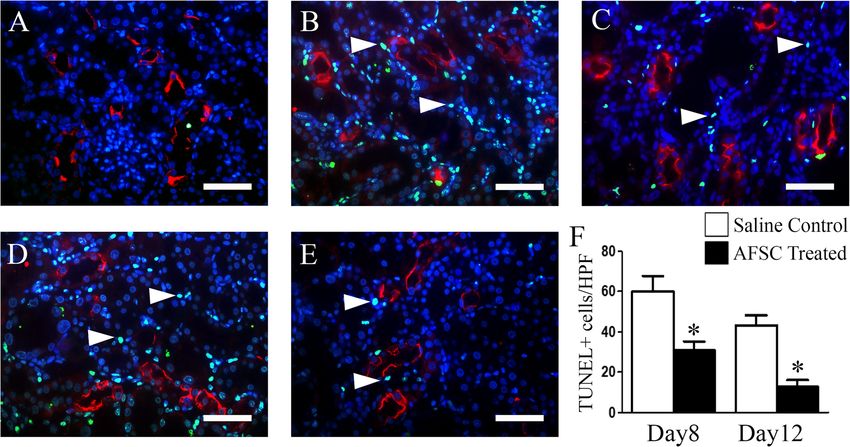
Minocha et al. Stem Cell Research & Therapy   (2019) 10:370                                                     Page 2 of 16

Background                                                    environment, with food and water provided ad libitum.
Acute renal failure (ARF) also known as acute kidney in-      All animal procedures in the study were conducted in
jury (AKI) is a grave clinical condition characterized by     accordance with the guidelines of Institutional Animal
sudden loss of renal function and can be induced by a         Ethics Committee (IAEC) and Committee for the Pur-
variety of factors including hypoxia, drugs, mechanical       pose of Control and Supervision of Experiments on Ani-
trauma, inflammation, surgery, cardiopulmonary bypass,        mals (CPCSEA), India. The protocol was approved by
and hemodynamic instability [1]. In some cases ARF re-        IAEC of Sanjay Gandhi Post Graduate Institute of Med-
covers spontaneously, while in others the recovery            ical Sciences, Lucknow, India.
process is either delayed or does not occur at all, thus
leading to chronic kidney disease (CKD). Therefore, it        Isolation and culture of amniotic fluid stem cells (AFSC)
becomes very necessary to restore the normal structure        Amniotic fluid samples were obtained from pregnant fe-
and function of the kidney after ARF in order to prevent      male Wistar rats at gestation day 16 and cultured as pre-
its progression into CKD.                                     viously described [5]. Briefly, from each gravid rat, 2–3
   Over the last decade, stem cell-based therapy has          ml of amniotic fluid was obtained, corresponding to cell
emerged as a promising approach for renal regeneration,       numbers ranging from 7 × 10³ to 7 × 10⁵ which was then
and of the various stem cells used, mesenchymal stromal       centrifuged at 300g for 5 min and the pellet obtained
cells (MSCs) have reached phase I/II clinical trials. How-    was resuspended in complete culture medium consisting
ever, even with MSC therapy, the results are conflicting,     of α-MEM, 16.5% fetal bovine serum, 2 mM Glutamax,
and no consistent benefit has been demonstrated till          100 U/ml penicillin, and 100 μg/ml streptomycin (all
date, perhaps due to their limited differentiation poten-     from Gibco, NY, USA) and incubated at 37 °C with 5%
tial in vivo [2]. Therefore, the search to identify a suit-   CO2 atmosphere. After 72 h of seeding, culture media
able stem cell type for renal regeneration is still on.       containing non-adherent cells were replaced. On day 7,
   Amniotic fluid stem cells (AFSC) represent a novel         the adherent cells were harvested by trypsinization with
class of stem cells with intermediate characteristics be-     TrypLE Express (Gibco, NY, USA) and further expanded
tween embryonic stem cells and adult stem cells. They         as above. The third passage cells were used throughout
have extended self-renewal capacity and multipotent dif-      the study.
ferentiation potential [3]. Major volume of amniotic fluid
is derived from fetal urine [4], and we have previously       Flow cytometry
shown that it harbors a stem cell population that ex-         Flow cytometry was performed on three independent
presses high percentage of renal progenitor markers and       amniotic fluid samples (n = 3) (obtained from three inde-
possesses renal differentiation potential [5, 6]. Although    pendent gravid rats) to characterize AFSC for the ex-
previous studies have shown the renal regenerative po-        pression of (i) cell-surface mesenchymal markers using
tential of AFSC in cisplatin- or glycerol-induced AKI         CD90, CD73, CD105, hematopoietic marker: CD45, and
[7–11], the underlying mechanisms responsible for the         MHC-Class II and (ii) intracellular renal progenitor
renoprotective effect are largely unknown.                    markers: Wilms’ tumor protein 1 (WT1), Paired Box 2
   It has been shown that autophagy has a protective role     (PAX2), and SIX Homeobox2 (SIX2) as previously de-
in cisplatin-induced AKI by inhibiting apoptosis [12].        scribed [5]. All flow-cytometric acquisitions were per-
Autophagy also plays a critical role in maintaining           formed on BD-FACS CantoII and analyzed using FACS
homeostasis of renal tubular cells [13], and impairment       Diva software. The specific dilutions and details of the
of autophagy in renal tubular cells has been implicated       antibodies used are listed in Table 1.
in the pathogenesis of various kidney diseases, including
AKI [14, 15]. Thus, restoration and promotion of au-          Development of ARF model and AFSC therapy
tophagy is considered as a promising therapeutic strat-       ARF was induced in male Wistar rats weighing 200–225
egy in ARF.                                                   g by an intra-peritoneal injection of cisplatin (Sigma Al-
   Therefore, the aim of the present study was to investi-    drich, MO, USA) at a dose of 7 mg/kg of body weight
gate the renoprotective effects of AFSC in cisplatin-         after fasting for 12 h. After 5 days of cisplatin injection, a
induced rat model of ARF and to evaluate the role of au-      significant increase in the blood biochemical parameters
tophagy in AFSC-mediated amelioration of ARF.                 and renal damage was observed; hence, at this point, the
                                                              rats were randomized into two groups: AFSC-treated
Methods                                                       group (n = 10) and saline-treated group (n = 10) for the
Animals and ethics statement                                  evaluation of efficacy of stem cell therapy. On day 5 after
Adult Wistar rats weighing 200–225 g were used in the         cisplatin injection, AFSC (2 × 10⁶cells/rat) suspended in
study. The animals were maintained on a 12-h light-           500 μl of normal saline (in AFSC-treated group) or
dark cycle in a constant temperature and humidity             500 μl saline alone (saline-treated group) was injected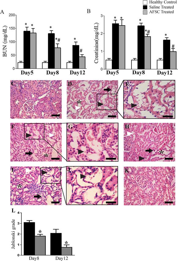
Minocha et al. Stem Cell Research & Therapy   (2019) 10:370                                                  Page 3 of 16

Table 1 List of antibodies with their dilutions, sources, and catalog numbers
Antibody                                                      Dilution          Source
Mouse monoclonal CD90                                         1:100 (FCM)       BD Biosciences, CA, USA
(Cat# 554894)
Mouse monoclonal CD73                                         1:100 (FCM)       BD Biosciences, CA, USA
(Cat# 551123)
Rat monoclonal CD105                                          1:100 (FCM)       Santa Cruz Biotechnology, CA, USA
(Cat# sc-71042)
Mouse monoclonal CD45                                         1:100 (FCM)       BD Biosciences, CA, USA
(Cat# 554878)
Mouse monoclonal MHC Class II                                 1:100 (FCM)       Abcam, MA, USA
(Cat# ab22266)
Mouse monoclonal WT1                                          1:100 (FCM)       My BioSource, CA, USA
(IgG clone #6F-H2)
Rabbit monoclonal Pax2                                        1:100 (FCM)       Abcam, MA, USA
(Cat# ab79389)
Goat polyclonal Six2                                          1:100 (FCM)       Santa Cruz Biotechnology, CA, USA
(Cat# sc-67834)
Rabbit monoclonal Phospho-AMPKα                               1:1000 (WB)       Cell Signaling Technology (CST), MA, USA
(Cat# 2535)
Mouse monoclonal Phospho-p70 S6 Kinase                        1:1000 (WB)       CST, MA, USA
(Cat# 9206)
Rabbit monoclonal ATG5                                        1:1000 (WB)       CST, MA, USA
(Cat#12994)
Rabbit monoclonal ATG7                                        1:1000 (WB)       CST, MA, USA
(Cat# 8558)
Rabbit monoclonal Beclin1                                     1:1000 (WB)       CST, MA, USA
(Cat# 3495)
Rabbit polyclonal LC3B                                        1:1000 (WB)       CST, MA, USA
(Cat# 2775)
Mouse monoclonal cleaved-Caspase-9                            1:1000 (WB)       CST, MA, USA
(Cat# 9508)
Rabbit polyclonal SQSTM1/p62                                  1:1000 (WB)       Abcam, MA, USA
(Cat# ab91526)
Rabbit polyclonal SQSTM1/p62                                  1:200 (IHC)       MBL International, MA, USA
(Cat# PM045)
Rabbit polyclonal p53                                         1:1000 (WB)       Abcam, MA, USA
(Cat# ab131442)
Rabbit polyclonal Bcl-2                                       1:1000 (WB)       Abcam, MA, USA
(Cat# ab196495)
Rabbit monoclonal Bax                                         1:1000 (WB)       Abcam, MA, USA
(Cat# ab32503)
Rabbit polyclonal PUMA                                        1:1000 (WB)       Abcam, MA, USA
(Cat# ab9643)
Rabbit polyclonal cleaved- Caspase-3                          1:1000 (WB)       CST, MA, USA
(Cat# 9661)                                                   1:100 (IHC)
Rabbit monoclonal GAPDH                                       1:1000 (WB)       CST, MA, USA
(Cat# 5174)
Biotinylated LTL                                              1:500 (IHC)       Vector Laboratories, CA, USA
(Cat# B-1325)
Goat Anti-mouse IgG (H&L)(PE)                                 1:200 (FCM)       Abcam, MA, USA
(Cat#ab97041)
Goat Anti-rabbit IgG (H&L)(FITC)                              1:200 (FCM)       Abcam, MA, USA
(Cat#ab6717)
Streptavidin, Alexa Fluor 568 conjugate                       1:500 (IHC)       Thermo Fisher Scientific, MA, USA
(Cat#S11226)
Minocha et al. Stem Cell Research & Therapy   (2019) 10:370                                                        Page 4 of 16

Table 1 List of antibodies with their dilutions, sources, and catalog numbers (Continued)
Antibody                                                      Dilution                        Source
Goat Anti-Rabbit (Alexa Flour 488)                            1:500 (IHC)                     Abcam, MA, USA
(Cat# ab150077)
Goat Anti-Rabbit (HRP)                                        1:5000 (WB)                     Abcam, MA, USA
(Cat# ab205718)
Goat Anti-mouse (HRP)                                         1:5000 (WB)                     Abcam, MA, USA
(Cat# ab205719)

intravenously through tail vein in each rat (Additional              analyzer Selectra Pro M (ELITech, France) and commer-
file 1: Figure S1A). In addition, a group of healthy con-            cially available assay kits from ELITech diagnostic
trol rats (n = 5) was also included in the study to com-             (France) according to the standard protocols provided
pare the histology and renal function with the AFSC-                 by the manufacturers.
and saline-treated groups.
   To determine the homing ability of AFSC to cisplatin-             Histopathological analysis
injured kidney, the AFSC were first transduced with                  The cortical kidney tissues obtained from the left kidney
CellLight Nucleus-GFP, BacMam 2.0 (Thermo Fisher                     of at least three independent rats from each group were
Scientific, MA, USA) at a concentration of 45 PPC (par-              fixed in 10% formalin, embedded in paraffin, cut into 5-
ticles per cell) according to the manufacturer’s instruc-            μm-thick sections, and then mounted on slides. Sections
tion and then administered intravenously in cisplatin-               were then deparaffinized, rehydrated, and stained with
induced ARF rats (n = 3) at a concentration of 2 × 10⁶               hematoxylin and eosin (H&E) to evaluate the histo-
cells/rat suspended in 500 μl of normal saline. After                pathological changes in saline-, AFSC-, and chloroquine-
3 days, the rats were sacrificed and kidney tissues were             treated kidney tissue sections. The H&E stained sections
obtained that were embedded in paraffin, sliced into 5-              were then analyzed under a light microscope (Olym-
μm-thick sections, and then incubated with Lotus tetra-              pusBX51) equipped with a digital camera. Quantitative
gonolobus Lectin (LTL) for 2 h at room temperature                   assessment of renal tubular necrosis was done using the
followed by incubation with Streptavidin Alexa Fluor                 Jablonski grading scores [16].
568 conjugated secondary antibody for 1 h at room
temperature. Nuclei were stained with Hoechst dye. Im-               Western blotting
ages were acquired using a fluorescence microscope                   The cortical kidney tissues obtained from the right kid-
(Olympus BX61) equipped with Nuance Multispectral                    ney of at least three independent rats from each group,
Imaging System (CRi Inc., MA, USA). GFP-positive cells               viz, healthy control, saline-treated, AFSC-treated, and
were counted in five renal sections per rat (n = 3), and             chloroquine-treated rats, were homogenized in RIPA
data was expressed as the number of GFP+ cells per 105               Lysis buffer supplemented with 1X protease (cOmplete,
renal cells.                                                         EDTA-free protease inhibitor cocktail, Roche) and phos-
   To confirm the protective effects of autophagy follow-            phatase inhibitor cocktail (PhosSTOP, Roche). Kidney
ing cisplatin-induced AKI, chloroquine, an autophagy in-             tissue homogenate was then incubated on ice for 30 min
hibitor, was used. Chloroquine diphosphate salt (Sigma               and then centrifuged at 6000 rpm for 15 min at 4 °C.
Aldrich, MO, USA) was administered to rats (n = 10)                  The supernatant was removed and stored at − 80 °C.
intra-peritoneally at a dose of 60 mg/kg in distilled water          Thirty-microgram homogenate protein was loaded and
1 day prior to therapy and then daily till sacrifice (Add-           separated by sodium dodecyl sulphate-polyacrylamide
itional file 1: Figure S1B).                                         gel electrophoresis (SDS-PAGE). After electrophoresis,
   Animals were euthanized by CO2 overdose after 5 days              the proteins were transferred to nitrocellulose mem-
(day 5), 8 days (day 8), and 12 days (day 12) of cisplatin           brane. The membranes were blocked with 5% non-fat
injection. Both the kidneys were excised, and blood sam-             milk for 1 h at room temperature and incubated with
ples were collected to perform the histological analysis             primary antibodies (Table 1), viz phospho-AMP-
and determination of the blood biochemical parameters                activated protein kinase (phospho-AMPK), phospho-p70
respectively.                                                        S6 Kinase (phospho-p70S6K), Autophagy related 5
                                                                     (ATG5), Autophagy related 7 (ATG7), Beclin1,
Determination of blood urea nitrogen (BUN) and                       Microtubule-associated protein 1A/1B-light chain 3
creatinine levels                                                    (LC3B), cleaved caspase-9, Sequestosome 1 (SQSTM1/
Renal function was assessed by measuring the BUN and                 p62), p53, B cell lymphoma 2 (Bcl-2), B cell lymphoma
creatinine levels. After blood collection, serum levels of           2-associated X protein (Bax), p53 upregulated modulator
BUN and creatinine were measured using biochemical                   of Apoptosis (PUMA), and cleaved caspase-3 at 4 °C
Minocha et al. Stem Cell Research & Therapy (2019) 10:370 Page 5 of 16
overnight. GAPDH was used as loading control. Primary Statistical analysis
antibodies were detected by corresponding horseradish Values were expressed as mean ± standard error (SE).
peroxidase (HRP)-conjugated secondary antibodies using One-way analysis of variance (ANOVA) with Bonferroni
Clarity Western ECL Substrate (Bio-Rad, CA, USA). multiple-comparison post hoc test was used to compare
Semi-quantitative densitometric analysis was performed statistically significant differences between groups. Stat-
using ImageJ software. istical analysis was performed using GraphPad Prism
software version 5 (GraphPad, CA, USA), and p value <
0.05 was considered statistically significant.
Immunofluorescence
The paraffin-embedded kidney tissue sections were
deparaffinized, rehydrated, and subjected to antigen re-
Results
Expression of mesenchymal and renal progenitor markers
trieval in sodium citrate buffer (10 mM sodium citrate,
by AFSC
pH 6.0) at 95 °C for 30 min and then kept at room
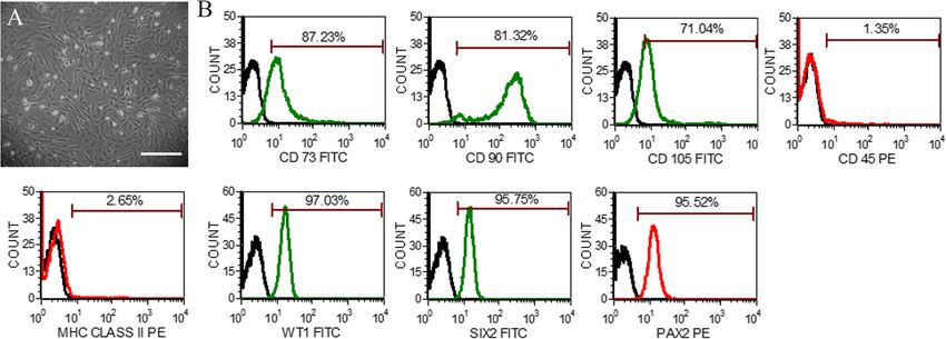
The AFSC exhibited uniform spindle-shaped morph-
temperature for cooling. The sections were then blocked
ology in culture at passage 3 (Fig. 1a). Flow cytometric
in 5% goat serum containing 1% bovine serum albumin
analysis showed that AFSC expressed mesenchymal
(BSA) for 1 h at room temperature followed by primary
markers, viz, CD73 (87.23% ± 5.16), CD90 (81.32% ±
antibody incubation, viz, p62 and cleaved-caspase3, for
3.32), and CD105 (71.04% ± 5.09), whereas the expres-
2 h at room temperature in a humidified chamber. After
sion of CD45 (1.35% ± 1.76) and MHC Class II (2.65% ±
rinsing the slides with PBS, sections were then incubated
1.51) was found to be less than 5%. Furthermore, AFSC
with Alexa Fluor 488 conjugated secondary antibody for
also expressed high percentage of renal progenitor
1 h at room temperature followed by nuclear staining
markers, viz, WT1 (97.03% ± 2.24), PAX2 (95.52% ±
with Hoechst. Images were acquired using a fluorescence
3.05), and SIX2 (95.75% ± 3.18), as revealed by flow cy-
microscope (Olympus BX61) equipped with Nuance
tometry (Fig. 1b).
Multispectral Imaging System (CRi Inc., MA, USA). Sec-
tions stained without primary antibody served as nega-
tive controls. AFSC therapy promotes improvement of renal function
and histology in ARF rats
On the 5th day of cisplatin injection, rats exhibited a sig-
Terminal deoxynucleotidyl-transferase-mediated dUTP nificant increase in the BUN and serum creatinine levels
nick end labeling (TUNEL) assay as compared to healthy controls (p < 0.05). On day 8, a
Apoptotic scores in kidney tissue sections were mea- significant decrease in the BUN and serum creatinine
sured by TUNEL assay using an in situ cell death de- levels was observed in the AFSC-treated group as com-
tection kit (Roche, Mannheim, Germany) according to pared to the saline-treated group (p < 0.05), but both
the manufacturer’s instruction. Briefly, the kidney tis- groups had significantly higher levels as compared to
sue sections were deparaffinized, rehydrated, and sub- healthy controls (p < 0.05). On day 12, the blood bio-
jected to antigen retrieval in citrate buffer followed by chemical parameters in the AFSC-treated group became
labeling with TUNEL reaction mixture for 1 h at comparable to that of healthy controls, but they were
37 °C. LTL was used to identify the proximal tubules. still significantly higher in the saline-treated group as
Immunostaining with LTL was performed after compared to healthy controls (p < 0.05) (Fig. 2a, b).
TUNEL staining. Sections were then washed in PBS Histopathological analysis of kidney tissues revealed that,
and blocked for 1 h at room temperature in 5% goat on the 5th day of cisplatin injection, the kidneys of ARF
serum and 1% BSA, followed by primary antibody in- rats exhibited severe tubular necrosis with the loss of
cubation, viz, LTL, for 2 h at room temperature in a brush border, hyaline cast formation, and tubular dilata-
humidified chamber. After rinsing the slides with tion (Fig. 2d, e). On day 8, the kidneys exhibited signifi-
PBS, sections were then incubated with Streptavidin cant attenuation of tubular injury in the AFSC-treated
Alexa Fluor 568 conjugated secondary antibody for 1 group as compared to the saline-treated group (Fig. 2f,
h at room temperature followed by counterstaining h). The Jablonski’s histological score also revealed sig-
with Hoechst dye (Sigma Aldrich, MO, USA). nificantly lower necrosis in the kidneys of AFSC-treated
TUNEL-positive nuclei were then counted in 10 ran- rats as compared to saline-treated rats (p < 0.05) (Fig. 2l).
domly selected non-overlapping high power fields On day 12, the kidneys of saline-treated rats still showed
(HPF) (40×) in the LTL-positive cortical regions necrotic tubular cells and hyaline casts, but the kidneys
under Nuance Multispectral Imaging System (CRi of AFSC-treated rats showed a lower index of cellular
Inc., MA, USA), and the apoptotic index was damage indicating attenuation of renal injury by infused
expressed as average number of TUNEL+ cells/HPF. cells (Fig. 2i, k) and there was a significant difference inMinocha et al. Stem Cell Research & Therapy (2019) 10:370 Page 6 of 16
Fig. 1 Morphology and phenotypic characterization of AFSC. a Representative photomicrographs of amniotic fluid stem cells (AFSC) in culture
showing spindle-shaped morphology in passage 3 (scale bar: 100 μm). b Phenotypic characterization of AFSC by flow cytometry showing the
expression of cell-surface markers, viz, CD73, CD90, CD105, MHC Class II, and CD45, and intracellular renal progenitor markers, viz, WT1, SIX2, and
PAX2 (green or red lines detected with FITC- and PE-conjugated antibodies, respectively, and black lines represent isotype controls). PE,
phycoerythrin; FITC, fluorescein isothiocyanate
the Jablonski grading score between the AFSC-treated significantly lower as compared to that of saline-treated
group and saline-treated group (p < 0.05) (Fig. 2l). rats on day 8 (p < 0.01), which further decreased signifi-
The homing capacity of AFSC to the injured kidney cantly (p < 0.01) on day 12 (Fig. 4).
was evaluated by the presence of GFP-labeled cells in
the renal parenchyma 3 days after their administration. AFSC mediate activation of autophagy in cisplatin-
Their frequency averaged 2.2 ± 0.77 per 105 renal cells. induced ARF rats
AFSC were predominantly found to localize in the peri- Since induction of autophagy rescues from cisplatin-
tubular areas and rarely within the tubular epithelium induced injury [12], we next studied if AFSC-
(Additional file 1: Figure S2). mediated protection involved autophagy induction.
AMPK and mammalian target of rapamycin (mTOR)
Administration of AFSC reduces apoptosis in kidney are the critical regulators of autophagy [17]. We dem-
tissues onstrated the effect of AFSC therapy on the levels of
Western blot analysis was performed to determine the phospho-AMPK and on the downstream target of
levels of apoptosis-related signaling pathway proteins mTOR signaling pathway, i.e., phospho-p70S6K. The
on day 8 and day 12 after cisplatin injection. A sig- levels of phospho-AMPK were found to be signifi-
nificant upregulation in the expression levels of pro- cantly upregulated in the AFSC-treated group as com-
apoptotic proteins, viz, PUMA (p < 0.05 for both day pared to the saline-treated group (p < 0.01), while the
8 and day 12), Bax/Bcl2 ratio (p < 0.001 for day 8; p < levels of phospho-p70S6K were found to be signifi-
0.01 for day 12), cleaved caspase-3 (p < 0.001 for both cantly reduced after AFSC administration as com-
day 8 and day 12), and cleaved caspase-9 (p < 0.01 for pared to the saline-treated group (p < 0.01 for day 8;
day 8; p < 0.05 for day 12), was observed in the p < 0.001 for day 12). These results indicate that
saline-treated group as compared to healthy controls. AFSC therapy inhibits mTOR downstream target, i.e.,
However, after administration of AFSC, there was a phospho-p70S6K, and activates phospho-AMPK,
marked downregulation of all the pro-apoptotic pro- thereby activating autophagy in the renal tubular epi-
teins in the AFSC-treated group as compared to the thelial cells in response to cisplatin. To determine the
saline-treated group (p53: p < 0.05 for both day 8 and level of autophagy activation following AFSC therapy,
day 12; PUMA: p < 0.05 for day 8; p < 0.01 for day 12; we examined the expression of autophagy-related pro-
Bax/Bcl2 ratio: p < 0.01 for day 8; p < 0.001 for day teins, viz, ATG5 and ATG7, and other critical au-
12; cleaved caspase-3: p < 0.05 for day 8; p < 0.01 for tophagy markers including Beclin-1, LC3-II, and p62
day 12; cleaved caspase-9: p < 0.05 for both day 8 and on both day 8 and day 12. The levels of autophagy-
day 12) (Fig. 3). related proteins, i.e., ATG5 and ATG7, were found to
Corroborating with our western blot data, TUNEL- be significantly increased in the AFSC-treated group
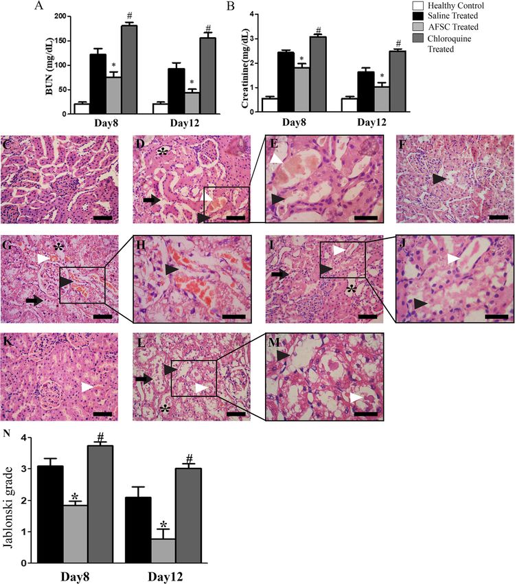
positive cells in the kidneys of AFSC-treated rats were as compared to the saline-treated group (p < 0.05).Minocha et al. Stem Cell Research & Therapy (2019) 10:370 Page 7 of 16 Fig. 2 Effect of AFSC therapy on renal function and histology in rats with cisplatin-induced ARF. a Levels of blood urea nitrogen (BUN) and b serum creatinine measured in healthy controls, saline-treated and AFSC-treated ARF rats on days 5, 8, and 12 after cisplatin injection. Values are expressed as mean ± SE (*p < 0.001 versus healthy control group; #p < 0.05 versus saline-treated group). c Kidney section of healthy control rat showing normal architecture of tubules and glomeruli (scale bar, 50 μm). d Kidney section after 5 days of cisplatin injection showing tubular dilatation (asterisk), necrotic tubules (black arrowhead), intra-tubular cast (white arrowhead), and loss of brush border (black arrow) (scale bar, 50 μm). e Magnified image of the boxed area in d showing necrotic tubules (black arrowhead) (scale bar, 30 μm). f Kidney section of saline- treated ARF rat on day 8 after cisplatin injection showing severe tubular dilatation (asterisk), loss of brush border (black arrow), intra-tubular cast (white arrowhead), and necrotic tubules (black arrowhead) (scale bar, 50 μm). g Magnified image of the boxed area in f showing necrotic tubules (black arrowhead) (scale bar, 30 μm). h Kidney section of AFSC-treated ARF rat on day 8 after cisplatin injection showing signs of recovery as revealed by mild tubular dilatation (asterisk) and fewer necrotic tubules (black arrowhead) and intra-tubular cast (white arrowhead) (scale bar, 50 μm). i Kidney section of saline-treated ARF rat on day 12 after cisplatin injection showing few intratubular hyaline casts (white arrowhead), tubular dilatation (asterisk), loss of brush border (black arrow), and necrotic tubules (black arrowhead) (scale bar, 50 μm). j Magnified image of the boxed area in i showing necrotic tubules (black arrowhead) (scale bar, 30 μm). k Kidney section of AFSC-treated rats on day 12 after cisplatin injection showing almost normal architecture of the tubules and preservation of the integrity of the cellular structure (scale bar, 50 μm). l Jablonski grading score for the assessment of renal tubular cell necrosis in saline- and AFSC-treated kidneys on day 8 and day 12 after cisplatin injection. Values expressed as mean ± SE (*p < 0.05 versus saline-treated group)
Minocha et al. Stem Cell Research & Therapy      (2019) 10:370                                                                        Page 8 of 16

 Fig. 3 AFSC inhibit apoptosis in kidneys with ARF. a Representative immunoblots showing the expression of apoptotic proteins, viz, p53, Bax, Bcl-
 2, PUMA, cleaved caspase-3, and cleaved caspase-9, in kidney tissues of healthy control, saline-treated, and AFSC-treated groups on day 8 and day
 12 after cisplatin injection. b Bar diagrams showing semi-quantitative densitometric determination of ratio of expression of Bax/Bcl-2 and
 expression of apoptotic proteins, viz, p53, PUMA, cleaved caspase-3, and cleaved caspase-9, by referring each gene to GAPDH which was taken as
 internal control. Values expressed as mean ± SE of three independent blots (*p < 0.05, **p < 0.01, ***p < 0.001)

The levels of autophagy markers, viz, LC3B-II and                          decreased in the AFSC-treated group as compared to
Beclin-1, were also found to be significantly increased                    the saline-treated group (p < 0.05) (Fig. 5).
in the AFSC-treated as compared to the saline-treated                        In addition, the immunofluorescence staining for p62
group (p < 0.01 for day 8; p < 0.05 for day 12) while                      and cleaved-caspase 3 in kidney tissue sections also re-
the levels of p62 were found to be significantly                           vealed that there was decreased accumulation of p62
Minocha et al. Stem Cell Research & Therapy       (2019) 10:370                                                                         Page 9 of 16

 Fig. 4 Effect of AFSC therapy on apoptosis of renal tubular epithelial cells in cisplatin-injured kidney. Representative immunofluorescence
 photomicrographs showing TUNEL-positive cells (green, indicated by white arrowheads) in the cortical region of the kidney tissue of a healthy
 control, b saline-treated group (day 8), c AFSC-treated group (day 8), d saline-treated group (day12), and e AFSC-treated group (day 12). The
 sections were co-stained with LTL (red) to mark the proximal tubule and Hoechst for nuclei (blue) (scale bar, 30 μm). f Quantification of TUNEL-
 positive cells in kidney sections of saline-treated and AFSC-treated groups on day 8 and day 12 after cisplatin injection. Values expressed as
 mean ± SE (*p < 0.01)

substrate along with decreased expression of cleaved-                       creatinine levels in the chloroquine group as compared
caspase3 in the AFSC-treated group as compared to the                       to the therapy-treated group (p < 0.001) on both day 8
saline-treated group on both day 8 and day 12, which                        and day 12 (Fig. 7a, b). Consistent with our functional
corroborated with our western blot results (Additional                      data, the histological examination also showed that the
file 1: Figure S3).                                                         tubular damage following cisplatin-induced renal injury
                                                                            was further aggravated by chloroquine treatment and
Amniotic fluid stem cells prevent cisplatin-induced                         more tubules showed dilatation and distortion, loss of
apoptosis by activating autophagy in-vivo                                   brush border, hyaline casts, and sloughed debris in the
In order to confirm whether the preventive effects of                       lumen space as compared to other groups (Fig. 7d–m).
AFSC on cisplatin-induced apoptosis are dependent on                        Furthermore, there was a significant difference in the
autophagy, we used an autophagic inhibitor, i.e., chloro-                   Jablonski grading score between the chloroquine-treated
quine, and analyzed its effect on the autophagic flux                       group and therapy-treated group (p < 0.001) on both day
marker: p62, and the apoptosis marker: cleaved caspase-                     8 and day 12 (Fig. 7n). Collectively, these results link the
3. The protein levels of the autophagic substrate p62 and                   induction of autophagy by AFSC with their beneficial ef-
the apoptotic marker cleaved caspase-3 were found to be                     fects in cisplatin-induced renal injury.
significantly upregulated in the chloroquine-treated
group as compared to the therapy-treated group on both                      Discussion
day 8 and day 12 (p < 0.001), indicating that inhibition of                 The present study demonstrated that culture-expanded
autophagy by chloroquine (as evident by p62 accumula-                       rat AFSC express mesenchymal and renal progenitor
tion) leads to enhanced renal apoptosis (as evident by                      markers. The administration of AFSC in cisplatin-
upregulated expressions of cleaved caspase-3) (Fig. 6).                     induced rat model of ARF resulted in improvement of
  We also analyzed the effect of chloroquine on renal                       renal function and attenuation of renal damage. The in-
function and histology on both day 8 and day 12 after                       fused AFSC activated autophagy, which led to reduction
cisplatin injection. Administration of chloroquine in-                      in renal cell apoptosis and acceleration in renal recovery.
duced a severe loss of renal function, which was ob-                        The protective effects of AFSC were blunted by the use
served by a significant increase in the BUN and serum                       of an autophagic inhibitor, i.e., chloroquine, suggesting
Minocha et al. Stem Cell Research & Therapy (2019) 10:370 Page 10 of 16 Fig. 5 (See legend on next page.)
Minocha et al. Stem Cell Research & Therapy     (2019) 10:370                                                                      Page 11 of 16

 (See figure on previous page.)
 Fig. 5 AFSC mediate activation of autophagy in response to cisplatin-induced acute renal failure. a Representative immunoblots showing the
 expression of the autophagic proteins, viz, phospho-AMPK, phospho-p70S6K, ATG5, ATG7, Beclin1, LC3B, and p62, in kidney tissues of healthy
 control, saline-treated, and AFSC-treated groups on day 8 and day 12 after cisplatin injection. b Bar diagrams showing semi-quantitative
 densitometric analysis for comparison of autophagic protein expression by referring each gene to GAPDH which was taken as internal control.
 Values expressed as mean ± SE of three independent blots (*p < 0.05, **p < 0.01, ***p < 0.001)

that induction of autophagy is essential for the protect-                 transdifferentiate into cells expressing proximal and dis-
ive role of AFSC in cisplatin-induced renal injury. To                    tal tubular agglutinins [11]. We also performed tracking
the best of our knowledge, this is the first study which                  experiments to determine the homing ability of the ad-
demonstrates activation of autophagy as one of the im-                    ministered AFSC and found that GFP-positive cells pre-
portant mechanisms responsible for the renoprotective                     dominantly localized in the peritubular areas of the
effects of AFSC in cisplatin-induced AKI.                                 injured kidney, which may represent only a small frac-
   In our recent study, we characterized rat AFSC for the                 tion of the infused cells, as a large fraction of the intra-
expression of renal progenitor markers, viz, WT1, PAX2,                   venously infused cells, get trapped into the lungs, liver,
SIX2, CITED1, and SALL1, and evaluated their in vitro                     and spleen as reported for other cell types [20–22].
potential to differentiate into renal proximal tubular                      Recently, our group showed the renoprotective effect
epithelial-like cells and juxtaglomerular-like cells [5]. In              of fetal kidney stem cells in ischemia and cisplatin-
the present study, we have further evaluated the thera-                   induced rat models of ARF via their anti-inflammatory,
peutic potential of AFSC in alleviating cisplatin-induced                 anti-apoptotic, anti-oxidative, and angiogenic properties
ARF and the underlying mechanisms for the renoprotec-                     [23, 24]. The present study also highlights that AFSC
tive effects. However, previous studies have reported the                 promote amelioration of cisplatin-induced kidney injury
renoprotective effects of AFSC in different pre-clinical                  via their anti-apoptotic properties. Apoptosis of tubular
models of renal diseases [9, 11, 18], but their mechanism                 cells is the characteristic feature of cisplatin nephrotox-
of action still remains unclear. AFSC have been shown                     icity and has been observed both in vitro and in vivo
to exert their renoprotective effects by their ability of                 [25, 26]. Several therapeutic interventions targeting the
homing to the injured kidney and paracrine release of                     apoptotic pathways involved in AKI have demonstrated
various soluble factors that create a regenerative micro-                 the beneficial effects on both in vitro cultured renal
environment [7, 19] or through their ability to                           tubular cells [27] and in vivo animal models of cisplatin-

 Fig. 6 Effect of chloroquine administration on autophagy and apoptosis. a Representative immunoblots showing the expression of autophagic
 marker p62 and apoptosis marker cleaved caspase-3 in kidney tissues of healthy control, saline-treated, AFSC-treated, and chloroquine-treated
 groups on day 8 and day 12 after cisplatin injection. b Bar diagrams showing semi-quantitative densitometric analysis for comparison of
 expression of p62 and cleaved caspase-3 by referring each gene to GAPDH which was taken as internal control. Values expressed as mean ± SE of
 three independent blots (*p < 0.05, **p < 0.01, ***p < 0.001)
Minocha et al. Stem Cell Research & Therapy   (2019) 10:370                                                 Page 12 of 16

 Fig. 7 (See legend on next page.)

induced renal injury [21, 28]. In the present study, we       also confirmed that infusion of AFSC reduced apoptosis
demonstrated that the number of TUNEL-positive cells          in the kidneys injured by cisplatin. Cisplatin administra-
increased significantly after cisplatin administration.       tion also activates p53 which in turn promotes apoptosis
However, infusion of AFSC resulted in a significant re-       by upregulating the expression of PUMA, one of the
duction of these TUNEL-positive cells. The decreased          major downstream mediators for the apoptotic actions
expression of cleaved caspase-3 and cleaved caspase-9         of p53 [29]. Knockout studies have shown suppression
Minocha et al. Stem Cell Research & Therapy        (2019) 10:370                                                                          Page 13 of 16

 (See figure on previous page.)
 Fig. 7 Effect of chloroquine administration on renal function and histology in rats with cisplatin-induced ARF. a Levels of blood urea nitrogen
 (BUN) and b serum creatinine measured in healthy controls, saline-treated, AFSC-treated, and chloroquine-treated ARF rats on days 8 and 12
 following cisplatin injection. Values expressed as mean ± SE (*p < 0.05 versus saline-treated group; #p < 0.001 versus therapy-treated group).
 Representative photomicrographs showing histology of cortical kidney tissue sections from healthy control (c), day 8 saline-treated ARF group (d)
 showing necrotic tubules (black arrowhead) and intratubular hyaline casts (white arrowhead) in higher magnification (e), day 8 AFSC treated ARF
 group (f), day 8 chloroquine-treated ARF group (g) showing necrotic tubules (black arrowhead) in higher magnification (h), day 12 saline-treated
 ARF group (i) showing necrotic tubules (black arrowhead) and intratubular hyaline casts (white arrowhead) in higher magnification (j), day 12
 AFSC-treated ARF group (k), and day 12 chloroquine-treated ARF group (l) showing necrotic tubules (black arrowhead) and intratubular hyaline
 casts (white arrowhead) in higher magnification (m). The kidney sections of day 8 and day 12 chloroquine-treated ARF rats show severe tubular
 necrosis (black arrowhead) with intratubular hyaline casts (white arrowhead), tubular dilatation (asterisk), and loss of brush border (black arrow) as
 compared to other groups. n Jablonski grading score for the assessment of tubular necrosis in saline-treated, AFSC-treated, and chloroquine-
 treated groups on day 8 and day 12 after cisplatin injection. Values expressed as mean ± SE (*p < 0.05 versus saline-treated group; #p < 0.001
 versus AFSC-treated group)

of cisplatin-induced apoptosis in PUMA knockout cells,                        p62 expression levels. The AFSC therapy significantly in-
indicating that inhibition of PUMA inhibits apoptosis                         creased LC3-II expression level and decreased the ex-
[30]. We observed that administration of AFSC attenu-                         pression level of p62. The LC3-II levels directly correlate
ated the activation of p53 and its downstream target                          with the autophagosome number while the levels of p62
PUMA, suggesting the protective effect of AFSC in                             inversely correlates with the autophagic activity [36].
cisplatin-induced AKI. Cisplatin administration activates                     The stem cell therapy also significantly increased the ex-
Bax, reduces Bcl2, and shifts the Bax/Bcl2 ratio in a pro-                    pression of other autophagy-related proteins, i.e., ATG5,
apoptotic direction. Bax deletion has been shown to con-                      ATG7, and Beclin-1. ATG5 and ATG7 proteins form
fer resistance to cisplatin in animals, further highlighting                  the critical component of the autophagic pathway that
the pathological role of Bax in cisplatin nephrotoxicity                      are involved in the elongation and closure of the autop-
[31]. Our study demonstrated that administration of                           hagosomal membrane [37]. Beclin-1 is the mammalian
AFSC upregulated the anti-apoptotic protein, Bcl2, and                        homolog of ATG6 and is involved in the vesicle nucle-
downregulated the pro-apoptotic protein Bax, which                            ation, an early event during autophagosome formation
corresponds with reduced apoptosis and improved renal                         [38]. Previous studies have shown that knockdown of
function.                                                                     the autophagy proteins: Beclin-1 and ATG5, leads to en-
   Under basal or physiological conditions, autophagy                         hanced activation of caspases and tubular cell apoptosis
works as a cellular housekeeper and helps in eliminating                      during cisplatin treatment [39]. The expression levels of
damaged organelles and intracellular pathogens and con-                       phospho-AMPK were found to be significantly elevated,
tributes to the maintenance of cellular homeostasis and                       while that of phospho-p70S6K were found to be signifi-
quality control of proteins and sub-cellular organelles.                      cantly downregulated by AFSC therapy treatment, sug-
However, under pathological conditions or cell stress,                        gesting that AFSC may activate autophagy via activation
autophagy is induced and serves as a protective mechan-                       of AMPK pathway and inhibition of the mTOR signaling
ism for cell survival [32]. The role of autophagy in the                      pathway. Previous studies have also shown that
pathogenesis of AKI still remains controversial, as there                     cisplatin-induced tubular cell apoptosis can be effectively
are contradictory reports regarding the same [33]. Some                       ameliorated by AMPK activation and inhibition of
reports have suggested a cytoprotective role of autoph-                       mTOR signaling pathway [40–42]. A recent study by
agy during cisplatin treatment [34] while the others have                     Wang et al. showed that human umbilical cord mesen-
shown that autophagy may be involved in apoptosis of                          chymal stem cell-derived exosomes (hucMSC-Ex) pre-
the proximal tubular cells following cisplatin treatment                      vent cisplatin-induced apoptosis under both in vitro and
[35]. However, our results establish a renoprotective role                    in vivo conditions through the activation of autophagy
of autophagy in cisplatin-induced kidney injury. Our ob-                      via inhibition of the mTOR signaling pathway and its
servation is in concordance with previous studies that                        downstream target, i.e., p70S6K [43]. In the same line,
have reported that autophagy triggers a pro-survival re-                      Jia et al. also reported that hucMSC-Ex prevents
sponse in cisplatin-induced AKI [12, 34]. However,                            cisplatin-induced renal injury through activation of au-
whether AFSC can activate autophagy to prevent renal                          tophagy via. trophic factor 14-3-3ζ which interacts with
tissue injury has not been explored. Here, we provide                         ATG-16 L [44], indicating that stem cell secretomes have
evidence for the first time that AFSC activate autophagy                      therapeutic effects on renal injury via autophagy activa-
in response to cisplatin-induced AKI. The autophagic re-                      tion. These studies suggest that paracrine mediators of
sponse to cisplatin was identified by monitoring the au-                      renal cell autophagy may include components of AFSC’s
tophagic flux which was determined by the LC3-II and                          secretome such as miRNA and other soluble proteins
Minocha et al. Stem Cell Research & Therapy   (2019) 10:370                                                                   Page 14 of 16

that link the efficacy of AFSC therapy with activation of     pharmacological therapies for ARF. Moreover, alterna-
autophagy.                                                    tive ways for blocking autophagy, like use of autophagy
  To further investigate whether autophagy was involved       gene-knockout animal models, would be more suitable
in the protective effects of AFSC against cisplatin-          to determine the conclusive evidence for the involve-
induced apoptosis, chloroquine, a pharmacological in-         ment of autophagy in amelioration of cisplatin-induced
hibitor of autophagy, was administered. Chloroquine, a        kidney injury.
widely used anti-malarial drug, is known to block the
last phase of autophagy by inhibiting the autophagosome       Supplementary information
fusion with lysosome and thus slows down the lysosomal        Supplementary information accompanies this paper at https://doi.org/10.
                                                              1186/s13287-019-1476-6.
acidification [45]. In the present study, we observed that
chloroquine administration inhibited the protective ef-        Additional file 1: Figure S1. Experimental Schedule. Figure S2. In-vivo
fects of AFSC therapy and led to a further worsening in        tracking of GFP-labelled AFSC in cisplatin-injured kidney tissue. Figure
the renal structure and function caused by cisplatin by        S3. AFSC therapy mediates activation of autophagy and inhibition of
                                                               apoptosis in response to cisplatin-induced renal injury.
significantly increasing the expression levels of p62 and
cleaved caspase-3, thereby suggesting the renoprotective
                                                              Abbreviations
role of autophagy in this disease model. Our observation      AFSC: Amniotic fluid stem cells; ARF: Acute renal failure; AKI: Acute kidney
corroborates with a previous study which showed that          injury; CKD: Chronic kidney disease; MSC: Mesenchymal stem cells;
administration of chloroquine abolished the protective        MEM: Minimum essential media; FITC: Fluorescein isothiocyanate;
                                                              PE: Phycoerythrin; BUN: Blood urea nitrogen; H&E: Hematoxylin and eosin;
effects of neferine against cisplatin-induced apoptosis       SDS-PAGE: Sodium dodecyl sulphate-polyacrylamide gel electrophoresis;
[46]. However, a recent study by Mauthe et al. (2018)         HRP: Horseradish peroxidase; TUNEL: Terminal deoxynucleotidyl-transferase-
showed that hydroxychloroquine at a dose of 60 mg/kg          mediated dUTP nick end labeling; HPF: High power field; mTOR: Mammalian
                                                              target of rapamycin; hucMSC-Ex: Human umbilical cord mesenchymal stem
in mice leads to multiple structural alterations like golgi   cell-derived exosomes; SE: Standard error; ANOVA: One-way analysis of
disorganization in the kidney and intestinal cells in an      variance; LTL: Lotus tetragonolobus Lectin; 3-MA: 3-Methyladenine
autophagy-independent manner [47]. This may hold true
                                                              Acknowledgements
for the present study as well, that effects of chloroquine    The authors would like to extend their sincere thanks to Mrs. Shobhita
may not be only limited to inhibition of autophagy, but       Katiyar for assisting us in performing the flow cytometry experiments. The
autophagy-independent effects of chloroquine may also         authors will also like to express their gratitude to Mr. Pradeep Paul and Dr.
                                                              Ruchi Gupta who assisted in processing the kidney tissues for H&E staining
be responsible that may contribute to worsening of renal      and analysis of renal histology, respectively.
injury after chloroquine application. Therefore, further
studies are needed in future to determine other ultra-        Authors’ contributions
                                                              EM designed and performed the experiments, collected and analyzed the
structural alterations that may be caused by chloroquine      data, and wrote the manuscript; RAS designed the experiments, analyzed the
application for blocking autophagy and their functional       data, wrote the manuscript, and contributed reagents; MJ performed the
consequences. Likewise, other pharmacological inhibi-         experiments; CPC analyzed the data, provided reagents, and wrote the
                                                              manuscript; SN conceived and designed the study, analyzed the data,
tors of autophagy such as 3-methyladenine (3-MA) have         contributed reagents, wrote the manuscript, and finally approved it. All
been equally shown to exhibit off-target effects [48];        authors have read and approved the final manuscript.
therefore, alternative ways for blocking autophagy, like
                                                              Funding
the use of autophagy gene-knockout animal models,             This work was supported by an Extramural Grant (BT/PR16863/MED/31/338/
would be more suitable to determine the actual involve-       2016) of Dept of Biotechnology (DBT), Govt. of India, sanctioned to SN and
ment of autophagy in amelioration of cisplatin-induced        Wellcome Trust DBT India Alliance Fellowship grant (IA/I/16/1/502374)
                                                              sanctioned to CPC and to RAS (IA/I/16/2/502691). EM is the recipient of the
acute renal failure. However, this was beyond the scope       Department of Science and Technology (DST), Govt. of India, INSPIRE PhD
of the present study.                                         fellowship (IF150226).

                                                              Availability of data and materials
Conclusions
                                                              All relevant data related to this study has been included in the article.
In conclusion, the present study demonstrates that ad-
ministration of AFSC in cisplatin-induced ARF results in      Ethics approval and consent to participate
rapid recovery of renal function and histology by activa-     The study was approved by Institutional Animal Ethics Committee (IAEC) of
                                                              Sanjay Gandhi Post Graduate Institute of Medical Sciences, Lucknow, India.
tion of autophagy and inhibition of apoptosis. To the
best of our knowledge, this is the first study that high-     Consent for publication
lights autophagy as one of the important mechanisms by        Not applicable.

which AFSC induce a renoprotective effect in cisplatin-       Competing interests
induced ARF. However, further studies on the long-term        The authors declare that they have no competing interests.
effects of AFSC in different pre-clinical models of ARF
                                                              Author details
would be important for its translational implications into    1
                                                               Stem Cell Research Centre, Department of Hematology, Sanjay Gandhi Post
clinical settings for the development of novel                Graduate Institute of Medical Sciences, Rae Bareli Road, Lucknow, UP 226014,
Minocha et al. Stem Cell Research & Therapy (2019) 10:370 Page 15 of 16
India. 2Department of Endocrinology, Sanjay Gandhi Post Graduate Institute 22. Yuan L, Wu M-J, Sun H-Y, Xiong J, Zhang Y, Liu C-Y, et al. VEGF-modified
of Medical Sciences, Lucknow, India. human embryonic mesenchymal stem cell implantation enhances
protection against cisplatin-induced acute kidney injury. Am J Physiol Renal
Received: 4 June 2019 Revised: 22 October 2019 Physiol. 2011;300(1):F207–18.
Accepted: 30 October 2019 23. Gupta AK, Jadhav SH, Tripathy NK, Nityanand S. Fetal kidney stem cells
ameliorate cisplatin induced acute renal failure and promote renal
angiogenesis. World J Stem Cells. 2015;7(4):776–88.
References 24. Gupta AK, Jadhav SH, Tripathy NK, Nityanand S. Fetal kidney cells can
1. Thiele RH, Isbell JM, Rosner MH. AKI associated with cardiac surgery. Clin J ameliorate ischemic acute renal failure in rats through their anti-
Am Soc Nephrol. 2015;10(3):500–14. inflammatory, Anti-Apoptotic and Anti-Oxidative Effects. PLoS One. 2015;
2. Mu D, Zhang X-L, Xie J, Yuan H-H, Wang K, Huang W, et al. Intracoronary 10(6):e0131057.
transplantation of mesenchymal stem cells with overexpressed integrin-
25. Lieberthal W, Triaca V, Levine J. Mechanisms of death induced by cisplatin
linked kinase improves cardiac function in porcine myocardial infarction. Sci in proximal tubular epithelial cells: apoptosis vs. necrosis. Am J Phys. 1996;
Rep. 2016;6:19155.
270(4 Pt 2):F700–8.
3. Siegel N, Rosner M, Hanneder M, Valli A, Hengstschläger M. Stem cells in
26. Ramesh G, Reeves WB. TNFR2-mediated apoptosis and necrosis in cisplatin-
amniotic fluid as new tools to study human genetic diseases. Stem Cell Rev.
induced acute renal failure. Am J Physiol Renal Physiol. 2003;285(4):F610–8.
2007;3(4):256–64.
27. Price PM, Safirstein RL, Megyesi J. Protection of renal cells from cisplatin
4. Underwood MA, Gilbert WM, Sherman MP. Amniotic fluid: not just fetal
toxicity by cell cycle inhibitors. Am J Physiol Renal Physiol. 2004;286(2):
urine anymore. J Perinatol. 2005;25(5):341–8 Available from: http://www.
F378–84.
nature.com/articles/7211290.
28. Hamar P, Song E, Kokeny G, Chen A, Ouyang N, Lieberman J. Small
5. Minocha E, Chaturvedi CP, Nityanand S. Renogenic characterization and in vitro
interfering RNA targeting Fas protects mice against renal ischemia-
differentiation of rat amniotic fluid stem cells into renal proximal tubular- and
reperfusion injury. Proc Natl Acad Sci U S A. 2004;101(41):14883–8.
juxtaglomerular-like cells. In Vitro Cell Dev Biol Anim. 2019 Feb;55(2):138–47.
29. Li Y-Z, Lu D-Y, Tan W-Q, Wang J-X, Li P-F. p53 initiates apoptosis by
6. Siegel N, Rosner M, Unbekandt M, Fuchs C, Slabina N, Dolznig H, et al.
transcriptionally targeting the antiapoptotic protein ARC. Mol Cell Biol. 2008;
Contribution of human amniotic fluid stem cells to renal tissue formation
28(2):564–74.
depends on mTOR. Hum Mol Genet. 2010;19(17):3320–31.
30. Jiang M, Wei Q, Wang J, Du Q, Yu J, Zhang L, et al. Regulation of PUMA-
7. Rota C, Imberti B, Pozzobon M, Piccoli M, De Coppi P, Atala A, et al. Human
alpha by p53 in cisplatin-induced renal cell apoptosis. Oncogene. 2006;
amniotic fluid stem cell preconditioning improves their regenerative
25(29):4056–66.
potential. Stem Cells Dev. 2012;21(11):1911–23.
8. Hauser PV, De Fazio R, Bruno S, Sdei S, Grange C, Bussolati B, et al. Stem 31. Wei Q, Dong G, Franklin J, Dong Z. The pathological role of Bax in cisplatin
cells derived from human amniotic fluid contribute to acute kidney injury nephrotoxicity. Kidney Int. 2007;72(1):53–62.
recovery. Am J Pathol. 2010;177(4):2011–21. 32. Jiang M, Wei Q, Dong G, Komatsu M, Su Y, Dong Z. Autophagy in proximal
9. Monteiro Carvalho Mori Da Cunha MG, Zia S, Arcolino FO, Carlon MS, tubules protects against acute kidney injury. Kidney Int. 2012;82(12):1271–83.
Beckmann DV, Pippi NL, et al. Amniotic fluid derived stem cells with a renal 33. Decleves A-E, Sharma K, Satriano J. Beneficial effects of AMP-activated
progenitor phenotype inhibit interstitial fibrosis in renal ischemia and protein kinase agonists in kidney ischemia-reperfusion: autophagy and
reperfusion injury in rats. PLoS One. 2015;10(8):1–21. cellular stress markers. Nephron Exp Nephrol. 2014;128:98–110.
10. Al-Husseiny F, Sobh MA, Ashour RH, Foud S, Medhat T, El-Gilany A-H, et al. 34. Periyasamy-Thandavan S, Jiang M, Wei Q, Smith R, Yin X-M, Dong Z.
Amniotic fluid-derived Mesenchymal stem cells cut short the acuteness of Autophagy is cytoprotective during cisplatin injury of renal proximal tubular
cisplatin-induced nephrotoxicity in Sprague-Dawley rats. Int J stem cells. cells. Kidney Int. 2008;74(5):631–40.
2016;9(1):70–8. 35. Inoue K, Kuwana H, Shimamura Y, Ogata K, Taniguchi Y, Kagawa T, et al.
11. Perin L, Sedrakyan S, Giuliani S, Da Sacco S, Carraro G, Shiri L, et al. Cisplatin-induced macroautophagy occurs prior to apoptosis in proximal
Protective effect of human amniotic fluid stem cells in an immunodeficient tubules in vivo. Clin Exp Nephrol. 2010;14(2):112–22.
mouse model of acute tubular necrosis. PLoS One. 2010;5(2):e9357. 36. Loos B, du Toit A, Hofmeyr J-HS. Defining and measuring autophagosome
12. Takahashi A, Kimura T, Takabatake Y, Namba T, Kaimori J, Kitamura H, et al. flux-concept and reality. Autophagy. 2014;10(11):2087–96.
Autophagy guards against cisplatin-induced acute kidney injury. Am J 37. Gomez-Puerto MC, Folkerts H, Wierenga ATJ, Schepers K, Schuringa JJ,
Pathol. 2012;180(2):517–25. Coffer PJ, et al. Autophagy proteins ATG5 and ATG7 are essential for the
13. Liu S, Hartleben B, Kretz O, Wiech T, Igarashi P, Mizushima N, et al. maintenance of human CD34(+) hematopoietic stem-progenitor cells. Stem
Autophagy plays a critical role in kidney tubule maintenance, aging and Cells. 2016;34(6):1651–63.
ischemia-reperfusion injury. Autophagy. 2012;8(5):826–37. 38. Jiang M, Liu K, Luo J, Dong Z. Autophagy is a renoprotective mechanism
14. De Rechter S, Decuypere J-P, Ivanova E, van den Heuvel LP, De Smedt H, during in vitro hypoxia and in vivo ischemia-reperfusion injury. Am J Pathol.
Levtchenko E, et al. Autophagy in renal diseases. Pediatr Nephrol. 2016; 2010;176(3):1181–92.
31(5):737–52. https://doi.org/10.1007/s00467-015-3134-2. 39. Yang C, Kaushal V, Shah SV, Kaushal GP. Autophagy is associated with
15. Kaushal GP, Shah SV. Autophagy in acute kidney injury. Kidney Int. 2016; apoptosis in cisplatin injury to renal tubular epithelial cells. Am J Physiol
89(4):779–91 Available from: https://www.ncbi.nlm.nih.gov/pubmed/26924 Renal Physiol. 2008;294(4):F777–87.
060. 40. Li J, Gui Y, Jiafa R, Liu X, Feng Y, Zeng Z, et al. Metformin protects against
16. Jablonski P, Howden BO, Rae DA, Birrell CS, Marshall VC, Tange J. An cisplatin-induced tubular cell apoptosis and acute kidney injury via AMPKα-
experimental model for assessment of renal recovery from warm ischemia. regulated autophagy induction. Sci Rep. 2016;6:23975.
Transplantation. 1983;35(3):198–204. 41. Liu H, Gu L, Tu Y, Hu H, Huang Y, Sun W. Emodin ameliorates cisplatin-
17. Russell RC, Yuan H-X, Guan K-L. Autophagy regulation by nutrient signaling. induced apoptosis of rat renal tubular cells in vitro by activating autophagy.
Cell Res. 2014;24(1):42–57. Acta Pharmacol Sin. 2016;37(2):235–45.
18. Sun D, Bu L, Liu C, Yin Z, Zhou X, Li X, et al. Therapeutic effects of human 42. Mi X, Wang Z, Han Y, Ren S, Hu J, Chen C, et al. The protective effects of
amniotic fluid-derived stem cells on renal interstitial fibrosis in a murine maltol on cisplatin-induced nephrotoxicity through the AMPK-mediated
model of unilateral ureteral obstruction. PLoS One. 2013;8(5):e65042. PI3K/Akt and p53 signaling pathways. Sci Rep. 2018;1:8.
19. Sedrakyan S, Da Sacco S, Milanesi A, Shiri L, Petrosyan A, Varimezova R, et al. 43. Wang B, Jia H, Zhang B, Wang J, Ji C, Zhu X, et al. Pre-incubation with
Injection of amniotic fluid stem cells delays progression of renal fibrosis. J hucMSC-exosomes prevents cisplatin-induced nephrotoxicity by activating
Am Soc Nephrol. 2012;23(4):661–73. autophagy. Stem Cell Res Ther. 2017;8(1):75.
20. Cheng K, Rai P, Plagov A, Lan X, Kumar D, Salhan D, et al. Transplantation of 44. Jia H, Liu W, Zhang B, Wang J, Wu P, Tandra N, et al. HucMSC exosomes-
bone marrow-derived MSCs improves cisplatinum-induced renal injury delivered 14–3-3ζ enhanced autophagy via modulation of ATG16L in
through paracrine mechanisms. Exp Mol Pathol. 2013;94(3):466–73. preventing cisplatin-induced acute kidney injury. Am J Transl Res. 2018;10(1):
21. Yao W, Hu Q, Ma Y, Xiong W, Wu T, Cao J, et al. Human adipose-derived 101–13 Available from: https://www.ncbi.nlm.nih.gov/pubmed/29422997.
mesenchymal stem cells repair cisplatin-induced acute kidney injury 45. Ye H, Chen M, Cao F, Huang H, Zhan R, Zheng X. Chloroquine, an
through antiapoptotic pathways. Exp Ther Med. 2015;10(2):468–76. autophagy inhibitor, potentiates the radiosensitivity of glioma initiating cellsYou can also read