Management and Treatment of Kidney and Ureteral Stone
←
→
Page content transcription
If your browser does not render page correctly, please read the page content below
Asian Journal of Research and Reports in Urology

                            3(3): 12-23, 2020; Article no.AJRRU.58148

    Management and Treatment of Kidney and Ureteral
                                             Stone
      Pranesh Pandurang Memane1, Sayali Sunil Chavan1, Nayana Bhikaji Mirke1
                                                and Pradnya Nilesh Jagtap1*
          1
           Department of Pharmacology, PDEA’S Seth Govind Raghunath Sable College of Pharmacy,
                                                                  Saswad, Pune, 412301, India.

                                                                                            Authors’ contributions

  This work was carried out in collaboration among all authors. All authors read and approved the final
                                                                                          manuscript.

                                                                                                 Article Information
                                                                                                                 Editor(s):
                                                      (1) Dr. Rameshwari Thakur, Muzaffarnagar Medical College, India.
                                                      (2) Dr. Muhammad Ujudud Musa, Federal Medical Centre, Nigeria.
                                                                                                               Reviewers:
                                                                 (1) Ahmet Tahra, Istanbul Medeniyet University, Turkey.
                                                                       (2) Suhera M. Aburawi, University of Tripoli, Libya.
                                            Complete Peer review History: http://www.sdiarticle4.com/review-history/58148

                                                                                            Received 04 May 2020
                                                                                           Accepted 10 July 2020
   Review Article
                                                                                           Published 28 July 2020

ABSTRACT

 Kidney and ureteral stone are common trouble worldwide with substantial morbidities and
 economic costs. This review describes focusing on management and treatments of stones. Most
 ureteral stones can be observed with a reasonable expectation of uneventful stone passage. When
 an active ureteral stone treatment is warranted, the best procedure to choose is dependent on
 several factors like stone size and location, patient's preference, available equipment and related
 costs. Current trends in extracorporeal shockwave lithotripsy (ESWL),percutaneous
 nephrolithotomy (PCNL) and ureterorenoscopy (URS). ESWL was recommended as the first-line
 treatment for small and intermediate-sized stones in the lower pole, URS and PCNL is
 recommended in large size stone treatment. Alpha-blockers are commonly used to improve stone
 passage through so-called medical expulsive therapy (MET). Immunosuppressive medications and
 calcium channel blocker use in medical therapy for distal ureteral lithiasis.In the management of
 kidney stones 2-3 lit/day fluid intake ensures the avoidance of kidney stones formation. Dietary
 modifications, lifestyle changes, and medical management are essential. This review focuses on
 management and treatment of kidney and ureteral stones.

Keywords: Kidney stone; urolithiasis; nephrolithiasis; renal calculi.
_____________________________________________________________________________________________________

*Corresponding author: E-mail: Pnj1511@gmail.com;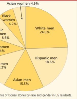
Memane et al.; AJRRU, 3(3): 12-23, 2020; Article no.AJRRU.58148

ABBREVIATIONS                                               parsley helped increase urine volume, decrease
                                                            urinary calcium excretion, and raise the acidity of
ESWL     : Extracorporeal Shockwave Lithotripsy             urine [9]; it also act as a natural diuretic, and
URS      : Ureteroscopy                                     prevent kidney stone formation [10,11,12].
PCNL     : Percutaneous Nephrolithotomy
MET      : Medical expulsive therapy                        When a drug therapy does not resolve the
                                                            symptoms, the placement of a ureteral catheter
                                                            or a nephrostomy tube has routinely represented
1. INTRODUCTION                                             the next step. These easy manoeuvres can offer
                                                            a prompt relief from pain for the patient and they
Kidney stone disease is one of the oldest and               are usually followed by ureteroscopy (URS) or
most common problems of the urinary system.                 extracorporeal shockwave lithotripsy (ESWL),
Race, gender and ethnicity play a part in who               which currently represents the mainstay of
may get kidney stones [1]. The yearly relative              treatment for symptomatic ureteral stones [13].
incidence of urolithiasis is about 10-15% in the            There are numerous ways to treat renal tract
western world but can be as prominent in Middle             calculi, depending on their size, location, volume,
East 20-25%. The recurrence rate without                    anatomical factors and patient comorbidities.
preventive treatment is approximately 10% at 1              Historically, it was open surgical techniques;
year, 33% at 5 years, and 50% at 10 years. In               shock wave lithotripsy (SWL) was introduced in
India, more or less 5-7 million patients suffer             1980, followed by percutaneous nephrolithotomy
from stone disease and leastwise 1/1000 of                  (PCNL) and         subsequently endourological
Indian universe necessarily hospitalization due to          techniques      with   the     popularisation    of
kidney stone disease [2]. Nephrolithiasis has a             ureteroscopy (URS) [14]. Shock wave lithotripsy
higher prevalence in hot, arid, or dry climates,            (SWL) and ureteroscopy (URS) have become the
such as the mountains, desert, or tropical areas.           most common treatment modalities. In this
Worldwide, regions of high stone prevalence                 paper, indications, results, complications, and
include the US, British Isles, Scandinavian and             innovations of these and other treatments of
Mediterranean countries, northern India and                 ureteral stones are reviewed.
Pakistan, northern Australia, central Europe,
portions of the Malay peninsula, and China [3].             2. MANAGEMENT OF STONE
Stones stuck in the ureter, which is the tube that
transports urine from the kidney to the bladder,            Several guidelines are available to provide a
often cause pain and make people see a doctor.              clinical framework for diagnosis, follow-up, and
Depending on which part of the ureter the stone             prevention of kidney stone disease [15]. In 2007
is stuck in and the size of the stone, it will open         the joint EAU and AUA Nephrolithiasis Guideline
pass into the bladder on its own over weeks. If             Panel joined efforts in developing internationally
the stone does not come out by itself, people               endorsed guidelines focusing on the changes in
often need to have procedures done to remove                ureteral stone management [16]. The guidelines
the stone [4,5]. Stone incidence depends on                 state that observation, with or without medical
geographical, climatic, ethnic, dietary and genetic         expulsive therapy, should be offered to patients
factors. The recurrence risk is basically                   with uncomplicated distal ureteral stones that are
determined by the disease or disorder causing               ≤10 mm in diameter. The guidelines also state
the stone formation. Accordingly, the prevalence            that active surveillance can be offered for
rates for urinary stones vary from 1% to 20% [6].           asymptomatic, non-obstructing calyceal stones
Nephrolithiasis is currently more prevalent in              [17]. Acute medical treatment for renal or ureteric
men than in women (13% Vs 7%, respectively),                colic includes conservative therapy, such as
and it is three to four times more likely to present        hydration, analgesia (intravenous pain relief with
in white than nonwhite patients [7]. Medical                morphine or the NSAID ketorolac), and
evaluation for and treatment of kidney stones               antiemetics [3]. Patients who present with acute
places a significant economic burden on society.            nephrolithiasis   most     often   require    fluid
The Urologic Diseases in America project                    administration, aggressive pain management,
estimated an annual cost of more than $2 billion            and treatment for nausea or vomiting. Most
in the United States alone [8]. The first step in           ureteral stones measuring 5.0 mm or less will
the treatment for acute renal colic caused by               typically pass spontaneously within a few
obstructing ureteral stones is medical relief of            weeks,but larger stones usually require
symptoms. It is very important to insert                    intervention—in some cases, surgery [7].
information about parsley in diet section. Parsley          Ureteral stones with a diameter less than 5 mm
may prevent kidney stones. It was found that                will pass in up to 68% of cases; however, for

                                                       13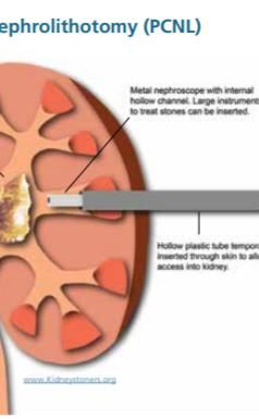
Memane et al.; AJRRU, 3(3): 12-23, 2020;; Article no.AJRRU.58148
                                                                                                     no.

stones with a greater diameter the overall                   volume of 2 L/day, dietary sodium restriction
chances of spontaneous passage are lower [13].   [1          (about 100 meq/day), oxalate restriction
Incidences of nephrocalcinosis increase with                 (avoidance of dark roughage, tea, nuts, soya
age, whereas
         ereas nephrolithiasis is mostly a disease           bean, sweet potato), increased citrus
                                                                                                citrus-fruit intake,
of the third to fifth decades of life [18].
                                      [1 Complete            avoidance of a meat-rich rich diet, and a moderate
management means not only proper evaluation                  intake of dietary
                                                                            ary calcium (up to an equivalent of
and treatment, but also prophylaxis to prevent               one glass of milk per day). A high intake of fluids
recurrence, which is impossible without the                  has been shown to be effective when used as
knowledge of the composition   on of the offending           the only method to prevent stone recurrence
stone [19,12]. ]. The modern western lifestyle               [22,23].]. Stone disease has been increasingly
provides a host of factors that impair urine                 linked to systemic conditions, althalthough it is not
composition and thereby increase the risk of                 clear if stone disease is a cause of these
stone formation. In our everyday life, we do not             disorders or if it is a consequence of the same
drink enough water and only twice or thrice a                conditions that lead to these disorders.
day, we eat food that is too rich in calories and            Overweight/obesity, hypertension and diabetes
table salt, but have deficiencies in fiber and               have all been shown to be associated with an
alkali. Last but not least, we do not exercise               increased risk of stone disease [24].
enough. Recent work showed that being                        Management          of   stone     disease       needs
overweight is a crucial risk factor with significant         individualization. Clinical presentation, proper
impact on stone formation [20]. ]. Management of a           history, and laboratory tests help to identify
kidney stone depends on its size, location, and              whether one needs urgent surgical or medical
composition and the presence of anatomical                   treatment [23]. ]. Management of urolithiasis in
malformation and complications. The presence of              transplanted patients
                                                                                tients is similar to that in the
a complication (complicated stone)—    — infection or        general population and includes factors such as
obstruction—maymay       necessitate        immediate        transplant function, coagulative status, and
intervention,
          on, whereas uncomplicated stones can               anatomical obstacles due to the iliacal position of
be managed conservatively with adequate fluid                the organ, directly influence the surgical strategy
intake and analgesia. If a stone does not pass               [4,6]. Therapeutic nutritionon recommendations for
spontaneously then definitive treatment is                   the secondary prevention of urolithiasis are
needed to remove it [21].                                    widely used. General nutrition guidelines are
                                                             useful in promoting public health and for
All patients with stones should be offered                   developing nutrition plans that reduce the risk for
conservative treatment, whether or not additional            or attenuate the effects of diseases that are
treatments with drugs are to be offered.                     affected
                                                                fected by nutrition. Nutrition therapy is the
Conservative     treatment  for    hypercalciuric            application of nutritional assessment, diagnosis,
individuals with normal bone density includes a              intervention, and counseling to prevent or
high fluid intake to ensure a minimum
                                mi          urine            manage disease [25].

                   Fig. 1. Ethnic diversity in the prevalence of kidney stones [7]

                                                        14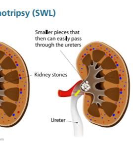
Memane et al.; AJRRU, 3(3): 12-23, 2020;; Article no.AJRRU.58148
no.
Fig. 2. Emergency management of ureteral stones [13]
[
3. TREATMENT OF STONE 5-10
10 mg daily or doxazosin 4 mg daily
were used with similar efficacy [1 [13].
Development in surgical equipment and If the patient has a stone present without
technologies, starting in the 1980s, has provided signs and symptoms of infection, he or she
numerous tools for the intra- and extra-corporeal
extra can be managed conservatively with
fragmentation of urinary calculi for treatment [26].
[ opioids and nonsteroidal anti anti-inflammatory
Treatment depends on the type of stone, stone drugs (NSAIDs). NSAIDs have been
composition, how bad it is and the length of time shown to offer effective pain relief from
you have had symptoms. There are different acute kidney stone related pain with fewer
treatments to choose from. It is important to talk side effects than opioids a
and
to your health care provider about whatwha is best acetaminophen [3,6].
for you [1,6].
6]. Until the 1980s, treatment of the Medical expulsive therapy (MET) has
upper urinary tract often involved extensive open recently emerged as an appealing option
surgical procedures. In the last 20 years the for the initial management of ureteral
treatment of stone disease has undergone stones [13]. ]. There is evidence to support
tremendous changes, especially with the that medical expulsive therapy, namely
introduction of the extracorporeal shock-wave
shock alpha-blockers,
blockers, may increase ureteral uretera
lithotripsy (ESWL) and refinements in endo- endo stone passage rate and decrease the time
urological procedures such as percutaneous to stone passage, particularly in distal
nephrolithotomy (PCNL) and ureterorenoscopy ureteral stones < 10mm in size. However,
(URS) techniques, which exclusively depend if a 4- to 6-week
week trial of MET has been
upon the use of various kind of energies to attempted without successful stone
fragment the stone [27,19]. ]. You can simply wait passage, the patient should undergo
for the stone to pass. Smaller stones are more definitive surgical management [3,5]. [3 The
likely than larger stones to pass on their own. addition of steroids to either calcium
Waiting four to six weeks for the stone to pass is channel blockers or a
a-adrenergic
safe as long as the pain is bearable [1]. antagonists added only a small
incremental benefit [16,23].
3.1 Medication
Immunosuppressive medications,
Certain medications
ons have been shown to particularly cyclosporine, increase urine
improve the chance that a stone will pass. concentration and serum and u urine uric
The most common medication prescribed acid levels, which may promote stone
for this reason is tamsulosin. Tamsulosin formation. The treatment of these patients
(Flomax) relaxes the ureter, making it is challenging, due to immunosuppressive
easier for the stone to pass. You may also drugs used [4].
need pain and anti-nausea
nausea medicine
me as Many patients require narcotic medications
you wait to pass the stone [1,23]. [ to control pain adequately. Antiemetic
tamsulosin 0.4 mg taken daily for one agents (such as the H1 -rec receptor blocker
month. However, in several trials terazosin dimenhydrinate 42) should be
15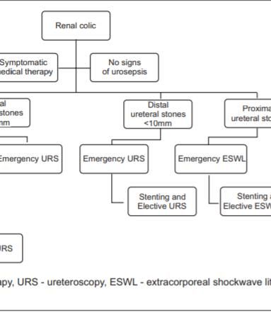
Memane et al.; AJRRU, 3(3): 12-23, 2020; Article no.AJRRU.58148
administered to control nausea and 3.2 Specific Recommendations for
vomiting [7]. Different Types of Stones
Nifedipine: This is a calcium channel
blocker commonly used in the treatment of 3.2.1 Calcium oxalate stones
hypertension and angina. It acts as a
suppressing mechanism of the fast In patients with the common form of
component of ureteral contraction leaving nephrolithiasis, avoiding high-dose vitamin C
the peristaltic rhythm unchanged. Its use in supplements is the only known strategy that
medical therapy for distal ureteral lithiasis reduces endogenous oxalate production. Firstly,
has been tested in various studies [13]. foods that contain high amounts of oxalate
It has been seen that the combination of should be avoided e.g. spinach, rhubarb, and
restricted intake of animal protein (52 potatoes [17]. Increased citrus fruit intake is
g/day), restricted salt intake (50 mmoL, or recommended to prevent stone recurrence [3].
2,900 mg/day of sodium chloride), and For calcium oxalate and calcium phosphate
normal calcium intake (30 mmoL/day, or stones thiazide diuretics (with sodium restriction)
1,200 mg/day) was associated with a lower may be used to reduce urine calcium [29].
incidence of stone recurrence in men with Thiazide diuretics—decrease urinary calcium
hypercalciuria, compared with traditional excretion by augmenting tubular reabsorption of
low-calcium intake (10 mmoL, or 400 calcium, but do not decrease intestinal
mg/day). Patients should therefore be absorption in absorptive hypercalciuria; the effect
advised to avoid excessive intake of may be attenuated or lost after two or more years
animal protein [17,21]. of treatment [21]. High fluid intake may be
Insulin resistance is the most important beneficial not only to prevent CaOx overgrowth,
factor of metabolic syndrome and kidney but also to reduce plaque formation itself.
stone formation, since insulin resistance Thiazide diuretics, which lower urine calcium,
decreases the production and transport of may reduce plaque as well as urine CaOx SS
ammonia, resulting in a low urine pH [15]. [30].Thiazide diuretics have shown to reduce the
addition to conventional treatment of a recurrence rates by up to 70% [23]. Magnesium
calcium channel blocker (nifedipine) to and citrate are inhibitors of crystallization since
relax ureteral muscles, short term they can reduce the saturation of calcium oxalate
prednisone (for five days) to reduce local by complexing oxalate and calcium, respectively
oedema through its anti-inflammatory [18].
action, antibiotics to prevent and treat
urinary tract infection, and paracetamol to 3.2.2 Calcium phosphate stones
raise the pain threshold and reduce the
need for narcotics boosted the rate of Calcium phosphate stones share the same risk
passage of stones and led to fewer lost factors as with calcium oxalate stones like higher
work days, emergency room visits, and concentrations of urine calcium and lower
surgical interventions, with a similar side concentrations of urine citrate [17]. Potassium
effect profile [21]. phosphate—may suppress calcitriol synthesis
Thiazides (trichlormethiazide 2 or 4 mg and thereby decrease calcium absorption [21].
four times a day, or hydrochlorothiazide 50 CaP SFs are usually treated with fluids and
mg twice or four times a day) correct the thiazide diuretics to lower urine calcium
“renal leak” of calcium, and restore normal excretion. Urine citrate excretion can be reduced,
parathyroid function, intestinal calcium as in idiopathic CaOx SFs, but because
absorption, and urinary calcium. Potassium potassium citrate salts can increase urine pH and
citrate or bicarbonate (20 meq twice a day) CaP SS, careful follow-up is needed. No clinical
given with thiazide prevents hypokalaemia trials have documented treatment outcomesfor
and improves citrate excretion [22]. CaP SFs [30]. In patients who also have
Thiazides should be considered hypocitraturia) or the use of a combination
appropriate for both calcium oxalate and thiazide/potassium-sparing diuretic, such as
calcium phosphate stone formers [24]. amiloride/hydrochlorothiazide in patients who do
Significant side effects are thus associated not require citrate repletion. Monitoring of urine
with thiazide therapy in at least 5% of the pH is also critical because elevation of the urine
[
cases 27]. Potential side-effects pH greater than 6.5 can lead to supersaturation
hypokalemia, glucose intolerance, of calcium phosphate and possible change in
dyslipidemia, and hyperuricemia [28]. stone recurrence composition [28].
16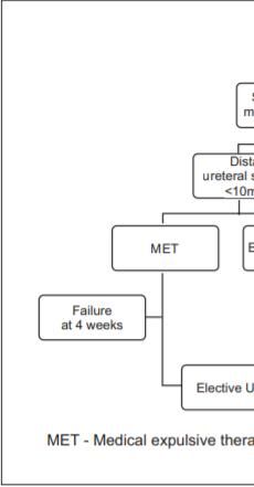
Memane et al.; AJRRU, 3(3): 12-23, 2020; Article no.AJRRU.58148
3.2.3 Uric acid stones alkalinisation (urine ph > 7.0) with potassium
citrate. In addition, specific agents such as, α -
The mainstay of prevention of uric acid stone mercaptopropionylglycine or d-penicillamine that
formation entails increasing urine pH. While form soluble complexes with cystine are used
acidifying the urine can be challenging, [21,30,23]. Although often well-tolerated,
alkalinising the urine can be readily achieved by infrequent side effects include the following: bone
increasing the intake of foods rich in alkali (e.g. marrow suppression, proteinuria with
fruits and vegetables). Supplementation with nephropathy, hepatotoxicity, aplastic anemia,
bicarbonate or citrate salts (preferably potassium drug-induced lupus, abdominal pain, diarrhea,
citrate) can be used to reach the recommended nausea and vomiting, and anorexia [28].
pH goal of 6–7 throughout the day and night [17]. Potassium citrate therapy provides an alkali load
For uric acid nephrolithiasis and other disorders that leads to increased urine pH for cystine stone
of the metabolic syndrome, a common formers, a urine pH of 7.0 should be achieved
pathophysiological cause is on the horizon – [24]. Captopril, a commonly used
insulin resistance. Patients with recurrent uric antihypertensive that contains a thiol-group, is
acid stones were found to be severely insulin another theoretical pharmacologic target for
resistant compared to healthy controls [20]. cystinuria. However, it does not appear in the
Allopurinol-to inhibit uric acid synthesis and urine in sufficient quantities to affect cysteine
decrease urinary uric acid excretion [21,23]. solubility and several small studies have yielded
Allopurinol also has serious side effects. One equivocal data on its ability to decrease urinary
study reported a 3.5% incidence of side effects in cystine level [28].
1835 hospitalized patients who received
allopurinol; 1.7% developed skin rash and 1.8% 3.3 Surgery
developed a variety of other complications of
which the most serious was liver necrosis [27]. Surgery may be needed to remove a stone from
Prevention and even dissolution of UA stones the ureter or kidney if:
depends upon the ability to increase urine pH The stone fails to pass.
above 6.0, which is accomplished by The pain is too great to wait for the stone to
administration of 20–30 mEq potassium alkali, 2 pass.
or 3 times daily [30]. If urine pH is not increased,
The stone is affecting kidney function. Small
xanthine oxidase inhibition of uricosuria may be
stones in the kidney may be left alone if they
ineffective; at high urine pH, xanthine oxidase
are not causing pain or infection. Some
inhibition is redundant in addressing recurrent
people choose to have their small stones
uric acid stones [28].
removed. They do this because they are
3.2.4 Struvite stones afraid the stone will unexpectedly start to
pass and cause pain [1].
These stones require complete removal by a
urologist. New stone formation can be avoided About 10-20% of all kidney stones need
by the prevention of UTIs [17]. Acetohydroxamic radiological or surgical intervention to remove the
acid, a urease inhibitor, has been shown to stone [21]. If there is spontaneous passage of
reduce the urinary saturation of struvite but is stones, most pass within 4 to 6 weeks. Surgical
associated with high frequency of side effects intervention is indicated in the presence of
(deep vein thrombosis, haemolytic anaemia), persistent obstruction, failure of stone
which limits its use [21,23]. Patients treated for progression, sepsis, or persistent or increasing
struvite stones may still be at risk for recurrent colic [3]. Surgical intervention may be required if
UTI after stone removal, and in some patients stones are too large to pass spontaneously
surgical stone removal is not feasible. The use of (typically ≥ 8 mm); if they cause acute renal
a urease inhibitor, AHA, may be beneficial in obstruction; or if they are located at a site with a
these patients, although the extensive side effect potential for complications or can lead to
profile may limit its use [24]. For patients with persistent symptoms without evidence that they
struvite calculi undergoing endourologic are passing [7]. The type of surgery chosen
procedures, preoperative antibiotics are depends on the size and location of the stone, as
commonly used [28]. well as the operators experience, patient
preference, available equipment and related
3.2.5 Cystine stones costs. Presence or absence of obstruction [13].
Treatment must include increasing urine output Surgeries to remove stones in the kidneys or
to about 3.5–5 liters /day and adequate ureters are:
17Memane et al.; AJRRU, 3(3): 12-23, 2020; Article no.AJRRU.58148
1. Shock wave lithotripsy (SWL) the recurrence rate after ESWL, endo-urological
2. Ureteroscopy (URS) procedures, and open surgery [19]. Shock wave
3. Percutaneous Nephrolithotomy (PCNL) lithotripsy is less efficacious if the stone is dense
(attenuation value of more than 1000 Housnfield
3.3.1 Shock wave lithotripsy (SWL) units) on helical computed tomography and might
The first successful ESWL treatment was adversely affect ovarian function when used for
accomplished in 1980 in Germany by Dr distal ureteric stones in women [21].It can have
Christian Chaussy using a Dornier HM1 side effects. In human and animal models it can
lithotripter. Owing to its effectiveness and its rare cause acute renal injury [34]. Although the
side effects, ESWL was quickly approved by the application of ESWL in children was first reported
US Food and Drug Administration (FDA) for in 1986. When considering ESWL for kidney
clinical use [16,14,31]. Shockwave lithotripsy as stones in children, it appears to be a different
first-line therapeutic option, applied rapidly after condition from that in adults [35]. ESWL can
the onset of renal colic, has deserved very become more efficient and safer. In addition, new
limited attention so far [13]. Shock Wave concepts, novel ideas, and future research are
Lithotripsy is used to treat stones in the kidney leading the way toward a brighter future for
and ureter. Shock waves are focused on the ESWL [31]. The advantages of ESWL are that it
stone using X-rays or ultrasound to pinpoint the is minimally invasive, usually well tolerated, and
stone. Repeated firing of shock waves on the a stent is not usually required. The risks of ESWL
stone usually causes the stone to break into include bruising, pain or urinary obstruction from
small pieces. These smaller pieces pass out in fragments of stone, haematuria and, rarely,
the urine over a few weeks [1,26,30]. However, perinephric haematoma, the disadvantages of
intervention success rates depend on stone ESWL are that effectiveness decreases with
composition, size, properties and location of the increasing body mass index, it is not suitable in
stone as well as the orchestration type and pregnancy [36].
frequency of shock.This procedure has been
optimized, and new instruments have been 3.3.2 Ureteroscopy (URS)
developed to increase utility to urologists and to
improve tolerability for the patient [2,13]. Calculi Rigid URS was first applied therapeutically for
between 10 mm and 20 mm are in general the treatment of mid distal ureteral calculi in the
treated with extracorporeal shock wave lithotripsy 1980s. Although large (>10F) diameter
(ESWL) or ureteroscopy as first-line therapy. ureteroscopes were used, success rates of 90%
However for ESWL, the results for lower pole were achieved [16,17]. Ureteroscopy represents
stones are inferior (55%) to upper and mid pole a safe and minimally invasive procedure in the
stones (71.8% and 76.5%, respectively) [3,7]. management of ureteral stones [13].
ESWL is a valuable, noninvasive method for Ureteroscopy is used to treat stones in the
stone management, but some limitations exist in kidney and ureter. URS involves passing a very
transplanted patients. Usually, ESWL is a small telescope, called an ureteroscope*, into the
reasonable treatment for calculi smaller than 1.5 bladder, up the ureter and into the kidney. Rigid
cm [4]. Success depends on the efficacy of the telescopes are used for stones in the lower part
lithotripter and the size, location and composition of the ureter near the bladder. Flexible
(hardness) of the stones, patient’s habitus telescopes are used to treat stones in the upper
performance of SWL [6]. Lowering shock wave ureter and kidney. The ureteroscope lets the
frequency from 120 to 60-90 shock waves/min urologist see the stone without making an
improves SFR. Tissue damage increases with incision (cut). Once the urologist* sees the stone
shock wave frequency [32]. Reducing the shock- with the ureteroscope, it can be fragmented
wave rate to 60/min improves stone using a laser and a small, basket-like device
disintegration and reduces tissue damage [33]. grabs smaller stones and removes them. The
The major advantages of SWL are that it is the procedure is more invasive than ESWL [1,3,37].
least invasive technique to treat renal and Ureteroscopy using the holmium:yttrium-
ureteral stones and potentially can be performed aluminum-garnet (YAG) laser (photothermal
with only sedation and analgesics in most lithotripsy) is effective for stones of all
patients [16]. ESWL is usually an outpatient compositions and sizes, with a success rate of
procedure performed with analgesia or sedation. 97-100%. 29 [21]. Flexible ureteroscopy is
The use of ESWL has increased but still PCNL recommended for adequate access and
has many advantages over ESWL and in some desintegration of calculi and can be done by
cases, URS [17]. Medical treatment could lower electrohydraulic lithotripsy, ultrasonic lithotripsy
18Memane et al.; AJRRU, 3(3): 12-23, 2020;; Article no.AJRRU.58148
no.
or holmium laser in the ureter and kidney, with a are present or when patient factors such as
success rate of 67–100% 100% [4]. Technical pregnancy, coagulopathy,
gulopathy, or morbid obesity
improvements
rovements including endoscope miniature- preclude lithotripsy [34].
]. The high success rate
sation, improved deflection mechanism, and low morbidity of UL for stones of >2 cm
enhanced optical quality and tools, and render it preferable to other methods, including
introduction of disposables have led to an ESWL and PCNL. It should be considered as a
increased use of URS for both, renal and ureteral standard approach to treat large renal calculi
ca
stones [6]. ureteroscopy is more efficacious,
efficacious less [32]. One disadvantage of ureteroscopy is that a
expensive than shock wave lithotripsy for distal ureteral stent, which causes considerable
ureteral stones but is more time consuming and discomfort in some patients, is often necessary
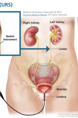
technically demanding [8,21]. ]. ureteroscopy is to prevent obstruction from ureteral oedema or
useful when complex or lower pole renal calculi stone fragments [33].
Fig. 3. Shock wave lithotripsy (SWL) [1]
Fig. 4. Ureteroscopy (URS) [1]
19Memane et al.; AJRRU, 3(3): 12-23, 2020;; Article no.AJRRU.58148
no.
3.3.3 Percutaneous nephrolithotomy (PCNL) large stones [2]. PCNL was developed to reduce
the morbidity and mortality associated with open
PCNL was established as a minimally invasive renal surgery, However, it represents the most
treatment option for kidney stone removal in the morbid of the minimally invasive endoscopic
1970s [33]. The first report of percutaneous surgeries for renal stones [17]. 1,000 consecutive
stone surgery for upper-tract
tract stones in adults in patients
atients who underwent percutaneous removal of
1976.experience has dramatically increased with renal and ureteral stones. Removal was
the increasing application of PCNL in subsequent successful for 98.3 per cent of the targeted renal
years. Nine years later, in 1985, the first stones and 88.2 per cent of the ureteral stones.
paediatric series of PCNL was reported [35]. [ Percutaneous techniques are an effective way to
Patientss with larger or more complex stones may handle the majority of renal calculi [29]. [2
require percutaneous nephrolithotomy [7]. Traditionally, PCNL has been considered
Percutaneous Nephrolithotomy is the best advantageous for treating large lower-pole
lower
treatment for large stones in the kidney. General stones, but recent series showed that UL
anesthesia is needed to do a PCNL. PCNL achieves comparable outcomes [[32]. PCNL as
involves making a half-inchinch incision(cut) in the the first-line
line treatment in children with staghorn
back or side, just large enough to allow a rigid calculi, open surgery
ery is recommended among the
telescope (nephroscope) to be passed into the secondary options, with ESWL [35 35]. PCNL is still
hollow center part of the kidney where the stone a challenging procedure and can be associated
is located. An instrument passed through the with significant complications. increase morbidity
nephroscope breaks up the stone and suctions associated with larger instruments like blood
out the pieces, graspers, or basket extraction. loss, postoperative pain and potent potential renal
This procedure usually requires two days in damage, infection, other organ damage and
hospital [1,34,36].]. Patients with stones >20 mm problems due to residual stones [4,6,
4,6,16,32]. The
should primarily be treated with PCNL unless advantages of PCNL are that it provides
specific indications for an alternate procedure are excellent access to large stones and has a high
present. While PCNL is the first-lineline therapy for rate of stone clearance [36].
Fig. 5. Percutaneous nephrolithotomy (PCNL) [1]
20Memane et al.; AJRRU, 3(3): 12-23, 2020; Article no.AJRRU.58148
Diet recurrence and in expulsion of small size stones.
Removal of stones from the kidney and ureter is
Drink enough fluids each day have shown that an important and extensive part of the care of
increasing urine volume to at least 2 L/day OR 2 patients with urinary tract stone disease. For
lit/day can reduce the recurrence of stone smaller ureteral stones in a reasonable
disease by up to 40–50%, Reduce the amount of asymptomatic patient, MET is an excellent initial
salt in your diet, Eat plenty of fruits and form of treatment. Regarding their expulsive
vegetables, Eat foods with low oxalate levels, Eat efficacy, alleviation of pain, and safety profile,
less meat, Eat the recommended amount of both calcium channel blockers and a-adrenergic
calcium [1,23,37]. Modern lifestyle, dietary habits antagonists can be suggested. In terms of active
and obesity emerge to be the promoters of stone removal, SWL, PCNL and URS have been
idiopathic stone disease. Modern diets containing shown to be useful alternatives and are effective
a lot of animal protein, refined carbohydrates and in treating the overwhelming majority of stones
salts act on the metabolism like an acid .This techniques have advantages and
concentration [2,38]. high fluid intake ensures disadvantages as well as different patterns of
optimal specific weight levels of urine < 1.010. A complications. All such factors need to be
purine-reduced diet decreases the risk of considered when choosing the most appropriate
spontaneous crystallization in urine [6]. A diet method. Patient satisfaction becomes
high in calcium (≥ 1,200 mg/d) reduces the risk increasingly important when choosing between
for calcium oxalate stone recurrence. oxalate-rich competing modalities of similar efficacy, and so it
foods like cucumber, green peppers, beetroot, is difficult to give priority to either of these
spinach, soya bean, chocolate, rhubarb, procedures. Operator’s experience, access to
popcorn, and sweet potato should be avoided adequate equipment and specific circumstances
[7,23]. Fish-oil supplementation showed are probably the most important determinants of
advantageous effects on the lithogenic serum which method will be most appropriate for each
and urine parameters [20]. Oral potassium citrate particular case.
(Kcit) has been shown to be useful in increasing
urinary citrate and reducing the stone recurrence. CONSENT
Surprisingly, fresh tomato juice was found to
have the highest citrate and low oxalate content It is not applicable.
[23]. Nutrition therapy, widely used for secondary
prevention of urolithiasis, is the application of ETHICAL APPROVAL
nutritional assessment, diagnosis, intervention,
and counseling to prevent or manage disease It is not applicable.
[25]. Patients with urolithiasis should be well
hydrated, and have a diet low in salt, oxalate and COMPETING INTERESTS
protein [36].
Authors have declared that no competing
4. CONCLUSION interests exist.
The incidence of urolithiasis is increasing REFERENCES
worldwide. Kidney stone disease remains a
major public health burden. Many aspects of 1. Kidney Stones: A Patient Guide, urology
renal stone formation remain unclear.The care foundation 1000 Corporate
increasing incidence of renal stones is adding to Boulevard, Linthicum, MD 21090 1-800-
the morbidity and huge economic losses 828-7866 UrologyHealth.org
worldwide of this pathology. The technological 2. Uthaya Chandirika Jayaraman, Annadurai
advances have helped with early diagnosis and Gurusamy. Review on uro-lithiasis patho-
treatment. High fluid intake and adopting healthy physiology and aesculapian discussion.
lifestyle measures are some of the cost-effective IOSR Journal of Pharmacy. 2018;8(2
measures of preventing renal stones. Treatment Version. 1):30-42.
is successful if attended in early stage itself. (e)-ISSN: 2250-3013,(p)-ISSN: 2319-4219.
Surgical treatment is more effective. URS is 3. Jodi Antonelli, MD, Naim Maalouf, MD:
associated with a significantly greater stone free BMJ Best practice Nephrolithiasis.
rate and fewer required re-treatment. Medical 4. Dean Markić, Kristian Krpina, Juraj Ahel,
therapy, when used judiciously in conjunction Antun Gršković, Josip Španjol, Nino
with dietary measures, can help in preventing Rubinić, Mauro Materljan, Ivana
21Memane et al.; AJRRU, 3(3): 12-23, 2020; Article no.AJRRU.58148
Mikolašević, Lidija Orlić, Sanjin Rački: White JR. Medical management of kidney
Treatment of kidney stone in a kidney- stones: AUA guideline. The Journal of
transplanted patient with mini- urology. 2014;192(2):316-324.
percutaneous laser lithotripsy: A Case 13. Luis Osorio, Estêvão Lima, Riccardo
Report: Case Rep Nephrol Dial. 2016;6: Autorino, Filinto Marcelo. Emergency
26–31. management of ureteral stones: Recent
DOI: 10.1159/000444251 advances; 2020.
Published online: March 2, 2016 Available:http://www.indianjurol.com
5. Campschroer T, Zhu X, Vernooij RWM, [IP: 106.77.50.253]
Lock MTWT. Alpha-blockers as medical 14. Francesca New & Bhaskar K. Somani: A
expulsive therapy for ureteral stones Complete World Literature Review of
(Review): Cochrane Database of Quality of Life (QOL) in Patients with
Systematic Reviews. 2018;4:Art. No.: Kidney Stone Disease (KSD): Curr Urol
CD008509. Rep. 2016;17:88.
DOI: 10.1002/14651858.CD008509.pub3 DOI: 10.1007/s11934-016-0647-622
6. Türk C, (Chair), Knoll T, (Vice-chair), 15. Yee Wong, Paul Cook, Paul Roderick,
Petrik A, Sarica K, Skolarikos A, Straub M, Bhaskar K. Somani. Metabolic syndrome
Seitz C. Guidelines Associates: S. and kidney stone disease: A systematic
Dabestani, T. Drake, N. Grivas, Y. review of literature. Journal of Endourology.
Ruhayel, A.K. Tepeler: EAU Guidelines on Mary Ann Liebert, Inc. 2016;30(3):246-
Urolithiasis: European Association of 253.
Urology; 2016. DOI: 10.1089/end.2015.0567
7. Catherine C. Wells, Kiran B. 16. Markus J. Bader, Brian Eisner, Francesco
Chandrashekar, Garikiparthy N. Porpiglia, Glen M. Preminger, Hans-Goran
Jyothirmayi, Vikesh Tahiliani, John C. Tiselius. Collaborative review – Stone
Sabatino, Luis A. Juncos. Kidney stones disease contemporary management of
current diagnosis and management. ureteral stones. European Association of
Clinician Reviews. 2012;22(2). Urology. Published by Elsevier B.V. All
8. Brian R. Matlaga, Jeroen P. Jansen, Lisa rights reserved. 2012;764–772
M. Meckley, Thomas W. Byrne, James E. DOI: 10.1016/j.eururo.2012.01.009
Lingeman. Treatment of ureteral and renal 17. Ramesh Aggarwal, Anshuman Srivastava,
stones: A systematic review and meta- Sachin Kumar Jain, Ritika Sud, Rati Singh.
analysis of randomized, controlled trials. Renal stones: A clinical review. EMJ
NIH Public Access Author Manuscript J European Medical Journal. Citation: EMJ
Urol. Author manuscript; available in PMC Urol. 2017;5[1]:98-103.
2013 September 16. Published in final [Received: 23.11.17]
edited form as: J Urol. 2012;188(1):130– [Accepted: 01.03.17]
137. 18. Khan SR. Animal models of kidney stone
DOI: 10.1016/j.juro.2012.02.2569 formation: An analysis. Springer-Verlag
9. Al-Yousofy F, Gumaih H, Ibrahim H, 1997, World J Urol. 1997;15:236-243.
Alasbahy A. Parsley! mechanism as 19. Mohd S. Ansari, Narmada P. Gupta,
antiurolithiasis remedy. Am J Clin Exp Ashok K. Hemal, Premnath N. Dogra,
Urol. 2017;5(3):55-62. Amlesh Seth, Monish Aron, Tej P. Singh.
[PMID: 29181438; PMCID: PMC5698599] Spectrum of stone composition: structural
10. Kreydiyyeh SI, Usta J. Diuretic effect and analysis of 1050 upper urinary tract calculi
mechanism of action of parsley. J from northern India: International Journal
Ethnopharmacol. 2002;79(3):353‐357. of Urology. 2005;12-16.
DOI: 10.1016/s0378-8741(01)00408-1 20. Michael Straub and Richard E. Hautmann:
11. InformedHealth.org [Internet]. Cologne, Developments in stone prevention:
Germany: Institute for Quality and Current Opinion in Urology. 2005;15:119–
Efficiency in Health Care (IQWiG); 2006-. 126.
Preventing kidney stones; 2016. 21. Malvinder S. Parmar. Kidney stones:
[Updated 2019 Feb 28] Clinical review. BMJ. 2004;328:1420–4.
Available:https://www.ncbi.nlm.nih.gov/boo BMJ VOLUME 328 12 JUNE 2004
ks/NBK348941/ bmj.com
12. Pearle MS, Goldfarb DS, Assimos DG, 22. Charles Y. C. Pak. Kidney stones. The
Curhan G, Denu-Ciocca CJ, Matlaga BR, Lancet. 1998;351.
22Memane et al.; AJRRU, 3(3): 12-23, 2020; Article no.AJRRU.58148
23. Shriganesh R. Barnela, Sachin S. Soni, 30. Fredric L. Coe, Andrew Evan, Elaine
Sonali S. Saboo, Ashish S. Bhansali. Worcester. Kidney stone disease.
Medical management of renal stone. 2005;10(115).
Indian Journal of Endocrinology and The Journal of Clinical Investigation.
Metabolism. 2012;16(2). Available:http://www.jci.org,
DOI: 10.4103/2230-8210.93741 DOI: 10.1172/JCI26662
24. Margaret Sue Pearle, David S. Goldfarb, 31. Naeem Bhojani, James E. Lingeman.
Dean G. Assimos, Gary Curhan, Cynthia Shockwave lithotripsy–New concepts and
optimizing treatment parameters. Urol Clin
J. Denu-Ciocca, Brian R. Matlaga, Manoj
N Am. 2013;40:59–66.
Monga, Kristina Lea Penniston, Glenn M.
Preminger, Thomas M. T. Turk, James Available:http://dx.doi.org/10.1016/j.ucl.20
Robert White. Medical management of 12.09.00.
kidney stones: AUA guideline: The Journal 32. Demetrius H. Bagley, Kelly A. Healy, Nir
Kleinmann. Ureteroscopic treatment of
of Urology. 2014;192:1-9.
larger renal calculi (>2 cm): Arab Journal
Available:http://dx.doi.org/10.1016/j.juro.20 of Urology. 2012;10:296-300.
14.05.006 33. Thomas Knoll, Noor Buchholz, Gunnar
25. Kristina L. Penniston, Stephen Y. Nakada. Wendt-Nordahl. Extracorporeal shockwave
Diet and alternative therapies in the lithotripsy vs. percutaneous nephro-
management of stone disease. Urol Clin N lithotomy vs. flexible ureterorenoscopy for
Am. 2013;40:31–46. lower-pole stones. Arab Journal of
26. Yaw A. Nyame, Shubha De, Carl Urology. 2012;10:336-341.
Sarkissian. Robert Brown, Ganesh Kartha, 34. Nicole L. Miller, James E Lingeman.
Paurush Babbar, Manoj Monga. Kidney Management of kidney stones. BMJ.
stone models for in vitro lithotripsy 2007;334:468-72.
research: A Comprehensive Review: DOI: 10.1136/bmj.39113.480185.80
Journal of Endourology. 35. Rahim Horuz, Kemal Sarica. The
DOI: 10.1089/end.2014.0850 management of staghorn calculi in
children. Arab Association of Urology.
27. Jaime Uribarri, Man S. Oh, Hugh J.
2012;10:330-335.
Carroll. Diagnosis and treatment the first
36. James Sewell, Darren J. Katz, Ohad
kidney stone. Annals of Internal Medicine.
Shoshany, Christopher Love. Urolithiasis –
1989;111:1006-1009.
Ten things every general practitioner
28. Brian H. Eisner, David S. Goldfarb, Gyan should know. UROLITHIASIS FOCUS,
Pareek. Pharmacologic treatment of The Royal Australian College of General
kidney stone disease: Urol Clin N Am. Practitioners. 2017;46(9).
2013;40:21–30 37. Osorio L, Lima E, Autorino R, Marcelo F.
29. Joseph W. Segura, Davide. Patterson, Emergency management of ureteral
Andrew J. Leroy, Hugh J. Williams, Jr., stones: Recent advances. Indian journal of
David M. Barrett, Ralph C. Benson, JR., urology: IJU. Journal of the Urological
Gerald R. May, Claire E. Bender. Society of India. 2008;24(4):461.
Percutaneous removal of kidney stones: 38. Aggarwal R, Srivastava A, Jain SK, Sud R,
review of 1,000 cases. The Journal of Singh R. Renal stones: A clinical
Urology. 134. review. EMJ Urol. 2017;5(1):98-103.
_________________________________________________________________________________
© 2020 Memane et al.; This is an Open Access article distributed under the terms of the Creative Commons Attribution License
(http://creativecommons.org/licenses/by/4.0), which permits unrestricted use, distribution, and reproduction in any medium,
provided the original work is properly cited.
Peer-review history:
The peer review history for this paper can be accessed here:
http://www.sdiarticle4.com/review-history/58148
23You can also read