The Hippo Pathway Regulates Homeostatic Growth of Stem Cell Niche Precursors in the Drosophila Ovary
←
→
Page content transcription
If your browser does not render page correctly, please read the page content below

RESEARCH ARTICLE

                                                          The Hippo Pathway Regulates Homeostatic
                                                          Growth of Stem Cell Niche Precursors in the
                                                          Drosophila Ovary
                                                          Didem P. Sarikaya, Cassandra G. Extavour*
                                                          Department of Organismic and Evolutionary Biology, Harvard University, Cambridge, Massachusetts, United
                                                          States of America

                                                          * extavour@oeb.harvard.edu

       a11111
                                                          Abstract
                                                          The Hippo pathway regulates organ size, stem cell proliferation and tumorigenesis in adult
                                                          organs. Whether the Hippo pathway influences establishment of stem cell niche size to ac-
                                                          commodate changes in organ size, however, has received little attention. Here, we ask
                                                          whether Hippo signaling influences the number of stem cell niches that are established dur-
    OPEN ACCESS
                                                          ing development of the Drosophila larval ovary, and whether it interacts with the same or dif-
Citation: Sarikaya DP, Extavour CG (2015) The             ferent effector signaling pathways in different cell types. We demonstrate that canonical
Hippo Pathway Regulates Homeostatic Growth of
                                                          Hippo signaling regulates autonomous proliferation of the soma, while a novel hippo-
Stem Cell Niche Precursors in the Drosophila Ovary.
PLoS Genet 11(2): e1004962. doi:10.1371/journal.          independent activity of Yorkie regulates autonomous proliferation of the germ line. More-
pgen.1004962                                              over, we demonstrate that Hippo signaling mediates non-autonomous proliferation signals
Editor: Gregory P. Copenhaver, The University of          between germ cells and somatic cells, and contributes to maintaining the correct proportion
North Carolina at Chapel Hill, UNITED STATES              of these niche precursors. Finally, we show that the Hippo pathway interacts with different
Received: September 19, 2014                              growth pathways in distinct somatic cell types, and interacts with EGFR and JAK/STAT
                                                          pathways to regulate non-autonomous proliferation of germ cells. We thus provide evidence
Accepted: December 18, 2014
                                                          for novel roles of the Hippo pathway in establishing the precise balance of soma and germ
Published: February 2, 2015
                                                          line, the appropriate number of stem cell niches, and ultimately regulating adult female
Copyright: © 2015 Sarikaya, Extavour. This is an          reproductive capacity.
open access article distributed under the terms of the
Creative Commons Attribution License, which permits
unrestricted use, distribution, and reproduction in any
medium, provided the original author and source are
credited.

Data Availability Statement: All relevant data are
                                                            Author Summary
within the paper and its Supporting Information files.      During development, organ growth must be carefully regulated to make sure that organs
Funding: This work was supported by National                achieve the correct final size needed for organ function. In organs that are made of many
Institutes of Health grant 1R01 HD073499 to CGE.            different types of cells, this growth regulation is likely to be particularly complex, because
DPS is supported by a post-graduate scholarship             it is important for organs to have appropriate proportions, or relative numbers, of the dif-
from the Natural Sciences and Engineering Research
                                                            ferent kinds of cells that make up the organ, as well as the correct number of total cells.
Council of Canada (NSERC) and a pre-doctoral
fellowship from Fonds de recherché du Québec—
                                                            One method that cells use to regulate organ growth is a signaling pathway called the
Santé (FRQS).” The funders had no role in study             Hippo pathway. However, Hippo signaling has been studied, to date, primarily in organ
design, data collection and analysis, decision to           systems that are made up of one cell type. In this study, we examine how Hippo signaling
publish, or preparation of the manuscript.

PLOS Genetics | DOI:10.1371/journal.pgen.1004962                 February 2, 2015                                                                           1 / 28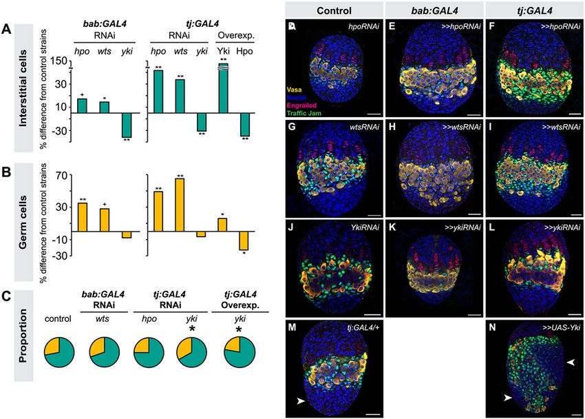
Hippo Regulates Homeostasis of the Ovarian Germ Line Stem Cell Niche

Competing Interests: The authors have declared
that no competing interests exist.                 can work to regulate the proportions of different types of cells, as well as the total number
                                                   of cells in an organ. To do this, we used the developing ovary of the fruit fly as a study sys-
                                                   tem. We found that (1) Hippo signaling regulates the proliferation of many different cell
                                                   types of the ovary; and (2) Hippo signaling activity in one cell type influences proliferation
                                                   of other cell types, thus ensuring appropriate proportions of different ovarian cell types.

                                                 Introduction
                                                 The Hippo pathway is a tissue-intrinsic regulator of organ size, and is also implicated in stem
                                                 cell maintenance and cancer [1,2,3]. An outstanding question in the field is whether the Hippo
                                                 pathway regulates proliferation of cells comprising stem cell niches during development in
                                                 order to ensure that adult organs have an appropriate number of stem cells and stem cell niches
                                                 [4]. The adult Drosophila ovary is an extensively studied stem cell niche system. In this organ,
                                                 specialized somatic cells regulate the proliferation and differentiation of germ line stem cells
                                                 (GSCs) throughout adult reproductive life [reviewed in 5]. The fact that GSCsare first estab-
                                                 lished in larval stages raises the question of how the correct numbers of GSCs, and their associ-
                                                 ated somatic niche cells, are achieved during larval development. To date, only the Ecdysone,
                                                 Insulin and EGFR pathways have been implicated in this process [6,7,8]. Here, we investigate
                                                 the role of the Hippo pathway in regulating proliferation of somatic cells and GSC niche pre-
                                                 cursors to establish correct number of GSC niches.
                                                     Our current understanding of the Hippo pathway is focused on the core kinase cascade and
                                                 upstream regulatory members. The Hippo pathway’s upstream regulation is mediated by a
                                                 growth signal transducer complex comprising Kibra, Expanded and Merlin [9,10,11,12] and
                                                 the planar cell polarity regulators Fat [13,14,15] and Crumbs [16,17]. Regulation of Hippo sig-
                                                 naling further upstream of these factors appears to be cell type-specific [18]. When the core ki-
                                                 nase cascade is active, the kinase Hippo (Hpo) phosphorylates the kinase Warts (Wts) [19,20].
                                                 Phosphorylated Wts then phosphorylates the transcriptional coactivator Yorkie (Yki), which
                                                 sequesters Yki within the cytoplasm [21]. In the absence of Hpo kinase activity, unphosphory-
                                                 lated Yki can enter the nucleus and upregulate proliferation-inducing genes [21,22,23,24]. The
                                                 Hippo pathway affects proliferation cell-autonomously in the eye and wing imaginal discs, glia,
                                                 and adult ovarian follicle cells in Drosophila [18,19,20,25,26], as well as in liver, intestine, heart,
                                                 brain, breast and ovarian cells in mammals [27,28,29,30,31,32]. Hippo pathway is often im-
                                                 properly regulated in cancers of these tissues, which display high levels and ectopic activation
                                                 of the human ortholog of Yki, YAP [27,28,33,34]. Upregulation of YAP is also commonly ob-
                                                 served in a variety of mammalian stem cell niches, where YAP can be regulated in a Hippo-in-
                                                 dependent way to regulate stem cell function [reviewed in 4]. Interestingly, germ line clones
                                                 lacking Hippo pathway member function do not cause germ cell tumors in the adult Drosophi-
                                                 la ovary, which has led to the hypothesis that Hippo signaling functions only in somatic cells
                                                 but not in the germ line [35,36].
                                                     More recently, it has become clear that the Hippo pathway can regulate proliferation non-
                                                 autonomously: Hippo signaling regulates secretion of JAK/STAT and EGFR ligands in Dro-
                                                 sophila intestinal stem cells [37,38,39], and of EGFR ligands in breast cancer cell lines [31], and
                                                 the resulting changes in ligand levels affect the proliferation of surrounding cells non-autono-
                                                 mously. How autonomous and non-autonomous effects of the Hippo pathway coordinate dif-
                                                 ferentiation and proliferation of multiple cell types has nonetheless been poorly investigated.
                                                 Moreover, most studies address the Hippo pathway’s role in adult stem cell function, but

PLOS Genetics | DOI:10.1371/journal.pgen.1004962        February 2, 2015                                                                          2 / 28
Hippo Regulates Homeostasis of the Ovarian Germ Line Stem Cell Niche

                                           whether Hippo signaling also plays a role in the early establishment of stem cell niches during
                                           development remains unknown.
                                               Here we use the Drosophila larval ovary as a model to address both of these issues. Adult
                                           ovaries comprise egg-producing structures called ovarioles, each of which houses a single GSC
                                           niche. The GSC niche is located at the anterior tip of each ovariole, and produces new oocytes
                                           throughout adult life. The niche cells include both GSC and differentiated somatic cells called
                                           cap cells [40]. Each GSC niche lies at the posterior end of a stack of seven or eight somatic cells
                                           termed terminal filaments (TFs). Somatic stem cells located close to the GSCs serve as a source
                                           of follicle cells that enclose each developing egg chamber during oogenesis [5]. All of these cell
                                           types originate during larval development, when the appropriate number of stem cells and
                                           their niches must be established. The larval ovary thus serves as a compelling model to address
                                           issues of homeostasis and stem cell niche development.
                                               TFs serve as beginning points for ovariole formation and thus establish the number of GSC
                                           niches [41]. TFs form during third instar larval (L3) development by the intercalation of termi-
                                           nal filament cells (TFCs) into stacks (TFs) (Fig. 1A; [41]). TFCs proliferate prior to entering a
                                           TF, and cease proliferation once incorporated into a TF [42]. The morphogenesis and prolifer-
                                           ation of TFCs during the third larval instar (L3) is regulated by Ecdysone and Insulin signaling,
                                           and by the BTB/POZ factor bric-à-brac (bab) [6,8,41,43,44,45]. Intermingled cells (ICs) arise
                                           from somatic cells that are in close contact with the germ cells (GCs) during L2, and proliferate
                                           throughout larval development [46] (Fig. 1A). ICs regulate GC proliferation and differentiation
                                           and are thought to give rise to escort cells in the adult niche [6,8,47]. Both Insulin and EGFR
                                           signaling promote the proliferation of ICs [6,48]. Finally, larval GCs give rise to GSCs and early
                                           differentiating oocytes. GCs proliferate during development and do not differentiate until mid-
                                           L3, when the GSCs are specified in niches that form posterior to the TFs [6,8,49], and the re-
                                           maining GCs begin to differentiate as oocytes. GCs secrete Spitz, an EGFR ligand, and promote
                                           proliferation of ICs [49]. In addition, activation of Insulin and Ecdysone signaling in ICs regu-
                                           lates timing of early GC differentiation and cyst formation [6,8], though the identity of the IC-
                                           to-GC signal is unknown. ICs can non-autonomously regulate the proliferation of GCs both
                                           positively and negatively through Insulin and EGFR signaling respectively [6,7,49].
                                               We previously showed that hpo and wts regulate TFC number in a cell-autonomous manner
                                           [50]. Here we demonstrate a role for canonical Hippo pathway activity in regulating both TFCs
                                           and ICs. We also provide evidence for three novel roles of Hippo pathway members in ovarian
                                           development: First, in contrast to a previous report suggesting that yki did not play a role in de-
                                           termining GC number [35], we show that non-canonical, hpo-independent yki activity regu-
                                           lates proliferation of the germ line. Second, we show that Hippo signaling regulates
                                           homeostatic growth of germ cells and somatic cells through the JAK/STAT and EGFR path-
                                           ways. Third, we show that the Hippo pathway interacts with the JAK/STAT pathway to regu-
                                           late TFC number, and with both the EGFR and JAK/STAT pathways to regulate IC number
                                           autonomously and GC number non-autonomously. These data elucidate how Hippo pathway-
                                           mediated control of ovarian development establishes an organ-appropriate number of stem
                                           cell niches, and thus ultimately influences adult reproductive capacity.

                                           Results
                                           Hippo pathway activity is cell type-specific in the larval ovary
                                           To determine whether Hippo signaling regulates proliferation of the GSC niche precursor cells,
                                           we first examined the expression pattern of Hippo pathway members in the larval ovary.
                                           Throughout larval development, Hpo was expressed ubiquitously in the ovary (S1A–C Fig.),
                                           and Yki was expressed in all somatic cells of the ovary (S1F–H Fig.). However, the subcellular

PLOS Genetics | DOI:10.1371/journal.pgen.1004962   February 2, 2015                                                                      3 / 28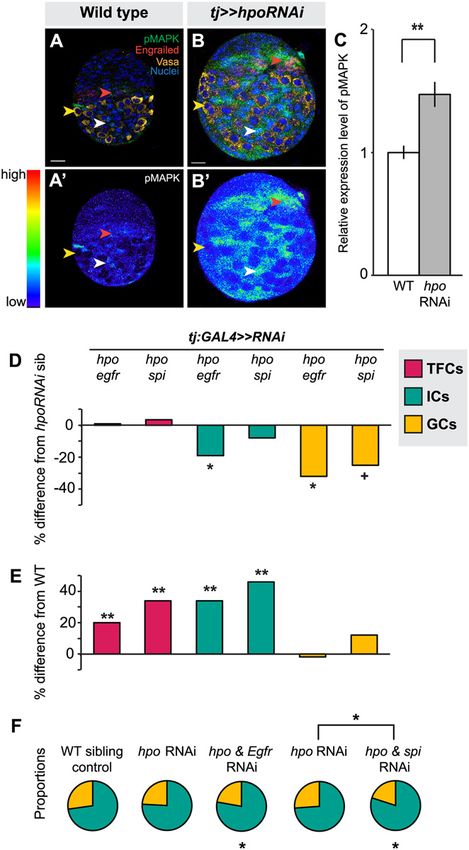
Hippo Regulates Homeostasis of the Ovarian Germ Line Stem Cell Niche

Fig 1. Hippo pathway activity is cell-type specific in the larval ovary. (A) Schematic of Drosophila larval ovarian development from first instar (L1) to the
larval-pupal (LP) transition stage. The L1 larval ovary consists of germ cells (GCs: yellow) surrounded by a layer of somatic cells. As the ovary grows (L2),
some somatic cells intermingle with GCs, becoming intermingled cells (ICs: green). Terminal filament cells (TFCs: pink) emerge during early L3, and begin
intercalating to form terminal filaments (TFs), whose formation continues until the LP stage. (B–I, B’–I’) Expression of Yorkie (B–E) and expanded-lacZ (F–I)
in larval ovarian cell types. B–I show merged images with Yki (B–E) or ex-lacZ (F–I) in green, nuclear marker Hoechst 33342 in cyan, TFC marker anti-
Engrailed (B–C, F–G) or IC marker anti-Traffic Jam (D) in red, and F-actin (E) or GC marker anti-Vasa (H–I) in white. B’-I’ show Yki (B’-E’) or ex-lacZ (F’-I’)
signal only. See S2I–J Fig. for quantification. (B) TFCs at early L3 that are intercalating into TFs display nuclear Yorkie localization. (C) Once incorporated

PLOS Genetics | DOI:10.1371/journal.pgen.1004962        February 2, 2015                                                                                   4 / 28
Hippo Regulates Homeostasis of the Ovarian Germ Line Stem Cell Niche

into TFs, TFCs display cytoplasmic Yorkie localization. (D) ICs have high levels of nuclear and cytoplasmic Yorkie. (E) Yorkie is detectable only at very low
levels in GCs. expanded-lacZ expression is detected in intercalating TFCs (F), ICs (H) and GCs (I) but not in TFCs once they are incorporated into TFs (G).
White arrowheads indicate an example of the specific cell types indicated in each column. Yellow arrowheads indicate cap cells posterior to TFs. Scale bar =
10 μm in B–I’.
doi:10.1371/journal.pgen.1004962.g001

                                               localization of Yki was dynamic during ovariole morphogenesis, and different in distinct so-
                                               matic cell types. We observed nuclear Yki expression in newly differentiating TFCs (identified
                                               by Engrailed expression and elongated cellular morphology) (Fig. 1B–B’, arrowhead; S2I Fig.),
                                               while late stage TFs had very little detectable nuclear Yki (Fig. 1C–C’, arrowhead; S2I Fig.).
                                               Since Yki localization in the nucleus indicates low or absent Hippo pathway activity [21], these
                                               data suggest that Hpo signaling may promote TFC and TFC-progenitor proliferation before
                                               TF formation, and then suppress proliferation in TFCs that have entered TFs. This is consistent
                                               with previous reports of the somatic proliferative dynamics of the larval ovary [42,48].
                                                  We also assessed Yki activity by analyzing expression of the downstream target genes ex-
                                               panded (ex) [21], diap1 (also called thread) [21] and bantam [22]. ex-lacZ (Fig. 1F–G, S2J Fig.)
                                               and diap1-lacZ (S2A–B, K Fig.) were expressed in early TFCs, but ceased expression once TFCs
                                               were incorporated into a TF. The bantam-GFP sensor is a GFP construct containing bantam
                                               miRNA target sites, such that low or absent GFP expression indicates bantam expression and
                                               activity. The sensor was not expressed in early differentiating TFCs, but was expressed in TFCs
                                               within a TF (S2E–F, L Fig.). These data are consistent with the subcellular localization of Yki in
                                               TFCs. Yki activity reporters were also expressed in cap cells of the GSC niche, which are imme-
                                               diately posterior to TFs (Fig. 1G–G’, yellow arrowhead).
                                                  In ICs, strong cytoplasmic and nuclear expression of Yki was observed throughout develop-
                                               ment (Fig. 1D–D’; S2I Fig.). Likewise, all Yki activity reporters examined were expressed in ICs
                                               (Fig. 1H–H’, S2C–C’, G–G’, J–L Figs.), consistent with continuous proliferation of these cells
                                               throughout larval development.

                                               Hippo signaling regulates terminal filament cell proliferation
                                               The expression patterns described above, and our previous observation that knockdown of hpo
                                               or wts increased TFC number [50], suggested that the Hippo pathway regulates TFC prolifera-
                                               tion. To further test this hypothesis, we manipulated activity of the core Hippo pathway mem-
                                               bers hpo, wts and yki in somatic cells using the bric-à-brac (bab) and traffic jam (tj) GAL4
                                               drivers [51,52]. bab:GAL4 is strongly expressed in TFCs during L3 but only weakly in other so-
                                               matic cell types [50,51]. tj:GAL4 is expressed primarily in somatic cells posterior to the TFs, in-
                                               cluding ICs, to a lesser extent in newly forming TF stacks during early and mid L3, and in
                                               posterior TFCs in late L3 (S3A–D Fig.) [53]. We note that the expression of Tj in intercalating
                                               TFCs is not detected with the Traffic-Jam antibody (S3E Fig.). Antibody staining against Hpo
                                               and Yki was used to confirm effectiveness of the RNAi-mediated knockdown under both
                                               GAL4 drivers (S1D–D’, I–I’ Fig.; see Methods for further details of RNAi validation in these
                                               and subsequent experiments).
                                                  Lowering Hippo pathway activity in somatic cells by expressing RNAi against hpo or wts
                                               under either GAL4 driver significantly increased TFC number (student’s t-test was used for
                                               this and all other comparisons: p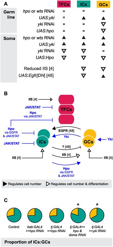
Hippo Regulates Homeostasis of the Ovarian Germ Line Stem Cell Niche

Fig 2. Hippo pathway influences proliferation of TFCs, thereby influencing ovariole number. Changes in (A) TFC or (B) TF number in LP ovaries
expressing UAS-induced RNAi against hpo, wts or yki, or overexpressing hpo or yki under the bab:GAL4 or tj:GAL4 drivers. Here and in Figs. 3–6, S4 and
S5, bar graphs show percent difference from control genotypes of the indicated cell type or structure in each of the experimental genotypes, which are those
that carry both UAS and GAL4 constructs. Control genotypes are either parental strains or siblings carrying a balancer chromosome instead of the GAL4
construct (see Methods). When statistical comparisons were performed to parental strains, values from the two parental strains were averaged and percent
difference from the average was plotted. Statistical significance was calculated using a student’s two-tailed t-test with unequal variance. ** p
Hippo Regulates Homeostasis of the Ovarian Germ Line Stem Cell Niche

(control, white squares), tj:GAL4 driving hpoRNAi (black diamonds), tj:GAL4 driving ykiRNAi (grey triangles). Error bars indicate confidence intervals. * p
Hippo Regulates Homeostasis of the Ovarian Germ Line Stem Cell Niche

                                           quantified ovariole number in adults with RNAi-mediated knockdown of Hippo signaling
                                           pathway members hpo, wts, salvador (sav), Merlin (Mer), or ex in somatic cells. In all cases
                                           adult ovariole number was significantly increased (p
Hippo Regulates Homeostasis of the Ovarian Germ Line Stem Cell Niche
detected only extremely low levels of Yki in GCs throughout development (Figs. 1E, E’, S1F–H,
S2I). The bantam-GFP sensor also suggested low or absent Yki activity in GCs (S2H–H’, L
Fig.). However, we did observe expression of the expanded-lacZ (Figs. 1I–I’, S2J) and diap1-
lacZ (S2D–D’, S2K Figs.) reporters in the GCs. We thus performed functional experiments to
evaluate the roles of Yki and other Hpo pathway members in GCs.
We disrupted Hippo pathway activity in GCs using the germ line-specific driver nos:GAL4
(S3E–H Fig.). In contrast to the overproliferation of somatic cell types observed in the experi-
ments described above, driving RNAi against hpo or wts in the germ line did not significantly
change GC number (Fig. 4A; S3 Table). However, driving yki RNAi in the germ line signifi-
cantly reduced GC number (pHippo Regulates Homeostasis of the Ovarian Germ Line Stem Cell Niche Fig 4. Yorkie activity regulates GC number. Changes in (A) GC number in ovaries expressing hpo, wts, ex, hpo/wts/ex triple, hipk, yki or sd RNAi, or overexpressing hpo, yki or ykiS168A under the nos:GAL4 driver. Bar graphs are as explained in Fig. 2 legend. * p
Hippo Regulates Homeostasis of the Ovarian Germ Line Stem Cell Niche

                                           changes in the other, which would ensure an appropriate number of operative stem cell niches
                                           [8]. To test this hypothesis, we analyzed GC number in conditions where Hippo pathway activ-
                                           ity was altered in the somatic cells. Non-autonomous positive regulation of GC number by ICs
                                           has been documented, but only in ways that also affect GC differentiation [49]. Whether ICs
                                           can positively regulate GC proliferation without affecting their differentiation thus remains un-
                                           known [6,8]. We found that increasing somatic cell number by driving hpo or wts RNAi in the
                                           soma also significantly increased GC number (p>wtsRNAi; Figs. 3C, S7; S3, S6 Tables) or
                                           as much as 70% (tj:GAL4>>hpoRNAi; Figs. 3C, S7; S3, S6 Tables), resulting in a consistent ratio
                                           of ICs to GCs (Figs. 3C, S7; S6 Table). However, increasing IC number by 150% via somatic
                                           overexpression of yki prompted only a 10% increase in GC number (p
Hippo Regulates Homeostasis of the Ovarian Germ Line Stem Cell Niche
Fig 5. The Hippo pathway interacts with the EGFR pathway to regulate IC and GC growth. (A)
Expression pattern of EGFR pathway activity marker pMAPK in wild type L3 ovary. Expression is mainly in
posterior IC cells. Scale bar = 10 μm and applies also to A’. (B) pMAPK expression in ovary expressing UAS:
hpoRNAi in the soma, exposed at same laser setting as (A). Scale bar = 10 μm and applies also to B’. (C)
Relative intensity of anti-pMAPK fluorescence in wild type compared to hpo knockdown experimental (n = 8).
PLOS Genetics | DOI:10.1371/journal.pgen.1004962 February 2, 2015 12 / 28Hippo Regulates Homeostasis of the Ovarian Germ Line Stem Cell Niche
Overall expression level of pMAPK is higher than controls, most prominently in the ICs. (D–E) Percent
difference in TF (red), IC (green), and GC (yellow) number in double RNAi (hpo and egfr, or hpo and spi)
compared to hpo single RNAi sibling controls (D), and wild type sibling controls (E). * pHippo Regulates Homeostasis of the Ovarian Germ Line Stem Cell Niche
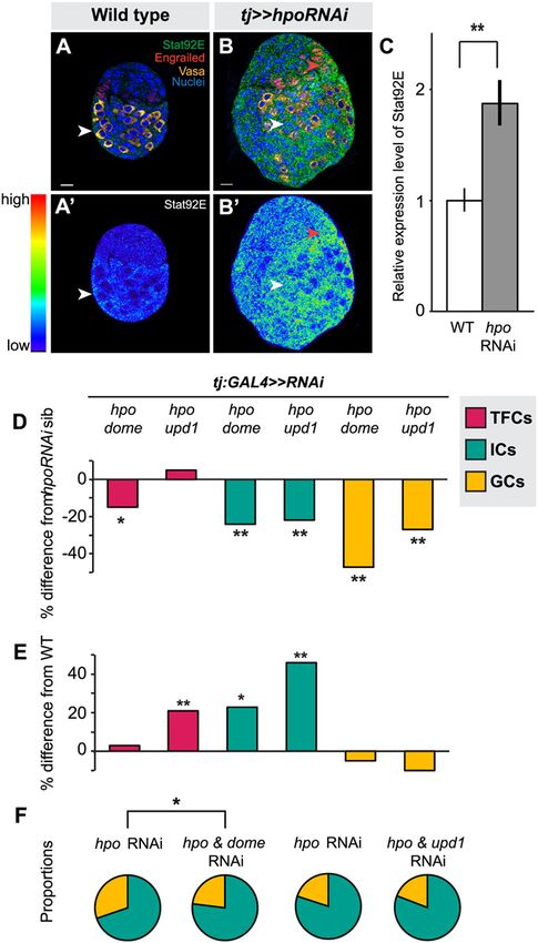
Fig 6. Hippo pathway interacts with JAK/STAT pathway to regulate TFC and IC proliferation, and non-
autonomous regulation of GC number. (A) Expression pattern of JAK/STAT pathway kinase Stat92E in
wild type L3 ovary. Scale bar = 10 μm and applies also to A’. (B) Stat92E expression in ovary expressing
UAS:hpoRNAi in the soma, exposed at same laser setting as (A). Scale bar = 10 μm and applies also to B’. (C)
Relative intensity of anti-Stat92E fluorescence in wild type compared to hpo knockdown experiments
PLOS Genetics | DOI:10.1371/journal.pgen.1004962 February 2, 2015 14 / 28Hippo Regulates Homeostasis of the Ovarian Germ Line Stem Cell Niche
(n = 10). (D–E) Percent difference in TF (red), IC (green), GC (yellow) number in double RNAi (hpo and
dome, or hpo and upd1) knockdowns compared to hpo single RNAi sibling controls (D), and wild type sibling
controls (E). * pHippo Regulates Homeostasis of the Ovarian Germ Line Stem Cell Niche
effect on TFC number (Fig. 5D). Third, loss of germ cell-less (gcl) function leads to reduced GC
and IC numbers [49], but has no effect on ovariole number [81]. Given that ovariole number is
largely determined by TFC number [50], it is likely that gcl ovaries have reduced ICs but not re-
duced TFCs. However, we note that both TFCs and ICs respond to hormonal cues provided by
Ecdysone and Insulin signaling [6,8]. This suggests that growth of these somatic cell types may
be accomplished through their response to systemic hormonal cues, rather than through non-
autonomous effects of one somatic cell type on another.
While the Hippo pathway regulates proliferation of both ICs and TFCs, each cell type had a
unique pattern of Hippo pathway activity during larval development, suggesting that the up-
stream regulatory cues of Hippo signaling are different for TFCs and ICs. In Drosophila, glial
cells and wing disc cells activate the Hippo pathway using different combinations of upstream
regulators [18], indicating that the Hippo pathway can interact with a unique set of upstream
regulatory genes depending on the cell type. Addressing these cell type-specific differences in
Hippo pathway activation in future studies will elucidate how the Hippo pathway is regulated
locally during development of complex organs to establish organ size.
Another notable difference between Hippo pathway operation in ICs and TFCs is its differ-
ential interactions with the EGFR and JAK/STAT pathways in distinct ovarian cell types. In
Drosophila intestinal stem cell development and stem cell-mediated regeneration [37,38,39,68],
as well as in eye imaginal discs [64,67], the Hippo pathway regulates proliferation of these tis-
sues via interactions with both the EGFR and JAK/STAT pathways. In contrast, the Hippo
pathway acts in parallel with but independently of both pathways to regulate the maturation of
Drosophila ovarian follicle cells [25,36]. We do not know what mechanisms determine whether
the Hippo pathway interacts with EGFR signaling, JAK/STAT signaling, or both in a given cell
or tissue type. One mechanism that may be relevant, however, is the differential activation of
specific ligands. For example, in the Drosophila eye disc, Hippo signaling interacts genetically
with EGFR activity induced by vein, but not by any of the other three Drosophila EGFR ligands
[31]. Similarly, constitutively active human YAP can upregulate transcription of vein, but not
the other three EGFR ligands, in Drosophila wing imaginal discs [31]. That fact that spiRNAi
driven in the soma does not rescue the hpoRNAi overproliferation phenotype in the ovary may
indicate that other ligands, such as vein, are required for this EGFR-Hippo signaling interac-
tion, or that the relevant EGFR ligands are expressed by GCs rather than the soma. Our results
suggest that the larval ovary could serve as a model to examine whether differential ligand use
within a single organ could modulate Hippo pathway activity during development.
Hippo signaling in germ cells of the larval ovary
Previous reports [35,36] suggested that the Hippo pathway components were dispensable for the
proliferation of adult GSCs. In contrast, we observed that yki controls proliferation of the larval
GCs, albeit independently of hpo and wts. These contrasting results are likely due to the fact that
Sun et al. [35] sought to detect conspicuous germ cell tumors in response to reduced Hippo path-
way activity, whereas we manually counted GCs and in this way detected significant changes in
GC number in response to yki knockdown or overexpression. Although hpo, wts, ex or hipk
RNAi (Fig. 4A) and hpo null clones (Fig. 4F) suggested that yki activity in GCs was independent
of the canonical Hippo kinase cascade, overexpression of hpo in GCs did decrease GC number
(Fig. 4A). Taken together, our data suggest that although sufficiently high levels of hpo are capa-
ble of restricting Yki activity in GCs, hpo does not regulate yki in GCs in wild type ovaries.
A growing body of evidence shows that hpo-independent mechanisms for regulating Yki are
deployed in stem cells of multiple vertebrate and invertebrate tissues. For example, in mamma-
lian epidermal stem cells, YAP is regulated in a Hpo-independent manner by an interaction
PLOS Genetics | DOI:10.1371/journal.pgen.1004962 February 2, 2015 16 / 28Hippo Regulates Homeostasis of the Ovarian Germ Line Stem Cell Niche
between alpha-catenin and adaptor protein 14–43 [82]. Similarly, the C-terminal domain of
YAP that contains the predicted hpo-dependent phosphorylation sites is dispensable for YAP-
dependent tissue growth in postnatal epidermal stem cells in mice [83]. Other known Hpo-in-
dependent regulators of Yki include the phosphatase PTPN14 and the WW domain binding
protein WBP2, which were identified in mammalian cancer cell lines [84,85]. The flatworm
Macrostomum ligano displays a requirement for hpo, sav, wts, mats and yki in regulating stem
cell number and proliferation, although it is unknown whether yki operates independently of
the core kinase cascade in this system [86]. In contrast, however, in the flatworm Schmidtea
mediterranea, while yki plays a role in regulating stem cell numbers, hpo, wts and Mer appear
dispensable for stem cell proliferation [87]. We hypothesize that, as in many other stem cell
systems, the Drosophila germ line may use Yki regulators that are not commonly used in the
soma to regulate proliferation. Further investigation into the Yki interacting partners in GCs
will be needed to understand how Yki may be regulated non-canonically in establishing stem
cell populations.
A novel role for Hippo signaling in germ line-soma homeostasis
One of the most striking aspects of growth regulation in the larval ovary is the homeostatic
growth of ICs and GCs during development. This homeostatic growth is critical to ensure es-
tablishment of an appropriate number of GSC niches that each contain the correct propor-
tions of somatic and germ cells. We have summarized the available data on the molecular
mechanisms that regulate the number of ICs and GCs (Fig. 7A) and our current understand-
ing of how these mechanisms operate within and between the cell types that comprise the
GSC niche (Fig. 7B). Previous work has shown that these mechanisms include the Insulin sig-
naling and EGFR pathways. Insulin signaling function in the soma regulates differentiation
and proliferation both autonomously in ICs and non-autonomously in GCs [6] (Fig. 7A, B).
The EGFR pathway regulates homeostatic growth of both IC and GC numbers as follows:
GCs produce the ligand Spitz that promotes survival of ICs, and ICs non-autonomously re-
presses GC proliferation via an unknown regulator that is downstream of the EGFR pathway
[49] (Fig. 7A, B). Our results add four critical new elements to the emerging model of soma-
germ line homeostasis in the larval ovary (Fig. 7B, blue elements). First, yki positively and
cell-autonomously regulates GC number independently of the canonical Hippo signaling
pathway. Second, canonical Hippo signaling negatively and cell-autonomously regulates TFC
number via JAK/STAT signaling, and IC number via both EGFR and JAK/STAT signaling.
Third, JAK/STAT signaling also negatively regulates IC and TFC number in a cell-autono-
mous manner. Finally, Hippo signaling contributes to non-autonomous homeostatic growth
of ICs and GCs in at least two ways: (1) Yki activity in GCs non-autonomously regulates IC
proliferation; and (2) Hippo signaling activity in ICs non-autonomously regulates GC prolif-
eration through the EGFR and JAK/STAT pathways. The latter relationship is, to our knowl-
edge, the first report of a non-autonomous mechanism that ensures that GC number
increases in response to increased IC number, without negatively affecting GSC niche differ-
entiation or function.
Finally, we note that although IC number and GC number had been previously observed to
affect each other non-autonomously [6,49], our experiments shed new light on the remarkable
degree to which specific proportions of each cell type are maintained, and demonstrate the
Hippo pathway’s involvement in this precise homeostasis. This proportionality was not main-
tained, however, in Hippo/ EGFR or Hippo/JAK/STAT pathway double knockdowns (Figs. 7,
S7). This suggests that Hippo pathway-mediated proportional growth of ICs and GCs requires
PLOS Genetics | DOI:10.1371/journal.pgen.1004962 February 2, 2015 17 / 28Hippo Regulates Homeostasis of the Ovarian Germ Line Stem Cell Niche
Fig 7. The Hippo pathway regulates coordinated growth of the soma and germ line. (A) Summary of
changes in TFC, IC and GC numbers when expression of genes from various growth pathways were altered
in our study and two other studies [6,49]. Black triangles indicate significant increase; white triangles indicate
significant decrease; = indicate no significant change. (B) Model of how Hippo pathway influences
coordinated proliferation of somatic cells and germ cells in the larval ovary. Contributions of the present study
PLOS Genetics | DOI:10.1371/journal.pgen.1004962 February 2, 2015 18 / 28Hippo Regulates Homeostasis of the Ovarian Germ Line Stem Cell Niche
are indicated in blue; elements of the model derived from other studies [6,49] are indicated in black. The
Hippo pathway interacts with JAK/STAT to regulate proliferation of TFCs, and interacts with EGFR and JAK/
STAT pathways to regulate autonomous proliferation of ICs and non-autonomous proliferation of GCs. In
addition, yki acts independently of hpo to influence proliferation of GCs in a non-canonical manner. (C)
Summary of representative IC (green)/GC (yellow) proportions observed in our experiments, further
elaborated in S7 Fig. Proportions of ICs and GCs are similar to controls when we knock down hpo or wts
alone in the soma, but disrupting both hpo and EGFR or JAK/STAT pathway members leads to loss of
proportional growth. Asterisk denotes pHippo Regulates Homeostasis of the Ovarian Germ Line Stem Cell Niche
(ykiB5 [21]) had the same effect on germ cell number as RNAi against these genes driven in the
germ line (Fig. 4F). (3) A null allele of expanded [54] had the same effect on TFC number, GC
number and IC number as RNAi against Hippo pathway activity (S4 Fig., S2 Table). (4) Two
different yki RNAi lines had the same effect on GC number (S3 Table). (5) Expression of
pMAPK and Stat92E in the larval was reduced by RNAi against egfr or spi and dome or upd1,
respectively (S8 Fig.). In addition, the wtsRNAi and domeRNAi lines we used here have been inde-
pendently validated by other studies [88,89].
Immunohistochemistry
Larvae were all reared at 25°C at 60% humidity. Larval fat bodies were dissected in 1xPBS with
0.1% Triton-X, and fixed in 4% PFA in 1xPBS for 20 minutes at room temperature or overnight
at 4°C. For tissues stained with the rat-Hippo antibody (courtesy of N. Tapon, London Research
Institute), fat body tissue was fixed in freshly made PLP fixative [17] for 20 minutes. Tissues
were stained as previously described [50]. Primary antibodies were used in the following con-
centrations: Mouse anti-Engrailed 4D9 (1:50, Developmental Studies Hybridoma Bank), guinea
pig anti-Traffic Jam (1:3000–5000, courtesy of D. Godt, University of Toronto), rabbit anti-
Vasa (1:500, courtesy of P. Lasko, McGill University), rabbit anti-Yorkie (1:400, courtesy of
K. Irvine, Rutgers University), rat anti-Hippo (1:100, courtesy of N. Tapon, London Research
Institute), chicken anti-Beta-galactosidase (1:200, Abcam), mouse anti-Alpha spectrin 3A9 (1:5,
Developmental Studies Hybridoma Bank), rabbit anti-dpErk (1:300, Cell Signaling), rabbit anti-
Stat92E (1:200, courtesy of E. Bach, New York University). We used goat anti-guinea pig Alexa
488, anti-mouse Alexa 488, Alexa 555, and Alexa 647, anti-rabbit Alexa 555, Alexa 647, anti-rat
Alexa 568, and anti-chicken Alexa 568 at 1:500 as secondary antibodies (Life Technologies). All
samples were stained with 10 mg/ml Hoechst 33342 (Sigma) at 1:500 to visualize nuclei, and
some samples were stained with 0.1 mg/ml FITC-conjugated Phalloidin (Sigma) at 1:200 to vi-
sualize cell outlines. For GAL4 crosses, we crossed virgin females carrying the GAL4 construct
with males carrying the UAS construct, and analyzed F1 LP stage larvae. Samples were imaged
with Zeiss LSM 700, 710 or 780 confocal microscopes at the Harvard Center for Biological Im-
aging. Each sample was imaged in z-stacks of 1 μm thickness. For expression level analysis, laser
settings were normalized to the secondary only control conducted in parallel to the experimental
stain. Expression levels were quantified using Image J (NIH) and were normalized to nuclear
stain intensity to control for staining level differences between samples.
Cell type, ovariole number and egg-laying quantification
White immobile pupae were collected from uncrowded tubes (Hippo Regulates Homeostasis of the Ovarian Germ Line Stem Cell Niche
the ovariole number of siblings carrying balancer chromosomes for bab:GAL4, and to the tj:
GAL4 parental line for the tj:GAL4 crosses.
Adult fecundity was measured by placing three females and one male in a vial for 24 hours,
and counting total egg number per vial. Five replicates (vials) were performed for each treat-
ment. The egg count was divided by the number of females to obtain the average egg number
per female per 24 hours.
Clonal analysis
P0 flies were mated (for ykiB5 clones: w1118; P{ry+t7.2 = neoFRT}42D P{w+mC = Ubi GFP(S65T)
nls}2R/CyO x hsFLP12 w ; P{ry+t7.2 = neoFRT}42D ykiB5/CyO; for hpoBF33 clones: P{ry+t7.2 =
hsFLP}1, w1118; P{ry+t7.2 = neoFRT}42D P{w+mC = Ubi GFP(S65T)nls}2R/CyO x y w ; P{ry+t7.2 =
neoFRT}42D hpoBF33/CyO (y+); for control w clones: P{ry+t7.2 = hsFLP}1, w1118; P{ry+t7.2 =
neoFRT}42D P{w+mC = Ubi GFP(S65T)nls}2R/CyO x w1118; P{ry+t7.2 = neoFRT}42D P{w+t ry+t =
white-un1}47A) and F1 eggs were collected for 8–12 hours at 25°C. L1 larvae were heat shocked
at 37°C for 1 hour 36–48 hours after egg laying. Late L3 to LP stage ovaries were dissected,
stained with 10 mg/ml Hoechst 4333 (Sigma) at 1:500, FITC-conjugated anti-GFP (1:500, Life
Technologies), and rabbit anti-Vasa (1:500, courtesy of P. Lasko, McGill University), and im-
aged. GFP-negative mutant GC clone size (number of cells per clone) and GFP++ wild type
twin spot clone size were counted manually.
Supporting Information
S1 Fig. Hippo pathway core components are expressed in the larval ovary. (A–C) Hippo
protein is expressed ubiquitously in the larval ovary throughout development. (D) Hippo ex-
pression is strongly reduced in ovaries expressing RNAi against hpo under the somatic driver
tj:GAL4, confirming specificity of the anti-Hpo antibody used in A–C and validating the RNAi
line used. The decrease in Hpo protein levels observed throughout the ovary is likely due to the
fact that the tj:GAL4 driver is initially expressed in all somatic cells of the ovary, as previously
reported [50,51]. (E) Secondary only control for Hippo antibody staining. Panels (B–E) were
imaged at the same laser confocal settings. A–E show merged images with Hpo (A–D) or goat
anti-Rat (E) in green, nuclear marker Hoechst 33342 in cyan, TFC marker anti-Engrailed in
red (B–D), and IC marker anti-Traffic Jam in orange (B and C). B’-I’ show Hpo (A’–D’) or
goat anti-Rat (E’) signal only. (F–H) Yorkie is detected in all somatic cells during larval ovarian
development. (I) Yorkie expression is undetectable in ovaries expressing RNAi against yki
using the somatic driver bab:GAL4, confirming specificity of the anti-Yki antibody used in
F–H and validating the RNAi line used. The decrease in Yki protein levels observed throughout
the ovary is likely due to the fact that the bab:GAL4 driver is initially expressed in all somatic
cells of the ovary, as previously reported [50,51]. (J) Secondary only control for Yki antibody.
F–J show merged images with Yki (F–I) or goat anti-rabbit (J) in green, nuclear marker
Hoechst 33342 in cyan, and TFC marker anti-Engrailed in red (G–I). F’–J’ show Yki (F’–I’) or
goat anti-Rabbit (J’) signal only. Panels in (H–J) were taken at the same laser confocal settings.
Green: Hippo or Yorkie; cyan: nuclei; red: Engrailed; orange: Traffic Jam. Scale bar = 10 μm.
(EPS)
S2 Fig. Expression pattern of Hippo pathway activity reporter lines in larval ovarian cell
types. Expression of (A–D, K) diap1-LacZ and (E–H, L) bantam-GFP reporters in larval ovari-
an cell types. (A) Engrailed-positive cells beginning to differentiate into disc-shaped TFCs ex-
press diap1-LacZ. (B) TFCs within a TF stack in mid-late L3 do not have strong diap1
expression. (C–D) ICs and GCs express diap1. A–D show merged images with diap1-lacZ in
PLOS Genetics | DOI:10.1371/journal.pgen.1004962 February 2, 2015 21 / 28Hippo Regulates Homeostasis of the Ovarian Germ Line Stem Cell Niche
green, nuclear marker Hoechst 33342 in cyan, TFC marker anti-Engrailed in red (A–B), and
GC marker anti-Vasa in white (C–D). A’-D’ show diap1-lacZ signal only. (E–H) Expression of
the bantam-GFP sensor line in larval ovarian cell types. The reporter line contains a GFP con-
struct with three bantam miRNA target sites, so that GFP mRNA is degraded when bantam is
expressed; GFP expression therefore indicates to little or no bantam expression. (E) Early TFCs
express bantam (GFP expression is not detected). (F) TFCs in a mature TF express little to no
detectable bantam (GFP expression is detected). (G) Low levels of GFP are detected in ICs, sug-
gesting that bantam is expressed. (H) GCs express little or no detectable bantam (GFP expres-
sion is detected). Arrowheads point to an example of the specific cell types in each column.
E–H show merged images with bantam-GFP sensor in green, nuclear marker Hoechst 33342
in cyan, and TFC marker anti-Engrailed in red (A). E’-H’ show bantam-GFP sensor signal
only. Green: β-gal (A–B) or GFP (E–H); cyan: nuclei; red: Engrailed; white: Vasa (C–D). Scale
bar = 10 μm. (I–L) Quantification of relative intensity of (I) Yki, (J) expanded-LacZ, (K) diap1-
LacZ, and (L) the bantam-GFP sensor in early and mid L3 TFCs, ICs, and GCs. Error bars de-
note confidence intervals. n = 5 per measurement.
(EPS)
S3 Fig. GFP expression driven by traffic-jam and nanos GAL4 during larval ovarian devel-
opment. (A–D) tj:GAL4 is expressed in most somatic cells in early larval development. Expres-
sion becomes confined to posterior cells in L3, persisting in a few TFCs and anterior patches of
somatic cells. Expression in TFCs is strongest while TF stacking is occurring (arrowheads).
GCs do not express tj:GAL4. (E) An anti-Traffic Jam antibody (green) detects a subset of the
cells that express the tj:GAL4 driver. (F–H) nos:GAL4 is specific to GCs throughout larval ovar-
ian development.. Green: GFP in A–D, F–I; Traffic Jam in E; blue: nuclei in all panels; red: En-
grailed in all panels; orange: Traffic Jam in F; white: Vasa in A, E and I. Scale bar = 10 μm.
(EPS)
S4 Fig. Homozygous mutants of Hippo pathway components significantly influence TFC,
IC, and GC number. Percent difference of (A) TFCs, (B) ICs, and (C) GCs of ex1 and ykiDBO2
homozygous mutants compared to w1118 control line. + p = 0.06, pykiRNAi and (C) nos:
GAL4>>UAS-yki larvae and their siblings (controls: B and D). Round spectrosomes (green),
indicating germ cells (red) that have not initiated oogenesis, are found in most GCs at this
stage in all four genotypes. Scale bar = 10 μm.
(TIF)
PLOS Genetics | DOI:10.1371/journal.pgen.1004962 February 2, 2015 22 / 28Hippo Regulates Homeostasis of the Ovarian Germ Line Stem Cell Niche
S7 Fig. ICs and GCs generally maintain homeostatic growth when Hippo pathway activity
is reduced in the soma. Pie charts show proportion of ICs (green) and GCs (yellow) when we
knocked down (A) Hippo pathway members alone, or in combination with (B) EGFR signaling
pathway components or (C) JAK/STAT signaling pathway components using bab:GAL4 and
tj:GAL4. denotes pHippo Regulates Homeostasis of the Ovarian Germ Line Stem Cell Niche
(indicated by in Figs. 5 and 6); orange shading indicates near-significant differences
0.05Hippo Regulates Homeostasis of the Ovarian Germ Line Stem Cell Niche
10. Genevet A, Wehr MC, Brain R, Thompson BJ, Tapon N (2010) Kibra Is a Regulator of the Salvador/
Warts/Hippo Signaling Network. Developmental Cell 18: 300–308. doi: 10.1016/j.devcel.2009.12.011
PMID: 20159599
11. Baumgartner R, Poernbacher I, Buser N, Hafen E, Stocker H (2010) The WW Domain Protein Kibra
Acts Upstream of Hippo in Drosophila. Developmental Cell 18: 309–316. doi: 10.1016/j.devcel.2009.
12.013 PMID: 20159600
12. Yu J, Zheng Y, Dong J, Klusza S, Deng WM, et al. (2010) Kibra functions as a tumor suppressor protein
that regulates Hippo signaling in conjunction with Merlin and Expanded. Developmental cell 18: 288–299.
doi: 10.1016/j.devcel.2009.12.012 PMID: 20159598
13. Willecke M, Hamaratoglu F, Kango-Singh M, Udan R, Chen CL, et al. (2006) The fat cadherin acts
through the Hippo tumor-suppressor pathway to regulate tissue size. Current Biology 16: 2090–2100.
PMID: 16996265
14. Silva E, Tsatskis Y, Gardano L, Tapon N, McNeill H (2006) The tumor-suppressor gene fat controls tis-
sue growth upstream of Expanded in the Hippo signaling pathway. Current Biology 16: 2081–2089.
PMID: 16996266
15. Bennett FC, Harvey KF (2006) Fat cadherin modulates organ size in Drosophila via the Salvador/
Warts/Hippo signaling pathway. Current Biology 16: 2101–2110. PMID: 17045801
16. Robinson BS, Huang J, Hong Y, Moberg KH (2010) Crumbs regulates Salvador/Warts/Hippo signaling
in Drosophila via the FERM-domain protein Expanded. Current Biology 20: 582–590. doi: 10.1016/j.
cub.2010.03.019 PMID: 20362445
17. Grzeschik NA, Parsons LM, Allott ML, Harvey KF, Richardson HE (2010) Lgl, aPKC, and Crumbs regu-
late the Salvador/Warts/Hippo pathway through two distinct mechanisms. Current Biology 20: 573–581.
doi: 10.1016/j.cub.2010.01.055 PMID: 20362447
18. Reddy BV, Irvine KD (2011) Regulation of Drosophila glial cell proliferation by Merlin-Hippo signaling.
Development 138: 5201–5212. doi: 10.1242/dev.069385 PMID: 22069188
19. Wu S, Huang J, Dong J, Pan D (2003) hippo encodes a Ste-20 family protein kinase that restricts cell
proliferation and promotes apoptosis in conjunction with salvador and warts. Cell 114: 445–456. PMID:
12941273
20. Udan RS, Kango-Singh M, Nolo R, Tao C, Halder G (2003) Hippo promotes proliferation arrest and ap-
optosis in the Salvador/Warts pathway. Nature Cell Biology 5: 914–920. PMID: 14502294
21. Huang J, Wu S, Barrera J, Matthews K, Pan D (2005) The Hippo signaling pathway coordinately regu-
lates cell proliferation and apoptosis by inactivating Yorkie, the Drosophila Homolog of YAP. Cell 122:
421–434. PMID: 16096061
22. Nolo R, Morrison CM, Tao C, Zhang X, Halder G (2006) The bantam microRNA is a target of the Hippo
tumor-suppressor pathway. Current Biology 16: 1895–1904. PMID: 16949821
23. Thompson BJ, Cohen SM (2006) The Hippo pathway regulates the bantam microRNA to control cell
proliferation and apoptosis in Drosophila. Cell 126: 767–774. PMID: 16923395
24. Wu S, Liu Y, Zheng Y, Dong J, Pan D (2008) The TEAD/TEF family protein Scalloped mediates tran-
scriptional output of the Hippo growth-regulatory pathway. Developmental Cell 14: 388–398. doi: 10.
1016/j.devcel.2008.01.007 PMID: 18258486
25. Polesello C, Tapon N (2007) Salvador-Warts-Hippo signaling promotes Drosophila posterior follicle cell
maturation downstream of Notch. Current Biology 17: 1864–1870. PMID: 17964162
26. Meignin C, Alvarez-Garcia I, Davis I, Palacios IM (2007) The Salvador-Warts-Hippo pathway is re-
quired for epithelial proliferation and axis specification in Drosophila. Current Biology 17: 1871–1878.
PMID: 17964161
27. Hall CA, Wang R, Miao J, Oliva E, Shen X, et al. (2010) Hippo pathway effector Yap is an ovarian can-
cer oncogene. Cancer Research 70: 8517–8525. doi: 10.1158/0008-5472.CAN-10-1242 PMID:
20947521
28. Zhou D, Zhang Y, Wu H, Barry E, Yin Y, et al. (2011) Mst1 and Mst2 protein kinases restrain intestinal
stem cell proliferation and colonic tumorigenesis by inhibition of Yes-associated protein (Yap) over-
abundance. Proceedings of the National Academy of Sciences of the United States of America 108:
E1312–1320. doi: 10.1073/pnas.1110428108 PMID: 22042863
29. Striedinger K, VandenBerg SR, Baia GS, McDermott MW, Gutmann DH, et al. (2008) The Neurofibro-
matosis 2 tumor suppressor gene product, Merlin, regulates human meningioma cell growth by signal-
ing through YAP. Neoplasia 10: 1204–1212. PMID: 18953429
30. Heallen T, Zhang M, Wang J, Bonilla-Claudio M, Klysik E, et al. (2011) Hippo pathway inhibits Wnt sig-
naling to restrain cardiomyocyte proliferation and heart size. Science 332: 458–461. doi: 10.1126/
science.1199010 PMID: 21512031
PLOS Genetics | DOI:10.1371/journal.pgen.1004962 February 2, 2015 25 / 28Hippo Regulates Homeostasis of the Ovarian Germ Line Stem Cell Niche
31. Zhang J, Ji J-Y, Yu M, Overholtzer M, Smolen GA, et al. (2009) YAP-dependent induction of amphiregulin
identifies a non-cell-autonomous component of the Hippo pathway. Nature Cell Biology 11: 1444–1450.
doi: 10.1038/ncb1993 PMID: 19935651
32. Zhao B, Li L, Lei Q, Guan K-L (2010) The Hippo-YAP pathway in organ size control and tumorigenesis:
an updated version. Genes and Development 24: 862–874. doi: 10.1101/gad.1909210 PMID:
20439427
33. Zhao B, Wei X, Li W, Udan RS, Yang Q, et al. (2007) Inactivation of YAP oncoprotein by the Hippo
pathway is involved in cell contact inhibition and tissue growth control. Genes and Development 21:
2747–2761. PMID: 17974916
34. Zhang X, George J, Deb S, Degoutin JL, Takano EA, et al. (2011) The Hippo pathway transcriptional
co-activator, YAP, is an ovarian cancer oncogene. Oncogene 30: 2810–2822. doi: 10.1038/onc.2011.8
PMID: 21317925
35. Sun S, Zhao S, Wang Z (2008) Genes of Hippo signaling network act unconventionally in the control of
germline proliferation in Drosophila. Developmental Dynamics 237: 270–275. PMID: 18095349
36. Yu J, Poulton J, Huang YC, Deng WM (2008) The Hippo pathway promotes Notch signaling in regula-
tion of cell differentiation, proliferation, and oocyte polarity. PloS ONEs 3: e1761. doi: 10.1371/journal.
pone.0001761 PMID: 18335037
37. Karpowicz P, Perez J, Perrimon N (2010) The Hippo tumor suppressor pathway regulates intestinal
stem cell regeneration. Development 137: 4135–4145. doi: 10.1242/dev.060483 PMID: 21098564
38. Ren F, Wang B, Yue T, Yun E-Y, Ip YT, et al. (2010) Hippo signaling regulates Drosophila intestine
stem cell proliferation through multiple pathways. Proceedings of the National Academy of Sciences of
the United States of America 107: 21064–21069. doi: 10.1073/pnas.1012759107 PMID: 21078993
39. Shaw RL, Kohlmaier A, Polesello C, Veelken C, Edgar BA, et al. (2010) The Hippo pathway regulates
intestinal stem cell proliferation during Drosophila adult midgut regeneration. Development 137:
4147–4158. doi: 10.1242/dev.052506 PMID: 21068063
40. King RC (1970) Ovarian Development in Drosophila melanogaster. New York: Academic Press. 227
p.
41. Godt D, Laski FA (1995) Mechanisms of cell rearrangement and cell recruitment in Drosophila ovary
morphogenesis and the requirement of bric à brac. Development 121: 173–187. PMID: 7867498
42. Sahut-Barnola I, Dastugue B, Couderc J-L (1996) Terminal filament cell organization in the larval ovary
of Drosophila melanogaster: ultrastructural observations and pattern of divisions. Roux’s Archives of
Developmental Biology 205: 356–363.
43. Bartoletti M, Rubin T, Chalvet F, Netter S, Dos Santos N, et al. (2012) Genetic basis for developmental
homeostasis of germline stem cell niche number: a network of Tramtrack-Group nuclear BTB factors.
PloS ONE 7: e49958. doi: 10.1371/journal.pone.0049958 PMID: 23185495
44. Hodin J, Riddiford LM (1998) The ecdysone receptor and ultraspiracle regulate the timing and progres-
sion of ovarian morphogenesis during Drosophila metamorphosis. Development, Genes and Evolution
208: 304–317. PMID: 9716721
45. Green DA II, Extavour CG (2014) Insulin Signaling Underlies Both Plasticity and Divergence of a Re-
productive Trait in Drosophila. Proceedings of the Royal Society of London Series B: Biological Sci-
ences 281: 20132673. doi: 10.1098/rspb.2013.2673 PMID: 24500165
46. Li MA, Alls JD, Avancini RM, Koo K, Godt D (2003) The large Maf factor Traffic Jam controls gonad mor-
phogenesis in Drosophila. Nature Cell Biology 5: 994–1000. PMID: 14578908
47. Song X, Call GB, Kirilly D, Xie T (2007) Notch signaling controls germline stem cell niche formation in
the Drosophila ovary. Development 134: 1071–1080. PMID: 17287246
48. Green DA II, Extavour CG (2012) Convergent Evolution of a Reproductive Trait Through Distinct Devel-
opmental Mechanisms in Drosophila. Developmental Biology 372: 120–130. doi: 10.1016/j.ydbio.2012.
09.014 PMID: 23022298
49. Gilboa L, Lehmann R (2006) Soma-germline interactions coordinate homeostasis and growth in the
Drosophila gonad. Nature 443: 97–100. PMID: 16936717
50. Sarikaya DP, Belay AA, Ahuja A, Green DA II, Dorta A, et al. (2012) The roles of cell size and cell num-
ber in determining ovariole number in Drosophila. Developmental Biology 363: 279–289 doi: 10.1016/j.
ydbio.2011.12.017 PMID: 22200592
51. Cabrera GR, Godt D, Fang PY, Couderc JL, Laski FA (2002) Expression pattern of Gal4 enhancer trap
insertions into the bric a brac locus generated by P element replacement. Genesis 34: 62–65. PMID:
12324949
52. Hayashi S, Ito K, Sado Y, Taniguchi M, Akimoto A, et al. (2002) GETDB, a database compiling expres-
sion patterns and molecular locations of a collection of Gal4 enhancer traps. Genesis 34: 58–61. PMID:
12324948
PLOS Genetics | DOI:10.1371/journal.pgen.1004962 February 2, 2015 26 / 28Hippo Regulates Homeostasis of the Ovarian Germ Line Stem Cell Niche
53. Tanentzapf G, Devenport D, Godt D, Brown NH (2007) Integrin-dependent anchoring of a stem-cell
niche. Nature Cell Biology 9: 1413–1418. PMID: 17982446
54. Stern C, Bridges CB (1926) The mutants of the extreme left end of the second chromosome of Dro-
sophila melanogaster. Genetics 11. PMID: 17246473
55. Dietzl G, Chen D, Schnorrer F, Su KC, Barinova Y, et al. (2007) A genome-wide transgenic RNAi library
for conditional gene inactivation in Drosophila. Nature 448: 151–156. PMID: 17625558
56. Ni JQ, Liu LP, Binari R, Hardy R, Shim HS, et al. (2009) A Drosophila resource of transgenic RNAi lines
for neurogenetics. Genetics 182: 1089–1100. doi: 10.1534/genetics.109.103630 PMID: 19487563
57. Jia J, Zhang W, Wang B, Trinko R, Jiang J (2003) The Drosophila Ste20 family kinase dMST functions
as a tumor suppressor by restricting cell proliferation and promoting apoptosis. Genes and Develop-
ment 17: 2514–2519. PMID: 14561774
58. Badouel C, Gardano L, Amin N, Garg A, Rosenfeld R, et al. (2009) The FERM-domain protein Expand-
ed regulates Hippo pathway activity via direct interactions with the transcriptional activator Yorkie. De-
velopmental Cell 16: 411–420. doi: 10.1016/j.devcel.2009.01.010 PMID: 19289086
59. Oh H, Reddy BV, Irvine KD (2009) Phosphorylation-independent repression of Yorkie in Fat-Hippo sig-
naling. Developmental Biology 335: 188–197. doi: 10.1016/j.ydbio.2009.08.026 PMID: 19733165
60. Oh H, Irvine KD (2008) In vivo regulation of Yorkie phosphorylation and localization. Development
(Cambridge, England) 135: 1081–1088. doi: 10.1242/dev.015255 PMID: 18256197
61. Poon CL, Zhang X, Lin JI, Manning SA, Harvey KF (2012) Homeodomain-interacting protein kinase
regulates Hippo pathway-dependent tissue growth. Current Biology 22: 1587–1594. doi: 10.1016/j.cub.
2012.06.075 PMID: 22840515
62. Goulev Y, Fauny JD, Gonzalez-Marti B, Flagiello D, Silber J, et al. (2008) SCALLOPED interacts with
YORKIE, the nuclear effector of the Hippo tumor-suppressor pathway in Drosophila. Current Biology
18: 435–441. doi: 10.1016/j.cub.2008.02.034 PMID: 18313299
63. Zhang L, Ren F, Zhang Q, Chen Y, Wang B, et al. (2008) The TEAD/TEF family of transcription factor
Scalloped mediates Hippo signaling in organ size control. Developmental Cell 14: 377–387. doi: 10.
1016/j.devcel.2008.01.006 PMID: 18258485
64. Reddy BV, Irvine KD (2013) Regulation of Hippo signaling by EGFR-MAPK signaling through Ajuba
family proteins. Developmental Cell 24: 459–471. doi: 10.1016/j.devcel.2013.01.020 PMID: 23484853
65. Huang JM, Nagatomo I, Suzuki E, Mizuno T, Kumagai T, et al. (2013) YAP modifies cancer cell sensi-
tivity to EGFR and survivin inhibitors and is negatively regulated by the non-receptor type protein tyro-
sine phosphatase 14. Oncogene 32: 2220–2229. doi: 10.1038/onc.2012.231 PMID: 22689061
66. Herranz H, Hong X, Cohen SM (2012) Mutual repression by Bantam miRNA and Capicua links the
EGFR/MAPK and Hippo pathways in growth control. Current Biology 22: 651–657. doi: 10.1016/j.cub.
2012.02.050 PMID: 22445297
67. Reddy BV, Rauskolb C, Irvine KD (2010) Influence of Fat-Hippo and Notch signaling on the proliferation
and differentiation of Drosophila optic neuroepithelia. Development 137: 2397–2408. doi: 10.1242/dev.
050013 PMID: 20570939
68. Staley BK, Irvine KD (2010) Warts and Yorkie mediate intestinal regeneration by influencing stem cell
proliferation. Current Biology 20: 1580–1587. doi: 10.1016/j.cub.2010.07.041 PMID: 20727758
69. Ohsawa S, Sato Y, Enomoto M, Nakamura M, Betsumiya A, et al. (2012) Mitochondrial defect drives
non-autonomous tumour progression through Hippo signalling in Drosophila. Nature 490: 547–551.
doi: 10.1038/nature11452 PMID: 23023132
70. Sweitzer SM, Calvo S, Kraus MH, Finbloom DS, Larner AC (1995) Characterization of a Stat-like DNA
binding activity in Drosophila melanogaster. The Journal of Biological Chemistry 270: 16510–16513.
PMID: 7622453
71. Yan R, Small S, Desplan C, Dearolf CR, Darnell JE (1996) Identification of a Stat gene that functions in
Drosophila development. Cell 84: 421–430. PMID: 8608596
72. Flaherty MS, Salis P, Evans CJ, Ekas LA, Marouf A, et al. (2010) chinmo is a functional effector of the
JAK/STAT pathway that regulates eye development, tumor formation, and stem cell self-renewal in
Drosophila. Developmental Cell 18: 556–568. doi: 10.1016/j.devcel.2010.02.006 PMID: 20412771
73. Brown S, Hu N, Hombria JC (2003) Novel level of signalling control in the JAK/STAT pathway revealed
by in situ visualisation of protein-protein interaction during Drosophila development. Development 130:
3077–3084. PMID: 12783781
74. Agaisse H, Petersen UM, Boutros M, Mathey-Prevot B, Perrimon N (2003) Signaling role of hemocytes
in Drosophila JAK/STAT-dependent response to septic injury. Developmental Cell 5: 441–450. PMID:
12967563
PLOS Genetics | DOI:10.1371/journal.pgen.1004962 February 2, 2015 27 / 28You can also read