The Effects of Houttuynia cordata Thunb and Piper ribesioides Wall Extracts on Breast Carcinoma Cell Proliferation, Migration, Invasion and ...
←
→
Page content transcription
If your browser does not render page correctly, please read the page content below

molecules
Article
The Effects of Houttuynia cordata Thunb and
Piper ribesioides Wall Extracts on Breast Carcinoma
Cell Proliferation, Migration, Invasion and Apoptosis
Subhawat Subhawa, Teera Chewonarin                   and Ratana Banjerdpongchai *
 Department of Biochemistry, Faculty of Medicine, Chiang Mai University, Chiang Mai 50200, Thailand;
 subhawat_s@cmu.ac.th (S.S.); teera.c@cmu.ac.th (T.C.)
 * Correspondence: ratana.b@cmu.ac.th; Tel.: +66-53-93-5325; Fax: +66-53-894-031

 Academic Editors: Halina Ekiert and Agnieszka Szopa

 Received: 11 February 2020; Accepted: 2 March 2020; Published: 6 March 2020

 Abstract: Houttuynia cordata Thunb. (HCT) and Piper ribesioides Wall. (PR) are common herbs that
 are widely distributed throughout East Asia and possess various biological properties including
 anti-cancer effects. However, in breast cancer, their mechanisms responsible for anti-carcinogenic
 effects have not been clarified yet. In this study, the inhibitory effects of HCT and PR ethanolic extracts
 on breast cancer cell proliferation, migration, invasion and apoptosis were examined. In MCF-7
 and MDA-MB-231 cells, HCT and PR extracts at low concentrations can inhibit colony formation
 and induce G1 cell cycle arrest by downregulating cyclinD1 and CDK4 expression. Additionally,
 HCT and PR extracts also decreased the migration and invasion of both breast cancer cell lines
 through inhibition of MMP-2 and MMP-9 secretion. Moreover, the induction of apoptosis was
 observed in breast cancer cells treated with high concentrations of HCT and PR extracts. Not only
 stimulated caspases activity, but HCT and PR extracts also upregulated the expression of caspases
 and pro-apoptotic Bcl-2 family proteins in breast cancer cells. Altogether, these findings provide the
 rationale to further investigate the potential actions of HCT and PR extracts against breast cancer
 in vivo.

 Keywords: Houttuynia cordata Thunb; Piper ribesioides Wall; extracts; anti-cancer; anti-proliferation;
 breast cancer; cancer progression; migration; invasion

1. Introduction
     Global cancer statistics from 2019 report that breast cancer is the most frequently diagnosed
cancer and the leading cause of cancer death among females worldwide [1]. There are many signaling
pathways related to the initiation of carcinogenesis, the reproductive maintenance systems and
their involvement in cells, through the highly proliferative cells during tumorigenesis. Moreover,
these are related to the metastasis of cancer cells and can be caused by violence in breast cancer cells.
Despite significant advances in the diagnosis and treatment of breast cancer, several major unresolved
clinical and scientific problems remain, for example: Prevention; tumor progression; recurrence;
and metastasis or treatment [2]. Moreover, breast cancer cells can metastasize anywhere in the body,
although they mainly metastasize to bones, lungs, regional lymph nodes, the liver and the brain,
with the most common site being bones [3]. The metastasis mechanism contains many subsequent
steps. First, cancer cells begin to invade from the primary tumor site and migrate intravasate into the
blood or lymphatic vessels. Second, metastatic cancer cells change some phenotypes that lead to the
downregulation of cell-cell adhesion molecules, such as E-cadherins, while concurrently upregulating
the expression of mesenchymal markers such as N-cadherin and activating metalloproteinases (MMPs).
Third, the extracellular matrix (ECM) is degraded mainly through MMPs such as MMP-2 or MMP-9.

Molecules 2020, 25, 1196; doi:10.3390/molecules25051196                        www.mdpi.com/journal/molecules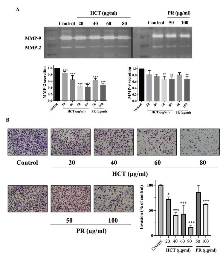
Molecules 2020, 25, 1196                                                                              2 of 23

Invasion is preceded by the degradation of the ECM to enable the infiltration of cancer cells [4].
The knowledge of its mechanisms is still fragmentary and must be summarized to improve our
therapeutic approach and influence the long-term and effective control of breast cancer progression.
      Clinically, breast cancer can be divided into distinct subtypes, including prognostic, which can be
classified by severity [5]. There are many breast cancer treatments, such as chemotherapy, radiotherapy,
and targeted therapy. The mechanisms of these treatments can modulate pro- and anti-apoptotic
proteins by upregulating the expression of pro-apoptotic proteins, such as p53-effector related to p21,
BAX and Noxa. Conversely, they can also lead to the downregulation of anti-apoptotic proteins [6].
Yet these methods, like all those using drugs, will result in side effects. Currently, herbal medicines play
a role in the prevention of breast cancer and have fewer side effects than using chemotherapeutic drugs
since herbs are plants that most people take orally as a dietary supplement. Therefore, the development
of herbal extracts to be used as a dietary supplement to prevent cancer and treat breast cancer is required.
      Houttuynia cordata Thunb and Piper ribesioides Wall are widespread herbs found in Northern
Thailand. There is a long history of herbal medicine plants widely distributed in East Asia,
with Chinese people having utilized herbs and plants to treat various diseases for a long time.
Moreover, they are intriguing natural products which are widely used as food supplements and to
promote health [7]. Furthermore, H. cordata possesses anti-cancer, anti-diabetics, and anti-inflammation
properties. However, for P. ribesioides, there has been little investigation or research conducted
regarding such features. P. ribesioides contains various phytochemicals, including camphene, sabinene,
and β-caryophyllene [8]. This study aimed to study and investigate the effects of anti-proliferation,
anti-invasion, anti-migration and apoptosis induction of both plants ethanolic extracts on two different
breast cancer cell types, including MCF-7 (non-invasive breast cancer cell) and MDA-MB-231 (invasive
breast cancer cell). Based on the potential actions on breast cancer cells, both extracts can be developed as
anti-cancer agents in order to prolong life among breast cancer patients. However, the anti-carcinogenic
activity against the breast cancer and toxicity tests of both extracts need to be verified using animal
models and clinical trials.

2. Results

2.1. Identification of Phytochemical Compositions in H. cordata and P. ribesioides Extracts
     Many phytochemical compounds in both plants have been reported, such as phenolic acids and
alkaloids [7,8]. We investigated the total phenolic acid contents, flavonoids and antioxidant activity,
as shown in Table 1. The phenolic acid compositions and flavonoids in P. ribesioides were higher
than H. cordata. Moreover, both extracts from plants possessed radical scavenging activity by using
DPPH assay. The values of IC50 of H. cordata and P. ribesioides were 234.6 ± 11.9 and 153.8 ± 4.4 when
compared to vitamin C (Table 1). Additionally, the HPLC chromatography exhibited related results
to the total phenolic content, total flavonoids, and DPPH-radical scavenging activity. To determine
and quantify phenolic acids and flavonoids, both ethanolic Houttuynia cordata Thunb. (HCT) and
Piper ribesioides Wall. (PR) extracts were analyzed using the standard curve compared to 11 phenolic
acids and flavonoids standards. Six phenolic acids were compared as gallic, vanilic, ferulic, p-coumaric,
chlorogenic and rosmarinic acids; and five flavonoids were compared as catechin, rutin, quercetin,
apigenin, and luteolin. Eleven phenolic acids and flavonoids in both extracts were identified according
to HPLC analysis (Table 2 and Figure 1), in which chlorogenic acid and rutin were predominant phenolic
acids and flavonoids in HCT extract, respectively. For PR extract, the predominant phenolic acid
and flavonoids were unidentifiable due to the limitations of the standards. However, the researchers
had investigated more active compounds compared to alkaloid derivatives, such as piperine by
HPLC analysis. We found that piperine was the predominant compound in ethanolic PR extract
(Table 2 and Figure 1). Further studies had been characterized for volatile substances in both ethanolic
plant extracts by using the GC-MS (Gas chromatography–mass spectrometry) method as shown in
Figure 2A,B. The HCT extract contained 9.42% of decanoic acid, 10.5% of n-hexadecanoic acid, 11.67% of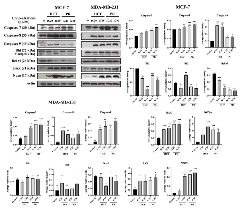
Molecules 2019, 24, x                                                                                           3 of 23

  Molecules
10.5%   of 2019, 24, x
            n-hexadecanoic      acid, 11.67% of 9,12-octadecatrienoic acid and 14.74% of cholest-5-en-3-ol 3 of 23

(Table 3). For PR extract, the major compound was piperine (49.51%), as shown in Table 4. The
  10.5% 2020,
Molecules of n-hexadecanoic
               25, 1196         acid, 11.67% of 9,12-octadecatrienoic acid and 14.74% of cholest-5-en-3-ol     3 of 23
researchers, therefore, studied the anti-cancer potential effect of both ethanolic plant extracts in
  (Table 3). For PR extract, the major compound was piperine (49.51%), as shown in Table 4. The
further experiments.
  researchers, therefore, studied the anti-cancer potential effect of both ethanolic plant extracts in
9,12-octadecatrienoic
  further experiments.acid and 14.74% of cholest-5-en-3-ol (Table 3). For PR extract, the major compound
      Table 1. (49.51%),
was piperine     Determination    of total
                            as shown         phenolic
                                        in Table       andresearchers,
                                                   4. The    flavonoid contents and
                                                                        therefore,    DPPHthe
                                                                                   studied  radical scavenging
                                                                                              anti-cancer  potential
      capacity
effect of        of  H. cordata
           both1.ethanolic      and  P.  ribesioides
                             plantofextracts         extracts.
                                               in further      Results are presented  as mean ± SD  from  three
       Table      Determination       total phenolic   andexperiments.
                                                            flavonoid contents and DPPH radical scavenging
      independent experiments.
         capacity of H. cordata and P. ribesioides extracts. Results are presented as mean ± SD from three
      Table    1. Determination
         independent  experiments.of total phenolic and flavonoid contents and DPPH radical scavenging
                        Total Phenolic Content
                                                        TotalResults
      capacity of H. cordata and P. ribesioides extracts.      Flavonoid   (mg
                                                                       are presentedDPPH-Radical
                                                                                     as mean ± SDScavenging
                                                                                                        from three
           Plants          (mgPhenolic
                         Total   Gallic Acid/g
                                          Content
      independent experiments.                           Catechin/g   Extract)
                                                        Total Flavonoid (mg          Activity  (IC50   (μg/mL))
                                                                                  DPPH-Radical Scavenging
             Plants              Extract)
                            (mg Gallic   Acid/g
                                                         Catechin/g Extract)        Activity (IC50 (μg/mL))
                            Total Extract)
                                  Phenolic
       H.Plants
            cordata             119.8 ± 3.0 Content            Total±Flavonoid
                                                               93.8   1.3              DPPH-Radical
                                                                                            234.6 ± 11.9Scavenging
                          (mg Gallic Acid/g Extract)       (mg Catechin/g Extract)       Activity (IC50 (µg/mL))
          H. cordata            119.8 ± 3.0                   93.8 ± 1.3                  234.6 ± 11.9
              P.
         H. cordata                119.8 ± 3.0                          ± 1.3                          ± 11.9
                               203.8  ± 10.8                   95.493.8
                                                                    ± 2.5                       234.6
                                                                                             153.8 ± 4.4
      P.ribesioides
               P.
          ribesioides             203.8 ± 10.8                     95.4 ± 2.5                   153.8 ± 4.4
                                203.8 ± 10.8                  95.4 ± 2.5                   153.8 ± 4.4
          ribesioides

          Figure
           Figure1.
          Figure 1.1.HPLC
                     HPLCchromatograms
                     HPLC chromatograms of ethanolic
                                        of ethanolic
                          chromatograms of           H. cordata
                                                     H.cordata
                                           ethanolicH.          extract
                                                        cordataextract  (A)
                                                                extract(A)  and
                                                                            andP.P.
                                                                        (A)and       ribesioides
                                                                                 P.ribesioides
                                                                                     ribesioides extract
                                                                                                 extract
                                                                                               extract   (B).
                                                                                                         (B).
                                                                                                       (B).

           (A)
            (A) GC-MS
                 GC-MSof
                       of80%
                          80% ethanolic
                              ethanolic extract of H.
                                        extract of H. cordata
                                                       cordata

 Molecules 2019, 24, x; doi:                       Figure 2. Cont.                 www.mdpi.com/journal/molecules
Molecules 2019, 24, x; doi:                                                         www.mdpi.com/journal/molecules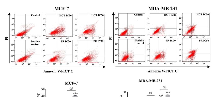
Molecules 2019, 24, x 4 of 23
Molecules 2020, 25, 1196 4 of 23
(B) GC-MS of 80% ethanolic extract of P. ribesioides
Figure
Figure 2. GC-MS chromatograms
2. GC-MS chromatograms of of both
both ethanolic
ethanolic (A) H. cordata
(A) H. cordata and
and (B) P. ribesioides
(B) P. ribesioides extracts.
extracts.
Phytochemicals in both plant extracts were analyzed using DB-5MS column with Agilent
Phytochemicals in both plant extracts were analyzed using DB-5MS column with Agilent technologytechnology
GC
GC 7890A
7890A coupled
coupled to
to Agilent
Agilent technology
technology MSD
MSD 5975C
5975C (EI).
(EI).
Table 2. Phenolic acid compositions and flavonoids of both ethanolic H. cordata and P. ribesioides.
Table 2. Phenolic acid compositions and flavonoids of both ethanolic H. cordata and P. ribesioides.
Retention Times (Rt) H. cordata P. ribesioides Compounds
Retention Times (Rt) H. Cordata P. Ribesioides Compounds
3.186 3.186 0.435 ± 0.01
0.435 ± 0.01 0.8270.827 ± 0.08 Gallic acid
± 0.08 Gallic acid
7.908 7.908 1.64 ± 0.43
1.64 ± 0.43 1.1281.128 ± 0.13
± 0.13 CatechinCatechin
9.012 9.012 25.5 ± 3.41
25.5 ± 3.41 n.d. n.d. Chlorogenic
Chlorogenic acid acid
10.904 0.844 ± 0.14 0.543 ± 0.03 Vanilic acid
10.904 0.844 ± 0.14 0.543 ± 0.03 Vanilic acid
13.132 0.621 ± 0.02 1.199 ± 0.02 Ferulic acid
13.132 0.621 ± 0.02 1.199 ± 0.02 Ferulic acid
13.990 0.116 ± 0.02 n.d. p-Courmaric acid
13.990 0.116 ± 0.02 n.d. p-Courmaric acid
17.167 44.00 ± 5.61 n.d. Rutin
18.288 17.167 44.00
1.49 ± 5.61
± 0.04 n.d.
1.89 ± 0.01 Rutin
Rosmarinic acid
21.361 18.288 1.49
0.196 ± ±0.05
0.04 1.890.465
± 0.01± 0.05Rosmarinic Quercetin
acid
22.329 21.361 0.196
n.d. ± 0.05 0.4650.124
± 0.05
± 0.01 Quercetin Apigenin
22.329 n.d. 0.124 ± 0.01
n.d. = non-detectable.
Apigenin
n.d. = non-detectable.
Table 3. Phytochemical compounds identified in H. cordata by using GC-MS.
Table 3. Phytochemical compounds identified in H. cordata by using GC-MS.
No. Retention Time Name of Compounds % of Total
No. Retention Time Name of Compounds % of Total
1 1 7.926 7.926 Decanoicacid
Decanoic acid 9.42
9.42
2 10.329 Dodecanoic acid 2.57
2 10.329 Dodecanoic acid 2.57
3 12.578 Tetradecanoic acid 0.54
3 12.578 Tetradecanoic acid 0.54
4 12.979 Octadecane 0.9
4 12.979 Octadecane 0.9
5 14.558 n-Hexadecanoic acid 10.5
5 6 14.55814.878 n-Hexadecanoic
Eicosane acid 10.5
0.59
6 7 14.87816.034 Eicosane
9,12-Octadecadienoic acid 0.59
1.78
7 8 16.03416.091 9,12-Octadecadienoic
9,12-Octadecatrienoicacid
acid 1.78
11.67
8 9 16.09116.257 9,12-Octadecatrienoic
Octadecanic acidacid 11.67
2.93
9 10 16.25721.716 Octadecanic acid
9,17-Octadecadienal 2.93
0.78
10 11 21.71633.629 alpha-Tocopherol
9,17-Octadecadienal 5.33
0.78
11 12 33.62934.711 Sesamin
alpha-Tocopherol 2.01
5.33
12 13 34.71142.287 Cholest-5-en-3-ol
Sesamin 14.74
2.01
13 42.287 Cholest-5-en-3-ol 14.74
Molecules 2019, 24, x; doi: www.mdpi.com/journal/molecules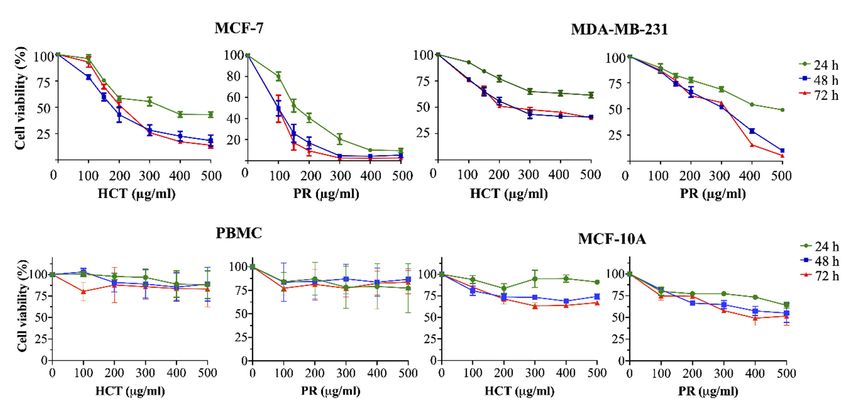
Molecules 2020, 25, 1196                                                                                                  5 of 23

                  Table 4. Phytochemical compounds identified in P. ribesioides by using GC-MS.

                   No.            Retention Time             Name of Compounds                   % of Total
                    1                   10.324                 Dodecanoic acid                      0.05
                    2                   12.584                Tetradecanoic acid                    0.43
                    3                   14.581               n-Hexadecanoic acid                    6.82
                    4                   16.051              9,12-Octadecanoic acid                  3.84
                    5                   16.103                 cis-Vaccenic acid                     5.1
                    6                   16.269                 Octadecanoic acid                    1.94
                    7                   26.540                      Piperine                        49.51
                    8                   42.292                    β-Sitrosterol                     3.46

2.2. Cytotoxic Effects of H. cordata and P. ribesioides Extracts on Breast Cancer Cells, PBMCs and MCF-10A
      HCT was cytotoxic against many types of cancer cells, for example MCF-7 [9] and
MDA-MB-231 [10]. Yet, there is still no report of the PR toxic effect on both types of breast cancer
cells. To investigate the effects on cell viability, both HCT and PR extracts were prepared in various
concentrations (100–500 µg/mL) and were incubated in both MCF-7, MDA-MB-231, peripheral blood
mononuclear cells (PBMCs), and MCF-10A (human breast epithelial cell line) for 24, 48, and 72 h.
In addition, IC20 and IC50 values (inhibitory of concentration at 20% and 50%) were evaluated by
using results of cell viability. As shown in Figure 3, both ethanolic extracts decreased the viability of
both breast cancer cells, but not PBMCs, in dose- and time-dependent manners. Although the viability
of human mammary epithelial cell line MCF10A tends to decrease when treated with increasing doses
of HCT and PR extracts (Figure 3), IC20 and IC50 of both extracts in normal cells MCF10A were
higher than in breast cancer cells MCF-7 and MDA-MB-231 (Table 5). These findings indicate that
breast cancer cells were more sensitive to ethanolic HCT and PR extracts than normal mammalian
cells. Furthermore, IC20 and IC50 of PR extracts in breast cancer cells were lower than in HCT extract
(Table 5), implying that both cancer cells were more sensitive to PR extract. Besides, the results also
show that the viability of the cells treated with both ethanolic extracts at 24 h was significantly different
from at 48 and 72 h (Figure 3). However, there were no significant differences between breast cancer
Molecules 2019, 24, x                                                                                 6 of 23
cell viability at 48 and 72 h of incubation (Figure 3).

      Figure 3.
              3. Cytotoxic
                  Cytotoxiceffects
                                effectsofofherbal
                                            herbalextracts onon
                                                    extracts  breast cancer
                                                                breast      cells,
                                                                       cancer      PBMCs
                                                                                cells,     andand
                                                                                       PBMCs    MCF-10A
                                                                                                    MCF-10Acell line. The
                                                                                                                 cell line.
      The cells
      cells werewere
                  treatedtreated   with various
                             with various          concentrations
                                             concentrations         (100–500
                                                             (100–500  µg/mL)µg/mL)     of H. and
                                                                                of H. cordata cordata and P. ribesioides
                                                                                                  P. ribesioides extracts
      extracts
      for 24, 48for
                  and24,7248h.and
                               Cell72  h. Cellwas
                                    viability    viability was by
                                                    evaluated   evaluated
                                                                  comparingby comparing     with 0.5%
                                                                               with 0.5% DMSO           DMSO
                                                                                                   treated        treated
                                                                                                            control   cell,
      control
      after 24,cell,  after7224,h 48
                 48 and           of and    72 h of Results
                                     incubation.     incubation.  Results are
                                                             are presented  as presented
                                                                                mean ± SDasfrom       ± SDindependent
                                                                                               meanthree     from three
      independent experiments.
      experiments.

2.3. Effects of H. Cordata and P. Ribesioides Extracts on Growth Inhibition in Breast Cancer Cells
     H. cordata plays an important role in cell proliferation and cell cycle progression [9,11]. However,
the anti-proliferation effect of P. ribesioides has not yet been studied. This anti-proliferation with both
H. cordata and P. ribesioides extracts in both breast cancer cells was verified. The cells were treated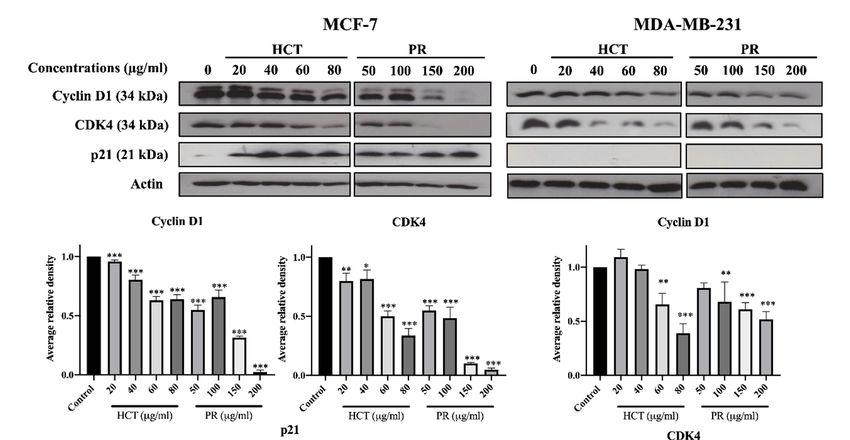
Molecules 2020, 25, 1196                                                                                         6 of 23

      Table 5. Determination of IC20 and IC50 values of H. cordata and P. ribesioides extracts of breast cancer
      cells, peripheral blood mononuclear cells (PBMCs) and MCF-10A cell line. All cells were treated with
      various concentrations (100–500 µg/mL) of H. cordata and P. ribesioides extracts for 24, 48 and 72 h.
      Results are presented as mean ± SD for three independent experiments.

                                                           MCF-7
                              H. cordata Extract (µg/mL)                      P. ribesioides Extract (µg/mL)
                       24 h               48 h              72 h           24 h           48 h            72 h
     IC20          129.3 ± 6.9          82.4 ± 7.1     129.7 ± 3.5    93.2 ± 7.2        56.4 ± 9.7     66.4 ± 12.2
     IC50          347.4 ± 10.8        187.9 ± 18.2    210.8 ± 3.1   167.0 ± 11.0      99.5 ± 12.0     98.4 ± 14.1
                                                      MDA-MB-231
                       24 h               48 h              72 h           24 h           48 h            72 h
     IC20          184.6 ± 14.0        66.9 ± 5.3      64.8 ± 3.8    174.6 ± 13.8      146.3 ± 5.6     149.5 ± 8.5
     IC50          667.8 ± 11.7        279.6 ± 4.4    290.7 ± 11.9   484.5 ± 14.3      264.1 ± 12.2    254.1 ± 8.0
                                                           PBMCs
                       24 h               48 h              72 h           24 h           48 h            72 h
     IC20                                                    >1000 µg/mL
     IC50                                                    >1000 µg/mL
                                                       MCF-10A
                       24 h               48 h              72 h           24 h           48 h            72 h
     IC20          514.6 ± 5.3         372.3 ± 9.1     275.9 ± 2.1    353.1 ± 4.5      256.3 ± 6.5     264.5 ± 2.5
     IC50         >1000 µg/mL          893.8 ± 8.4     679.5 ± 7.9    879.5 ± 2.6      523.0 ± 1.7     442.9 ± 1.0

2.3. Effects of H. cordata and P. ribesioides Extracts on Growth Inhibition in Breast Cancer Cells
      H. cordata plays an important role in cell proliferation and cell cycle progression [9,11]. However,
the anti-proliferation effect of P. ribesioides has not yet been studied. This anti-proliferation with both
H. cordata and P. ribesioides extracts in both breast cancer cells was verified. The cells were treated with
various concentrations of HCT and PR, at concentrations not more than IC20, to prevent cell death from
such concentrations. The colony formation assay was measured, and HCT and PR dose-dependently
inhibited colony formation in both MCF-7 and MDA-MB-231 cells (Figure 4A). MCF-7 was more
sensitive to both HCT and PR compared to MDA-MB-231 cells. However, the low concentrations of
HCT extract were more effective than in PR extract, since the cell cycle is disrupted and involved in the
proliferation of cancer cells in carcinogenesis [12]. Consequently, the cell cycle assay was verified to
demonstrate the proliferation-inhibitory effects of HCT and PR (Figure 4B). After treatment with both
ethanolic extracts, MCF-7 cells were arrested at G1 stages in a dose-dependent manner and significantly
increased in the G1 phase. For MDA-MB-231 cells, only PR treatment at 200 µg/mL significantly
increased in the G1 phase. These results suggest that both HCT and PR inhibited the proliferation of
both breast cancer cells by cell cycle arrest (Figure 4A). To confirm that HCT and PR-induced G1 arrest
in breast cancer cells, both breast cancer cells were treated with HCT and PR extracts with the same
condition for 24 hours’ treatment, and cell cycle-related proteins such as p21, cyclin D1 and CDK4 [13]
were detected. The results show that cyclin D1 and CDK4 decreased in both MCF-7 and MDA-MB-231
cells and correlated with the results from the colony formation assay and cell cycle analysis. It was
also found that there was an increase of p21 in MCF-7, but in MDA-MB-231, p21 was not detectable
(Figure 5). These findings show that both ethanolic HCT and PR extracts inhibited cell growth and
arrested the G1 phase (Figure 4B), by decreasing the expression of cyclin D1 and CDK4 and altering
the p21 expression level, as shown in Figure 5.
Molecules 2020, 25, 1196 7 of 23
Figure 4. Effect of both H. cordata and P. ribesioides extracts on breast cancer cell growth inhibition and
cell cycle disruption. (A) Colony formation assay. MCF-7 and MDA-MB-231 were treated with non-toxic
concentrations of both Houttuynia cordata Thunb. (HCT) and Piper ribesioides Wall. (PR) extracts for 24 h,
washed by PBS and cultured for seven days. The colonies were counted and compared with the control.
(B) For cell cycle assay, the cells were treated with the same condition in the colony formation assay
and analyzed by flow cytometry after the cells were stained with propidium iodide. Cell numbers are
presented as a percentage of the total analyzed cells. Each value is presented as mean ± SD from three
independent experiments. Data are the means ± SD. * p < 0.05, ** p < 0.01, and *** p < 0.001 vs. control.Molecules 2020,
Molecules 2019, 25,
24, 1196
x 88 of 23
of 23
Figure 5. Effect of both H. cordata and P. ribesioides extracts inhibited cell cycle related protein expressions
on breast cancer, by using Western blotting to investigate cyclin D1, CDK4, and p21. β-Actin acted
Figure 5. Effect of both H. cordata and P. ribesioides extracts inhibited cell cycle related protein
as the loading control. Each value is presented as mean ± SD from three independent experiments.
expressions on breast cancer, by using Western blotting to investigate cyclin D1, CDK4, and p21. β-
Data are the means ± SD. * p < 0.05, ** p < 0.01, and *** p < 0.001 vs. control.
Actin acted as the loading control. Each value is presented as mean ± SD from three independent
experiments.
2.4. Effect Data are
of H. cordata andthe means ± SD.
P. ribesioides on*pBreast
< 0.05,Cancer
**p < 0.01, and ***pand
Migration < 0.001 vs. control.
Invasion
Cancer
2.4. Effect cell
of H. migration
Cordata and
and P. invasiononare
Ribesioides veryCancer
Breast important characteristics
Migration and Invasion of breast cancer cells at
the metastasis steps. Cancer cells secrete metalloproteinases (MMPs), such as MMP-9, to degrade
Cancer cell migration
the extracellular matrix [2].and invasionin
Moreover, are veryH.important
both cordata andcharacteristics
P. ribesioides,ofnobreast cancer
research cells at
studies the
have
metastasis steps. Cancer cells secrete metalloproteinases (MMPs), such as MMP-9,
previously been undertaken on the inhibition of migration and invasion in breast cancer cells. To exploreto degrade the
extracellular
the matrix
effect of both [2]. Moreover,
H. cordata in both on
and P. ribesioides H.cancer
cordatacells
andmigration
P. ribesioides, no research
and invasion, bothstudies
MCF-7haveand
previously been
MDA-MB-231 undertaken
cells on the
were treated inhibition
with both H. of migration
cordata and P.and invasion
ribesioides at in breast cancer
non-toxic cells. To
concentrations
explore
in the effect
incomplete of both
media. H. HCT
Both cordataand
andPR P. significantly
ribesioides on inhibited
cancer cells bothmigration
MCF-7 andand invasion,
MDA-MB-231 both
MCF-7 and
migration in MDA-MB-231
a dose-dependent cellsresponse,
were treated with with
compared both the
H. control
cordata using
and P. theribesioides at non-toxic
wound healing assay
(Figure 6). The researchers found that both ethanolic extracts decreased MMP-2 and MMP-9and
concentrations in incomplete media. Both HCT and PR significantly inhibited both MCF-7 MDA-
secretions
MB-231
in migration(Figure
MDA-MB-231 in a dose-dependent
7A). Meanwhile, response, compared
both ethanolic with in
extracts thethecontrol
sameusing the wound
concentrations healing
inhibited
assay
the (Figureof6).
invasion The researchers
MDA-MB-231 found
cell by usingthat both ethanolic extracts
a Transwell-invasion assaydecreased
(Figure 7B).MMP-2 and MMP-9
HCT inhibited the
secretions in MDA-MB-231 (Figure 7A). Meanwhile, both ethanolic
breast cancer cells migration to a better degree than PR. These results indicated that both HCT extracts in theand same
PR
concentrations inhibited the invasion of MDA-MB-231 cell by using a Transwell-invasion
extracts inhibited breast cancer cell migration and invasion though the inhibition of MMPs secretion. assay
(Figure 7B). HCT inhibited the breast cancer cells migration to a better degree than PR. These results
indicated that both HCT and PR extracts inhibited breast cancer cell migration and invasion though
the inhibition of MMPs secretion.
Molecules 2019, 24, x; doi: www.mdpi.com/journal/moleculesMolecules 2019, 24, x 9 of 23
Molecules 2020, 25, 1196 9 of 23
Figure 6. Effect of H. cordata and P. ribesioides extracts on breast cancer cell migration. Cells were
treated6.with
Figure non-toxic
Effect doses for
of H. cordata and24P.h,ribesioides
and the wound healing
extracts assaycancer
on breast was investigated. TheCells
cell migration. percentage
were
of effectiveness
treated for inhibition
with non-toxic on24
doses for migration
h, and the inwound
bar graphs. Each
healing value
assay wasis investigated.
presented as mean ± SD from
The percentage
three
of independent
effectiveness experiments.
for inhibition Data are the
on migration means
in bar graphs. * pMolecules2019,
Molecules 25,x1196
2020,24, 1010ofof23
23
Effectof
Figure7.7. Effect
Figure H.cordata
ofH. cordataand
andP.P. ribesioides
ribesioides extracts
extracts on
on breast
breast cancer
cancer cell
cell invasion.
invasion. MMP-2
MMP-2andand-9-9
secretions assay (A) and invasion assay (B) were analyzed. MMP-2 and -9 were quantitated
secretions assay (A) and invasion assay (B) were analyzed. MMP-2 and -9 were quantitated compared compared
withthe
with thecontrol.
control. Each
Eachvalue
valueisispresented
presentedas mean±±SD
asmean SDfrom
fromthree
threeindependent
independentexperiments.
experiments.Data
Dataare
are
the means ± SD. * p < 0.05, ** p < 0.01, and *** p < 0.001 vs.
the means ± SD. *p < 0.05, **p < 0.01, and ***p < 0.001 vs. control. control.
2.5. Effects of H. cordata and P. ribesioides Extracts on Apoptosis Induction in Breast Cancer Cells
2.5. Effects of H. Cordata and P. Ribesioides Extracts on Apoptosis Induction in Breast Cancer Cells
Various reports have investigated HCT extracts-induced apoptosis in many types of cancer,
Various
such as human reports have
primary investigated
colorectal cancerHCT extracts-induced
cells apoptosis
[14]. Nevertheless, for PR in many
there types
is no of cancer,
study such
on apoptosis
as human primary
induction. However, colorectal
there arecancer
reportscells
about[14]. Nevertheless,
anti-cancer forother
effect of PR there
Piper is no study
species [15].on apoptosis
Furthermore,
induction. However, there are reports about anti-cancer effect of other Piper
the relationship of doses and types of cell death remains intriguing, for example, in low doses it couldspecies [15].
Furthermore, the relationship
produce apoptosis, and at highof dose
dosestheand types of cell
necroptosis aredeath remainsby,
stimulated intriguing, for example,
e.g., shikonin in low
[16]. To further
doses it could produce apoptosis, and at high dose the necroptosis are stimulated
investigate apoptosis induction by both ethanolic HCT and PR extracts, the apoptosis cell death by, e.g., shikonin
[16].
was To further using
evaluated investigate apoptosis
annexin induction
V-fluorescein by both ethanolic
isothiocyanate HCT and PRiodide
(FITC)/propidium extracts,
(PI)the apoptosis
staining and
cell death was evaluated using annexin V-fluorescein isothiocyanate (FITC)/propidium
analyzed by flow cytometry. Each IC20 and IC50 of the ethanolic HCT and PR extracts were treated iodide (PI)
staining and analyzed by flow cytometry. Each IC20 and IC50 of the ethanolic HCT and PR extracts
Molecules 2019, 24, x; doi: www.mdpi.com/journal/moleculesMolecules 2020, 25, 1196 11 of 23
Molecules 2019, 24, x 11 of 23
were
in treated
both breastincancer
both breast cancer
cells, and cells,were
the cells and the
thencells were to
induced then induced
undergo to undergo
apoptosis. Theapoptosis.
percentageThe
of
percentage of apoptotic cells was significantly increased in both cancer cells in concentration-
apoptotic cells was significantly increased in both cancer cells in concentration-dependent manners
dependent
(Figure manners (Figure
8). Compared 8). concentrations
with the Compared withofthe theconcentrations of the extracts,
extracts, the MCF-7 the MCF-7
cell line was cell line
more sensitive
was more
than sensitive than MDA-MB-231.
MDA-MB-231.
Figure 8. Effect of H. cordata and P. ribesioides extracts on apoptosis induction in breast cancer cells.
Figure
Flow 8. Effect of
cytometric H. cordata
analysis and P. ribesioides
of apoptosis inductionextracts
on MCF-7on apoptosis inductioncells
and MDA-MB-231 in breast cancer cells.
were treated with
Flow cytometric analysis of apoptosis induction on MCF-7 and MDA-MB-231 cells
various concentrations of HCT and PR ethanolic extracts for 24 h. The annexin V-FITC/PI staining were treated with
various concentrations
apoptotic of HCT
cells were analyzed andflow
using PR ethanolic
cytometry.extracts for 24 h.
Camptothecin The 5annexin
(CTP; nM) wasV-FITC/PI
used as a staining
positive
apoptotic
control. cells
Bar wereshowed
graphs analyzed using
the flow cytometry.
summarized Camptothecin
data of three (CTP;
independent 5 nM) was used
experiments from as a positive
MCF-7 (left)
control. Bar graphs showed the summarized data of three independent experiments
and MDA-MB-231 cells (right), performed in duplicate compared with the untreated control. Each from MCF-7 (left)
and MDA-MB-231
value is presented as cells (right),
mean ± SDperformed
from three in independent
duplicate compared with the
experiments. untreated
Data are the control.
means ±EachSD.
p < 0.05,
*value ** p < 0.01,
is presented as and
mean < 0.001
***±pSD from vs.
three # p < 0.01
independent
control. and ## p < Data
experiments. 0.001are
vs. the means
IC20 ± SD.
of each *p <
extract.
0.05,
ns, **psignificantly
not < 0.01, and different.
***p < 0.001 vs. control. #p < 0.01 and ##p < 0.001 vs. IC20 of each extract. ns, not
significantly different.
In addition, the activity of caspases was determined to investigate caspase-dependent apoptosis
In addition,
cell death by using the activity of caspases
a colorimetric was
assay kit. determined
Caspase to investigate
activities are used tocaspase-dependent
demonstrate the pathways apoptosis of
cell apoptotic
the death by using
cell deatha colorimetric assay
pathway [17]. kit. cell
Both Caspase
typesactivities are used
were treated with tothedemonstrate
same conditionsthe pathways
as in the
of the apoptotic
experiments cellcytometry,
of flow death pathway [17].activity
or caspase Both cellwastypes were
assayed bytreated
using awith the same conditions
spectrophotometric as in
microplate
the experiments
reader. The caspases of flow
-3, -8,cytometry, or caspase
and -9 activities activityincreased
significantly was assayed by using a spectrophotometric
in concentration-dependent manners
microplate
(Figure reader. The
9) compared caspases
to the untreated-3, control
-8, andcells,
-9 activities
and theysignificantly
correlated with increased in concentration-
the results from annexin
dependentbymanners
V-FITC/PI (Figure However,
flow cytometry. 9) compared to the untreated
caspases-3 controlincells,
is not detectable and they
the MCF-7 cell correlated with the
line, corresponding
results
to from annexin
a previous study [18]. V-FITC/PI by flow
The results cytometry.
indicate However,
that both ethanolic caspases-3 is not detectable
extracts induced apoptosis in in
theMCF-7
MCF-
7 cellMDA-MB-231
and line, corresponding via bothto aextrinsic
previousand study [18]. The
intrinsic results indicate
pathways. To confirmthat HCT
both ethanolic extracts
and PR induced
induced apoptosis
apoptosis, in MCF-7 and
apoptotic-protein MDA-MB-231
levels in Bcl-2 family via both extrinsic
proteins wereand intrinsic pathways.
investigated by Western Toblotting.
confirm
HCT and
When bothPR induced
breast cancerapoptosis,
cells wereapoptotic-protein
treated with thelevels in Bcl-2
IC20 and IC50family proteins were
concentrations investigated
of each ethanolic
by Western
extract, blotting.
the results showWhen both
that the breast cancer
anti-apoptotic cellsBcl-xl
protein weresignificantly
treated with the IC20whereas
decreased, and IC50 the
concentrationsproteins
pro-apoptotic of eachsignificantly
ethanolic extract,
increased theinresults show thatmanners
dose-dependent the anti-apoptotic
(Figure 10).protein Bcl-xl
These results
significantly
indicate decreased,
that both ethanolicwhereas the pro-apoptotic
extracts induced apoptosis usingproteins significantly
cytotoxic doses in both increased in dose-
breast cancer cells.
dependentismanners
Caspase-8 the marker (Figure 10).
of the These results
extrinsic pathway, indicate
whereasthatthe
both ethanolic
caspase-9 is extracts
responsibleinduced
in theapoptosis
intrinsic
using cytotoxic doses in both breast cancer cells. Caspase-8 is the marker of the extrinsic pathway,
Molecules 2019, 24, x; doi: www.mdpi.com/journal/moleculesMolecules 2020, 25, 1196 12 of 23
Molecules 2019, 24, x 12 of 23
whereas the caspase-9 is responsible in the intrinsic pathway [19,20]. Yet, these two caspases act as if
pathway [19,20]. Yet, these two caspases act as if they were at the initiation caspases [21]. Caspases-3, -6,
they were at the initiation caspases [21]. Caspases-3, -6, and -7 are the effector caspases or executioner
and -7 are the effector caspases or executioner caspases that are activated in the cascade of both intrinsic
caspases that are activated in the cascade of both intrinsic (mitochondrial) and extrinsic (death
(mitochondrial) and extrinsic (death receptor) pathways [22]. MCF-7 cells do not possess caspase-3
receptor) pathways [22]. MCF-7 cells do not possess caspase-3 proteins. Hence, to confirm the effect
proteins. Hence, to confirm the effect of apoptosis at the molecular pathways, the apoptosis-mediated
of apoptosis at the molecular pathways, the apoptosis-mediated proteins were determined. The Bcl-
proteins were determined. The Bcl-2 family consists of three main types: anti-apoptotic (such as
2 family consists of three main types: anti-apoptotic (such as Bcl-xl); pro-apoptotic proteins with
Bcl-xl); pro-apoptotic proteins with multiple Bcl-2 homology domains (such as BAX); and BH3-only
multiple Bcl-2 homology domains (such as BAX); and BH3-only proteins (such as Noxa, Bid) [23,24].
proteins (such as Noxa, Bid) [23,24]. The immunoblotting of caspase-7, -9, -8, Bid/tBid, BAX, and Noxa
The immunoblotting of caspase-7, -9, -8, Bid/tBid, BAX, and Noxa expression level was measured. It
expression level was measured. It was found that the expression of Bcl-xl decreased, whereas the
was found that the expression of Bcl-xl decreased, whereas the Bid/tBid protein levels were
Bid/tBid protein levels were modulated as a decrease of the pro-form and an increase of the truncated
modulated as a decrease of the pro-form and an increase of the truncated Bid (tBid), confirming the
Bid (tBid), confirming the involvement of the extrinsic pathway as the enhancement of caspase-8
involvement of the extrinsic pathway as the enhancement of caspase-8 activity in MCF-7. For MDA-
activity in MCF-7. For MDA-MB-231 cells, the caspase-3 was activated together with caspase-8 and 9
MB-231 cells, the caspase-3 was activated together with caspase-8 and 9 in both the caspases activities,
in both the caspases activities, and corresponds with the caspase protein expression levels. The Bcl-2
and corresponds with the caspase protein expression levels. The Bcl-2 protein expression levels
protein expression levels followed the same pattern in accordance with those of MCF-7 (Figure 10).
followed the same pattern in accordance with those of MCF-7 (Figure 10).
Figure 9. Caspases’ activities of breast cancer cells treated with H. cordata and P. ribesioides extracts.
Figure
Cells 9. Caspases’
were grown underactivities of breast
the same cancer
conditions andcells treated
caspases with H.
activities cordata
were and P.including
analyzed, ribesioidescaspase-8,
extracts.
Cells
-9, andwere grown in
-3 activities under
MCF-7the (excepted
same conditions andand
caspases-3) caspases activities were
MDA-MB-231. analyzed,
Data are the meansincluding
± SD.
caspase-8, -9, and -3 activities in MCF-7 (excepted
* p < 0.05, ** p < 0.01, and *** p < 0.001 vs. control. caspases-3) and MDA-MB-231. Data are the means
± SD. *p < 0.05, **p < 0.01, and ***p < 0.001 vs. control.
Molecules 2019, 24, x; doi: www.mdpi.com/journal/moleculesMolecules 2019, 25,
Molecules 2020, 24, 1196
x 13 of
13 of 23
23
Figure 10. The effects of H. cordata and P. ribesioides extracts on the expression of apoptotic involving
Figure 10.
proteins inThe effects
breast of H.
cancer cordata
cells. and P.
MCF-7 andribesioides extractscells
MDA-MB-231 on the expression
were of apoptotic
treated with IC20 and involving
IC50 of
proteins
each in breast
ethanolic cancer
extract forcells.
24 h, MCF-7 and cell
and whole MDA-MB-231
lysates werecells were treated
prepared with IC20
and subjected and IC50blotting.
to Western of each
ethanolic extract for 24 h, and whole cell lysates were prepared and subjected to
The immunoblots were investigated for caspase-7, caspase-8, caspase-9, Bid, tBid, Bcl-xl, BAX and Western blotting.
The immunoblots
Noxa. β-Actin acted were investigated
as the for caspase-7,
loading control. caspase-8,
The results caspase-9,toBid,
are normalized thetBid, Bcl-xl,
control. Data BAXare and
the
Noxa. β-Actin
means < 0.05,
± SD. * pacted as **thepMolecules 2020, 25, 1196 14 of 23
in the ethanolic extract of HCT. Chlorogenic acid (CGA) has been revealed to possess anti-cancer
potentials on breast cancer. Through the inhibition of protein kinase C signaling, CGA induces cell
cycle arrest and apoptosis in MCF-7 and MDA-MB-231 cells [29], which is consistent with effects of
HCT extract in this study. Thus, CGA is a promising bioactive phenolic compound of HCT extract on
cell cycle arrest and apoptosis induction in breast cancer cell lines. For Piper spp. (Piperaceae), such as
P. nigrum and P. tuberculatum, piperine is a standard alkaloid that is used to measure the efficiency
of solvent extraction for the plants [30,31]. Besides, there have also been reports about the effect of
piperine against breast cancer. Piperine suppresses the proliferation and migration of triple negative
breast cancer cell lines by reducing the levels of MMP-2 secretion [32,33]. In agreement with these
studies, PR extract, which contains a high amount of piperine, also decreased the colony formation and
migration of aggressive human breast cancer cell MDA-MB-231. Therefore, the researchers suggest that
piperine is likely to be the potential active compound that is responsible for the anti-cancer bioactivities
of PR extract.
Apart from HPLC, both plant extracts were subjected to GC-MS analysis for identifying the
phytochemical compositions including alkaloids and some volatile components, in which the
HPLC system could not be determined. The lipophilic phytochemicals in HCT extract consist
of alpha-tocopherol, decanoic acid, n-hexadecanoic acid and 9,12-octadecatrienoic acid, which are
all comparable to the results from previous studies [27,34–36]. Although camphene, sabinene,
and β-caryophyllene are common substances found in piperine plants [8], the researchers found
that piperine, n-hexadecanoic acid and cis-vaccenic acid were mainly presented in the PR extract.
This might be due to the different condition used in GC-MS analysis. Decanoic acid, a straight-chain
saturated fatty acid, has been previously reported as the antibacterial- and the anti-inflammatory
agent [36]. The derivatives or conjugated forms of decanoic acid were also found as contributing agents,
supporting the anti-cancer agents, a class of cell-permeable Bcl-2 binding peptides, by chemically
attaching to a decanoic acid as the cell-permeable moiety [37]. Besides, n-hexadecanoic acid that was
found in both extracts has been reported for its activity on apoptosis induction in breast cancer [38].
Regarding the phytochemical studies, it is possible that decanoic acid and its derivatives are the active
compounds of HCT and PR extracts on apoptosis induction.
Low concentrations (less than IC 20) of ethanolic HCT and PR extracts were found to significantly
inhibit cell growth and colony formation in both breast cancer cell types. These concentrations also
significantly induced the cell cycle arrest at the G1 phase, and the results were confirmed with cell
cycle progression histograms and the expression of cell cycle-related proteins by Western blot analysis.
The cyclin D1 and CDK4 proteins, which are cell cycle checkpoint proteins for the G1 phase, were found
to be significantly decreased. These resulted in the inhibition of colony formation and cell cycle
arrest [39]. The p21 protein expression levels in MCF-7 and MDA-MB-231 cell lines were different,
because MDA-MB-231 is often accompanied by a higher frequency of p53 gene mutations [40], and there
is also no evidence of p21 protein expression in such invasive cells [41,42]. These results indicate that
HCT and PR extracts induced G1-arrest in MCF-7 cells, which depended on p21 expression, but not in
the case of MDA-MB-231 cells. However, we still observed that the G1 arrest occurred in MDA-MB-23.
This result might be due to the activation of a p-21-independent pathway, such as the EGFR signaling
pathway [43]. There are several previous reports about HCT-induced cell cycle arrests in various types
of cancer, including lung cancer cell (A549 cell), [11], hepato-cellular carcinoma (HepG2 cell) [44],
and also breast cancer cells (MCF-7 cell) [9]. The inhibition of DNA synthesis, cell cycle arrest, and the
inhibition of proliferation are associated with increased p21(Waf1/Cip1) expression [45].
Breast cancer progression requires multiple steps, including migration, invasion, angiogenesis,
and metastasis [2]. In particular, the anti-migration and invasion effects of HCT and PR extracts have
not yet been studied in either MCF-7 or MDA-MB-231 cells. Matrix metalloproteinases (MMPs) are
the proteolytic enzymes that are important for cancer cell invasion. MMPs are capable of degrading
a range of extracellular matrix proteins, then allowing the cancer cells to migrate and invade, away
from the primary site [46]. Among various subtypes of MMP proteins, MMP-2 and MMP-9 areMolecules 2020, 25, 1196 15 of 23
frequently found to be related with cancer cell invasion and aggressiveness. MMP-2 and MMP-9 have
been reported as proteases for the degradation of type IV collagen, a major component of basement
membrane [47,48]. Kim et al. reported that MMP-2 secretion in MCF-7 is extremely low compared to
MDA-MB-231 [49]. Consistently, the results from the present study show that MMP-2 and -9 activities
in MCF-7 cell lines were not detectable (data not shown). Thus, MDA-MB-231 cells were selected
for investigating the mechanisms underlying the anti-migration and invasion properties of HCT and
PR extracts. To avoid the influence from cytotoxic effects of the extracts, the migration and invasion
potentials of MDA-MB-231 cells were examined in the serum-free media condition. The results indicate
that HCT and PR extracts at non-toxic doses could inhibit cancer cell migration and invasion by
decreasing the MMP-2 and -9 secretion. However, the underlying mechanisms related to the decreased
secretion of MMPs should be further determined.
Beyond the anti-migration and anti-invasion effects, HCT and PR extracts could induce apoptosis
in both MCF-7 and MDA-MB-231 cells via the caspase-dependent mechanism. Regarding the apoptosis
pathways, the extrinsic pathway, caspase-8, is the initiator caspases that activates downstream caspases,
including caspase-3, -7 [50]. Caspase-9 is activated by internal signaling, increasing Noxa and BAX
protein, leading to the interaction and inhibition function of Bcl-xl protein [51,52]. The active caspase-8
in the cross-talk apoptosis pathway is activated via the induction of caspase-8 to cleave Bid and become
truncated Bid (tBid) [53,54]. Both caspases then activated caspase-3 or -7 in a downstream cascade and
the cells underwent apoptosis [55,56]. Although there is a report about the effector caspase between
caspase-3 and caspase-7 [57], caspase-3 and caspase-7 play separate roles during apoptosis [22,58].
Thus, the researchers focused on the effects of both extracts on the aforementioned caspases types.
Western blotting analyses confirm that both HCT and PR extracts enhanced the expression of caspase-7,
-8 and -9 at protein levels. Moreover, caspase-3, -8 and -9 activities were inhibited in MDA-MB-231
cells treated with high doses of HCT and PR extracts. Similar results excluding caspase-3 activity were
observed in MCF-7, according to the lack of caspase 3 in MCF-7 cells [19].
Taken together, our results demonstrate the novel mechanisms associated with the inhibitory
effects of HCT and PR extracts on breast cancer cell growth, migration, and invasion. Both extracts
at sub-cytotoxic doses induced cell cycle arrest via the modulation of cyclin D1, CDK4 and p21
protein expressions. Besides, both extracts suppress breast cancer cell migration and invasion by
inhibiting MMPs secretion. Furthermore, the toxic concentrations of both extracts (IC20 and IC50 ) can
induce breast cancer cell apoptosis through the upregulated expression of caspases and pro-apoptotic
proteins, and the decreased expression of anti-apoptotic proteins. To enhance the value of the plants,
anti-carcinogenic effects of HCT and PR extracts against breast cancer should be further verified in the
animal model.
4. Materials and Methods
4.1. Reagents
Dulbecco’s Modified Eagle Medium—high glucose (DMEM-HG) (12800-58), Roswell Park
Memorial Institute (RPMI)-1640, DMEM/Ham’s F-12 (GIBCO-Invitrogen, Carlsbad, CA, USA),
fetal bovine serum (FBS), phosphate-buffered saline (PBS)and trypsin-EDTA solution were purchased
from Gibco (Grand Island, NY, USA) Dimethyl sulfoxide (DMSO), and sulforhodamine B (SRB)
was purchased from Sigma Chemical, Inc. (St Louis, MO, USA) The substrate of caspase-9
(LEHD-para-nitroaniline; LEHD-p-NA), caspase-8 (IETD-para-nitroaniline; IETD-p-NA), caspase-3
(DEVD-para-nitroaniline; DEVD-p-NA), and SuperSignal West Pico Chemiluminescent Substrate were
obtained from Invitrogen (Thermo Fisher Scientific Inc., Waltham, MA, USA). Primary antibodies
against caspase-9 (ab32539), caspase-8 (ab25901), caspase-7 (ab25900), BAX (ab32503), Bcl-xl (ab32370),
Bid (ab2388), Noxa (ab13654), actin (ab8227) and peroxidase-labeled secondary antibodies; anti-rabbit
IgG (ab97051), anti-mouse IgG (ab97046) were purchased from Abcam (Cambridge, UK). Protease
inhibitor was obtained from Roche Diagnostics, Mannheim, Germany.Molecules 2020, 25, 1196 16 of 23
4.2. Plant Sample Preparation
The leaves and stems of H. cordata (HCT) and P. ribesioides (PR) were provided from Prolac (Thailand)
Corporation, Ltd., Lamphun, Thailand. One-hundred grams of each plant powder were roughly
blended and then extracted in 1:10 ratios with 80% ethanol by stirring overnight. The supernatant from
extraction was directly filtered through Whatman filter paper No.1, followed by evaporation using a
rotating evaporator (40 ◦ C) and pressure at 100–150 mbar. After that, the fraction was lyophilized.
4.3. Measurement of Total Phenolic Compounds
The total phenolic compounds in these two plant extracts were determined by using a
Folin–Ciocalteu assay [59]. In total, 20 µL of each extract was mixed in 96 well plates with 100 µL
of Folin-Ciocalteu reagent and 80 µL of sodium carbonate (Na2 CO3 ) (75 g/L) solution. The reaction
mixture was incubated at room temperature for 30 min and then the absorbance was measured at
765 nm. A standard curve was attained using various concentrations of standard gallic acid. The total
phenolic content was expressed as milligrams of gallic acid equivalents (GAE) per gram dry weight of
the extracts.
4.4. Determination of Total Flavonoid Compound
The total flavonoid compound in HCT and PR was determined by using an aluminum chloride
colorimetric assay. Various diluted solutions of 25 µL were mixed with 125 µL distilled water and
7 µL of 5% sodium nitrite (NaNO2 ) in 96-well plates. The reaction mixture was incubated at room
temperature for 6 min. Then, 15 µL of 10% aluminum chloride (AlCl3 ) was added to the reaction
mixture and incubated for 5 min at room temperature. Following that, 500 µL of 1 M NaOH and
27.5 µL of distilled water were added. The absorbance was measured at 510 nm. The total flavonoid
content was calculated by catechin standard curve and expressed as milligrams of catechin equivalent
(CE) per gram weight of both plant extracts [60].
4.5. Determination of Anti-OxidantActivity by 2,2-Diphenyl-1-Picrylhydrazyl (DPPH) Assay
The free radical scavenging activity of HCT and PR was determined by DPPH radical
scavenging assay [61]. Both plant extracts were dissolved in DMSO at 100 µg/mL. After that,
20 µL of both plant extracts in various diluted solutions were mixed with 180 µL of 0.2 mM DPPH
(2,2-diphenyl-1-picrylhydrazyl) in 96 well-plates and incubated for 30 min at room temperature in the
dark. The absorbance of DPPH was measured at 517 nm. Mixing methanol with DMSO was used as
a blank control, and the scavenging activity of each plant extract was compared with the vitamin C
standard curve. IC50 of each extract was calculated and compared with the vitamin C standard curve.
4.6. Determination of Phenolic Compounds by Reversed Phase—High Performance Liquid Chromatography (HPLC)
This investigation was determined by HPLC using the column Phenomenex RP-Gemini NX
C18 (250 mm × 4.6 mm, 5 µm), and diameter was of particle size. The mobile phases were 0.1%
trifluoroacetic acid (TFA): water as solvent A and methanol as solvent B, at a flow rate of 1 mL/min,
and both were in the gradient elution program. Ten microliters of the samples were injected into the
column with a flow rate of 1.0 mL/min. Standards were monitored at 280 and 293 nm, respectively.
The contents of each phenolic compound were calculated by the HPLC peak area under the curve,
compared with the standard calibration curve [62].
4.7. Characterization of Phytochemicals by Using GC-MS Technique
A GC-MS analysis was carried out, using Agilent Technology GC 7890A coupled to Agilent
Technology MSD 5975C (EI) (Agilent, Santa Clara, CA, USA). Both plant extracts were performed on a
DB-5MS column fused with silica of 30 m × 0.25 mm ID × 0.25 µm film thickness. The oven temperature
was programmed from 80 ◦ C at 10 ◦ C/min, to 200 ◦ C at 12 ◦ C/min, to 260 ◦ C (30 min). Helium gasMolecules 2020, 25, 1196 17 of 23
(99.999%) was used as the carrier gas at a constant flow rate of 1mL/min, and an injection volume of 1 µL
was employed (split ratio of 10:1) at an injector temperature of 250 ◦ C; the ion-source temperature was
set at 280 ◦ C. The compounds were detected in the range 50–550 amu [63]. The molecular weight and
structure of the compounds of the test materials were ascertained by interpretation of the mass spectrum
of the GCMS, using the database of the National Institute of Standards and Technology (NIST).
4.8. Cell Culture
Human breast cancer cell lines (MDA-MB-231 and MCF-7) were generous gifts from Professor
Dr. Prachya Kongtawelert, Excellence Center of Tissue Engineering and Stem Cells, Department of
Biochemistry, Faculty of Medicine, Chiang Mai University. Both cells were cultured in Dulbecco’s
Modified Eagle Medium with 25 mM NaHCO3 , 100 Units/mL penicillin, and 100 µg/mL streptomycin,
and supplemented with 10% heat-inactivated fetal bovine serum, grown at 37 ◦ C under a 5% CO2
atmosphere. The cells were harvested, plated or sub-cultured when they obtained a 70–80% confluence
for preservation or cycle passages.
A buffy coat from volunteers at the Blood Bank Unit was separated by histopaque-1077, following
density gradient centrifugation standard protocol to obtain PBMCs. The PBMCs were cultured in
RPMI-1640 medium and supplemented with 10% FBS, 2 mM glutamine, 100 Units/mL penicillin,
and 100 µg/mL streptomycin at 37 ◦ C in a 5% CO2 atmosphere. The informed consent was signed,
and the approval was available from the Ethic Committee of the Maharaj Nakorn Chiang Mai
Hospital, Faculty of Medicine, Chiang Mai University (no. EXEMPTION-6146/2562, approved on
4 November 2019).
The human MCF10A mammary epithelial cell line was purchased from ATCC and cultured in
DMEM/Ham’s F-12 supplemented with 5% HS, 20 ng/mL EGF, 0.5 mg/mL hydrocortisone, 100 ng/mL
cholera toxin, 10 µg/mL insulin, 50 U/mL penicillin and 50 µg/mL streptomycin.
4.9. Cell Viability and Proliferation Assay
The procedure of SRB assay was performed by seeding the cells in a 96-well culture plate
(10,000 cells/well) in 100 µL complete medium. The herbal extracts treatment conditions were prepared
in 100 µL complete medium, and the solution was added to the cells and incubated for 24, 48 and 72 h
at 37 ◦ C under 5% CO2 condition. The cell viability was determined by the SRB assay and compared
to untreated cells. After cells were treated with herbal extracts, 40 µL of 50% TCA was added to
each well and incubated at 4 ◦ C for an hour. After that, plates were washed 4 times by slowing tap
water. Then, the plates were completely dried and 100 µL of 0.057% (w/v) SRB solution was added to
each well, incubated at room temperature for 30 min, and then the plate was washed with 1% (v/v)
acetic acids 4 times to remove unbound dye. After the plate was dried, 200 µL of 10 mM Tris-based
solution (pH 10.5) was added to dissolve the dye and the plate was shaken. The absorbance at 510 nm
was measured by a microplate reader (BioTek, Winooski, VT, USA) [64]. The cytotoxicity of both
ethanolic extracts (HCT and PR) was determined by SRB assay. Briefly, both MCF-7 and MDA-MB-231
cells were plated in a 96-well culture plate at 10,000 cells per well. After 24 h, the cells were treated
with various concentrations of HCT and PR (100–500 µg/mL) separately, and incubated for 24, 48,
and 72 h at 37 ◦ C, under 5% CO2 condition. The cell viability was measured by SRB assay as has been
described previously.
4.10. Apoptosis Determination by Flow Cytometry
After treatment with both ethanolic HCT and PR extracts at concentrations of IC20 and IC50 for
24 h, the cells were washed twice with PBS (centrifuged at 4 ◦ C, 500× g for 5 min) and stained with 50 µL
of binding buffer containing the reagent annexin V-FITC and PI for 15 min. Then, the binding buffer
250 µL was added to the stained cells and the samples were processed using a BD FACScan™ flow
cytometer (BD Biosciences, San Jose, CA, USA). Results were analyzed and reported as the percentage
of selected cells that are positive for either annexin V-FITC or PI or both [65].You can also read