Critical role of SMG7 in activation of the ATR CHK1 axis in response to genotoxic stress
←
→
Page content transcription
If your browser does not render page correctly, please read the page content below
www.nature.com/scientificreports
OPEN Critical role of SMG7 in activation
of the ATR‑CHK1 axis in response
to genotoxic stress
Kathleen Ho1,3, Hongwei Luo1,3, Wei Zhu1,2 & Yi Tang1*
CHK1 is a crucial DNA damage checkpoint kinase and its activation, which requires ATR and RAD17,
leads to inhibition of DNA replication and cell cycle progression. Recently, we reported that SMG7
stabilizes and activates p53 to induce G1 arrest upon DNA damage; here we show that SMG7 plays
a critical role in the activation of the ATR-CHK1 axis. Following genotoxic stress, SMG7-null cells
exhibit deficient ATR signaling, indicated by the attenuated phosphorylation of CHK1 and RPA32,
and importantly, unhindered DNA replication and fork progression. Through its 14-3-3 domain,
SMG7 interacts directly with the Ser635-phosphorylated RAD17 and promotes chromatin retention
of the 9-1-1 complex by the RAD17-RFC, an essential step to CHK1 activation. Furthermore, through
maintenance of CHK1 activity, SMG7 controls G2-M transition and facilitates orderly cell cycle
progression during recovery from replication stress. Taken together, our data reveals SMG7 as an
indispensable signaling component in the ATR-CHK1 pathway during genotoxic stress response.
An essential function of the mammalian cell division cycle is to ensure that a cell can duplicate its genome
accurately and efficiently during proliferation. This is a daunting task, as cells constantly encounter internal or
external conditions that can hinder DNA replication or cause physical damage to D NA1,2. Unsurprisingly, cells
employ conserved cell cycle checkpoint mechanisms, which help cope with DNA replication obstacles or dam-
age, in order to maintain genome integrity3. The critical nature of these mechanisms are illustrated by the fact
that loss or deficiency of their components can cause various human diseases associated with genome instability
including cancer2,4.
The ataxia-telangiectasia mutated (ATM) and ATM- and Rad3-related (ATR) checkpoint kinases, which
belong to the phosphatidylinositol 3-kinase-like kinase (PIKK) family, are two key regulators of the DNA dam-
age response3,5–8. While ATM primarily functions in DNA double-strand break (DSB) signaling, ATR responds
to various forms of DNA damage and replication stress caused by a wide range of genotoxic c onditions5,6. Acti-
vated ATM and ATR phosphorylate and activate downstream effectors to halt cell cycle progression and DNA
replication, so cells can repair damaged DNA or resolve any replication stress before entering the next phase
of the cell cycle. For example, upon DNA damage (e.g., DSB), ATM activates the tumor suppressor p53, which
1 phase6. CHK1 is a major downstream effector of ATR, and
can cause prolonged arrest of the cell cycle at the G
activation of the ATR-CHK1 axis leads to inhibition of DNA replication in S phase and G2 arrest5.
Activation of ATR is a complex and multi-step process that is initiated in response to stretches of replication
protein A (RPA)-coated single-strand DNA (ssDNA)5,9. This aberrant DNA structure arises frequently at stalled
replication forks when elongation and unwinding are uncoupled during replication, or at the DSB damage site
following resection. The RPA-ssDNA complex serves a recruiting platform to bring together ATR and its acti-
vators—topoisomerase II binding protein 1 (TOPBP1)10 and Ewing tumor-associated antigen 1 (ETAA1)11–13.
ATR does not bind RPA, and its recruitment is mediated by the direct interaction between its binding partner
ATR-interacting protein (ATRIP) and R PA14. On the other hand, the recruitment of TOPBP1 occurs indepen-
dently of ATR and relies on the 5′-ended ssDNA-double-stranded DNA (dsDNA) junction present in the stalled
replication fork or resected DSB. Two additional components critical for TOPBP1 recruitment and subsequent
ATR activation are the RAD17-RFC complex, which contains the checkpoint protein RAD17 and replication
factor C subunits 2–5, and the RAD9-RAD1-HUS1 (9-1-1) checkpoint clamp15–17. Facilitated by RPA-ssDNA,
RAD17-RFC loads to the 5′-ended ssDNA-dsDNA junction the 9-1-1 clamp complex, which further enables
the recruitment of TOPBP118–23. Unlike TOPBP1, ETAA1 binds RPA directly, and its recruitment appears to
1
Department of Regenerative and Cancer Cell Biology, Albany Medical College, 47 New Scotland Ave, Albany,
NY 12208, USA. 2Key Laboratory of State Ethnic Affairs Commission for Biological Technology, College of Life
Science, South-Central University for Nationalities, Wuhan 430074, Hubei, China. 3These authors contributed
equally: Kathleen Ho and Hongwei Luo. *email: tangy@amc.edu
Scientific Reports | (2021) 11:7502 | https://doi.org/10.1038/s41598-021-86957-x 1
Vol.:(0123456789)
www.nature.com/scientificreports/

          Scientific Reports |   (2021) 11:7502 |   https://doi.org/10.1038/s41598-021-86957-x   2

Vol:.(1234567890)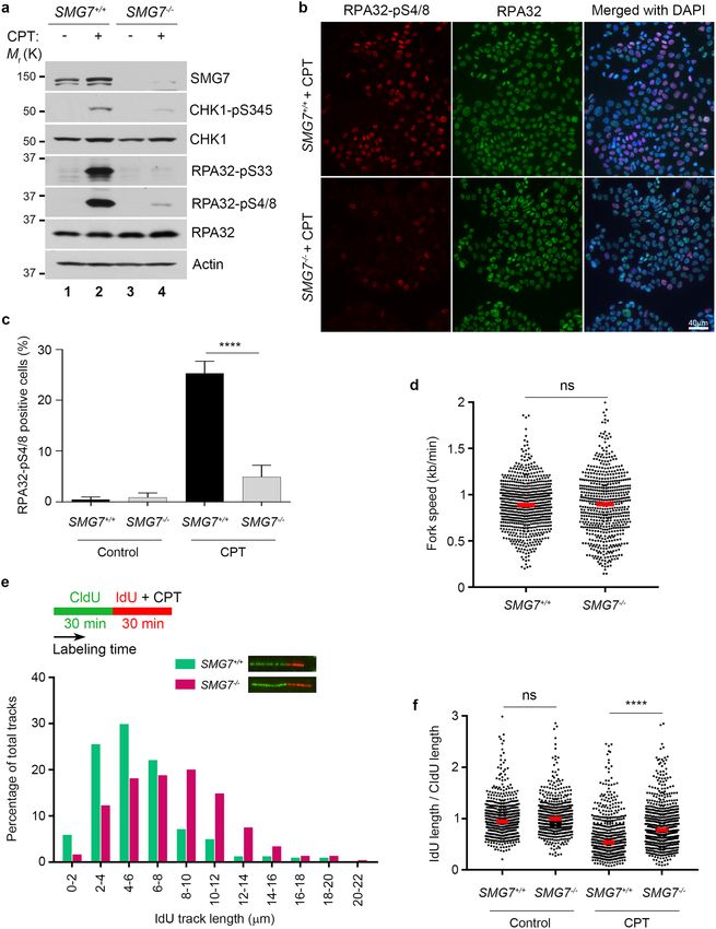
www.nature.com/scientificreports/
◂Figure 1. Loss of SMG7 impairs activation of CHK1 upon DNA damage. (a–c) Wild type and SMG7−/− cells
(a HCT116, b,c DLD1) were treated with ionizing radiation (a,b 10 Gy; c 20 Gy) and total cell extracts were
examined for proteins in the ATM and ATR pathways by western blot analysis using antibodies as indicated
including α-SMG7, α-CHK1-pS345, α-CHK1, α-CHK2-pT68, α-CHK2, α-γH2AX, α-H2AX, α-ATM-pS1981,
α-ATM, α-ATR-pT1989, α-ATR, α-RPA32-pS4/8, α-RPA32-pS33, α-RPA32, α-Actin and α-Tubulin.
Quantitation of relative levels of CHK1-pS345, CHK2-pT68, RPA-pS33, and RPA-pS4/8 in IR-treated cells
are shown in Fig. S1a,d,e). (d) Wild type and SMG7−/− HCT116 cells were treated with ionizing radiation
(10 Gy, 1 h) and pulsed with 25 μM BrdU for 20 min, and analyzed by immunostaining using α-BrdU antibody
(BU1/75). Representative images are shown. (e) Quantification of the percentage of BrdU-positive cells of
the total population from (d). Data are presented as Mean ± SEM (n = 3 independent experiments) and were
analyzed by ANOVA with Tukey post-test. n.s indicates not significant, P > 0.05. (f) Quantification of the relative
intensities of BrdU staining in BrdU + cells from (d) using MetaMorph software. Red bars represent the mean
intensities of each population. Data are shown as nuclear BrdU fluorescence intensities of cells pooled from
three independent experiments and analyzed by ANOVA with Tukey post-test. **** indicates P < 0.001 and n.s,
not significant, P > 0.05. Graphs were generated using GraphPad Prism.
be independent on the 5′-ended ssDNA-dsDNA junction and the 9-1-1 complex11–13. Once brought together
with ATR-ATRIP, TOPBP1 and ETAA1, both of which contain the ATR-activating domain (AAD), stimulate
the kinase activity of ATR in the phosphorylation of its target substrates including downstream effectors (e.g.,
CHK1)24,25 as well as upstream signaling components.
One such signaling molecule is RAD17, a member of the AAA+ family of ATPases that is critical for 9-1-1
loading in activation of the ATR-CHK1 axis, and is phosphorylated on two serine residues S635 and S645 in the
ATM/ATR SQ m otif26–28. As reported in early studies, while ATM can mediate RAD17 phosphorylation, ATR
is the major kinase for S635/645, and loss of these modifications impairs the RAD17 checkpoint f unction27–29.
Importantly, phosphorylation of RAD17 on S635/645, which occurs in a cell cycle-dependent manner in undam-
aged cells, is required to maintain 9-1-1 at the DNA damage site and activate CHK1 in response to replication
stress28–30. Interestingly, a recent study shows that RAD17 also regulates ATM activation in response to DSBs,
involving phosphorylation of RAD17 on T622 by A TM31. Thus, these studies clearly demonstrate that RAD17
plays a key role in checkpoint regulation, and its phosphorylation by ATR and ATM is a pivotal signaling step
in shaping the response of a cell to genotoxic stress.
Previously, we identified a critical role for the suppressor with morphological defects in genitalia 7 (SMG7) in
the G1/S checkpoint in response to DNA damage response, through promoting ATM-mediated p53 stabilization
and transcriptional a ctivation32. Here, based on our initial observation that loss of SMG7 impairs CHK1 activa-
tion by ATR, we explored the idea that SMG7 may have a more general role in regulation of the genotoxic stress
response. Interestingly, we found that the N-terminal 14-3-3 domain of SMG7 directly binds RAD17 and this
interaction is completely dependent on ATR phosphorylation of RAD17 on S635. Sequence analysis revealed at
the RAD17 C-terminal a conserved S635-containing SQ motif shared by other SMG7-binding partners includ-
ing p53 and UPF1, indicating that the SMG7 14-3-3 domain may serve as a common protein-binding module
in phospho-SQ-mediated signaling. Furthermore, we show that SMG7 constitutively associates with chroma-
tin and promotes the recruitment of RAD9 to the DNA damage site. Importantly, unlike wild-type cells, the
SMG7-deficient cells fail to inhibit S-phase DNA replication after genotoxic treatment, and their recovery from
replication stress is significantly compromised because of the inability of cells to maintain CHK1 activity. Taken
together, SMG7 has a critical function in activation of the ATR-CHK1 pathway, at least partly through engaging
the RAD17-mediated DNA damage signaling.
Results
SMG7 is required for activation of the ATR‑CHK1 pathway upon DNA damage. SMG7 is
known for its role in nonsense-mediated mRNA decay (NMD), a surveillance pathway that degrades premature
stop codon-containing mRNA transcripts including those derived from alternative splicing such as the p53β
isoform32–34. In our recent studies, we uncovered a novel function of SMG7 in stabilizing the tumor suppressor
p53 by promoting ATM phosphorylation of MDM232. Given that SMG7 contains the conserved 14-3-3 protein
domain involved in numerous signaling pathways35,36, we reasoned that SMG7 likely has additional roles in the
DNA damage response besides regulating the ATM-MDM2-p53 pathway. To explore this idea, we employed
HCT116 SMG7 knockout cells (SMG7−/−) generated in our previous studies and examined activation of CHK1
and CHK2, the two checkpoint kinases downstream of ATR and ATM, respectively7. As shown in Fig. 1a, fol-
lowing induction of DSBs (indicated by γ-H2AX) by ionizing radiation (IR), we observed similar levels of CHK2
phosphorylation on the ATM site T68 in both wild type and SMG7−/− cells. Interestingly, loss of SMG7 caused
a significant reduction of CHK1 phosphorylation on the ATR site S345—a well-established marker for CHK1
activation24,25 (Fig. 1a, lane 2 vs. lane 4 and Supplementary Fig. 1a), indicating that SMG7 has a specific role in
activation of the ATR-CHK1 axis upon DNA damage. To corroborate this finding, we generated SMG7 knock-
outs in human colorectal adenocarcinoma DLD1 cells using CRISPR-Cas9-mediated gene editing (Supplemen-
tary Fig. 1b and 1c). Notably, we made similar observations that deletion of SMG7 impaired ATR-dependent
CHK1 activation but had no effect on CHK2 (Fig. 1b, lane 2 vs. lane 4 and Supplementary Fig. 1d). Furthermore,
we found that although wild type and SMG7−/− cells exhibited minimal differences in the auto-phosphorylation
of ATR on T1989, an ATR activation m arker37,38, loss of SMG7 abrogated ATR-mediated phosphorylation of
RPA32 on S33 (a direct ATR site) and S4/8 (the DNA-dependent protein kinase sites dependent on S33 phos-
phorylation)39,40 (Fig. 1c, lane 2 vs. lane 4 and Supplementary Fig. 1e and 1f). Thus, these results show that SMG7
Scientific Reports | (2021) 11:7502 | https://doi.org/10.1038/s41598-021-86957-x 3
Vol.:(0123456789)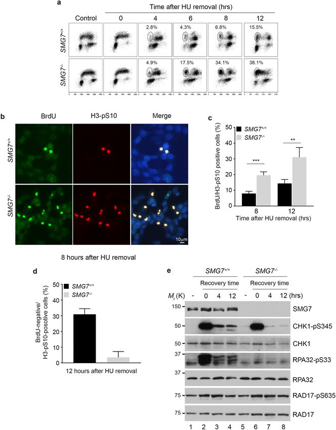
www.nature.com/scientificreports/
is required for activation of the ATR-CHK1 axis and phosphorylation of some other ATR signaling components
such as RPA32 but is dispensable for the intrinsic activity of ATR.
It is well known that the ATR-CHK1 pathway plays a key role in regulation of replication stress, and activa-
tion of CHK1 upon DNA damage inhibits DNA replication in S p hase41,42. To assess the downstream effects of
SMG7 on CHK1, we examined DNA synthesis in SMG7 cells after IR. To this end, we exposed HCT116 cells
−/−
to 10 Gy radiation and labeled them with BrdU to identify replicating cells, which comprise approximately 40%
of the total population regardless of SMG7 status (Fig. 1d,e). As expected, IR had no effect on the percentage of
S-phase cells (Fig. 1e), but significantly reduced the intensity of BrdU staining in wild type cells (Fig. 1d,f). The
lack of reduction of BrdU staining in SMG7−/− cells indicates that DNA synthesis proceeded without hindrance
despite DNA damage (Fig. 1f), consistent with impaired CHK1 activation in the absence of SMG7. We made
similar observations in DLD1 cells (Supplementary Fig. 1g), which express mutant p 5343,44, and importantly, re-
introduction of ectopic SMG7 restored CHK1-pS345 phosphorylation and strongly inhibited DNA replication
(Supplementary Fig. 1h,i). Taken together, these data indicate that SMG7 is critically important for regulation
of the genotoxic stress response through activation of the ATR/CHK1 pathway—a function that is separate from
p53-mediated cell cycle arrest following DNA damage.
RAD17 is a binding partner of SMG7. SMG7 contains an N-terminal 14-3-3-like domain and a C-ter-
minal low complex region (LCR), both of which are implicated in mediating protein–protein interactions35,45.
To clarify the molecular basis of CHK1 activation by SMG7, we took a proteomic approach to search for SMG7-
interacting proteins that potentially regulate the ATR-CHK1 pathway. Thus, we utilized the p53-null human lung
carcinoma H1299 cells, which have been widely used to study DNA damage and generate stable lines, to express
SMG7 containing N-terminal Flag and HA (hemagglutinin) tags (FH-SMG7) (Supplementary Fig. 2a). Through
tandem affinity purification using agarose beads conjugated with α-Flag and α-HA antibodies, followed by mass
spectrometric analysis, we identified from the immunoprecipitates RAD17 in addition to several known SMG7-
binding proteins including UPF1 and SMG5 (Supplementary Fig. 2b,c)46. By western blot analysis, we verified
the presence of RAD17 in the SMG7-specific protein complex (Fig. 2a), indicating that RAD17 is a previously
unknown SMG7-interacting protein. RAD17 contains several well-defined domains in its N-terminal region
that are critical for chromatin binding and 9-1-1 loading, and two SQ motifs (S635 and S645) at its C-terminal
domain (Fig. 2b)47. It is worth noting that SMG7 also interacts with RFC proteins, possibly via RAD17, and
exhibits strong binding activity towards phosphorylated RAD17 (Fig. 2c), suggesting that SMG7 may regulate
the RAD17-RFC clamp loader.
To assess the interaction of SMG7 with RAD17 further, we transiently expressed SMG7 and RAD17 in H1299
cells. As shown in Fig. 2d, RAD17 was only detected in the α-Flag immunoprecipitates from cells expressing
FH-SMG7 and HA-Rad17 but not from cells expressing HA-RAD17 alone (lane 3 vs. lane 2), indicating that
RAD17 was co-immunoprecipitated with SMG7. Notably, when transiently expressed, RAD17 was able to pull
down endogenous SMG7 in the co-immunoprecipitation assays (Fig. 2e), suggesting a strong and stable inter-
action between RAD17 and SMG7 in cells. To examine whether SMG7 physically interacts with RAD17, we
performed in vitro binding assays using purified recombinant proteins. As shown in Fig. 2f, SMG7 was pulled
down by immobilized GST-RAD17 but not GST proteins, indicating that SMG7 binds RAD17 directly. Moreo-
ver, using various GST-RAD17 fragments, we determined that SMG7 could interact with multiple regions of
RAD17 including the N-terminal 1–245 aa fragment containing the P-loop and Walker-B domain (Fig. 2g, lane
4 and Supplementary Fig. 2d) and the middle fragment containing the sensors-1 and 2 (lane 5). Thus, these data
demonstrate that RAD17 is a bona fide SMG7-binding protein.
ATR phosphorylation of RAD17 on S635 mediates its interaction with SMG7. To investigate
how SMG7 activates the ATR-CHK1 axis through RAD17, several lines of evidence prompted us to look into
whether phosphorylation of RAD17 regulates its interaction with SMG7. These include: (1) RAD17 is phospho-
rylated at two SQ sites S635 and S 64527,28; (2) these modifications are enhanced in response to genotoxic s tress27;
(3) SMG7 contains a 14-3-3 protein domain that binds phosphorylated serine; and (4) SMG7 exhibits strong
binding activity towards phosphorylated RAD17 (Fig. 2c). As reported in early studies, ATM and ATR can both
phosphorylate RAD1727; thus we tested whether ATM or ATR is required for the interaction of RAD17 with
SMG7. To this end, we treated H1299 cells stably expressing FH-SMG7 with the kinase inhibitors KU-55933
(ATM) or VE-822 (ATR)48,49, and found that inhibition of ATR abolished phosphorylation of RAD17 on S635
and its interaction with SMG7 whereas the ATM inhibitor had no detectable effect (Fig. 3a, lane 4 vs. lane 3 and
Supplementary Fig. 3a). Moreover, IR enhanced phosphorylation of RAD17 on S635 and its interaction with
SMG7, which was completely abrogated by treatment with the ATR inhibitor VE-822 (Fig. 3b, lane 4 vs. lane 3
and Supplementary Fig. 3b). These data indicate that ATR is the major kinase for Rad17 S635 phosphorylation
and required for the SMG7-RAD17 interaction.
Based on the strong correlation between RAD17 phosphorylation and its interaction with SMG7, we reasoned
that the phosphorylated serine in the SQ motif possibly serves as a binding site for the 14-3-3 domain of SMG7.
To test this hypothesis, we first mapped the RAD17-binding region of SMG7 and found that RAD17 indeed
binds the N-terminal fragment containing the 14-3-3 domain as well as the C-terminal LCR (Fig. 3c, lane 3 and
lane 6 and Supplementary Fig. 3). Sequence analysis of the two SQ motifs of RAD17 shows that the amino acids
adjacent to S635 but not S645 are highly conserved with those of two other known SMG7-binding proteins UPF1
and p53 (Fig. 3d), which is consistent with the finding that loss of S645 phosphorylation had no effect on SMG7
binding (Supplementary Fig. 3d). To examine whether RAD17 S635 contributes to SMG7 binding, we carried
out in vitro pulldown assays using purified wild type RAD17 proteins, which are readily phosphorylated on S635
when expressed in cells, and mutant RAD17 with S635 replaced with alanine (Fig. 3d). Interestingly, we found
Scientific Reports | (2021) 11:7502 | https://doi.org/10.1038/s41598-021-86957-x 4
Vol:.(1234567890)
www.nature.com/scientificreports/
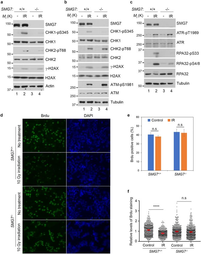
Figure 2. Identification of RAD17 as an SMG7-binding protein. (a) Verification of SMG7-binding proteins identified
from complex purification. The total cell lysates and α-Flag/HA immunoprecipitated materials from H1299 and
H1299-FH-SMG7 cells were assayed by western blot analysis using α-RAD17, α-UPF1, α-SMG5, and α-HA (SMG7)
antibodies. (b) Schematic presentation of the RAD17 domain structure containing P-loop, Walker B, Sensor 1, Sensor
2 and C-terminal highlighted SQ motifs. (c) As in (a), the total cell lysates and α-Flag immunoprecipitates from H1299
and H1299-FH-SMG7 cells were assayed by western blot analysis using α-RAD17, α-RAD17-pS645, α-SMG7, α-RFC2
and α-RFC3 antibodies. (d) H1299 cells were transfected with SMG7- and RAD17-expressing plasmid DNA, and
the cell extracts and α-Flag immunoprecipitated materials were analyzed by western blot with α-RAD17 and α-HA
(SMG7) antibodies. (e) The total cell extracts and α-Flag immunoprecipitated materials from H1299 cells transfected
with empty vector or FH-RAD17-expression plasmid DNA were examined by western blot analysis using α-SMG7 and
α-RAD17 antibodies. (f–g) SMG7 direct binding to RAD17 in vitro. GST, full-length GST-RAD17 (f), or truncated
GST-RAD17 fragment (g) fusion proteins were used in pulldown assays with purified FH-SMG7 proteins. FH-SMG7
was detected by western blot analysis using α-SMG7 antibody, and GST proteins visualized by Ponceau S staining in (f)
and Supplementary Fig. 2d.
Scientific Reports | (2021) 11:7502 | https://doi.org/10.1038/s41598-021-86957-x 5
Vol.:(0123456789)www.nature.com/scientificreports/
Figure 3. ATR-dependent RAD17 phosphorylation at S635 enhances SMG7 interaction. (a) The
FH-SMG7-H1299 cells were treated for 4 h with 10 µM ATM (KU-55933) or ATR (VE-822) inhibitor. The
total cell lysates and α-Flag immunoprecipitates were analyzed by western blot using α-SMG7, α-RAD17 and
α-RAD17-pS635 antibodies. Relative levels of immunoprecipitated RAD17 are quantitated in Fig. S3a. (b) Cells
treated with or without ATR inhibitor (VE-822, 10 µM for 4 h) were exposed to 10 Gy IR and harvested 1 h later.
The total cell lysates and α-Flag immunoprecipitates were analyzed by western blot using α-SMG7, α-RAD17
and α-RAD17-pS635 antibodies as in (a). Relative levels of immunoprecipitated RAD17 and RAD17-pS635
are quantitated in Fig. S3b. (c) GST and GST-SMG7 fragment fusion proteins were incubated with lysates from
293FT cells transiently expressing HA-RAD17, and RAD17 pulled down by the GST proteins was analyzed by
western blot with α-RAD17 antibody. (d) Alignment of the amino acid sequences adjacent to the serine residues
within the SQ motif of RAD17, UPF1 and p53 (top). The FH-RAD17 and FH-RAD17-S635A proteins purified
from transfected 293FT cells were analyzed by western blot using α-RAD17-pS635 and α-RAD17 antibodies
(bottom). (e) GST or GST-SMG7 fragments were incubated with purified FH-RAD17 or FH-RAD17-S635A
protein from (d) in GST pull-down assays. RAD17 and RAD17-pS635 were detected by western blotting with
the antibodies indicated. Relative levels of RAD17 binding and RAD17-pS635 binding are quantitated in Fig.
S3f. (f) H1299 cells were co-transfected with plasmids expressing FH-SMG7 and HA-RAD17 WT, S635A,
S645A or S635/645A, and the total cell extracts and α-Flag immunoprecipitated materials were examined by
western blot analysis using α-SMG7, α-RAD17 and α-RAD17-pS635 antibodies. Relative levels of RAD17 and
RAD17-pS635 binding to FH-SMG7 are quantitated in Fig. S3g.
Scientific Reports | (2021) 11:7502 | https://doi.org/10.1038/s41598-021-86957-x 6
Vol:.(1234567890)www.nature.com/scientificreports/
that loss of S635 phosphorylation markedly reduced RAD17 binding to the SMG7 14-3-3 domain (Fig. 3e, lane
3 vs. lane 7 and Supplementary Fig. 3e,f), but only had mild effects on its binding to the LCR region (Fig. 3e.
lane 4 vs. lane 8 and Supplementary Fig. 3f). Moreover, when exogenously expressed in cells, wild type RAD17
bound SMG7 much more strongly than the RAD17 S635A mutant (Fig. 3f, lane 2 vs. lane 4 and Supplementary
Fig. 3g), and the reduced binding was not further exacerbated by the S645A mutation (lane 4 vs. lane 8). It is
worth noting that the S635-phosphorylated RAD17 detected in the immunoprecipitated materials from cells
expressing S635A or S635A/S645A mutants is likely endogenous RAD17 (Fig. 3f, lanes 4 and 8), as transiently
expressed SMG7 was able to pull down endogenous S635-phosphorylated RAD17 (Supplementary Fig. 3h).
Taken together, these data indicate that ATR phosphorylation of RAD17 on S635 is critical for its interaction
with SMG7 under both normal and genotoxic stress conditions.
SMG7 promotes chromatin recruitment of RAD9 upon DNA damage. So far, our data suggest
that SMG7 activates the ATR-CHK1 axis upon DNA damage through interactions with RAD17, and this protein
binding activity appears to be very specific for RAD17, as we did not observe any interactions between SMG7
and ATR, CHK1, or TOPBP1 in HCT116 and DLD1 cells (Fig. 4a, and Supplementary Fig. 4a). A key function of
RAD17-RFC in the activation of ATR-CHK1 is to load the 9-1-1 c lamp16,21, and given that SMG7 interacts with
RFC proteins as well, we examined whether SMG7 regulates the recruitment or retention of RAD9 to the DNA
damage site. To this end, we performed immunofluorescence staining of cells following DNA damage, and found
that IR induced similar levels of DSBs (~ 90% γ-H2AX positive cells) in wild type and SMG7−/− cells (Fig. 4b and
Supplementary Fig. 4b). In addition, compared with wild-type cells, only a very mild reduction in the number
of SMG7−/− cells with γ-H2AX/RPA32 foci was observed, which suggests that loss of SMG7 may have, if any,
a slight effect on the formation of ssDNA following DSB resection. These data are largely consistent with our
observation that loss of SMG7 does not affect activation of ATM (Fig. 1a,b), which controls DSB r esection50,51.
As formation of RPA-ssDNA is the starting point of ATR activation, the presence of RPA32 foci indicates that
the initial molecular signal for ATR-CHK1 activation is present in wild type and SMG7−/− cells. Interestingly,
although IR strongly induced RAD9 foci co-localized with the RPA32 foci in wild type cells, the RAD9/RAP32-
colocolized foci were much less pronounced in the SMG7−/− cells (Fig. 4c and Supplementary Fig. 4c) and the
SMG7−/− cells also had significantly less RAD9/RAP32-colocolized foci fractions than wild type cells (Fig. 4d
and Supplementary Fig. 4c), suggesting that SMG7 is important for the recruitment or retention of the RAD9
complex to the DNA damage site.
To corroborate these findings, we carried out chromatin fractionation assays to examine RAD9 recruitment
upon DNA d amage52. Notably, we found that in unstressed cells, a small fraction of SMG7 was associated with
chromatin, and loss of SMG7 had no effect on the levels of the chromatin-bound RAD17 (Fig. 4e, lane 5 vs. lane
6 and Supplementary Fig. 4d,e). Interestingly, the chromatin-bound fraction of RAD9 was substantially reduced
in the SMG7−/− cells compared with wild type cells following IR (Fig. 4f, lane 2 vs. lane 4 and Supplementary
Fig. 4f), which is consistent with the results from immunofluorescence staining. Thus, these data show that SMG7
plays an important role in recruitment or retention of the RAD9 complex to the DNA damage site and reveals a
novel mechanism for SMG7 activation of the ATR-CHK1 axis.
SMG7 regulates CHK1 activation and fork progression following replication stress. The ATR-
CHK1 axis is a key regulator of replication stress, upon which activated CHK1 halts DNA replication in the S
phase of the cell cycle by inhibiting replication initiation and progression of the replication fork41,42,53. Thus, our
observation that loss of SMG7 impaired activation of CHK1 following IR (Fig. 1) suggests that SMG7 has an
important function in the regulation of the cellular response to replication stress induced by DNA lesions. To
assess the role of SMG7 in S phase, we utilized camptothecin (CPT), a topoisomerase I inhibitor that specifi-
cally induces DSBs on replicating DNA54. As expected, treatment with CPT induced γ-H2AX and RPA32 foci
similarly in both wild type and SMG7−/− cells (Supplementary Fig. 5a,b), suggesting that SMG7 does not affect
formation of RPA-bound ssDNA. Interestingly, loss of SMG7 significantly reduced CPT-induced phosphoryla-
tion of CHK1 on S345 (Fig. 5a, lane 2 vs. lane 4, Supplementary Fig. 5c). Moreover, phosphorylation of RPA32
on S33 and S4/8 following treatment with CPT was nearly abolished in the absence of SMG7 (Fig. 5a, lane 2 vs.
lane 4, Fig. 5b,c, and Supplementary Fig. 5c), indicating that SMG7 is critical for activation of the ATR/CHK1
signaling upon replication stress.
To examine how SMG7 regulates replication stress, we performed DNA fiber analysis, a well-established
double-labeling approach that uses the BrdU derivatives CldU and IdU to directly assess the replication f ork55.
This dual labeling strategy makes it possible to (1) identify active replication forks during the labeling times,
which proceed bi-directionally from their origins, and (2) determine the effect of replication stress on fork
progression by measuring the IdU-labeled DNA tracks. To this end, cells were pulse-labeled with CldU and
IdU (with or without CPT), followed by DNA spreading, immunostaining and microscopic analysis for the
presence and length of the distinct CIdU and IdU tracks. Under normal conditions, wild type and SMG7−/− cells
exhibited similar symmetric CIdU/IdU-labeled tracks (Supplementary Fig. 5d) and showed no significant differ-
ence in fork speed (Fig. 5d). Interestingly, in the presence of CPT, wild type cells had much shorter IdU-labeled
tracks compared with the SMG7−/− cells (Fig. 5e, distribution within 2–8 µm for wild type and 4–12 µm for the
SMG7−/− cells, respectively), which is consistent with previous studies showing that activation of CHK1 inhibits
fork progression56. Moreover, by analyzing hundreds of active replication forks individually, we found that treat-
ment with CPT resulted in a significant reduction in the ratio between IdU and CIdU-labeled tracks, which was
reversed by inhibition of CHK1 or loss of SMG7 (Supplementary Fig. 5e,f). Thus, these data indicate that by
promoting ATR signaling and CHK1 activation, SMG7 has a important role in the control of fork progression
in response to replication stress.
Scientific Reports | (2021) 11:7502 | https://doi.org/10.1038/s41598-021-86957-x 7
Vol.:(0123456789)www.nature.com/scientificreports/
Scientific Reports | (2021) 11:7502 | https://doi.org/10.1038/s41598-021-86957-x 8
Vol:.(1234567890)www.nature.com/scientificreports/
◂Figure 4. SMG7 promotes chromatin recruitment of RAD9 upon DNA damage. (a) Cell extracts from DLD1
SMG7−/− cells expressing FH-SMG7 and the α-Flag immunoprecipitates were examined by western blot analysis
using α-SMG7, α-RAD17, α-ATR, α-TOPBP1, α-CHK1 and α-Tubulin antibodies. (b,c) Wild type and SMG7−/−
cells were treated with ionizing radiation (10 Gy, 1 h), pre-extracted, and immunostained in (b) with α-RPA32
(green) and α-γH2AX (red) antibodies and in (c) with α-RPA32 (green) and α-RAD9 (sc-74464, red). Nuclei
were stained with DAPI. Representative images from immunofluorescence microscopy are shown. Wider fields
of view at are shown in Fig. S4b-c. (d) Quantification of yH2AX-, yH2AX/RPA- and RPA/RAD9- foci-positive
cells from experiments represented in (b,c). Data are presented as Mean ± SEM (n = 3 independent experiments
containing pooled data) and were analyzed by one-way ANOVA with Tukey post-test. **** indicates P < 0.001.
(e) Wild type and SMG7−/− DLD1 cells were subjected to fractionation, and the total cells extracts, soluble
nuclear and chromatin fractions were analyzed by western blot using α-SMG7 and α-RAD17 antibodies.
α-alpha-Tubulin and α-Histone H3 antibodies were used as cytoplasmic and chromatin markers, respectively.
Relative levels of chromatin-bound SMG7 and RAD17 are quantitated in Fig. S4d. (f) As in (e), the chromatin-
bound fractions from the control and irradiated cells were isolated and examined by western blot analysis using
α-SMG7, α-RAD17, α-RAD17-pS645, α-RAD9 (A300-890A) and α-H3 antibodies. Relative levels of chromatin-
bound RAD9 are quantitated in Fig. S4f.
SMG7 regulates CHK1‑S345 phosphorylation and modulates cell cycle progression during
recovery from replication stress. The crucial function of CHK1 in the replication stress response is
manifested not only in fork regulation but also during recovery of cells after removal of stress conditions56–61.
During the process of recovery, the ATR-mediated checkpoint signaling is attenuated in a regulated manner,
which ensures coordinated DNA replication and cell cycle progression after replication stress is r esolved62. Not
surprisingly, deficiency in CHK1 itself or components required for activation of the ATR-CHK1 axis impairs
normal cell cycle progression or cell survival after cells are relieved from a short-term replication stress of revers-
ible nature, such as a few hours of treatment with hydroxyurea (HU), a ribonucleotide reductase inhibitor that
induces replication stress by depleting cellular deoxyribonucleotides. For example, cells depleted of RAD17 or
expressing phosphorylation-defective S635/645A mutant RAD17 are unable to maintain controlled CHK1 acti-
vation and lose cell viability during recovery from HU treatment29. Thus, our finding that SMG7 promotes
CHK1 activation through robust binding to S635-phosphorylated RAD17 prompted us to examine whether
SMG7 is involved in facilitation of replication stress recovery. As expected, HU treatment strongly induced
CHK1 activation in a dose- and time-dependent manner, which was markedly reduced in the absence of SMG7
(Supplementary Fig. 6a,b). When we assessed ATR phosphorylation of CHK1 shortly after HU removal, we
found that levels of S345-phosphorylated CHK1 decreased over time in wild type cells, and loss of SMG7 further
accelerated this downregulation (Supplementary Fig. 6c). Consistent with previous studies29, these data suggest
that SMG7 plays an important role in maintaining CHK1 S345 phosphorylation in the early stages following
removal of replication stress.
Next, we examined cell cycle profile, and found that when the whole cell population was analyzed in a course
of 12-h recovery, the SMG7−/− cells showed deficient cell cycle progression (Supplementary Fig. 6d). To assess
the S-phase population specifically, we pulse-labeled cells with BrdU, and then treated them with HU, which
arrested BrdU-positive cells throughout the S phase and BrdU-negative cells at G1 (Fig. 6a, control vs. 0 h). Until
4 h after HU removal, we observed little difference in the S-phase progression of the BrdU-positive cells. The
gradual increase (from 4 to 8 h) in the wild type BrdU-positive population with 2 N DNA content, which results
from completion of mitosis, indicates a regulated and orderly entry into mitosis from G 2 after release from HU.
Interestingly, the SMG7−/− BrdU-positive cells completed the G 2-M transition in an accelerated fashion, and
subsequently accumulated at the 2 N position (Fig. 6a; SMG7−/− 34.1% vs. wild type 6.8% by 8 h). The acceler-
ated transition of the SMG7−/− BrdU-positive cells was clearly illustrated when the BrdU-positive population
was selected and analyzed by flow cytometry (Supplementary Fig. 6e). To assess the role of SMG7 in the G 2-M
progression directly during the 12-h course of recovery, we treated cells with the mitosis-arresting agent nocoda-
zole upon HU removal and assayed the presence of the mitotic marker S10-phosphorylated histone 3 (H3-pS10).
Notably, the BrdU-positive population of the SMG7−/− cells had a significantly higher H3-pS10-positive fraction
compared with that of wild type cells (Fig. 6b,c), suggesting that SMG7 is indeed critical for the G 2-M transition
during recovery from replication stress. It is worth noting that the BrdU-negative population of wild type cells
showed a higher H3-pS10-positive fraction than that of the SMG7−/− cells (Fig. 6d), and this is likely because
wild type cells appeared to progress from G 1 through S phase more quickly (Fig. 6a).
To determine the underlying mechanism by which SMG7 regulates the G 2-M transition, we examined the
status of CHK1 phosphorylation during recovery, as previous studies have shown that the mitotic entry from
G2 is controlled by CHK157,59,60. While treatment with a higher dose of HU (5 mM) for a prolonged period
of time (6 h) induced similar CHK1 S345 phosphorylation regardless of SMG7 status, importantly, wild type
cells retained markedly higher levels of S345-phosphorylated CHK1 than SMG7−/− cells after removal of HU
(Fig. 6e, lanes 3–4 vs. 7–8). The further reduced CHK1 S345 phosphorylation in the absence of SMG7 during
recovery is consistent with the observation that SMG7−/− cells showed an accelerated G2-M transition. It is also
interesting to note that loss of SMG7 abolished HU-induced phosphorylation of RPA32 on S33 but had no effect
on RAD17 phosphorylation on S635 before or after HU removal. Thus, these data indicate that SMG7 plays an
important role in regulating CHK1 S635 phosphorylation and modulating cell cycle progression during recovery
from replication stress.
Scientific Reports | (2021) 11:7502 | https://doi.org/10.1038/s41598-021-86957-x 9
Vol.:(0123456789)www.nature.com/scientificreports/
Scientific Reports | (2021) 11:7502 | https://doi.org/10.1038/s41598-021-86957-x 10
Vol:.(1234567890)www.nature.com/scientificreports/
◂Figure 5. SMG7 regulates ATR signaling and CHK1 activation and fork progression following replication
stress. (a–c) Wild type and SMG7−/− cells were treated with 1 µM CPT for 1 h. (a) Total cell extracts were
examined by western blot analysis using α-SMG7, α-CHK1, α-CHK1-pS345, α-RPA32-pS33, α-RPA32-pS4/8,
α-RPA32, and α-Actin antibodies. Relative levels of CHK1-pS345, RPA-pS33, and RPA-pS4/8 in CPT-treated
cells are quantitated in Fig. S5c. (b) Cells were immunostained with α-RPA32-pS4/8 (red) and α-RPA32 (green)
antibodies. Representative images from immunofluorescence microscopy are shown. (c) Quantification of
S4/8-positive cells from (b) using MetaMorph software. Data are presented as Mean ± SEM (n = 3 independent
experiments containing pooled data) and analyzed by one-way Anova with Tukey post-test; **** indicates
P < 0.001. Graphs were generated using GraphPad Prism. (d–f) DNA fiber analysis of untreated or CPT-treated
wild type and SMG7−/− cells as in (a). Cells were first labeled with 25 μM CldU for 30 min and then by 250 μM
IdU for 30 min in the absence or presence of CPT. DNA fibers were then spread, stained with α-BrdU antibodies
(B44 and BU1/75) and visualized using fluorescence microscopy. More than 500 CIdU-/IdU-labeled tracks from
two independent experiments were measured using ImageJ. (d) Quantification of replication fork speed using
the lengths of CldU- and IdU-labeled DNA fiber. Fork speed was estimated using 1 μm = 2.6 kbp as described in
Methods. Student’s t-test was performed to determine the statistical difference; n.s. not significant. (e) Scheme
for the CIdU/IdU labeling and CPT treatment (Top). Representative tracks were shown (Middle). Distribution
of the IdU-labeled tracks from the CPT-treated cells (Bottom). (f) Quantification of length ratios of the IdU-
labeled tracks to the CldU-labeled tracks per replication fork. Red bars represent the means of each population.
Data were analyzed by one-way Anova with Tukey post-test; **** indicates P < 0.001; n.s. not significant, P > 0.05.
Discussion
Here, we report a previously unknown function of SMG7 in regulation of the ATR-CHK1 pathway in response
to DNA damage and replication stress. For the first time, we have identified SMG7 as a binding partner to the
RAD9-RAD1-HUS1 clamp loader protein RAD17 and shown that SMG7 promotes the recruitment of RAD9
to RPA-coated ssDNA in the activation of CHK1. The interaction of SMG7 with RAD17 is highly dynamic and
tightly controlled by the ATR-dependent phosphorylation of RAD17 on S635, a conserved SQ site that is required
for RAD17 checkpoint function. Importantly, loss of SMG7 impairs ATR signaling and CHK1 activation upon
genotoxic stress and disables intra-S checkpoint function in control of replication fork progression. Furthermore,
SMG7 has an important role in maintaining CHK1 S345 phosphorylation and cell cycle progression. In summary,
our present studies identify SMG7 as an indispensable signaling component in activation of the ATR-CHK1
pathway through RAD17 binding and RAD9 recruitment, and suggests that by engaging phospho-SQ-mediated
signaling through its 14-3-3 domain, SMG7 has a more general and direct role in regulation of the genotoxic
stress response.
Activation of the ATR-CHK1 axis upon DNA damage is a complex process, which necessitates recruit-
ment of the 9-1-1 complex to the damage site by RAD17-RFC2,5,8. Given that loss of SMG7 markedly impairs
the formation of RAD9 foci in regions of RPA-bound ssDNA, our data provide an important insight into how
SMG7 engages in the ATR-CHK1 signaling pathway (Fig. 4c). Although the precise mechanism by which SMG7
promotes RAD9 recruitment/retention remains to be fully elucidated, SMG7 binding to RAD17 and other
RFC subunits likely has a direct impact on 9-1-1 association and/or recruitment (Fig. 2). For example, stud-
ies have shown that the N-terminal P-loop and Walker-B domains of RAD17 are critical for 9-1-1 binding
and recruitment47,63; thus the direct binding of SMG7 to the RAD17 N-terminal region may facilitate 9-1-1
recruitment or stabilize the RAD17 interaction with 9-1-1. Additionally, SMG7 may promote the recruitment/
retention of the RAD9 complex through interaction with the S635-phosphorylated RAD17. While early reports
suggest that ATR phosphorylation of RAD17 is not required for initial RAD9 loading, recent studies show that
phosphorylation of the RAD17 SQ motif indeed plays an important role in maintaining DNA damage-induced
RAD9 foci22,30. Therefore, it is likely that through interacting with S635-phosphorylated RAD17, SMG7 may
stabilize RAD9 foci following its recruitment, which allows full activation of the ATR-CHK1 axis. Besides act-
ing as a RAD9-RAD1-HUS1 clamp loader protein, RAD17 also functions in CHK1 recruitment by CLASPIN,
which is critical for ATR phosphorylation and activation of C HK129,64. Intriguingly, SMG7 and CLASPIN both
engage with RAD17 in a phosphorylation-dependent manner via interactions with phosphorylated RAD17, but
it is not known whether S635, S645 or both are required for the RAD17-CLASPIN interaction. While we show
that loss of SMG7 reduces RAD9 recruitment/retention (Fig. 4), our data does not rule out the possibility that
SMG7 may be involved in CLASPIN regulation of CHK1. It is possible that SMG7 may compete with CLASPIN
in RAD17 binding; however, given that both SMG7 and CLASPIN positively regulate CHK1, it is intriguing how
these two RAD17 binding proteins may affect each other with respect to different aspects of CHK1 activation. For
example, CLASPIN is critically important for the attenuation of the ATR-CHK1 signaling during r ecovery57,59
and it remains to be elucidated whether and how SMG7/phosphorylated RAD17 are involved in this regulation.
RAD17 is an important checkpoint regulator, and its phosphorylation by ATR and ATM appears to be a
pivotal signaling step in shaping the response of a cell to genotoxic stress27,29,31. As reported here and previously,
ATR is the major kinase that phosphorylates RAD17 on the SQ sites S635/645, and loss of these modifications
impairs the RAD17 checkpoint function27,29. Moreover, a recent study has shown that RAD17 activates ATM
in response to DSB through binding to Nijmegen breakage syndrome (NBS1) of the MRE11-RAD50-NBS1
(MRN) complex, which involves the phosphorylation of RAD17 on T622 by ATM31. Thus, our finding that SMG7
interacts with RAD17 in an ATR-dependent but ATM-independent manner reveals phosphorylation-mediated
SMG7 binding to RAD17 as a potential underlying mechanism by which SMG7 exerts its function in response
to genotoxic stress (Fig. 3a,b). Indeed, our data show that loss of SMG7 abrogates CHK1 activation but has no
effect on the ATM-CHK2 pathway, and support a model in which through binding to S635-phosphorylated
Scientific Reports | (2021) 11:7502 | https://doi.org/10.1038/s41598-021-86957-x 11
Vol.:(0123456789)www.nature.com/scientificreports/
Scientific Reports | (2021) 11:7502 | https://doi.org/10.1038/s41598-021-86957-x 12
Vol:.(1234567890)www.nature.com/scientificreports/
◂Figure 6. SMG7 maintains normal attenuation of the ATR-CHK1 axis during recovery from replication stress.
(a) Wild type and SMG7−/− HCT116 cells were pulsed with 25 μm BrdU followed by treatment with 5 mM HU
for 6 h. After HU treatment, cells were released into fresh normal media, and harvested at the indicated time
points. Cells were then fixed, stained with α-BrdU antibody (y-axis) and 7-AAD (x-axis) and analyzed by flow
cytometry. The BrdU-positive fractions containing G1-DNA content from total populations are circled (4, 6
and 8 h after HU removal). (b–d) Cells treated as in (a) were released into fresh media containing 1 μg/mL
nocodazole for different hours. Cells were then fixed and stained with α-BrdU (BU1/75) (green) and α-Histone
H3-pS10 (red) antibodies, and imaged using a fluorescence microscope. Representative images of cells 8 h after
HU removal are shown in (b). Cells were counted using ImageJ, and the percentage of BrdU/H3-pS10 double
positive (c) cells were quantified. Data are presented as Mean ± SD (n = 3) and analyzed by one-way ANOVA
(****P < 0.0001). (d) BrdU-negative/H3-pS10-positive cells were quantified. Data are presented as Mean ± SEM
(n = 3) and analyzed by Student’s t-test; P < 0.01. (e) Total cell extracts from wild type and SMG7−/− cells treated
as in (b–d) were examined by western blot analysis using α-SMG7, α-CHK1-pS345, α-CHK1, α-RPA32-pS33,
α-RPA32, α-RAD17 and α-RAD17-pS635 antibodies.
RAD17 in an ATR-dependent fashion, SMG7 acts on ATR-CHK1 signaling following DNA damage (Figs. 1a–c
and 3e,f). Importantly, consistent with its role in activation of the ATR-CHK1 axis, loss of SMG7 disables CHK1-
dependent intra-S checkpoint function in control of replication fork progression in response to CPT-induced
replication stress (Fig. 5). However, it appears that SMG7 is not significantly involved in regulation of CHK1
under basal conditions, as indicated by normal DNA replication and fork progression in SMG7−/− cells during
S phase (Figs. 1d,e and 5). Moreover, unlike RAD17-null cells, which rapidly lose viability following deletion
of RAD1765, SMG7−/− cells are able to grow and proliferate normally, consistent with the phenotype observed
in cells expressing the phosphorylation-defective S635/645A RAD17 mutant29. Thus, these data support the
notion that SMG7 functions in the ATR-CHK1 pathway mainly, if not exclusively, through interaction with the
S635-phosphorylated RAD17.
In addition to being indispensable for CHK1 activation upon DNA damage, our data further show that SMG7
may have an important role in regulating CHK1 and cell cycle progression during recovery from replication
stress. Unlike wild type cells, SMG7−/− cells fails to retain CHK1 S345 phosphorylation shortly after release from
replication stress, consistent with a previous study showing that loss of RAD17 phosphorylation on S635/645A
leads to a defect in maintaining Chk1 activation after HU withdrawal29. Furthermore, SMG7−/− cells exhibit
an accelerated transition from G2 to M after release from prolonged treatment (6 h) with a higher dose of HU
(5 mM), which is supported by data that SMG7−/− cells have even lower S345-phosphorylated CHK1 compared
to wild-type cells during recovery (Fig. 6 and Supplementary Fig. 6). As S635/645-phosphorylated RAD17
engages both SMG7 and CLASPIN, whose degradation is critical for CHK1 inactivation during recovery from
replication stress29, it is possible that SMG7 may regulate CHK1 by modulating CLASPIN function during
recovery from replication stress. It is important to note that in these experiments HU treatment (5 mM for
6 h) induced similar CHK1 S345 phosphorylation regardless of SMG7 status, suggesting that wild-type and
SMG7−/− cells have the same levels of initial CHK1 activation following HU treatment. As our data show that loss
of SMG7 does not abolish CHK1 S345 phosphorylation after HU treatment and only attenuates it in a dose- and
time-dependent manner, indicating that the mechanism for CHK1 activation still exists in SMG7−/− cells (Sup-
plementary Fig. 6a,b). It is worth noting that the level of CHK1 phosphorylation in S MG7−/− cells treated with
2 mM HU for 1 h is even higher than that in wild-type cells treated with 0.5 mM HU. A recent study shows that
the amount of HU and the time of its treatment has very dynamic effects on the recruitment pattern of several
critical proteins involved in CHK1 activation such as TOPBP1 and the 9-1-1 c omplex66. Thus, it is conceivable
that under specific conditions such as prolonged treatment with a high concentration (5 mM) of HU, CHK1
S345 phosphorylation may be maximized in both wild-type and S MG7−/− cells, which leads to similar levels of
S345-phosphorylated CHK1.
In response to genotoxic stress, ATR phosphorylates a plethora of proteins, and there is enormous interest in
understanding the mechanism for ATR activation towards the different substrates3,5,8. By examining CHK1 and
RPA32, a recent study has identified two distinct ATR activation modes, which are orchestrated by RAD17 and
NBS1 in phosphorylation of CHK1 and RPA32, r espectively67. Interestingly, our present data show that loss of
SMG7 abolishes ATR phosphorylation of CHK1 and RPA32 but has no effect on RAD17 (Figs. 1a–c, 4f, 5a and
6a,f), suggesting that SMG7 may regulate phosphorylation of ATR substrates differentially, and likely engages
either directly or indirectly other components (e.g., NBS1) of the ATR pathway besides RAD17. Future studies
aimed to elucidate the precise mechanisms of SMG7-mediated signaling will further our understanding of the
role of SMG7 in the regulation of genotoxic stress response.
Methods
Cell culture. The human colorectal adenocarcinoma HCT116, DLD1 and their SMG7-null derivative cell
lines were cultured in McCoy’s 5A medium (Corning, Manassas, VA, USA) supplemented with 10% fetal bovine
serum (Sigma, F2442). The H1299 and 293FT cells were cultured in Dulbecco’s modified Eagle’s medium
(DMEM, Corning/Cellgro) supplemented with 10% fetal bovine serum and combined antibiotics (100 I.U/ml
penicillin and 100 ug/ml streptomycin).
Generation of DLD1 SMG7−/− cell line. We carried out CRISPR-Cas9-mediated gene targeting in DLD1
cells based on the protocol described in the previous s tudy68. Briefly, two sgRNAs (target sequence: 5′- ATGTTC
GAATAATCAGATTG-3′ and 5′-GATTATAGGTGATCAATCCC-3′) designed using the online CRISPR Design
Scientific Reports | (2021) 11:7502 | https://doi.org/10.1038/s41598-021-86957-x 13
Vol.:(0123456789)www.nature.com/scientificreports/
Tool (http://tools.genome-engineering.org) were cloned into the Cas9-expressing plasmid PX458 (Addgene,
#48138). After transfection with the sgRNA-expressing plasmid DNA and pCin4-puro (Clontech, #6031-1),
cells were treated with 3 µg/ml puromycin for 24 h and plated in 96-well in the absence of puromycin. Two weeks
later, clones were expanded and screened for SMG7 targeting, which was further verified by DNA sequencing.
Plasmids construction. pCin4-FH-SMG7, pCin4-FH-RAD17 and pCin4-HA-RAD17 plasmids were con-
structed by cloning the cDNA encoding SMG7 or RAD17 generated by RT-PCR from mRNA extracted from
HCT116 cells into pCin4-Flag-HA or pCin4-HA vectors. pCin4-FH-RAD17-S635A, pCin4-HA-RAD17-S635A,
S645A and S635A/645A plasmids were generated by site-directed mutagenesis. GST-RAD17 full-length, GST-
RAD17 fragments and GST-SMG7 fragments expressing plasmids were generated by cloning of each DNA frag-
ments into pGEX-2TL(+) vector. All the plasmids were confirmed by DNA sequence analysis.
Antibodies and inhibitors. The following antibodies were from Bethyl: α-H2AX (A300-081A, 1:1000),
α-H2AX (A303-837A, 1:2000), α-RAD9 (A300-890A, WB 1:500, IF 1:50), α-RAD17-pSer645 (A300-153A,
1:1000), α-RFC2 (A300-142A, 1:1000), α-RFC3 (A300-188A, 1:1000), α-RPA32-pS4/8 (A300-245A, 1:1000),
α-RPA32-pS33 (A300-246A, 1:1000), α-RPA32 (A300-244A, 1:3000), α-SMG7 (A302-170A, 1:1000). The fol-
lowing antibodies were from Cell Signaling Technology: α-ATM-pS1981 (5883, 1:1000), α-ATM (2873, 1:1000),
α-CHK1-pS345 (2348, 1:1000), α-CHK2-pT68 (2661, 1:1000), α-CHK2 (2662, 1:1000), α-RAD17-pS635
(13404S, 1:1000), α-RAD17 (8561, 1:1000), α-RPA32 (2208S, IF 1:1000), α-Histone H3 (3638, 1:2000), α-rabbit
IgG-HRP (7074, 1:5000). The following antibodies were from Santa Cruz Technology: α-Actin-beta (sc-47778,
1:1000), α-ATR (sc-1887, 1:1000), α-CHK1 (sc-8408, 1:100), α-Histone H3 p-Ser10 (sc-8656, IF 1:200), α-RAD9
(sc-74464, IF 1:100), α-RAD17 (sc-17761, 1:500), α-Tubulin-alpha (sc-53029, 1:1000). Goat α-rat IgG-HRP
(3050-05, 1:5000) and donkey α-goat IgG-HRP (6420-05, 1:5000) were from Southern Biotech. α-ATR-pT1989
(GTX128145, 1:1000), α-HA (MMS-101P, 1:1000), α-SMG5 (12694-1-P, 1:1000), α-SMG7 (LS-C353204-100,
1:1000) and Sheep α-mouse IGG-HRP (NA931, 1:5000) were from GeneTex, Bio-Rad, Covance, Proteintech,
LifeSpan Biosciences and GE healthcare, respectively. Antibodies used for BrdU staining include α-BrdU clone
B44 (347580, 1:200; BD Biosciences) and α-BrdU clone BU1/75 (MCA2060, 1:200 BioRad). The following inhib-
itors were used: ATR inhibitor (VE-822; MedKoo Biosciences) and ATM inhibitor (KU-55933; Sigma-Aldrich).
Purification of the SMG7‑associated protein complex. The H1299-FH-SMG7 stable cell lines were
generated by transfection with pCin4-FH-SMG7, followed by selection with 1 mg/ml G418 for 2–3 weeks. To
purify the SMG7 protein complex, freshly grown H1299-FH-SMG7 cells (~ 5 × 10⁸) were harvested in cold PBS.
The cell pellets were suspended in 20 times volume of ice cold BC100 buffer (20 mM Tris–HCl pH 7.9, 100 mM
NaCl, 10% glycerol, 0.2 mM EDTA and 0.5% Triton X-100) with fresh proteinase inhibitor cocktail and incu-
bated on ice for 1 h with several times of brief vertex. After centrifugation at 15,000 rpm for 30 min at 4 °C, the
supernatants were cleared by passing through 0.45 μm syringe filters and the final cell extracts were subjected to
immunoprecipitation with anti-Flag antibody-conjugated M2 agarose beads (Sigma). The bound polypeptides
eluted with the Flag peptide were further affinity purified by anti-HA antibody-conjugated agarose, and the
final elutes from the HA-beads with HA peptides were resolved on an 8% SDS-PAGE gel for silver staining or
subjected to mass spectrometric analysis (by MS Bioworks LLC).
Immunoprecipitation and western blotting. Cells were washed twice with PBS, and then lysed in
BC100 lysis buffer supplemented with protease inhibitor cocktail and phosphatase inhibitors (Sigma) for 1 h on
ice with occasional vortex. Cell lysates were cleared by centrifugation at 14000 rpm for 20 min at 4 °C. For IP of
Flag-tagged proteins, cell lysates with or without Flag-tagged proteins were incubated with anti-Flag M2 beads
(Sigma) overnight at 4 °C. Beads were washed five times in lysis buffer and bound proteins were eluted with 1%
SDS or Flag peptides (Sigma) in lysis buffer. For direct western blotting, the cells were washed twice with PBS
and lysis in BC100 buffer containing 0.5% SDS and protease inhibitor cocktail and phosphatase inhibitors and
sonicated for 20 s on power setting 1 using a sonic dismembrator (Model 100, Fisher Scientific). After measuring
protein concentration using the Bio-Rad protein assay, equal amounts of proteins were loaded and separated on
a SDS-PAGE gel ranging from 8–12%, and transferred to 0.45 μM nitrocellulose membranes (GE healthcare).
Transfer quality was verified by Ponceau staining, followed by blocking with 5% milk/TBST/0.5 h, followed by
overnight incubation with primary antibody diluted in 5% milk or BSA. The membranes were washed and incu-
bated for 1 h in horseradish peroxidase (HRP)-conjugated secondary antibody diluted 1:1000–1:10,000. Clarity
western ECL substrate (Bio-Rad) and blue film (Crystalgen) were used to detect the proteins. Densitometry of
Western blots was performed using ImageJ software (https://imagej.net/Fiji). Western blot images were prepared
using Photoshop CS6 (https://www.adobe.com/products/photoshop.html).
Chromatin fractionation. Cell fractionation was performed largely according to the protocol described
in the previous study69. Cells from 10-cm dishes were washed with PBS twice, and suspended in 200 μl of Buffer
A (10 mM HEPES pH 7.9, 10 mM KCl, 1.5 mM M gCl2, 0.34 M sucrose, 10% glycerol, 10 mM NaF, 1 mM DTT,
protease inhibitor cocktail and phosphatase inhibitors), and Triton X-100 was added to 0.1%, then the cells were
left on ice for 5 min. The cytoplasmic fractions were separated from the nuclei by centrifugation at 1300 g for
4 min at 4 °C, and the cytoplasmic fractions were further clarified by centrifugation at 20,000 g for 5 min. The
nuclei were washed one time with Buffer A, and lysed in 100 μl of Buffer B (3 mM EDTA, 0.2 mM EGTA, 1 mM
DTT, protease inhibitor cocktail and phosphatase inhibitors) on ice for 30 min. The soluble nuclear fractions
were separated from the chromatin fractions by centrifugation at 1700 g for 4 min. The chromatin fractions were
Scientific Reports | (2021) 11:7502 | https://doi.org/10.1038/s41598-021-86957-x 14
Vol:.(1234567890)You can also read