REDOX STATUS, DNA AND HSA BINDING STUDY OF NATURALLY OCCURRING NAPHTHOQUINONE DERIVATIVES
←
→
Page content transcription
If your browser does not render page correctly, please read the page content below
EXCLI Journal 2020;19:48-70 – ISSN 1611-2156
                               Received: October 03, 2019, accepted: December 19, 2019, published: January 03, 2020

                                                 Original article:

    REDOX STATUS, DNA AND HSA BINDING STUDY OF NATURALLY
          OCCURRING NAPHTHOQUINONE DERIVATIVES
Milena D. Vukic1, Nenad L. Vukovic1,*, Ana Obradovic2, Milos Matic2, Maja Djukic1,
Edina Avdovic1,3
1
     Department of Chemistry, Faculty of Science, University of Kragujevac,
     Radoja Domanovića 12, 34000 Kragujevac, Serbia
2
     Department of Biology and Ecology, Faculty of Science, University of Kragujevac,
     Radoja Domanovića 12, 34000 Kragujevac, Serbia
3
     Department of Sciences, Institute for Information Technologies Kragujevac,
     University of Kragujevac, Jovana Cvijića bb, 34000 Kragujevac, Serbia
*
     Corresponding author: Nenad L. Vukovic, Department of Chemistry, Faculty of Science,
     University of Kragujevac, P.O. Box 60, 34000 Kragujevac, Serbia. Tel: +38134336223;
     Fax: +38134335040; E-mail: nvukovic@kg.ac.rs
http://dx.doi.org/10.17179/excli2019-1859
This is an Open Access article distributed under the terms of the Creative Commons Attribution License
(http://creativecommons.org/licenses/by/4.0/).

                                                      ABSTRACT
In the present work we modified the procedure for isolation of naphthoquinones α-methylbutyrylshikon (1), ace-
tylshikonin (2) and β-hydroxyisovalerylshikonin (3) from Onosma visianii Clem. We also investigated possible
mechanisms of 1, 2 and 3 as antitumor agents. Accordingly, we estimated concentrations of superoxide anion
radical (O2.-), nitrite (NO2 -) and glutathione in HCT-116 and MDA-MB-231 cell lines. Compounds 1 and 3 ex-
pressed significant prooxidative activity, while all tested compounds exhibited significant increase in nitrite levels.
Also, all examined compounds significantly increased the concentration of oxidized glutathione (GSSG), suggest-
ing significant prooxidative disbalance. The levels of reduced glutathione (GSH) were also elevated as a part of
antioxidative cell response. The data indicate that induced oxidative imbalance could be one of the triggers for
previously recorded decreased viability of HCT-116 and MDA-MB-231 cells exposed to tested naphthoquinone
derivatives. Moreover, we examined interactions mode of compounds 1, 2 and 3 with CT-DNA as one of the
crucial targets of many molecules that express cytotoxic activity. The results obtained by UV-visible, fluorescence
and molecular docking study revealed that 1, 2 and 3 bound to CT-DNA through minor groove binding. Further-
more, the interactions between HSA and 1, 2 and 3 were examined employing the same methods as for the CT-
DNA interaction study. Based on the obtained results, it can be concluded that naphthoquinones 1, 2 and 3 could
be effectively transported by human serum albumin. As a conclusion, this study provides further insight of anti-
tumor activity of selected naphthoquinones.

Keywords: Naphthoquinone derivatives, redox status, DNA interactions, HSA interactions, colon cancer, breast
cancer

                 INTRODUCTION                                     therapy of most cancers. Therefore, discover-
                                                                  ing novel, effective anticancer drugs is cur-
     According to the World Health Organiza-
                                                                  rently in focus of many investigations (Wel-
tion, cancer is one of the major causes of mor-
                                                                  lington, 2015). The colorectal and breast can-
tality. Radical approaches as surgery, radio-
therapy or chemotherapy are not efficient in                      cers are among the leading causes of cancer
                                                                  death (Stewart and Wild, 2014). Breast cancer
                                                                  is one of the most common cancer types and

                                                             48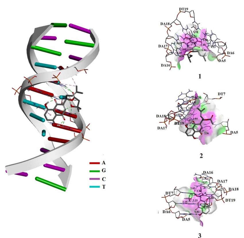
EXCLI Journal 2020;19:48-70 – ISSN 1611-2156
                        Received: October 03, 2019, accepted: December 19, 2019, published: January 03, 2020

represents the leading cause of mortality              new and more effective drugs that can target
among women worldwide (Murad et al.,                   specific site or conformation of DNA. Human
2016). Colorectal cancer, the main cause of            serum albumin (HSA) is the key protein in the
death in gastrointestinal cancers, is the third        blood plasma, the crucial soluble protein in
most common cancer worldwide in men and                the circulatory system. Likewise, HSA plays
women and the second largest cause of death            the main role in bioavailability, distribution
related to cancer. Risk factors implicated in          and elimination of several biologically active
the etiology of this type of cancer are related        moieties (drugs, toxins, natural products, etc.)
to bad alimentary habits, cigarette smoking,           from the body. Considering the importance of
low physical activity, inflammatory bowel              human serum albumin and deoxyribonucleic
disease, polyps, genetic factors, and aging            acid, studying their interaction with bioactive
(Granados-Romero et al., 2017).                        compounds may provide valuable infor-
     Cell metabolism and survival are strongly         mation on their therapeutic abilities.
dependent on the amount of different oxidants               Naphthoquinones, naturally occurring
present in cytoplasm. Reactive oxygen spe-             pigments, are secondary metabolites of many
cies (ROS), produced during mitochondrial              species belonging to Boraginaceae family, in-
respiratory chain activity, are implicated in          cluding Onosma genus. Previous chemical in-
several physiological process in all aerobic           vestigations showed that naphthoquinones
organisms (Cano et al., 2014). Oxidative               belonging to class of shikonin or alkannin de-
stress represents an imbalance between the             rivatives are concentrated in the roots of ge-
production of these aggressive radicals and            nus Onosma (Cadirci et al., 2007; Özgen et
their neutralization through compensating an-          al., 2004; Sagratini et al., 2008; Vukic et al.,
tioxidative mechanisms. This imbalance can             2018). These naphthoquinones are responsi-
lead to multiple cellular damages and devel-           ble for valuable ethnopharmacological usage
opment of many pathophysiological condi-               of these plants in treatments of various ail-
tions, including cancer (Jie et al., 2013). ROS        ments (Sezik et al., 1997; Davis, 1988). They
induce carcinogenesis through genetic and              exhibit a wide spectrum of biological proper-
epigenetic mechanisms. Elevated levels of              ties, including antitumor activity (Papa-
ROS detected in numerous tumors strongly               georgiou et al., 1999; Wang et al., 2015;
implicate the role of DNA damage caused by             Vukic et al., 2017; Kretschmer et al., 2012).
different oxidizing molecules in promoting             Currently, cytotoxic properties of the naph-
tumor development and progression (Liou                thoquinones are in the focus of interest of
and Storz, 2010).                                      many scientists, and it is already suggested
     Deoxyribonucleic acid (DNA) and human             that they are mainly based on their ability to
serum albumin (HAS) are important biom-                generate reactive oxygen species (ROS)
acromolecules. DNA is the major cellular tar-          (Papageorgiou et al., 1999; Duan et al., 2014;
get for many therapeutic agents, including             Gong and Li, 2011; Shahsavari et al., 2015).
plant secondary metabolites. Understanding                  Our previous studies showed that roots of
DNA-drug interactions has become an active             O. visianii are rich sources of naphthoqui-
research area as binding compounds have po-            nones and also provided a strong indication of
tential applications as anti-cancer agents.            the potential use of α-methylbutyrylshikon
Small molecules can bind to DNA double he-             (1), acetylshikonin (2) and β-hydroxyiso-
lix through three binding modes: electrostatic,        valerylshikonin (3) as cytotoxic agents on hu-
intercalation and groove binding (Sirajuddin           man colon cancer HCT-116 and human breast
et al., 2013). Essential to understanding of           cancer MDA-MB-231 cell lines (Vukic et al.,
these interactions, besides the characteriza-          2017). Considering that, in this study we de-
tion of the binding modes, is determination of
                                                       termined effects on redox status of α-methyl-
strength of binding. Apprehension of afore-
                                                       butyrylshikonin (1), acetylshikonin (2) and β-
mentioned parameter can lead to design of
                                                       hydroxyisovalerylshikon (3) on colon cancer

                                                  49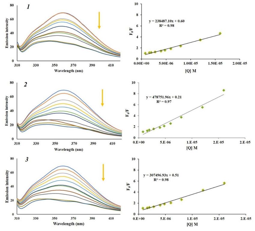
EXCLI Journal 2020;19:48-70 – ISSN 1611-2156
Received: October 03, 2019, accepted: December 19, 2019, published: January 03, 2020
cell line HCT-116 and human breast cancer Chemicals
cell line MDA-MB-231 as a potential mecha- Dulbecco’s Modified Eagle Medium
nism of proapoptotic and antiproliferative (DMEM), Fetal bovine serum (FBS), trypsin-
properties of these compounds. Additionally, EDTA and PBS were obtained from GIBCO,
in order to acquire more information on their Invitrogen, USA. Nitro blue tetrazolium
potential beneficial abilities, we examined in- (NBT) and Nicotinamide Adenine Dinucleo-
teractions of compounds 1, 2 and 3 with calf tide Phosphate (NADPH) were obtained from
thymus deoxyribonucleic acid (CT-DNA) SERVA, Germany. Sodium nitrite, phos-
and human serum albumin (HSA) by molecu- phoric acid, sulfanilamide, sulfanilic acid,
lar docking and spectroscopic methods – in- sulfosalicylic acid, and 5,5′-Dithiobis(2-nitro-
cluding fluorescence and UV-visible absorp- benzoic acid) (DTNB) were obtained from
tion. Also, we modified previously published Sigma-Aldrich, USA. Petroleum ether (boil-
procedure for isolation of α-methylbutyr- ing point ranges: 40 °C-60 °C), methylene
ylshikon (1), acetylshikonin (2) and -hy- chloride, ethyl acetate, chloroform, acetic
droxyisovalerylshikonin (3) from the roots of acid, dimethyl sulfoxide (DMSO), HPLC
O. visianii (Vukic et al., 2017). grade methanol, highly polymerized calf thy-
mus DNA (CT-DNA), human serum albumin
MATERIALS AND METHODS (HSA, lyophilized powder, free fatty acid
r0.007 %, purity r96 %, Catalogue No.
Instrumentation
A1887), phosphate buffered saline (PBS) tab-
Semi-preparative high performance liquid
lets, ethidium bromide, fetal bovine serum
chromatography was performed on Agilent
(FBS) and 7-Aminoactinomycin D (7-AAD)
1100 Series liquid chromatograph (Agilent
were purchased from Sigma-Aldrich, (Stein-
Technologies, Santa Clara, CA, USA)
heim, Germany). Water was treated in a
equipped with diode array detector (DAD;
Milli-Q water purification system (TGI Pure
520 nm, 450 nm), autosampler, and frac- Water Systems, Brea, CA, USA).
tion collector; conditions: injection volume,
600 L (2 mg/mL, methanol); column Zorbax Plant material
Eclipse XDB C18 (250 mm x 9.4 mm; 5 m); Roots of O. visianii Clem were collected
mobile phase (6 mL/min), water (40 %) and (Jun 2015) in the region of mountain Rumija
methanol (60 %). Sephadex-LH-20 purchased (southern Montenegro, altitude 650 m, 42º
from GE Helthcare (Uppsala, Sweden) was 06’ 10’’ N, 19º 11’ 37’’ E). A voucher herbar-
used for column chromatography. Preparative ium specimen was deposited at the Depart-
TLC was performed by using silica gel ment of Botany, Faculty of Biology, Univer-
P/UV254 with CaSO4 (Machery-Nagel, Ger- sity of Belgrade, Serbia (17130, BEOU).
many, 2 mm layer of adsorbent). Analytical
TLC was performed on silica gel (Silica gel Modified procedure for isolation of selected
60, layer 0.20 mm, Alugram Sil G, Mashery- naphthoquinones
Nagel, Germany). All spectroscopic measure- Petroleum ether-methylene chloride ex-
ments were performed with a double beam tract of the O. visianii roots was prepared as
UV-Vis spectrophotometer model Cary 300 previously described in our paper (Vukic et
(Agilent Technologies, Santa Clara, USA) al., 2017). Obtained extract was fractionated
with 1.0 cm quartz cells. Fluorescence meas- using preparative TLC eluted by mobile
urements were carried out using a RF-1501 phases consisting of petroleum ether:chloro-
PC spectrofluorometer (Shimadzu, Japan) form:ethyl acetate:acetic acid (5:2:2.5:0.5) to
equipped with a 150 W Xenon lamp source give eight fractions (F1-F8). Preliminary
with a 1.0 cm path length quartz cell. Color TLC analysis was conducted, and retention
reaction was measured spectrophotometri- times were compared to those of previously
cally on ELISA (2100C) 96-well microplate isolated compounds. Out of eight obtained
reader (Rayto, China).
50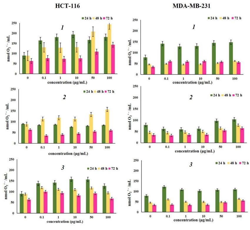
EXCLI Journal 2020;19:48-70 – ISSN 1611-2156
Received: October 03, 2019, accepted: December 19, 2019, published: January 03, 2020
fractions, presence of α-methylbutyrylshikon treated with 100 L of medium containing
(1), acetylshikonin (2) and -hydroxyiso- various doses of -methylbutyrylshikon, ace-
valerylshikonin (3) was confirmed in three tylshikonin and -hydroxyisovalerylshikonin
fractions (F4, F6 and F7) which were further with increasing concentrations (0.1 g/mL to
subjected to Sephadex LH20 column chroma- 100 g/mL) during 24 h, 48 h and 72 h, after
tography with methanol as eluent. After col- which the evaluation of levels of superoxide
umn chromatography, another TLC examina- anion radical, nitrites and glutathione was
tion was performed using petroleum performed. Non-treated cells were used as
ether:ethyl acetate (90:10) to confirm isolated control.
compounds. With the aim of obtaining high
purity, isolated α-methylbutyrylshikon (1), Determination of superoxide anion radical
acetylshikonin (2) and -hydroxyiso- (NBT assay)
valerylshikonin (3) were subjected to semi Concentrations of superoxide anion radi-
preparative HPLC on Zorbax Eclipse XDB cal (O2.-) were determined spectrophotometri-
C18 reversed phase column with isocratic elu- cally, using NBT assay based on the reduction
tion of mixture water and methanol (40:60). of nitroblue tetrazolium (NBT) to nitroblue-
Obtained spectra (UV, IR, 1H NMR and 13C formazan in the presence of O2.- (Auclair and
NMR) were in agreement with previously Voisin, 1985). The assay was performed by
published data (Vukic et al., 2017). adding 20 L of 5 mg/mL NBT to each well
and then the cells were incubated for 45 min
Estimation of redox status at 37 °C in 5 % CO2. To quantify the forma-
zan product, formazan was solubilized in
Stock solutions preparation 10 L of 2M KOH, and the resulting color re-
Tested naphthoquinones α-methylbutyr- action was measured spectrophotometrically
ylshikon (1), acetylshikonin (2) and -hy- on microplate reader at 570 nm. The amount
droxyisovalerylshikonin (3) were diluted in of reduced NBT was determined by the
DMSO at the concentration of 1 mg/mL, fil- change in absorbance at 570 nm (based on
tered through a 0.22 µm Millipore filter be- molar extinction coefficient for monoforma-
fore use. All treatment concentrations were zan 15000 M-1 cm-1) and the results were ex-
obtained by serial dilutions of stock solution. pressed as nmoL/mL.
Therefore, DMSO concentrations decreased
continuously so that the final concentration of Determination of nitrites (Griess assay)
DMSO in cell culture medium never ex- Spectrophotometric determination of ni-
ceeded 0.5 % (v/v). trites was performed using Griess method
(Griess, 1879). Nitrite standard solution (100
Cell preparation and culturing mM) was diluted from 100 M to 1.6 M (in
The human colon cancer adenocarcinoma triplicate) in a 96-well plate. Equal volumes
HCT-116 and human breast cancer MDA- of N-(1-naphthyl)ethylenediamine (1 mg/mL)
MB-231 cell lines were obtained from the and 5 % phosphoric acid solution of sulfanil-
American Tissue Culture Collection. These amide (10 mg/mL) were mixed to form the
cells were propagated and maintained in Griess reagent. Prepared cells were incubated
DMEM and supplemented with 10 % FBS for 5 - 10 min, and after that the absorbance
and antibiotics (100 IU/mL of penicillin and was measured at 550 nm by using microplate
100 g/mL of streptomycin). The cells were reader. The concentrations of nitrite were cal-
grown in 75 cm2 culture bottles and supplied culated from the standard curve for nitrite and
with 15 mL of DMEM at a confluence of expressed in nmol/mL.
70 % - 80 %. After a few passages, the cells
were seeded in a 96-well plate and cultured in
a humidified atmosphere with 5 % CO2 at
37 °C. Twenty-four hours later, the cells were
51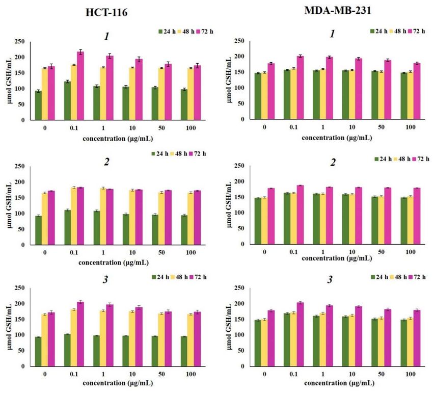
EXCLI Journal 2020;19:48-70 – ISSN 1611-2156
Received: October 03, 2019, accepted: December 19, 2019, published: January 03, 2020
Glutathione determination the absorbance at 260 nm to that at 280 nm.
Samples for measurement of concentra- The solution gave a ratio of 1.86 at A260/A280,
tions of reduced glutathione (GSH) and oxi- which indicates that DNA was free from pro-
dized glutathione (GSSG) were obtained by tein (Sirajuddin et al., 2013; Djukić et al.,
the following procedure: the cell suspension 2018). Prepared stock solutions were stored at
was centrifuged for 10 min at 1000 × g and 4 °C and used over no more than four days.
4 °C and after the removal of supernatant, the All measurements were performed at room
pellet was resuspended in 2.25 % sulfosali- temperature. Appropriate blanks were used to
cylic acid. Cell membranes were lysed by al- correct the fluorescence or UV-Vis back-
ternate freezing (- 80 °C) and thawing (37 °C) ground.
in three cycles for 15 min followed by 30 min The absorbance titration experiments
of centrifugation at 1000 × g. The supernatant were recorded at 235 - 800 nm using two sim-
was used for further analysis. The assay is ilar methods. The first consisted of maintain-
based on oxidation of a GSH by a DTNB rea- ing the constant concentrations of the -
gent, with an active thiol group, which forms methylbutyrylshikon (1), acetylshikonin (2)
a yellow product of 5′-thio-2-nitrobenzoic and -hydroxyisovalerylshikonin (3) (8.0 x
acid (Baker et al., 1990). Determination of 10-5 M), with the increasing concentration of
concentration of GSSG was based on GSH CT-DNA (from 0 to 1.73 x 10⁻⁴ M), and the
determination assay using glutathione second was based on keeping the constant
reductase after inhibition of spontaneous concentration of CT-DNA (1.66 x 10⁻⁵ M) and
GSH oxidation by 4-vinylpyridine (Baker et varying the concentrations of the -methyl-
al., 1990). Glutathione concentration was butyrylshikon (1), acetylshikonin (2) and -
expressed as mol/mL. hydroxyisovalerylshikonin (3) (from 0 to 2.4
Color reaction was measured spectropho- x 10-5 M). In addition, spectra containing only
tometrically on a microplate reader at 405 nm CT-DNA, -methylbutyrylshikon (1), acetyl-
following 10 min incubation. Concentrations shikonin (2) and -hydroxyisovalerylshi-
of reduced and oxidized glutathione were cal- konin (3) at the same concentration ratio used
culated from a standard curve constructed to evaluate interaction mode were recorded.
with determined concentrations. The fluorescence quenching spectra of ti-
DNA binding study tration EB-DNA with selected naphthoqui-
Characterization of the binding modes nones solutions were recorded in the range of
and determination of strength of binding for 550 - 700 nm with an excitation wavelength
selected naphthoquinone to CT-DNA were at ex = 520 nm. Fluorescence spectra were
estimated by fluorescence and UV-Vis ab- recorded by keeping the constant concentra-
sorption study. The 2.0 x 10⁻³ M stock solu- tions of DNA (1.72 x 10⁻⁵ M) and EB (1.2 x
tions of α-methylbutyrylshikon (1), acetylshi- 10-5 M), with the increasing concentration of
konin (2) and -hydroxyisovalerylshikonin α-methylbutyrylshikon (1), acetylshikonin (2)
(3) were prepared by dissolving each naph- and -hydroxyisovalerylshikonin (3) (from 0
thoquinone in small amounts of DMSO and to 2.4 x 10⁻⁵ M). Also, the fluorescence
diluting with phosphate buffer (pH 7.4) so the quenching spectra of titration Heochst
final DMSO concentration did not exceed 33342–DNA with naphthoquinone solutions
0.5 % v/v. The stock solutions of CT-DNA were recorded in the range of 380 - 600 nm
(2.0 x 10⁻³ M), EB (1.0 x 10⁻³ M) and Hoechst with an excitation wavelength at ex = 350
33342 (1.0 x 10⁻³ M) were prepared by dis- nm. The fluorescence spectra of naphthoqui-
solving with phosphate buffer solution. Con- nones were recorded under the same experi-
centration of CT-DNA was determined spec- mental conditions and no fluorescence emis-
trophotometrically at 260 nm using the molar sion was verified.
absorptivity (ε260 = 6600 L/mol cm). DNA pu-
rity was checked by monitoring the ratio of
52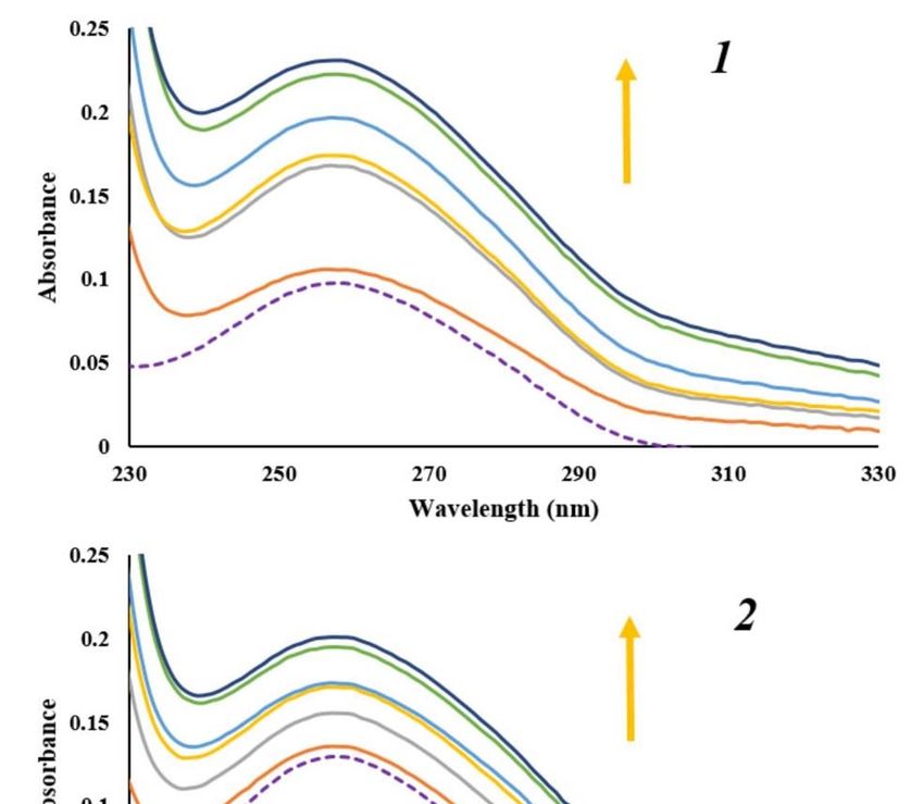
EXCLI Journal 2020;19:48-70 – ISSN 1611-2156
Received: October 03, 2019, accepted: December 19, 2019, published: January 03, 2020
Protein binding study Data Bank (Wenskowsky et al., 2018;
Binding modes and determination of Vandevenne et al., 2013). Preparation of HSA
strength of binding for selected naphthoqui- and DNA for docking simulations was done
nones to HSA were estimated by fluores- by removing the crystallized ligand, water
cence, UV-Vis absorption and molecular molecules, and cofactors in the Discovery
docking study. The stock solutions of α- Studio 4.0 (BIOVIA Discovery Studio 2016)
methylbutyrylshikon (1), acetylshikonin (2) (BIOVIA, Dassault Systèmes, 2017). The cal-
and -hydroxyisovalerylshikonin (3) were culations of Kollman charges and adding of
prepared in the same way as for the DNA the polar hydrogen were performed using
binding study. The stock solution of HSA (2.0 graphical user interface AutoDockTools
x 10-5 M) was prepared by dissolving in phos- (ADT). The optimization of the investigated
phate buffer solution (pH 7.4). Prepared stock molecules was performed by B3LYP-
solutions were stored at 4 °C and used over no D3BJ/6-311+G(d,p) level of theory using the
more than four days. All measurements were Gaussian09 software package (Frisch et al.,
performed at room temperature. Proper 2013). The HSA and DNA retained the rigid
blanks were used to correct the fluorescence structure during molecular docking simula-
or UV-Vis background. Absorbance titration tions in the ADT, while the investigated mol-
experiments were recorded at 235–800 nm ecules were flexible. For calculations of par-
with the constant concentration of the HAS tial charges, the Geistenger method was se-
(2.0 x 10⁻⁶ M), while the concentrations of α- lected. Lamarckian Genetic Algorithm (LGA)
methylbutyrylshikon (1), acetylshikonin (2) was used in all calculations. A docking box
and -hydroxyisovalerylshikonin (3) varied with a grid consisting of 60 x 60 x 60 points
from 0 to 1.6 x 10⁻⁵ M. Additionally, the ab- with 0.375 Å spacing was placed into the ac-
sorbances at 235 - 800 nm for solutions con- tive side of the receptor. All binding sites of
taining only selected naphthoquinones in the the HSA and DNA were thus covered, which
concentration ratio used for titration experi- enabled free movement of examined com-
ments were recorded. pounds.
The emission spectra were recorded be-
tween 300 and 460 nm upon excitation at ex Statistical analysis
= 295 nm. The naphthoquinone-HSA com- The data are expressed as mean values ±
plexes were prepared by incubating constant standard errors (SE). All experiments were
amount of HAS (2.0 x 10⁻⁶ M) with increasing performed in triplicate for each dose. One-
amounts of the α-methylbutyrylshikon (1), way analysis of variance (ANOVA) was per-
acetylshikonin (2) and -hydroxyisovaleryl- formed to determine significant differences
shikonin (3) (from 0 to 1.6 x 10⁻⁵ M). The flu- between means using SPSS for Windows,
orescence spectra of naphthoquinones were Version 17 (SPSS Inc., Chicago, IL, USA).
recorded under the same experimental condi-
tions and no fluorescence emission was de- RESULTS AND DISCUSSION
tected. The effects of selected naphthoquinones
Docking analysis on redox status in HCT-116 and MDA-MB-
Inhibitory activity of investigated naph- 231 cells
thoquinones against human serum albumin In our previous work we showed that -
(HSA) and deoxyribonucleic acid (DNA) was methylbutyrylshikonin (1), acetylshikonin (2)
tested using the AutoDock 4.2 software pack- and -hydroxyisovalerylshikonin (3) induce
age (Morris et al., 2009). Three-dimensional apoptosis in HCT-116 and MDA-MB-231
crystal structure of HSA (6EZQ) and DNA cells. Hence, in this paper we investigated the
(dodecamer d(CGCAAATTTGCG)2 [PDB: effects of naphthoquinones 1, 2 and 3 (Figure
102D]) was obtained from the RCSB Protein 1) on oxidative stress markers as a potential
53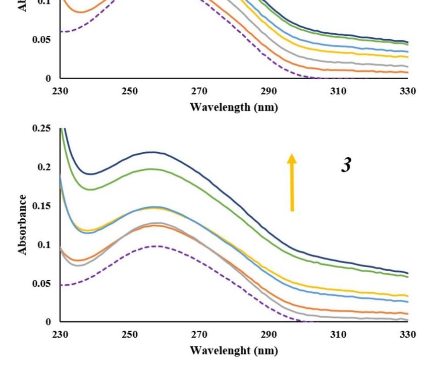
EXCLI Journal 2020;19:48-70 – ISSN 1611-2156
Received: October 03, 2019, accepted: December 19, 2019, published: January 03, 2020
mechanism of proapoptotic and antiprolifera- However, in MDA-MB-231 cell line naph-
tive properties (Vukic et al., 2017). thoquinone 2 increased the production of su-
peroxide anion after all three-time treatments.
In general, it can be concluded that the tested
naphthoquinones and 2show prooxidant
activity in HCT-116 and MDA-MB-231 cell
lines. Observed increased production of su-
peroxide anion radical after the treatment may
also be involved in expression of anti-prolif-
erative effects of these compounds and their
Figure 1: Chemical structures of -methylbutyr-
ability to induce apoptosis. From the structure
ylshikonin (1), acetylshikonin (2) and -hydroxy- point of view, quinone mechanisms of cyto-
isovalerylshikon (3) toxicity are closely related to their ability to
undergo one-electron reduction reaction cata-
lyzed by enzymes such as NADPH-cyto-
Reactive oxygen species (ROS) play key chrome P-450 reductase or mitochondrial
roles in many cell processes essential for NADH-ubiquinone oxidoreductase. This re-
maintaining cell homeostasis, and superoxide action forms semiquinone radical which can
anion radical is one of their key elements (Liu autoxidize in the presence of O2, generating
et al., 2017). Accordingly, in this study we quinone and superoxide anion radical (Papa-
have evaluated effects of various concentra- georgiou et al., 1999; Yang et al., 2014;
tions of naphthoquinones 1, 2 and 3 on super- Murakami et al., 2010).
oxide anion radical production by HCT-116 Nitric oxide (NO) is an important signal-
and MDA-MB-231 cells after 24 h, 48 h, and ing molecule in numerous physiological and
72 h treatment. The results presented in Fig- pathological processes, with a controversial
ure 2 showed that treatment with all com- role in genesis and progression of various
pounds at different concentrations increased cancer types (Liu et al., 2003). The data in
O2·- level in HCT-116 and MDA-MB-231 Figure 3 present nitrite concentrations in
cells compared to its production in non- HCT-116 and MDA-MB-231 cells incubated
treated cells. All investigated compounds with various concentrations of tested naph-
showed prooxidative properties during all thoquinones after 24 h, 48 h and 72 h. Treat-
time treatments, and the strongest effects ment with all compounds showed a signifi-
were detected after 24 h treatment. Overall, cant increase in production of NO by HCT-
compound 1 showed the strongest effect on 116 and MDA-MB-231 cells measured indi-
superoxide anion radical production in HCT- rectly through nitrite level compared to the
116 and MDA-MB-231 cells compared to level in non-treated cells. The strongest activ-
control after 24 h, 48 h and 72 h of treatment. ity was shown by -hydroxy-isovalerylshi-
Also, it can be observed that HCT-116 cells konin (3) after all treatments, and MDA-MB-
were more sensitive to treatment with the 231 cells were more sensitive to treatment
tested naphthoquinones compared to MDA- with the tested naphthoquinone derivatives. In
MB-231 cells. Acetylshikonin (2) increased general, for all investigated naphthoquinones,
production of O2·- in HCT-116 cells only after the highest production of NO for both cell
48 h of treatment, while after 24 h and 72 h of lines was observed after 48 h of treatment.
treatment the inhibition in production of su- The lowest level in production of NO was ob-
peroxide anion radical was observed, possibly served for -methylbutyrylshikonin (1) in
due to excessive production of nitric oxide both cell lines, which can be explained by
which reacted with O2·- , leading to formation generation of peroxynitrite due to elevated su-
of highly reactive peroxynitrite (ONOO-). peroxide anion production. Jaw-Jou Kang and
54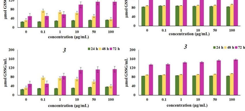
EXCLI Journal 2020;19:48-70 – ISSN 1611-2156
Received: October 03, 2019, accepted: December 19, 2019, published: January 03, 2020
associates point out that superoxide anion re- decreased activity of the NO-production path-
duces bioavailability of NO, which results in way or increased oxidative inactivation of NO
(Kang et al., 2006).
Figure 2: Effects of investigated naphthoquinones on HCT-116 and MDA-MB-231 cell lines, expressed
as the nmol O2 ̇ -/mL after 24 h, 48 h and 72 h of treatment. The cells were treated with α-methylbutyr-
ylshikonin (1), acetylshikonin (2) and -hydroxy-isovalerylshikonin (3) in concentration range from 0.1
to 100 g/mL. Results were expressed as the means ± SE from three independent determinations.
55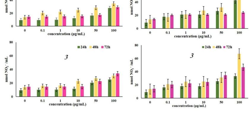
EXCLI Journal 2020;19:48-70 – ISSN 1611-2156
Received: October 03, 2019, accepted: December 19, 2019, published: January 03, 2020
Figure 3: Effects of investigated naphthoquinones on HCT-116 and MDA-MB-231 cell lines, expressed
as the nmol NO2- /mL after 24 h, 48 h and 72 h of treatment. The cells were treated with α-methylbutyr-
ylshikonin (1), acetylshikonin (2) and -hydroxy-isovalerylshikonin (3) in concentration range from 0.1
to 100 g/mL. Results were expressed as the means ± SE from three independent determinations.
Concentration of reduced and oxidized in detoxication of different metabolites, regu-
glutathione after 24 h, 48 h, and 72 h of incu- lates activity of some enzymes and cellular
bation with various concentrations of naph- macromolecules) (Chung et al., 2016). Also,
thoquinones 1, 2 and 3, presented in Figure 4 being a potent electron donor, GSH is one of
and Figure 5 showed increase in the levels of the major nonenzymatic antioxidant compo-
both reduced and oxidized glutathione com- nents (Jones, 2002). Increased levels of oxi-
pared to control. Glutathione -GSH and its ox- dized glutathione were noted in cells treated
idized form -GSSG, represent important with all tested naphthoquinones, after three
markers of the cell redox status (Traverso et experimental times (24 h, 48 h, and 72 h), sug-
al., 2013). As one of the strongest antioxida- gesting a strong antioxidative activity pro-
tive components present in cells, reduced glu- voked by considerable oxidative burst by the
tathione (GSH) exerts various roles (e.g., tested compounds. The strongest elevation of
maintains intracellular thiol status, involved both glutathione forms correlates to the high
56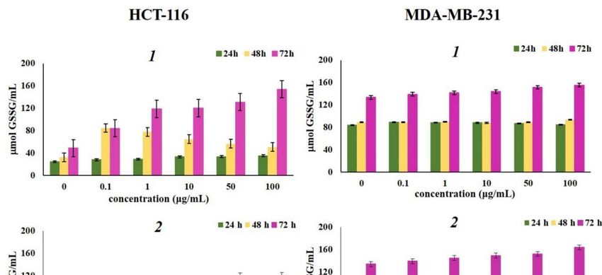
EXCLI Journal 2020;19:48-70 – ISSN 1611-2156
Received: October 03, 2019, accepted: December 19, 2019, published: January 03, 2020
concentration of superoxide anion radical. El- al., 2007). GSH is synthesized in cells de novo
evated levels of reduced glutathione detected and rapid induction of intracellular GSH syn-
in this study may indicate that 1, 2 and 3 in- thesis occurs in response to various stressors
duced de novo synthesis of GSH (Shizhong et (Shi et al., 1994).
Figure 4: Effects of investigated naphthoquinones on the concentration of reduced glutathione (GSH)
after 24 h, 48 h and 72 h of treatment. The HCT-116 and MDA-MB-231 cells were treated with α-methyl-
butyrylshikonin (1), acetylshikonin (2) and -hydroxyisovalerylshikonin (3) in concentration range from
0.1 to 100 g/mL. Results were expressed as the means ± SE from three independent determinations.
57EXCLI Journal 2020;19:48-70 – ISSN 1611-2156
Received: October 03, 2019, accepted: December 19, 2019, published: January 03, 2020
Figure 5: Effects of investigated naphthoquinones on the concentration of oxidized glutathione form
(GSSG) after 24 h, 48 h and 72 h of treatment. The HCT-116 and MDA-MB-231 cells were treated with
α-methylbutyrylshikonin (1), acetylshikonin (2) and -hydroxyisovalerylshikonin (3) in concentration
range from 0.1 to 100 g/mL. Results were expressed as the means ± SE from three independent
determinations.
DNA binding study induce production of NO2-. Hence, our next
It is well known that ROS overproduction step was to investigate the possibility that ob-
causes DNA damage through oxidation and served result could be the consequence of in-
DNA strand breaks. In addition, production of teraction of 1, 2 and 3 with calf thymus DNA
nitric oxide induces modifications of DNA by (CT-DNA).
directly altering DNA through creating reac-
tive nitrogen oxide species (RNOS) or indi- UV-Vis measurements
rectly by inhibiting various repair processes Observing the spectral changes of DNA in
(Colin et al., 2014; Graziewicz et al., 1996). the titration reaction with small molecules can
We have already pointed out that naphthoqui- provide evidence of binding mode of small
nones α-methylbutyrylshikon (1), acetylshi- molecule to CT-DNA. Effects of increasing
konin (2) and -hydroxyisovalerylshikonin amounts of CT-DNA on absorption spectra of
(3) have prooxidant effect in cancer cells and 1, 2 and 3 are given in Supplementary Figure
58EXCLI Journal 2020;19:48-70 – ISSN 1611-2156
Received: October 03, 2019, accepted: December 19, 2019, published: January 03, 2020
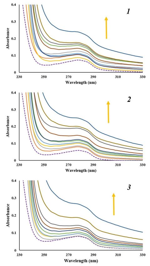
1. It is known that CT-DNA shows absorption DNA, it gives a significant increase in fluo-
maximum at wavelength of 260 nm due to pu- rescence emission due to its strong intercala-
rine and pyrimidine bases (Sirajuddin et al., tion between the adjacent DNA base pairs.
2013). Since there was no DNA absorption Observed fluorescence emission can be
above mentioned maximum, absorption max- quenched by the addition of a second mole-
imum of 1, 2 and 3 at wavelength range of cule capable to replace EB or by electron
460 - 600 nm followed. With increasing con- transfer (Sirajuddin et al., 2013; Cox et al.,
centration of CT-DNA, minor hyperchrom- 2009). The emission spectra of DNA-EB
ism with no obvious red or blue shift was ob- complex obtained in the absence and presence
served. Presented results revealed that inter- of increasing concentration of α-methylbutyr-
action between naphthoquinones and CT- ylshikon (1), acetylshikonin (2) and -hy-
DNA occurred. Intercalation binding mode of droxyisovalerylshikonin (3) were shown in
small molecules into the DNA helix results in Figure 7. Presented results show no signifi-
hypochromism and red shift. Hence, these re- cant decrease in the maximum of emission in-
sults indicate that naphthoquinones 1, 2 and 3 tensity of EB-DNA complex at excitation
do not bind to DNA through intercalation wavelength of 520 nm with the increasing
binding mode (Qiao et al., 2008). However, concentration of the naphthoquinones, which
spectral changes observed in Supplementary confirms that tested naphthoquinones do not
Figure 1 for tested naphthoquinones are a fea- intercalate into the DNA double helix. A
ture that represents non-covalent interactions, slight decrease can be result of forming new
particularly electrostatic and groove binding non-fluorescence EB-DNA-naphthoquinone
between small molecules and CT-DNA complex (Qiao et al., 2008; Bi et al., 2006).
(Sarwar et al., 2015).
In order to confirm interaction mode of Hoechst 33342 displacement assay
naphthoquinones 1, 2 and 3 to CT-DNA, ab- Unlike EB, Hoechst 33342 dye is a well-
sorption spectra of constant concentration of known minor groove DNA binder, and, like
CT-DNA were recorded, in the absence and other minor groove binders, it recognizes A-
presence of increasing amounts of naphtho- T rich sequences in DNA double helix (Wu et
quinones. As shown in Figure 6 with increas- al., 2007; Shi et al., 2015). Fluorescence emis-
ing concentration of 1, 2 and 3 concomitant sion of Hoechst 33342 bound to DNA is en-
hyperchromicity at the CT-DNA absorption hanced compared to free solution of Hoechst
maximum of 260 nm was observed. Observed 33342. As well as for EB, addition of a small
hyperchromic effect is a result of groove molecule can result in fluorescence quench-
binding interaction between tested naphtho- ing of Hoechst-DNA complex. In Figure 8,
quinones and CT-DNA. Also, absence of red emission spectra of Hoechst-DNA complex
or blue shift indicates that the structure of CT- obtained in the absence and presence of in-
DNA did not change much upon binding of creasing concentration of α-methylbutyr-
naphthoquinones. ylshikon (1), acetylshikonin (2) and -hy-
droxyisovalerylshikonin (3) are presented. In-
Fluorescence measurements tensity of the emission band of Hoechst-DNA
In order to further investigate binding at 480 nm at excitation wavelength of 350 nm
mode of naphthoquinones 1, 2 and 3, we em- decreased with increasing concentration of
ployed fluorescence quenching study involv- the tested naphthoquinones. This dye dis-
ing dye displacement. placement experiment, alongside with results
obtained by UV-Vis absorption spectroscopy,
Ethidium bromide displacement assay suggests that naphthoquinones 1, 2 and 3 are
Ethidium bromide (EB) shows weak fluo- minor groove binders of DNA.
rescence in the solvent, but when bonded to
59EXCLI Journal 2020;19:48-70 – ISSN 1611-2156
Received: October 03, 2019, accepted: December 19, 2019, published: January 03, 2020
Figure 6: Absorption spectra of CT-DNA (1.77 x 10⁻⁵ M) before (purple dashed line) and after addition
of α-methylbutyrylshikon (1), acetylshikonin (2) and β-hydroxyisovalerylshikonin (3) (0 - 1.80 x 10⁻⁵ M).
Arrow shows the absorbance changes upon increasing concentration of α-methylbutyrylshikon (1), ac-
etylshikonin (2) and β-hydroxyisovalerylshikonin (3).
60EXCLI Journal 2020;19:48-70 – ISSN 1611-2156
Received: October 03, 2019, accepted: December 19, 2019, published: January 03, 2020
Figure 7: The fluorescence emission spectra of DNA-EB fixed concentration (DNA (1.72 x 10⁻⁵ M) and
EB (1.2 x 10⁻⁵ M)), in the absence and presence of increasing concentration of α-methylbutyrylshikon
(1), acetylshikonin (2) and β-hydroxyisovalerylshikonin (3) (from 0 to 2.4 x 10⁻⁵ M). Arrow shows the
intensity change upon the increase of the naphthoquinone concentration. Purple dashed line represents
the emission spectra of 1, 2 and 3 in the absence of DNA-EB.
61EXCLI Journal 2020;19:48-70 – ISSN 1611-2156
Received: October 03, 2019, accepted: December 19, 2019, published: January 03, 2020
Figure 8: The fluorescence emission spectra of Hoechst-DNA fixed concentration (DNA (1.66 x 10⁻⁵ M)
and Hoechst (1.2 x 10⁻⁵ M)), in the absence and presence of increasing concentration of α-methylbutyr-
ylshikon (1), acetylshikonin (2) and β-hydroxyisovalerylshikonin (3) (from 0 to 2.4 x 10⁻⁵ M). Arrow shows
the intensity change upon the increase of the naphthoquinone concentration. Purple dashed line repre-
sents the emission spectra of 1, 2 and 3 in the absence of DNA-Hoechst. Right: corresponding plots of
F0/F versus [Q].
The fluorescence quenching data of value is considered to be 10-8 s, KSV is the
DNA-Hoechst were analyzed by the Stern– Stern–Volmer quenching constant, and [Q] is
Volmer constant obtained by Eq (1): the concentration of free quencher (Lakowicz,
2006). The quenching constants (KSV) were
1 τ Q 1 Q 1
obtained from slope and their values were
3.96 x 10⁴, 4.35 x 10⁴ and 4.19 x 10⁴ dm3 mol-1
where F0 and F are the fluorescence inten- for naphthoquinones 1, 2 and 3, respectively.
sities before and after addition of quencher, Out of the calculated constants we can con-
respectively. Kq is the biomolecular quench- clude that all tested naphthoquinones have
ing constant, 0 is the life time of the fluoro- similar ability to insert between DNA base
phore in the absence of quencher and and its pairs.
62EXCLI Journal 2020;19:48-70 – ISSN 1611-2156
Received: October 03, 2019, accepted: December 19, 2019, published: January 03, 2020
Protein binding study tions between them. Due to the Trp-214 resi-
Exploring protein - drug interactions is due, HSA fluorescence emission spectra at
closely related to drug characteristics such as excitation wavelength of 295 nm showed a
distribution, transportation and metabolism in peak at around 360 nm. Hence, in the wave-
the treatment of many diseases. Therefore, ab- length range 300 - 450 nm, fluorescence emis-
sorption and fluorescence quenching experi- sion spectra of HSA with increasing concen-
ments were employed to explore the effect of tration of naphthoquinones 1, 2 and 3 (from 0
binding of the naphthoquinones 1, 2 and 3 to 1.6 x 10⁻⁵ M) were recorded. From obtained
with the carrier protein HSA. spectra (Figure 10), decrease in the fluores-
cence intensity of the HAS was observed, fol-
UV-Vis measurements lowed by a slight shift to the shorter wave-
Titration experiment of HAS with in- length, with increasing concentration of naph-
creasing concentration of naphthoquinones 1, thoquinones, indicating the changes in the lo-
2 and 3 (from 0 to 1.6 x 10⁻⁵ M) monitored by cal microenvironment around the Trp-214
UV-Vis spectroscopy and corresponding ab- residue in HSA by formation of non-fluores-
sorption spectra are reported in Figure 9. Ab- cent HSA-naphthoquinone complexes. Addi-
sorbance spectra of HAS resulted in a weak tionally, obtained results were in agreement
peak around 280 nm as a result of the cumu- with previously published data for naphtho-
lative π‐π* transition of Trp, Tyr, and Phe res- quinone shikonin (He et al., 2005).
idues (Cao et al., 2019; Yan et al., 2018). The fluorescence quenching data were an-
Binding of naphthoquinones to HSA resulted alyzed by the Eq (1), similarly as described
in a continuous increase in absorbance maxi- above for CT-DNA binding studies and ob-
mum at 280 nm, indicating the changes in the tained Stern–Volmer quenching constants KSV
local environment in HSA. and the quenching rate constants Kq, are given
in Table 1. From the slope of the regression
Fluorescence measurements line in the derived plot of F0/F versus [Q] (in-
In order to confirm results obtained by sets of Figure 10), the Ksv values for the com-
UV-Vis absorption spectroscopy, we moni- plexes were determined to be in order 105
tored interactions of HSA with selected naph- dm3mol-1, indicating a strong affinity of tested
thoquinones by employing fluorescence spec- naphthoquinones to HSA. As shown in Table
troscopy. Decrease or shift in emission maxi- 1 bimolecular quenching constants were in
mum of protein in the presence of increasing the order of 1013 dm3moL-1s-1, indicating that
concentration of a small molecule could indi- formation of HSA-naphthoquinone complex
cate new complex formation, random colli- is a static process (Makarska-Bialokoz and
sions, energy transfer and excited state reac- Lipke, 2019; Panigrahi et al., 2015).
Table 1: Quenching constant (KSV), the quenching rate constants (Kq), binding constant (Kb), and num-
ber of binding sites (n) for naphthoquinone-HAS interactions
Compound Ksv Kq Kb n
(dm3mol-1) (dm3mol-1s-1) (dm3mol-1)
α-methylbutyrylshikon (1) 2.38x10⁵ 2.38x10¹³ 2.42x10⁷ 1.43
Acetylshikonin (2) 4.79x10⁵ 4.79x10¹³ 4.17x10⁷ 1.41
β-hydoxyisovalerylshikonin (3) 3.07x10⁵ 3.07x10¹³ 3.63x10⁷ 1.44
63EXCLI Journal 2020;19:48-70 – ISSN 1611-2156
Received: October 03, 2019, accepted: December 19, 2019, published: January 03, 2020
Figure 9: Absorption spectra of HSA (2.00 x 10⁻⁶ M) before (purple dashed line) and after addition of α-
methylbutyrylshikon (1), acetylshikonin (2) and β-hydroxyisovalerylshikonin (3) (0 - 1.60 x 10⁻⁵ M). Arrow
shows the absorbance changes upon increasing concentration of α-methylbutyrylshikon (1), acetylshi-
konin (2) and β-hydroxyisovalerylshikonin (3).
64EXCLI Journal 2020;19:48-70 – ISSN 1611-2156
Received: October 03, 2019, accepted: December 19, 2019, published: January 03, 2020
Figure 10: The fluorescence emission spectra of HSA fixed concentration (2.00 x 10⁻⁶ M), in the ab-
sence (blue line) and presence of increasing concentration of α-methylbutyrylshikon (1), acetylshikonin
(2) and β-hydroxyisovalerylshikonin (3) (from 0 to 1.6 x 10⁻⁵ M) Insets: plots of F0/F versus [naphthoqui-
none]
Binding constant and number of binding where F0 and F are the fluorescence inten-
sites sities in the absence and presence of the
The number of binding sites (n) and the quencher, respectively, Kb is binding constant
binding constant (Kb) presented in Table 1 or the apparent association constant for small
were obtained from the intercept and slope of molecule-protein interaction, n is the number
the plots of log(F0 - F)/F versus log[Q] de- of binding sites per protein and [Q] is the con-
rived from the Eq (2): centration of quencher (Lakowicz, 2006).
From the number of binding sites calculated
to be approximately 1, formation of a ground
Q 2
state complex with a single binding site in
HSA for all tested naphthoquinones was con-
firmed. The values of the binding constants
65EXCLI Journal 2020;19:48-70 – ISSN 1611-2156
Received: October 03, 2019, accepted: December 19, 2019, published: January 03, 2020
(Kb) showed that acetylshikonin bound more binding sites. Based on the docking results,
strongly than other investigated naphthoqui- the most stable conformers with the lowest
nones. free energy of binding were obtained. Ob-
tained values of the pairwise interaction ener-
Molecular docking analysis of naphthoqui- gies (Ei), constant of inhibition (Ki), free en-
nones with DNA and HSA ergy of binding (ΔGbind) as well as the non-
Molecular docking studies were per- covalent interactions for the investigated
formed for the evaluation of the inhibitory na- models are presented in Supplementary Ta-
ture of investigated naphthoquinones 1, 2, and bles 1 and 2. The most stable conformation
3 against DNA and HSA. In this study, the complexes of naphthoquinones (1, 2, and 3)
binding energy of complexes of the DNA- with DNA and HSA were presented in Fig-
naphthoquinones and of the HSA-naphtho- ures 11 and 12 respectively.
quinones was determined, as well as the pre-
diction of potential investigated compounds-
Figure 11: Interactions between α-methylbutyrylshikon (1), acetylshikonin (2) and β-hydroxyiso-
valerylshikonin (3) and DNA. The conventional hydrogen bonds are labeled using green dashed lines
and hydrophobic interactions are labeled with purple dashed lines.
66EXCLI Journal 2020;19:48-70 – ISSN 1611-2156
Received: October 03, 2019, accepted: December 19, 2019, published: January 03, 2020
Figure 12: Interactions between α-methylbutyrylshikon (1), acetylshikonin (2) and β-hydroxyiso-
valerylshikonin (3) and amino acids of HSA. The conventional hydrogen bonds are labeled using green
dashed lines.
The obtained results presented in Figure showed longer distances (Supplementary Ta-
11 indicate that naphthoquinones bind in AT- ble 2). These interactions were defined with
rich region of DNA via conventional hydro- high pairwise interaction energy and large
gen-bonding and π-alkyl as significant non- atomic distances (≥3 Å). The relative free en-
covalent interactions (Supplementary Table ergy of binding for compounds 1, 2, and 3
1). Obtained values of the pairwise interaction with HSA were -13.17, -29.08, and -7.74 kJ
energies (Ei), constant of inhibition (Ki), free mol-1, respectively. These results confirm the
energy of binding (ΔGbind) as well as the non- experimental ones, that naphthoquinone 2
covalent interactions for the investigated forms the most stable complex with corre-
models are presented in Supplementary Table sponding protein (Supplementary Table 2).
1. The resulting relative binding energies of 1,
2, and 3 with DNA were -22.99, -20.98, and -
CONCLUSION
20.31 kJ mol-1, respectively, which is in ac-
cordance with results obtained by employing In this study we reported that naturally oc-
UV-Vis absorption and fluorescence spec- curring naphthoquinones α-methylbutyrylshi-
troscopy study and indicates similar DNA- kon (1), acetylshikonin (2) and β-hydroxyiso-
binding affinity of tested naphthoquinones. valerylshikonin (3) isolated from O. visianii
The investigated naphthoquinone deriva- induced disturbance in oxidative homeostasis
tives contain several polar groups: one ester, of colon and breast cancer cells and increased
two carbonyl and two or three hydroxyl the levels of superoxide anion radical and ni-
groups. A large number of polar groups in the trites. All examined compounds significantly
investigated compounds increases the number increased the concentration of oxidized gluta-
of possible interactions with the amino acids thione (GSSG), suggesting an activation of
of the HSA protein. The obtained results are cell antioxidant mechanisms. Also, all exam-
shown in Supplementary Table 2 and indicate ined compounds significantly increased the
that the conventional hydrogen bond is the concentration of reduced glutathione (GSH),
most important type of interaction. In addi- indicating de novo synthesis and further con-
tion, significant non-covalent interactions in firming excessive prooxidative conditions.
naphthoquinones-HSA complexes are alkyl- Naphthoquinone 1 exerted the strongest
π, σ-π, π-π, π-cation as well as π-π stacked (in prooxidative effects in both cell lines, while
complexes 2 and 3). All atomic distances of compound 2 showed the highest increase in
the conventional hydrogen bonds were in the nitrite production compared to non-tretaed
range of 1.5 - 3.0 Å, while other interactions cells. The levels of oxidized glutathione were
highest in the treatment with compound 1,
67EXCLI Journal 2020;19:48-70 – ISSN 1611-2156
Received: October 03, 2019, accepted: December 19, 2019, published: January 03, 2020
which corresponded to the highest concen- Bi S, Qiao C, Song D, Tian Y, Gao D, Sun Y, et al.
tration of superoxide anion radical among Study of interactions of flavonoids with DNA using
acridine orange as a fluorescence probe. Sens Actuator
these three derivatives. The results obtained B-Chem. 2006;119:199–208.
in this experiment indicate a significant
prooxidative role of the examined compounds BIOVIA, Dassault Systèmes. Discovery Studio
in HCT-116 and MDA-MB-231 cells, which Modeling Environment, Release 2017, San Diego, CA:
Dassault Systèmes, 2017.
could be one of the potential mechanisms of
growth inhibition and apoptosis induction in Cadirci E, Suleyman H, Aksoy H, Halici Z, Ozgen U,
these cell types. Likewise, as CT-DNA is a Koc A, et al. Effects of Onosma armeniacum root
possible biomolecule target for antitumor ac- extract on ethanol-induced oxidative stress in stomach
tivity, naphthoquinones-CT-DNA tissue of rats. Chem Biol Interact. 2007;170:40–8.
interactions study revealed favored binding of Cano I, Selivanov V, Gomez-Cabrero D, Tegnér J,
the all three compounds at the A-T region in Roca J, Wagner PD, et al. Oxygen pathway modeling
minor groove. As minor groove binders are estimates high reactive oxygen species production
known to be an important class of derivatives above the highest permanent human habitation. PloS
One. 2014;9:e111068.
in anticancer therapy, naturally occurring
naphthoquinones 1, 2 and 3 may be further Cao X, Yang Z, He Y, Xia Y, He Y, Liu J. Multi-
exploited to understand their potential as an- spectroscopic exploration and molecular docking
tiproliferative drugs. In addition, the analysis on interaction of eriocitrin with bovine serum
albumin. J Mol Recognit. 2019;32:e2779.
intersection study with HSA showed that the
tested compounds could be transported and Chung BY, Choi SR, Moon IJ, Park CW, Kim YH,
distributed through the cells. According to the Chang SE. The glutathione derivative, GSH monoethyl
data presented in this paper, tested ester, may effectively whiten skin but GSH does not.
naphthoquinones could have a significant Int J Mol Sci. 2016;17 629.
beneficial role in developing new strategies Colin DJ, Limagne E, Ragot K, Lizard G, Ghiringhelli
against colon and breast cancer. F, Solary É, et al. The role of reactive oxygen species
and subsequent DNA-damage response in the
Conflict of interest emergence of resistance towards resveratrol in colon
cancer models. Cell Death Dis. 2014;5:e1533.
The authors confirm that this article
content has no conflict of interest. Cox PJ, Psomas G, Bolos CA. Characterization and
DNA-interaction studies of 1,1-dicyano-2,2-ethylene
Acknowledgments dithiolate Ni(II) mixed-ligand complexes with 2-
amino-5-methyl thiazole,2-amino-2-thiazoline and
This work was financially supported by
imidazole. Crystal structure of [Ni(i-MNT)(2a-5mt)2].
the Ministry of Education, Science and Tech- Bioorg Med Chem Lett. 2009;17:6054–62.
nological Development of the Republic of
Serbia (Grant OI172016 and Grant III41010). Davis PH. Flora of Turkey and the East Aegean
Islands. Vol 6. Edinburgh: Edinburgh Univ. Press,
1988.
REFERENCES
Auclair C, Voisin E. Nitroblue tetrazolium reduction. Djukić M, Jeremić MS, Jelić R, Klisurić O, Kojić V,
In: Greenwald RA (ed): Handbook of methods for Jakimov D, et al. Further insights into ruthenium(II)
oxygen radical research (pp 123-32). Boca Raton, FL: piano-stool complexes with N-alkyl imidazoles. Inorg
CRC Press, 1985. Chim Acta. 2018;483:359–70.
Baker MA, Cerniglia GJ, Zaman A. Microtiter plate Duan D, Zhang B, Yao J, Liu Y, Fang J. Shikonin
assay for the measurement of glutathioneand targets cytosolic thioredoxin reductase to induce ROS-
glutathione disulfide in large numbers of biological mediated apoptosis in human promyelocytic leukemia
samples. Anal Biochem. 1990;190:360-5. HL-60 cells. Free Radic Biol Med. 2014;70:182-93.
Frisch MJ, Trucks GW, Schlegel HB, Scuseria GE,
Robb MA, Cheeseman JR, et al. Fox, Gaussian 09.
Revision C.01. Inc, Wallingford, CT, 2013.
68EXCLI Journal 2020;19:48-70 – ISSN 1611-2156
Received: October 03, 2019, accepted: December 19, 2019, published: January 03, 2020
Gong K, Li W. Shikonin, a Chinese plant-derived Makarska-Bialokoz M, Lipke A. Study of the binding
naphthoquinone, induces apoptosis in hepatocellular interactions between uric acid and bovine serum
carcinoma cells through reactive oxygen species: A albumin using multiple spectroscopic techniques. J
potential new treatment for hepatocellular carcinoma. Mol Liq. 2019;276:595-604.
Free Radic Biol Med. 2011;51:2259–71.
Morris GM, Huey R, Lindstrom W, Sanner MF, Belew
Granados-Romero J, Valderrama-Treviño A, RK, Goodsell DS, et al. AutoDock4 and AutoDock-
Contreras-Flores E, Barrera-Mera B, Herrera Enríquez Tools4: automated docking with selective receptor
M, Uriarte-Ruíz K, et al. Colorectal cancer: a review. flexibility. J Comput Chem. 2009;30:2785–91.
Int J Res Med Sci. 2017;5:4667-76.
Murad H, Hawat M, Ekhtiar A, AlJapawe A, Abbas A,
Graziewicz M, Wink DA, Laval F. Nitric oxide inhibits Darwish H, et al. Induction of G1-phase cell cycle
DNA ligase activity: potential mechanisms for NO- arrest and apoptosis pathway in MDA-MB-231 human
mediated DNA damage. Carcinogenesis. 1996;17: breast cancer cells by sulfated polysaccharide extracted
2501-5. from Laurenciapapillosa. Cancer Cell Int. 2016;16:
1475-2867.
Griess P. Bemerkungen zu der Abhandlung der HH.
Weselky und Benedikt “Ueber einige Azover- Murakami K, Haneda M, Iwata S, Yoshino M. Effect
bindungen”. Ber Dt Chem Ges. 1879;12:426-8. of hydroxy substituent on the prooxidant action of
naphthoquinone compounds. Toxicol in Vitro. 2010;
He W, Li Y, Tian J, Liu H, Hu Z, Chen X. Spectro- 24:905-9.
scopic studies on binding of shikonin to human serum
albumin. J Photochem Photobiol A Chem. 2005;174: Özgen U, Coşkun M, Kazaz C, Seçen H. Naphtho-
53–61. quinones from the roots of Onosma argentatum Hub.-
Mor. (Boraginaceae). Turk J Chem. 2004;28:451–4.
Jie L, Wuliji O, Wei L, Zhi-Gang J, Ghanbari HA.
Oxidative stress and neurodegenerativedisorders. Int J Panigrahi GK, Suthar MK, Verma N, Asthana S,
Mol Sci. 2013;14:24438-75. Tripathi A, Gupta SK, et al. Investigation of the inter-
action of anthraquinones of Cassia occidentalis seeds
Jones DP. Redox potential of GSH/GSSG couple: with bovine serum albumin by molecular docking and
assay and biological significance. Meth Enzymol. spectroscopic analysis: Correlation to their in vitro
2002;348:93-112. cytotoxic potential. Food Res Int. 2015;77:368-77.
Kang JJ, Lee PJ, Chen YJ, Lee CC, Li CH, Cheng HW, Papageorgiou VP, Assimopoulou AN, Couladouros
et al. Naphthazarin and methylnaphthazarin cause EA, Hepworth D, Nicolaou KC. The chemistry and
vascular dysfunction by impairment of endothelium- biology of alkannin, shikonin, and related naphthazarin
derived nitric oxide and increased superoxide anion natural products. Angew Chem Int Ed Engl. 1999;38:
generation. Toxicol In Vitro. 2006;20:43-51. 270-301.
Kretschmer N, Rinner B, Deutsch AJ, Lohberger B, Qiao C, Bi S, Sun Y, Song D, Zhang H, Zhou W. Study
Knausz H, Kunert O, et al. Naphthoquinones from of interactions of anthraquinones with DNA using eth-
Onosma paniculata induce cell-cycle arrest and idium bromide as a fluorescence probe. Spectrochim
apoptosis in melanoma cells. J Nat Prod. 2012;75:865– Acta A Mol Biomol Spectrosc. 2008;70:136–43.
9.
Sagratini G, Cristalli G, Giardinà D, Gioventù G,
Lakowicz JR. Principles of fluorescence spectroscopy, Maggi F, Ricciutelli M, et al. Alkannin/shikonin
3rd ed. New York: Plenum Press, 2006. mixture from roots of Onosma echioides(L.) L:
Extraction method study and quantification. J Sep Sci.
Liou GY, Storz P, Reactive oxygen species in cancer. 2008;31:945–52.
Free Radic Res. 2010;44:479-96.
Sarwar T, Husain MA, Rehman SU, Ishqi HM, Tabish
Liu H, Liu X, Zhang C, Zhu H, Xu Q, Bu Y, et al. M. Multi-spectroscopic and molecular modelling
Redox imbalance in the development of colorectal studies on the interaction of esculetin with calf thymus
cancer. J Cancer. 2017;8:1586-97. DNA. Mol BioSyst. 2015;11:522-31.
Liu Q, Chan ST, Mahendran R. Nitric oxide induces Sezik E, Yeşİlada E, Tabata M, Honda G, Takaishi Y,
cyclooxygenase expression and inhibits cell growth in Fujita T, et al. Traditional medicine in Turkey VIII.
colon cancer cell lines. Carcinogenesis. 2003:24:637- Folk medicine in east anatolia; Erzurum, Erzincan,
42. Agri, Kars, Igdir provinces. Econ Bot. 1997;51:195–
211.
69You can also read