A Microphysiological Approach to Evaluate Effectors of Intercellular Hedgehog Signaling in Development - Frontiers
←
→
Page content transcription
If your browser does not render page correctly, please read the page content below

ORIGINAL RESEARCH
                                                                                                                                           published: 09 February 2021
                                                                                                                                         doi: 10.3389/fcell.2021.621442

                                            A Microphysiological Approach to
                                            Evaluate Effectors of Intercellular
                                            Hedgehog Signaling in Development
                                            Brian P. Johnson 1,2,3,4*† , Ross A. Vitek 1 , Molly M. Morgan 1 , Dustin M. Fink 5 ,
                                            Tyler G. Beames 4,5 , Peter G. Geiger 1 , David J. Beebe 1 and Robert J. Lipinski 4,5*
                           Edited by:       1
                                             Department of Biomedical Engineering, University of Wisconsin, Madison, WI, United States, 2 Department of Pharmacology
                   Sebastian Dworkin,       and Toxicology, Michigan State University, East Lansing, MI, United States, 3 Department of Biomedical Engineering, Institute
          La Trobe University, Australia    for Quantitative Health Science and Engineering, Michigan State University, East Lansing, MI, United States, 4 Molecular and
                        Reviewed by:        Environmental Toxicology Center, University of Wisconsin, Madison, WI, United States, 5 Department of Comparative
                         Marko Piirsoo,     Biosciences, School of Veterinary Medicine, University of Wisconsin, Madison, WI, United States
            University of Tartu, Estonia
                       Gaofeng Xiong,
 University of Kentucky, United States      Paracrine signaling in the tissue microenvironment is a central mediator of
                  *Correspondence:          morphogenesis, and modeling this dynamic intercellular activity in vitro is critical to
                       Brian P. Johnson     understanding normal and abnormal development. For example, Sonic Hedgehog (Shh)
                   bjohnson@msu.edu
                                            signaling is a conserved mechanism involved in multiple developmental processes
                      Robert J. Lipinski
               robert.lipinski@wisc.edu     and strongly linked to human birth defects including orofacial clefts of the lip and
                   † Present                palate. SHH ligand produced, processed, and secreted from the epithelial ectoderm
                              address:
                     Brian P. Johnson,      is shuttled through the extracellular matrix where it binds mesenchymal receptors,
    Department of Pharmacology and          establishing a gradient of transcriptional response that drives orofacial morphogenesis.
Toxicology, Department of Biomedical
 Engineering, Institute for Quantitative    In humans, complex interactions of genetic predispositions and environmental insults
Health Science and Engineering, East        acting on diverse molecular targets are thought to underlie orofacial cleft etiology.
           Lansing, MI, United States
                                            Consequently, there is a need for tractable in vitro approaches that model this complex
                     Specialty section:
                                            cellular and environmental interplay and are sensitive to disruption across the multistep
          This article was submitted to     signaling cascade. We developed a microplate-based device that supports an epithelium
                    Molecular Medicine,
                                            directly overlaid onto an extracellular matrix-embedded mesenchyme, mimicking the
                 a section of the journal
   Frontiers in Cell and Developmental      basic tissue architecture of developing orofacial tissues. SHH ligand produced from
                                 Biology    the epithelium generated a gradient of SHH-driven transcription in the adjacent
         Received: 26 October 2020          mesenchyme, recapitulating the gradient of pathway activity observed in vivo. Shh
         Accepted: 08 January 2021
        Published: 09 February 2021
                                            pathway activation was antagonized by small molecule inhibitors of epithelial secretory,
                               Citation:
                                            extracellular matrix transport, and mesenchymal sensing targets, supporting the use
  Johnson BP, Vitek RA, Morgan MM,          of this approach in high-content chemical screening of the complete Shh pathway.
    Fink DM, Beames TG, Geiger PG,
                                            Together, these findings demonstrate a novel and practical microphysiological model
   Beebe DJ and Lipinski RJ (2021) A
       Microphysiological Approach to       with broad utility for investigating epithelial-mesenchymal interactions and environmental
      Evaluate Effectors of Intercellular   signaling disruptions in development.
 Hedgehog Signaling in Development.
       Front. Cell Dev. Biol. 9:621442.     Keywords: gene environment interaction, chemical screening, paracrine signaling, cleft lip and palate, embryonic
       doi: 10.3389/fcell.2021.621442       morphogenesis, epithelial mesenchymal cross-talk, 3D extracellular matrix, signaling gradient

Frontiers in Cell and Developmental Biology | www.frontiersin.org                  1                                          February 2021 | Volume 9 | Article 621442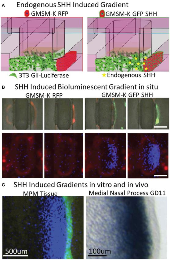
Johnson et al.                                                                                        Microphysiological Modeling of Hedgehog Signaling

INTRODUCTION                                                                 cascade. These targets range from secretory ligand modification
                                                                             and paracrine shuttling to downstream sensing and transduction
Paracrine signaling factors play key roles in embryonic                      events (Jeong and McMahon, 2002; Lauth et al., 2007; Petrova
morphogenesis by establishing complex temporospatial gene                    et al., 2013). However, efforts to identify Shh pathway effectors are
expression patterns that drive cell differentiation and tissue               limited by the simplicity of traditional in silico and in vitro assays
outgrowth. The temporally dynamic and multicellular nature of                and the time and cost of complex in vivo models. An ideal system
developmental paracrine signaling poses challenges to studying               would replicate key cellular and molecular interactions that,
this biology both in vivo and in vitro. Sonic hedgehog (Shh)                 when disrupted, give rise to most isolated birth defects and be
is a classic example of an intercellular paracrine signaling                 amenable to screening-based approaches to test environmentally
pathway that is critically important for normal embryonic and                relevant drug/chemical libraries (Knudsen et al., 2017).
fetal development. For example, Shh activity drives orofacial                    We present a novel microphysiological model (MPM)
morphogenesis (Lan and Jiang, 2009; Kurosaka, 2015), while                   that recapitulates developmental epithelial-mesenchymal
targeted pathway disruption results in orofacial clefts (OFCs) of            organization and is suited for chemical screening. To model
the lip and palate in animal models (Lipinski et al., 2010; Heyne            embryonic facial growth processes in vitro, a layer of oral
et al., 2015a). Normal development of the upper lip and palate               epithelium is overlaid on mesenchymal 3-dimensional (3D)
requires the orchestrated proliferation and fusion of embryonic              microtissues that are supported by an extracellular matrix
facial growth centers primarily composed of cranial neural crest-            (ECM). We show that cellular organization and gradients of
derived mesenchyme overlaid by an epithelial layer (Ferguson,                SHH-driven signaling of orofacial development are recapitulated
1988; Jiang et al., 2006). This tissue architecture facilitates              in this model and that it is well-equipped to screen for
epithelium-secreted SHH ligand producing a gradient of pathway               chemicals that modulate various distinct targets across the Shh
activation in the cranial neural crest-derived mesenchyme (Lan               signaling pathway. This microphysiological system provides a
and Jiang, 2009; Hu et al., 2015; Kurosaka, 2015).                           novel approach for identifying environmental influences that
    Most birth defects are thought to be caused by interacting               contribute to OFC susceptibility and a tractable foundation to
genetic and environmental influences (Beames and Lipinski,                   examine complex gene-environment interaction.
2020). Isolated congenital malformations linked to Shh
pathway disruption, including OFCs, holoprosencephaly, and
                                                                             MATERIALS AND METHODS
hypospadias, are particularly etiologically complex (Murray,
2002; Carmichael et al., 2012; Krauss and Hong, 2016; Beames                 Chemicals/Reagents
and Lipinski, 2020). Among the most common human                             SHH-N peptide (R&D Systems) and SAG (CAS #2095432-
birth defects, OFCs have been studied extensively, though                    58-7, Selleckchem) were used to exogenously induce Shh
our understanding of causative factors remains inadequate.                   pathway activity. The potent Smoothened inhibitors cyclopamine
Efforts to resolve OFC etiology using genetic approaches have                (CAS #4449-51-8) and vismodegib (CAS #879085-55-9) were
identified dozens of associated risk loci (Leslie and Marazita,              purchased from LC Laboratories. Additional Shh pathway
2013), but recognized sequence variants are rarely causative.                disruptors assessed include U18666A (CAS #3039-71-2, Tocris),
Furthermore, isolated OFC cases generally do not follow                      RU-SKI 43 (CAS # 1782573-67-4, Tocris), the anti-SHH
Mendelian inheritance patterns, suggesting an important role                 monoclonal neutralizing 5E1 antibody (Developmental Studies
for environmental influences in OFC susceptibility (Murray,                  Hybridoma Bank at the University of Iowa), piperonyl butoxide
2002; Roessler et al., 2003; Graham and Shaw, 2005; Juriloff and             (CAS #51-03-6, Toronto Research Chemicals), and GANT61
Harris, 2008; Lidral et al., 2008; Vieira, 2008). Identifying specific       (CAS # 500579-04-4, Tocris). The negative control compound
environmental factors that disrupt the signaling pathways that               benzo[a]pyrene was purchased from Sigma-Aldrich (CAS #50-
drive orofacial morphogenesis and may contribute to OFC risk                 32-8). All chemicals were dissolved in DMSO or water.
is a route to prevention strategies and, therefore, an important
focus of investigation.                                                      Maintenance and Engineering of Cell Lines
    The Shh signaling pathway is inherently sensitive to                     The embryonic murine mesenchymal cell line 3T3 Shh-Light2,
disruption by environmental chemicals. We have shown that the                human fetal oral epithelial (GMSM-K), and mouse cranial neural
natural alkaloid cyclopamine inhibits Shh signaling, decreases               crest mesenchymal (O9-1) cell lines (Gilchrist et al., 2000; Taipale
mesenchymal proliferation, and prevents tissue outgrowth and                 et al., 2000; Ishii et al., 2012) were used as indicated. The Shh-
fusion, leading to cleft lip and/or palate in mouse models                   Light2 variant of the 3T3 cell line expresses a Gli-driven firefly
(Heyne et al., 2015a; Everson et al., 2017). Numerous other                  luciferase reporter enabling real-time evaluation of the SHH-
environmental chemicals have been found to disrupt Shh                       pathway activation (Taipale et al., 2000). We also used two
pathway signaling, including cyclopamine-like dietary alkaloids,             types of GMSM-K cell lines: a SHH-null variant and a variant
natural and synthetic pharmaceuticals, and a common pesticide                that stably overexpresses human full-length SHH (Fan et al.,
component (Lipinski et al., 2007; Lipinski and Bushman, 2010;                2004). In addition, each cell line was engineered for in situ
Wang et al., 2012; Everson et al., 2019; Rivera-González et al.,             visualization; GMSM-K SHH-null cells express RFP and GMSM-
2021). Importantly, Shh signaling is sensitive to disruption by              K SHH overexpressing cells express GFP. GMSM-K and O9-1
a variety of mechanistically distinct chemicals that affect signal           cells were maintained in DMEM with 10% FBS and 1% penicillin-
transduction at multiple molecular targets within the signaling              streptomycin and maintained in an incubator at 37◦ C and 5%

Frontiers in Cell and Developmental Biology | www.frontiersin.org        2                                     February 2021 | Volume 9 | Article 621442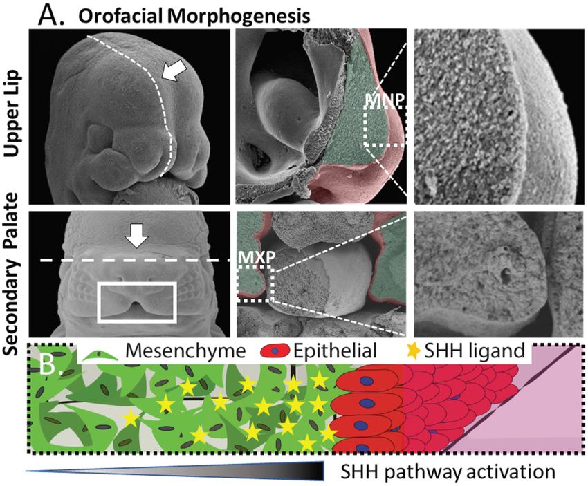
Johnson et al.                                                                                    Microphysiological Modeling of Hedgehog Signaling

CO2. 3T3 Shh-Light2 cells were similarly maintained in media              inducing gravity-driven perfusion of the microtissue was created
containing the selection agents G418 (0.4 mg/mL, Invivogen, San           by adding 100–150 µL media to the center well. Perfused media
Diego, CA) and zeocin (0.15 mg/mL, Invivogen).                            collected in the half moon reservoirs in the bottom well, which
                                                                          were aspirated each day. Media was changed every 1–2 days, and
Device Design and Construction                                            cultures were dosed daily for 3 days or as indicated after seeding.
Devices were designed and modeled with computer aided
design (CAD) modeling software (Solidworks, Dessault Systems,             Animal Studies
Vélizy-Villacoublay, France). SprutCAM software was used to               This study was conducted in strict accordance with the
generate toolpaths, and devices were CNC milled (Tormach Inc.,            recommendations in the Guide for the Care and Use of
Waunakee, WI, USA) from clear 96-well non-tissue culture-                 Laboratory Animals of the National Institutes of Health. The
treated plates (Corning Inc., Corning, NY, USA). After milling,           protocol was approved by the University of Wisconsin School
each plate was cleaned by sonication for 15 min in 100%                   of Veterinary Medicine Institutional Animal Care and Use
isopropyl alcohol. Milled plates were washed with water, dried            Committee (protocol number G005396). C57BL/6J mice were
with compressed air, then heated to 70◦ C. While plate devices            purchased from The Jackson Laboratory and housed under
were heating, 0.19 mm thick polystyrene sheets (Goodfellow,               specific pathogen-free conditions in disposable, ventilated cages
Huntingdon, England) were cut just slightly larger than the               (Innovive, San Diego, CA, USA). Rooms were maintained at 22 ±
milled plate devices, sprayed with 70% ethanol, and rinsed with           2◦ C and 30–70% humidity on a 12-h light, 12-h dark cycle. Mice
water. Sheets were dried with compressed air and added to the             were fed 2,920× Irradiated Harlan Teklad Global Soy Protein-
70◦ C hot plate. Once the devices and sheets reached temperature,         Free Extruded Rodent Diet (Envigo Teklad Global, Indianapolis,
35 µL of acetonitrile was added to milled bonding ports in the            IN, USA) until day of plug, when dams received 2,919 Irradiated
upper left-hand corner of the device to bond the polystyrene              Teklad Global 19% Protein Extruded Rodent Diet (Envigo Teklad
sheet to the milled plate. Excess acetonitrile was aspirated from         Global). Mice were set up for timed pregnancies as previously
adjacent corners and channels to avoid plate etching. This process        described (Heyne et al., 2015b). Scanning electron microscopy
was repeated for each of the three remaining corner holes in the          and hematoxylin and eosin (H&E) staining were conducted as
milled plate resulting in a bonded seal completely around the             previously described (Dunty et al., 2002; Heyne et al., 2015a).
device. Bonded devices were cooled at room temperature. Excess
polystyrene was trimmed from the outside of the device using a            In situ Hybridization (ISH)
handheld razor blade to complete device construction. Devices             ISH analysis was performed as previously described (Heyne
were then treated with UV light for 15 min and transferred to a           et al., 2016) using an established high-throughput technique
biosafety hood for cell culture. Device design and construction           (Abler et al., 2011). Embryos were processed whole or embedded
are illustrated in Figure 2.                                              in 4% agarose gel and cut in 50 µm sections using a vibrating
                                                                          microtome. Embryos were imaged using a MicroPublisher 5.0
Seeding of Devices                                                        camera connected to an Olympus SZX-10 stereomicroscope
All experimental cultures were seeded and maintained in DMEM              for whole mount imaging or a Nikon Eclipse E600 microscope
supplemented with 1% FBS and 1% penicillin-streptomycin. In               for imaging sections. ISH riboprobe primer sequences: Ptch1-
luciferase assays, 2 mM VivoGlo luciferin (Promega, Madison,              fwd GACGTGAGGACAGAAGATTG and Ptch1-rev + T7
WI, USA) and 25 mM HEPES were included in the culture                     leader CGATGTTAATACGACTCACTATAGGGAACTGGGCA
media. To improve hyaluronic acid attachment to the device,               GCTATGAAG.
each device well was filled with 3 µL of polyethyleneimine for
10 min. The wells were then aspirated and filled with 3 µL                Evaluation of Gli-Driven Luciferase
of glutaraldehyde (GA) for 30 min. Following GA treatment,                For in situ quantification of SHH-induced luciferase activity,
each device well was washed three times with water. Devices               culture media included 2 mM VivoGlo luciferin (Promega),
were air dried in a biosafety hood before cells were loaded into          which is an injectable in vivo-grade substrate that is cleaved
device wells. While devices were air drying, hyaluronic acid was          by luciferase, producing a luminescent signal. Persistent
prepared according to the manufacturer’s protocol. Hystem-C, a            exposure showed no adverse cytotoxic effect nor reduced
hyaluronic acid collagen gel solution (Sigma-Aldrich, St. Louis,          luminescent signal in response to SHH ligand (data not shown).
MO, USA) was mixed 1:1 with a 100,000 mesenchymal/fibroblast              Luminescence was measured prior to dosing, as well as after the
cells/µL solution. The hyaluronic acid:cell solution (1.75–3 uL)          dosing period to enable normalization to any baseline differences
was added to each device well; therefore, at most 150,000 cells           in luminescence across replicates. Luminescence was quantified
were seeded per well. Microtissues were allowed to polymerize at          on a Chemicdoc luminescent imager (BioRad, Hercules, CA,
room temperature for 45 min, then media was added to the top              USA) or Pherastar plate reader (BMG, Offenburg, Germany),
of cultures. One day after mesenchymal seeding, 10 µL of a 4,000          and magnified images of the SHH-mediated gene expression
GMSM-K cells/µL solution was loaded into one or both side                 gradient were enabled by placing a 2X dissecting microscope
channels of the well, and, where indicated, 5 µL of cells were also       lens (Leica, Wetzlar, Germany) in the optical path between the
seeded directly on top of the mesenchymal cells to increase signal        camera and the plate. Quantification was performed in ImageLab
for screening. 30 min later, wells and channels of the devices were       (Biorad) or ImageJ implemented through Fiji (Schindelin et al.,
flushed with media to remove unattached cells. A hydraulic head           2012). Cytotoxicity was assessed via multiple means including

Frontiers in Cell and Developmental Biology | www.frontiersin.org     3                                    February 2021 | Volume 9 | Article 621442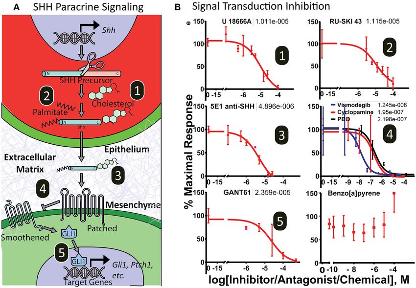
Johnson et al.                                                                                    Microphysiological Modeling of Hedgehog Signaling

microscopic evaluation of epithelia, recovery of luminescent              Development of Microtissues That
activity after chemical wash-out, or evaluation of Renilla                Recapitulate Orofacial Organization in a
luciferase activity using a dual-luciferase assay (endpoint only,
                                                                          Throughput-Compatible Format
Promega). Doses that induced cytotoxicity were not included in
                                                                          To create biomimetic microtissues in a format that is compatible
regression analysis.
                                                                          with drug/chemical screening we designed devices using
Histopathology                                                            microfluidic principles and manufactured the devices using
Microtissues were fixed in 10% formalin for 24 h and a razor was          CNC micromilling of microtiter plates. Devices were milled
used to remove the bonded thin polystyrene sheet of each device           directly into polystyrene cell culture plates (20 devices/plate)
so that a sharp probe could be used to extract the microtissues.          to maintain throughput compatibility, and a thin sheet (190
Mouse tissues were dissected and fixed in 10% formalin for 24 h.          um) of optically clear polystyrene was bonded to the bottom
Samples were embedded in paraffin then sectioned at 5 µm.                 of the plate to seal the culture chambers (Figure 2A). The
Tissues were stained with H&E to identify the cytoplasm and               devices are manufactured solely from polystyrene to avoid pitfalls
nuclei of cells, then imaged.                                             of using polydimethylsiloxane (PDMS), which is a common
                                                                          material used for organotypic models and has been previously
Statistical Analyses                                                      shown to sequester hydrophobic molecules including many
Quantification of SHH-induced bioluminescent signaling was                drugs/chemicals (Regehr et al., 2009; Guckenberger et al., 2015).
done in ImageLab software (BioRad Inc). For single comparisons,           Device design includes a central chamber (Figure 2B, left) that
Student’s t-test was used to identify significant differences in          is loaded with a hydrogel-ECM/mesenchymal cell suspension
treatment vs. control (p < 0.05). For dose-response experiments,          to form the body of the microtissue. Surface tension causes
data was background subtracted and normalized to the vehicle              the matrix to pin at 200 µm tall × 1 mm wide openings in
control (100% activity). A three-parameter non-linear regression          the bottom of the chamber (phase barrier) instead of flowing
curve-fit was generated in Graphpad Prism to determine                    out into adjacent flanking microchannels (Figure 2B, center).
antagonism/inhibition and IC50 values were determined for                 The matrix, once polymerized, forms a portion of the wall of
each chemical curve fit. Data are representative of at least two          the flanking microchannel. Epithelial cells suspended in media
independent experiments.                                                  are pipetted into the flanking channels. Laminar flow, with a
                                                                          high linear flow rate through the center of the channel and
                                                                          a low linear flow rate at the edges of the channel, leaves
RESULTS                                                                   cells coating the matrix while cells remaining in the center
Design and Engineering of a                                               of the channel are removed via flow (Figure 2B, right). The
Microplate-Based Microphysiological                                       resulting microtissue consists of a 3D mesenchymal matrix
                                                                          overlaid with epithelial cells perpendicular to the imaging plane.
Model                                                                     A culture method was developed through many iterations, which
We sought to create an in vitro platform that recapitulates key
                                                                          resulted in a method whereby a biomimetic mesenchyme was
aspects of epithelial-mesenchymal interaction in development
                                                                          generated by embedding 50,000 cells/µL murine embryonic
while remaining suitable for drug/chemical screening. The
                                                                          fibroblast cells (O9-1 or 3T3) in a hyaluronic acid/collagen
medial nasal process (MNP) and maxillary process (MXP)
                                                                          gel. Hyaluronic acid was used as the microtissue matrix
that form the upper lip and secondary palate, respectively,
                                                                          due to its importance in the developing palate (Ferguson,
share morphology as well as intercellular signaling events
                                                                          1988). The microtissues were then overlaid with GMSM-K
that orchestrate their development (Figure 1). These structures
                                                                          oral epithelial cells through flanking microchannels to coat the
are composed of a dense 3D cranial neural crest-derived
                                                                          side of the microtissue. To evaluate if the microtissues appear
mesenchyme covered by an ectoderm-derived epithelium
                                                                          phenotypically similar to the developing orofacial processes
(Ferguson, 1988; Jiang et al., 2006) (Figure 1A). The tissue
                                                                          in vivo, we compared microtissues to the developing lip and
outgrowth and fusion of these structures is driven by a
                                                                          palate of embryonic mice at gestational days 11 and 14,
continuous epithelial-mesenchymal interaction that is essential
                                                                          respectively. The H&E stains of the murine medial nasal
for the growth and fusion required to close the upper lip
                                                                          and maxillary processes (Figure 2C, left, center respectively)
and palate. Deficient outgrowth and/or subsequent fusion of
                                                                          and the organotypic microtissues (Figure 2C, right) appeared
these tissues results in orofacial clefts of the lip and/or palate.
                                                                          morphologically similar, where the dense mesenchyme was
Coordinated expansion of these facial growth centers is driven
                                                                          overlaid with epithelium.
in part by a gradient of epithelium-secreted SHH ligand that
induces pathway activation and drives proliferation in the
proximal mesenchyme through Gli-driven gene transcription
                                                                          SHH-Induced Gli-Luciferase Enables
(Lan and Jiang, 2009; Hu et al., 2015; Kurosaka, 2015). Both              Exogenous or Endogenous Real-Time in
the tissue architecture and epithelial-mesenchymal signaling              situ Quantification
informed the development of a microtissue design consisting of            Shh signaling drives the cell proliferation and tissue outgrowth
a dense 3D mesenchyme with an epithelial layer perpendicular to           that are critical for orofacial development (Yamada et al., 2005;
the imaging plane to study epithelial-mesenchymal interactions            Kurosaka, 2015; Everson et al., 2017). To enable the real-time
(depicted in Figure 1B).                                                  evaluation of SHH-driven gene activation, we used a variant

Frontiers in Cell and Developmental Biology | www.frontiersin.org     4                                    February 2021 | Volume 9 | Article 621442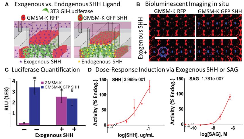
Johnson et al.                                                                                                     Microphysiological Modeling of Hedgehog Signaling

 FIGURE 1 | Morphogenesis of the orofacial processes. (A) Formation of the upper lip (upper panels) and secondary palate (lower panels) occurs through outgrowth
 and fusion of embryonic growth centers including the medial nasal process (MNP) and maxillary process (MXP). These tissues consist of a dense 3D mesenchyme
 covered with ectodermal epithelium. (B) Generalized epithelial-mesenchymal tissue architecture of the orofacial processes with high proximal SHH-induced activation.

of the 3T3 cell line that produces a luminescent signal upon                          (Figure 3D, left panel). Next, microtissues were exposed to
Shh pathway activation. By incorporating a live cell-compatible                       the synthetic Smoothened agonist, SAG, which also showed
luciferase substrate into the media, we sought to test if SHH-                        a concentration-dependent increase in luminescence (EC50
driven Gli transcriptional activity could be identified from both                     = 178nM, on-linear regression curve fit- three parameter)
exogenously added and endogenously secreted SHH ligand in                             (Figure 3D, right panel).
situ (Figure 3A). To incorporate endogenous SHH signaling
into the microtissues, we applied a variant of the human fetal
oral epithelial GMSM-K cells that stably overexpress GFP and                          SHH Ligand From the Epithelium
SHH (GMSM-K GFP SHH+ cells). The matrix-embedded 3T3                                  Stimulates a Gradient of Pathway
cells were cultured adjacent to GMSM-K GFP SHH+ cells or                              Activation in the Mesenchyme
RFP-overexpressing GMSM-K cells that are SHH- (GMSM-K                                 Formation of a gradient of Shh pathway activity is involved
RFP cells), then exposed to a vehicle control or 0.8 µg/mL                            in the morphogenesis of many tissues, including the upper
of exogenous SHH. After 72 h, microtissues were evaluated                             lip and palate (Lan and Jiang, 2009; Kurosaka, 2015; Everson
for luminescence. 3T3 cells co-cultured with GMSM-K GFP                               et al., 2017) and limbs (Li et al., 2006). To test whether
SHH+ cells exhibited a 19-fold higher (p < 0.05, Student’s t-                         SHH gradients observed during orofacial morphogenesis in vivo
test) signal of Gli-driven luciferase compared to 3T3 cells co-                       are recapitulated in vitro, microtissues were generated with
cultured with GMSM-K RFP cells. When the GMSM-K GFP                                   non-SHH-expressing GMSM-K RFP cells or SHH-expressing
SHH+ co-cultures were exposed to exogenous SHH, there                                 GMSM-K GFP SHH+ cells overlaid on one edge of the
was no significant change in Gli-driven luciferase activity. In                       mesenchyme (one flanking channel) for 48 h (Figure 4A). A
contrast, the GMSM-K RFP co-cultures exhibited a 14-fold                              merged brightfield and fluorescent image of the microtissues
higher (p < 0.05, Student’s t-test) signal of Gli-driven luciferase                   is shown in Figure 4B indicating RFP and GFP expression in
when exposed to exogenous SHH (Figures 3B,C). To test dose-                           the epithelia. Microtissues were then evaluated for Gli-driven
responsiveness of the reporter activity, microtissues were exposed                    luciferase using a luminescence imager and showed a gradient
to four concentrations of exogenous SHH which produced a                              of luciferase activity in the mesenchyme with higher activity
concentration-dependent increase in Gli-driven luciferase (EC50                       proximal to the epithelium (Figure 4B). To better compare in
= 0.4 µg/mL, non-linear regression curve fit- three parameter)                        vitro to in vivo gradients, we sought to improve resolution of

Frontiers in Cell and Developmental Biology | www.frontiersin.org                 5                                         February 2021 | Volume 9 | Article 621442
Johnson et al.                                                                                                       Microphysiological Modeling of Hedgehog Signaling

 FIGURE 2 | Development of a microplate-based microphysiological culture model. (A) Devices incorporate 3 wells of a 96-well plate, where the top and bottom wells
 are connected through two subsurface microchannels (red) which flank a microtissue well (green) milled into the center well. An array of 20 devices is CNC milled into
 each plate (bottom view). (B) Cross-sectional view of microtissue formation including the empty microtissue well (left panel), addition of the mesenchymal/ECM matrix
 (center panel), and epithelial overlay (right panel). (C) H&E stained formalin-fixed, paraffin-embedded sections of the mouse MNP (left panel) and MXP (center), and
 O9-1 mesenchymal/GMSM-K epithelial microtissue (right panel), scale bar = 50µm.

the luminescent imaging. In a separate experiment, additional                          Sensitivity to Chemical Disruption Across
optics were added to the light path in the luminescent imager.                         the Shh Signaling Cascade
Using landmarks of the microfluidic device captured by both the                        To test the utility of the platform for screening drug or
luminescent imager and the microscope, we were able to overlay                         chemical modulators of the complete Shh signaling cascade,
these images at higher resolution for comparison (Figure 4C,                           microtissues were exposed to various small molecule inhibitors
right). A photomicrograph of in situ hybridization of the SHH-                         that target distinct aspects of Shh signal transduction, including
responsive gene Ptch1 in the MNP of a GD10.25 mouse embryo                             epithelial secretion processes (cholesterol modification and
is provided as an in vivo reference (Figure 4C, left). Similar                         palmitoylation), ligand bioavailability, and mesenchymal signal
to previous studies (Everson et al., 2017), there was a gradient                       transduction (Smoothened, Gli proteins). Figure 5A illustrates
of SHH-responsive gene expression in the mesenchyme that                               the target of each inhibitor. After 3 days of exposure, cultures
decreased as distance from the epithelium increased (Figure 4C).                       were evaluated for luminescence (Figure 5B). Cytotoxicity,

Frontiers in Cell and Developmental Biology | www.frontiersin.org                  6                                          February 2021 | Volume 9 | Article 621442
Johnson et al.                                                                                                     Microphysiological Modeling of Hedgehog Signaling

 FIGURE 3 | Vital in situ quantification of endogenous and exogenous SHH ligand-induced pathway activity. (A) Experimental schematic shows incorporation of
 non-SHH-secreting and SHH-secreting epithelia and exogenously or endogenously derived SHH ligand. (B) Brightfield (red) and darkfield (blue) image of
 bioluminescent signal under different experimental conditions in a single plate. (C) Quantification of bioluminescent signal in (B) (*p < 0.05 vs. GMSM-K without
 exogenous SHH, Student’s t-test). (D) Dose-response quantification of exogenously added SHH ligand (left) and SAG (right). Non-linear regression curve fit (Graphpad
 Prism). EC50 values shown above.

monitored by loss of fluorescent signal, washout and luminescent                      microtissues (non-linear regression curve fit- three parameter)
signal recovery, or a lytic cytotoxicity assay, was assessed after                    (Supplementary Figure 1).
dosing, and data from cytotoxic doses were not included in
analysis. Inhibitors of SHH posttranslational modification in
epithelia affecting SHH secretion include cholesterol (U1886A)                        DISCUSSION
and palmitoylation (Ruski-43) inhibitors, which showed IC50
values at 10.1 µM and 11.2 µM, respectively. Importantly,                             Here we describe a novel microphysiological culture system
these inhibitors do not exhibit inhibition in a standard 2-                           that recapitulates key cellular and molecular aspects of
dimensional (2D) monoculture assay of 3T3-Gli Luc cells                               developmental Shh paracrine signaling and demonstrate its
exposed exogenously to SHH ligand (Supplementary Figure 1).                           utility for examining chemical influences that may contribute to
The anti-SHH monoclonal 5E1 antibody, which binds and                                 birth defects. This elegantly simple microphysiological system
neutralizes secreted SHH ligand, also inhibited Gli-driven                            mimics both the 3D epithelial-mesenchymal interactions and
luciferase signaling in the microtissues with an IC50 of                              critical molecular processes of Shh pathway-driven orofacial
4.9 µM. Reception of SHH ligand and the subsequent signaling                          development, including formation of an in vivo-like gradient
cascade that results in Gli activation in mesenchymal cells can                       of pathway activity. Application of a battery of mechanistically
also be inhibited by structurally diverse ligands at multiple                         diverse chemical inhibitors further demonstrated the sensitivity
molecular targets. The Smoothened antagonists piperonyl                               of the MPM to Shh pathway effectors exhibiting distinct
butoxide, cyclopamine, and vismodegib inhibited luciferase                            mechanistic targets throughout the inter- and intracellular
activity with IC50 values of 219, 195, and 12.5 nM, respectively                      signal transduction cascade. These observations underscore
(non-linear regression curve fit- three parameter). The Gli                           several important elements of both the developmental fidelity
inhibitor GANT61 also inhibited Gli-driven luciferase in a                            and investigative utility of this microphysiological approach to
dose-dependent manner with an IC50 value of 23.6 µM (non-                             modeling paracrine signaling in development.
linear regression curve fit- three parameter). The chemical                              In vitro models are often used to elucidate transduction
benzo[a]pyrene, which can induce cleft palate in rodents                              mechanisms and identify xenobiotic pathway modulators,
through a mechanism independent of Shh signaling, showed                              although common culture systems typically fail to recapitulate
no concentration-dependent inhibitory activity. As expected,                          the complex intercellular signaling pathways that produce
inhibitors of intracellular Shh signal transduction did exhibit                       morphogen gradients and involve crosstalk between different
inhibition in a standard 2D monoculture assay of 3T3-Gli                              cell types (Li et al., 2018). Orofacial morphogenesis requires
Luc cells with IC50 values at or above those seen in the                              paracrine signaling involving epithelial secretion of SHH

Frontiers in Cell and Developmental Biology | www.frontiersin.org                 7                                         February 2021 | Volume 9 | Article 621442
Johnson et al.                                                                                                      Microphysiological Modeling of Hedgehog Signaling

 FIGURE 4 | Vital epithelial-mesenchymal SHH gradient in situ. (A) Experimental schematic shows incorporation of non-SHH-secreting and SHH-secreting epithelia
 and endogenously produced SHH ligand. (B) Brightfield (greyscale) and fluorescent red (530/560 nm) and green (488/515 nm) images indicating GMSM-K overlayed
 epithelia (upper panels) and bioluminescent signal shows high activity proximal only to SHH secreting epithelia (lower panels), scale bar = 1mm. (C) Fluorescent and
 bioluminescent images taken at higher magnification were integrated to better illustrate gradient (left panel) and compared to ISH staining of SHH-responsive Ptch1
 gene in the medial nasal process of a GD10.25 mouse embryo. Scale bars in luminescent (left) and ISH image (right) are 500 and 100 µm, respectively.

ligand and transport through a 3D matrix of SHH-sensing                                increased cellular complexity by incorporating epithelium and
mesenchyme (Lan and Jiang, 2009; Kurosaka, 2015), a paradigm                           mesenchyme that are predictably engineered in direct contact
observed in many developmental contexts. 2D cultures can                               facilitating analysis. Biologically active SHH ligand secreted
be designed to incorporate a localized population of SHH                               from the epithelium induced pathway activity in the adjacent
producers, but the distribution of secreted ligand has been                            mesenchyme with maximal induction of downstream Shh
shown to differ between 2D and 3D cultures (Cederquist et al.,                         target genes occurring nearest the epithelial signaling source,
2019). The microphysiological approach described here achieves                         mimicking in vivo gene expression gradients. Thus, this MPM,

Frontiers in Cell and Developmental Biology | www.frontiersin.org                  8                                          February 2021 | Volume 9 | Article 621442
Johnson et al.                                                                                                        Microphysiological Modeling of Hedgehog Signaling

 FIGURE 5 | Dose-response curves of SHH pathway inhibitors. (A) Illustration of the Sonic hedgehog inter- and intracellular transduction pathway with important
 molecular targets numbered. (B) Microtissues show dose-response inhibition of endogenously derived SHH-induced pathway activity by xenobiotics targeting
 epithelial secretory SHH ligand cholesterol modification (1) and SHH palmitoylation modification (2); extracellular SHH ligand trafficking (3); and mesenchymal receptor
 SHH sensing and signal transduction by Patched/Smoothened (4) and Gli-driven transcription (5). The chemical 3-benzo[a]pyrene did not antagonize the pathway.
 Non-linear regression curve fit (Graphpad Prism). EC50 values shown above.

along with other recently developed 3D in vitro organoid models                         treatment of a 2D cell monolayer with pre-modified SHH
(Cederquist et al., 2019), may aid in our understanding of                              ligand or cells modified to constitutively drive downstream
how concentration gradients are formed and, more specifically,                          pathway activation (e.g., Ptch1 knockout or Gli overexpression)
address persistent questions of how secreted SHH ligand is                              (Chen et al., 2002; Lipinski and Bushman, 2010), entirely
shuttled through extracellular spaces while remaining available                         circumventing upstream intercellular signaling events, such as
to bind transmembrane receptors to initiate signal transduction                         the modification of SHH ligand by cholesterol. Here, application
(Wierbowski et al., 2020). A novel organoid model consisting                            of mechanistically distinct antagonists demonstrated that Shh
of human progenitor/stem cells designed to model disruption                             signaling in the MPM is sensitive to disruption at multiple points
of palatal fusion, which occurs later in development was also                           in the inter- and intracellular signaling cascade. Shh pathway
described recently (Belair et al., 2017). Predictably, a follow-                        activity was potently inhibited by blocking cholesterol trafficking
up screen did not detect any effects of the SHH antagonist                              (U18666A), SHH palmitoylation (RU-SKI), receptor binding by
vismodegib on the viability or fusion of their organoid                                 inactivating secreted SHH ligand (5E1), inhibiting Smoothened
model (Belair et al., 2018). These models can therefore be                              (vismodegib, cyclopamine, piperonyl butoxide), and targeting
viewed as complementary and could be employed in parallel                               Gli activation (GANT61). Because our engineered approach
to screen for chemical disruption over a greater range of                               to SHH expression bypasses endogenous SHH transcriptional
orofacial development.                                                                  regulators, this approach is unlikely to capture influences that act
   The Shh pathway illustrates the importance of designing                              further upstream, such as those that have been hypothesized for
in vitro models that recapitulate the complexity of the inter-                          fetal alcohol exposure (Ahlgren et al., 2002; Hong et al., 2020).
and intracellular signaling cascades, rather than just a single                         However, these results demonstrate that the MPM approach
point of sensitivity. While Smoothened has been given much                              described here offers broader sensitivity to mechanistically
attention as a molecular target, the Shh pathway is sensitive                           distinct pathway effectors than typical in vitro approaches.
to small molecule modulation at several steps. For example,                                 We sought with this MPM to address a significant limitation
distal cholesterol synthesis inhibitors cause palate and limb                           of microfluidic devices: technical complexity. Even with the
malformations consistent with Shh pathway disruption (Chevy                             appropriate expertise, the technical aspects of advanced systems
et al., 2002), but can fail to impact Shh signaling in simpler                          can contribute to variability between users or prevent widespread
in vitro cultures (Supplementary Figure 1). Traditional in vitro                        dissemination and use (Paguirigan and Beebe, 2008). Machining
assays examining Shh pathway activity may involve exogenous                             devices directly into microplates enabled rapid prototyping

Frontiers in Cell and Developmental Biology | www.frontiersin.org                   9                                           February 2021 | Volume 9 | Article 621442
Johnson et al.                                                                                     Microphysiological Modeling of Hedgehog Signaling

based on operational feedback and provided a familiar user                 facial growth processes can be biomimetically modeled in
format without introducing new materials into the experiment.              vitro by culturing an oral ectodermal monolayer over 3D-
Operationally, engineering devices that leverage the physical              embedded mesenchymal cells and that the microtissues
properties of fluids at the microscale enabled the simple creation         and expression patterns phenotypically resemble orofacial
of a microtissue ideally suited to quantify the biology of                 morphogenesis. The simplicity of this device makes it adaptable
interest without greatly increasing the expertise required for use.        with respect to cell types, pathways, and endpoints of interest.
Employing a hydraulic head to facilitate perfusion rather than a           The microplate design also makes this platform amenable
mechanical pump allowed us to retain a throughput-compatible               to throughput screening, while recent advances in gene-
format, which is a key advantage for conducting chemical                   editing technology open the door to its use in investigating
screens, and incorporation of live-cell endpoints greatly reduced          gene-environment interactions. Leveraging the physiological
handling time and enabled us to monitor activity without                   relevance and high tractability of this approach illustrate its
sacrificing the culture. Furthermore, the use of polystyrene rather        potential value, particularly for investigating biologically and
than polydimethylsiloxane (PDMS), a common component                       etiologically complex outcomes including human birth defects
of microfluidic devices that sequesters hydrophobic molecules              like orofacial clefts.
(Regehr et al., 2009), makes this device appropriate for screening
a diverse set of compounds.                                                DATA AVAILABILITY STATEMENT
    The operational simplicity of this microphysiological device
should enable ready use by other groups for a variety                      The original contributions presented in the study are included
of biological applications in toxicology, pharmacology, and                in the article/Supplementary Material, further inquiries can be
regenerative medicine. The MPM’s design affords substantial                directed to the corresponding author.
flexibility and even a “plug and play” paradigm with respect
to cells and ECMs, allowing it to model specific developmental             ETHICS STATEMENT
environments. For example, although we specifically modeled
epithelial-mesenchymal Shh signaling in this study, an iteration           The animal study was reviewed and approved by University of
of this device was recently used instead to model neurovascular            Wisconsin School of Veterinary Medicine Institutional Animal
development (Kaushik et al., 2020). Particular attention may               Care and Use Committee.
be given to Shh-associated congenital malformations, including
OFCs, holoprosencephaly, and hypospadias, that are often
etiologically complex and difficult to model in in vitro systems           AUTHOR CONTRIBUTIONS
(Murray, 2002; Carmichael et al., 2012; Krauss and Hong, 2016;
                                                                           BJ devised the device design and construction. BJ, RV, and
Beames and Lipinski, 2020). Full integration of gene-editing
                                                                           RL planned the experiments. BJ, RV, and PG conducted the
techniques such as CRISPR into the MPM could open the door
                                                                           experiments. DF prepared specialized cells/reagents. BJ, RV, MM,
to studying these complex etiologies in a biologically faithful
                                                                           TB, and RL wrote the manuscript. BJ and MM drew the figures.
system and even allow for “personalized toxicology” by providing
                                                                           DB and RL supervised the research and helped develop the
a platform to identify environmental factors that preferentially
                                                                           idea. All authors contributed to the article and approved the
interact with personal/familial mutations. Another emerging
                                                                           submitted version.
strategy for toxicity testing utilizes in silico models built from
data collected from in vitro and in vivo models to predict adverse
effects associated with toxicant exposures. A computational                FUNDING
model to predict the effects of chemicals on the growth and fusion
of the palate was recently reported (Hutson et al., 2017), and             Research reported in this publication was supported by
informing the development and refinement of in silico models               the National Institute of Environmental Health Sciences of
and conducting secondary screening of molecules identified by              the National Institutes of Health under award numbers
in silico screening is another logical niche for MPMs like the one         K99ES028744, R01ES026819, and T32ES007015, EPA Science
described herein.                                                          to Achieve Results (STAR) grant number 83573701, and the
    The development of model systems that are physiologically              University of Wisconsin Carbone Cancer Center Support Grant
relevant but also amenable to mechanistic studies and chemical             P30 CA014520.
screening is needed to bridge the gap between existing
in vitro and in vivo models (Beames and Lipinski, 2020).                   SUPPLEMENTARY MATERIAL
Addressing this need, we present the engineering, construction,
and implementation of a novel microphysiological culture                   The Supplementary Material for this article can be found
model of epithelial-mesenchymal interactions as applied                    online at: https://www.frontiersin.org/articles/10.3389/fcell.2021.
to the Shh signaling pathway. We show that embryonic                       621442/full#supplementary-material

Frontiers in Cell and Developmental Biology | www.frontiersin.org     10                                    February 2021 | Volume 9 | Article 621442
Johnson et al. Microphysiological Modeling of Hedgehog Signaling
REFERENCES Heyne, G. W., Plisch, E. H., Melberg, C. G., Sandgren, E. P., Peter, J. A., and
Lipinski, R. J. (2015b). A simple and reliable method for early pregnancy
Abler, L. L., Mehta, V., Keil, K. P., Joshi, P. S., Flucus, C. L., Hardin, H. A., detection in inbred mice. J. Am. Assoc. Lab. Anim. Sci. 54, 368–371.
et al. (2011). A high throughput in situ hybridization method to characterize Hong, M., Christ, A., Christa, A., Willnow, T. E., and Krauss, R. S. (2020).
mRNA expression patterns in the fetal mouse lower urogenital tract. J. Vis. Exp. Mutation and fetal alcohol converge on Nodal signaling in a mouse model of
19:2912. doi: 10.3791/2912 holoprosencephaly. Elife 9: e60351. doi: 10.7554/eLife.60351.sa2
Ahlgren, S. C., Thakur, V., and Bronner-Fraser, M. (2002). Sonic hedgehog Hu, D., Young, N. M., Li, X., Xu, Y., Hallgrímsson, B., and Marcucio, R. S.
rescues cranial neural crest from cell death induced by ethanol exposure. (2015). A dynamic Shh expression pattern, regulated by SHH and BMP
Proc. Natl. Acad. Sci. U. S. A. 99, 10476–10481. doi: 10.1073/pnas.162 signaling, coordinates fusion of primordia in the amniote face. Development
356199 142, 567–574. doi: 10.1242/dev.114835
Beames, T. G., and Lipinski, R. J. (2020). Gene-environment interactions: aligning Hutson, M. S., Leung, M. C. K., Baker, N. C., Spencer, R. M., and Knudsen, T. B.
birth defects research with complex etiology. Development 147:dev191064. (2017). Computational model of secondary palate fusion and disruption. Chem.
doi: 10.1242/dev.191064 Res. Toxicol. 30, 965–979. doi: 10.1021/acs.chemrestox.6b00350
Belair, D. G., Wolf, C. J., Moorefield, S. D., Wood, C., Becker, C., and Abbott, Ishii, M., Arias, A. C., Liu, L., Chen, Y. B., Bronner, M. E., and Maxson, R. E.
B. D. (2018). A Three-dimensional organoid culture model to assess the (2012). A stable cranial neural crest cell line from mouse. Stem Cells Dev. 21,
influence of chemicals on morphogenetic fusion. Toxicol. Sci. 166, 394–408. 3069–3080. doi: 10.1089/scd.2012.0155
doi: 10.1093/toxsci/kfy207 Jeong, J., and McMahon, A. P. (2002). Cholesterol modification of Hedgehog
Belair, D. G., Wolf, C. J., Wood, C., Ren, H., Grindstaff, R., Padgett, W., et al. (2017). family proteins. J. Clin. Invest. 110, 591–596. doi: 10.1172/JCI0216506
Engineering human cell spheroids to model embryonic tissue fusion in vitro. Jiang, R., Bush, J. O., and Lidral, A. C. (2006). Development of the upper
PLoS ONE 12:e0184155. doi: 10.1371/journal.pone.0184155 lip: morphogenetic and molecular mechanisms. Dev. Dyn. 235, 1152–1166.
Carmichael, S. L., Shaw, G. M., and Lammer, E. J. (2012). Environmental doi: 10.1002/dvdy.20646
and genetic contributors to hypospadias: a review of the epidemiologic Juriloff, D. M., and Harris, M. J. (2008). Mouse genetic models of cleft lip with
evidence. Birth Defects Res. Part A Clin. Mol. Teratol. 94, 499–510. or without cleft palate. Birth Defects Res. Part A Clin. Mol. Teratol. 82, 63–77.
doi: 10.1002/bdra.23021 doi: 10.1002/bdra.20430
Cederquist, G. Y., Asciolla, J. J., Tchieu, J., Walsh, R. M., Cornacchia, D., Resh, M. Kaushik, G., Gupta, K., Harms, V., Torr, E., Evans, J., Johnson, H. J., et al. (2020).
D., et al. (2019). Specification of positional identity in forebrain organoids. Nat. Engineered perineural vascular plexus for modeling developmental toxicity.
Biotechnol. 37, 436–444. doi: 10.1038/s41587-019-0085-3 Adv. Healthc. Mater. 9:e2000825. doi: 10.1002/adhm.202000825
Chen, J. K., Taipale, J., Young, K. E., Maiti, T., and Beachy, P. A. (2002). Small Knudsen, T., Klieforth, B., and Slikker, W. (2017). Programming
molecule modulation of Smoothened activity. Proc. Natl. Acad. Sci. U. S. A. 99, microphysiological systems for children’s health protection. Exp. Biol.
14071–14076. doi: 10.1073/pnas.182542899 Med. 242, 1586–1592. doi: 10.1177/1535370217717697
Chevy, F., Illien, F., Wolf, C., and Roux, C. (2002). Limb malformations of rat Krauss, R. S., and Hong, M. (2016). Gene–environment interactions
fetuses exposed to a distal inhibitor of cholesterol biosynthesis. J. Lipid Res. 43, and the etiology of birth defects. Curr. Top. Dev. Biol. 116, 569–580.
1192–1200. doi: 10.1194/jlr.M200082-JLR200 doi: 10.1016/bs.ctdb.2015.12.010
Dunty, W. C. Jr., Zucker, R. M., and Sulik, K. K. (2002). Hindbrain and cranial Kurosaka, H. (2015). The Roles of Hedgehog signaling in upper lip formation.
nerve dysmorphogenesis result from acute maternal ethanol administration. Biomed Res. Int. 2015:901041. doi: 10.1155/2015/901041
Dev. Neurosci. 24, 328–342. doi: 10.1159/000066748 Lan, Y., and Jiang, R. (2009). Sonic hedgehog signaling regulates reciprocal
Everson, J. L., Fink, D. M., Yoon, J. W., Leslie, E. J., Kietzman, H. W., Ansen- epithelial-mesenchymal interactions controlling palatal outgrowth.
Wilson, L. J., et al. (2017). Sonic hedgehog regulation of Foxf2 promotes Development 136, 1387–1396. doi: 10.1242/dev.028167
cranial neural crest mesenchyme proliferation and is disrupted in cleft lip Lauth, M., Bergström, A., Shimokawa, T., and Toftgård, R. (2007).
morphogenesis. Development 144, 2082–2091. doi: 10.1242/dev.149930 Inhibition of GLI-mediated transcription and tumor cell growth by
Everson, J. L., Sun, M. R., Fink, D. M., Heyne, G. W., Melberg, C. G., Nelson, K. F., small-molecule antagonists. Proc. Natl. Acad. Sci. U.S.A. 104, 8455–8460.
et al. (2019). Developmental toxicity assessment of piperonyl butoxide exposure doi: 10.1073/pnas.0609699104
targeting sonic hedgehog signaling and forebrain and face morphogenesis in Leslie, E. J., and Marazita, M. L. (2013). Genetics of cleft lip and cleft palate. Am. J.
the mouse: an in vitro and in vivo study. Environ. Health Perspect. 127:107006. Med. Genet. C Semin. Med. Genet. 163C, 246–258. doi: 10.1002/ajmg.c.31381
doi: 10.1289/EHP5260 Li, P., Markson, J. S., Wang, S., Chen, S., Vachharajani, V., and Elowitz, M. B.
Fan, L., Pepicelli, C. V., Dibble, C. C., Catbagan, W., Zarycki, J. L., Laciak, R., (2018). Morphogen gradient reconstitution reveals Hedgehog pathway design
et al. (2004). Hedgehog signaling promotes prostate xenograft tumor growth. principles. Science 360, 543–548. doi: 10.1126/science.aao0645
Endocrinology 145, 3961–3970. doi: 10.1210/en.2004-0079 Li, Y., Zhang, H., Litingtung, Y., and Chiang, C. (2006). Cholesterol modification
Ferguson, M. W. (1988). Palate development. Development 103, 41–60. restricts the spread of Shh gradient in the limb bud. Proc. Natl. Acad. Sci. U. S.
Gilchrist, E. P., Moyer, M. P., Shillitoe, E. J., Clare, N., and Murrah, V. A. 103, 6548–6553. doi: 10.1073/pnas.0600124103
A. (2000). Establishment of a human polyclonal oral epithelial cell line. Lidral, A. C., Moreno, L. M., and Bullard, S. A. (2008). Genetic factors and
Oral Surg. Oral Med. Oral Pathol. Oral Radiol. Endod. 90, 340–347. orofacial clefting. Semin. Orthod. 14, 103–114. doi: 10.1053/j.sodo.2008.
doi: 10.1067/moe.2000.107360 02.002
Graham, J. M. Jr., and Shaw, G. M. (2005). Gene-environment interactions in rare Lipinski, R. J., and Bushman, W. (2010). Identification of Hedgehog signaling
diseases that include common birth defects. Birth Defects Res. Part A Clin. Mol. inhibitors with relevant human exposure by small molecule screening. Toxicol.
Teratol. 73, 865–867. doi: 10.1002/bdra.20193 In Vitro 24, 1404–1409. doi: 10.1016/j.tiv.2010.04.011
Guckenberger, D. J., de Groot, T. E., Wan, A. M., Beebe, D. J., and Young, E. Lipinski, R. J., Dengler, E., Kiehn, M., Peterson, R. E., and Bushman,
W. K. (2015). Micromilling: a method for ultra-rapid prototyping of plastic W. (2007). Identification and characterization of several dietary alkaloids
microfluidic devices. Lab Chip 15, 2364–2378. doi: 10.1039/C5LC00234F as weak inhibitors of hedgehog signaling. Toxicol. Sci. 100, 456–463.
Heyne, G. W., Everson, J. L., Ansen-Wilson, L. J., Melberg, C. G., Fink, D. M., doi: 10.1093/toxsci/kfm222
Parins, K. F., et al. (2016). Gli2 gene-environment interactions contribute to Lipinski, R. J., Song, C., Sulik, K. K., Everson, J. L., Gipp, J. J., Yan, D., et al. (2010).
the etiological complexity of holoprosencephaly: evidence from a mouse model. Cleft lip and palate results from Hedgehog signaling antagonism in the mouse:
Dis. Model. Mech. 9, 1307–1315. doi: 10.1242/dmm.026328 phenotypic characterization and clinical implications. Birth Defects Res. Part A
Heyne, G. W., Melberg, C. G., Doroodchi, P., Parins, K. F., Kietzman, H. W., Clin. Mol. Teratol. 88, 232–240. doi: 10.1002/bdra.20656
Everson, J. L., et al. (2015a). Definition of critical periods for Hedgehog pathway Murray, J. C. (2002). Gene/environment causes of cleft lip and/or
antagonist-induced holoprosencephaly, cleft lip, and cleft palate. PLoS ONE palate. Clin. Genet. 61, 248–256. doi: 10.1034/j.1399-0004.2002.
10:e0120517. doi: 10.1371/journal.pone.0120517 610402.x
Frontiers in Cell and Developmental Biology | www.frontiersin.org 11 February 2021 | Volume 9 | Article 621442Johnson et al. Microphysiological Modeling of Hedgehog Signaling Paguirigan, A. L., and Beebe, D. J. (2008). Microfluidics meet cell biology: bridging Wang, J., Lu, J., Mook, R., Zhang, M., Zhao, S., Barak, L., et al. (2012). The the gap by validation and application of microscale techniques for cell biological insecticide synergist piperonyl butoxide inhibits Hedgehog signaling: assessing assays. Bioessays 30, 811–821. doi: 10.1002/bies.20804 chemical risks. Toxicol. Sci. 128, 517–523. doi: 10.1093/toxsci/kfs165 Petrova, E., Rios-Esteves, J., Ouerfelli, O., Glickman, J. F., and Resh, M. D. (2013). Wierbowski, B. M., Petrov, K., Aravena, L., Gu, G., Xu, Y., and Salic, A. Inhibitors of Hedgehog acyltransferase block Sonic Hedgehog signaling. Nat. (2020). Hedgehog Pathway activation requires coreceptor-catalyzed, lipid- Chem. Biol. 9, 247–249. doi: 10.1038/nchembio.1184 dependent relay of the Sonic Hedgehog ligand. Dev. Cell. 55, 450–467. Regehr, K., Domenech, M., Koepsel, J., Carver, K., Ellison-Zelski, S., Murphy, doi: 10.1016/j.devcel.2020.09.017 W., et al. (2009). Biological implications of polydimethylsiloxane-based Yamada, Y., Nagase, T., Nagase, M., and Koshima, I. (2005). Gene expression microfluidic cell culture. Lab Chip 9, 2132–2139. doi: 10.1039/b903043c changes of sonic hedgehog signaling cascade in a mouse embryonic Rivera-González, K. S., Beames, T. G., and Lipinski, R. J. (2021). Examining the model of fetal alcohol syndrome. J. Craniofacial Surg. 16, 1055–1061. developmental toxicity of piperonyl butoxide as a Sonic hedgehog pathway doi: 10.1097/01.scs.0000183470.31202.c9 inhibitor. Chemosphere 264:128414. doi: 10.1016/j.chemosphere.2020.128414 Roessler, E., Du, Y. Z., Mullor, J. L., Casas, E., Allen, W. P., Gillessen-Kaesbach, Conflict of Interest: BJ holds equity in Onexio Biosystems L.L.C. DB holds equity G., et al. (2003). Loss-of-function mutations in the human GLI2 gene in Bellbrook Labs, L.L.C., Tasso, Inc., Stacks to the Future, L.L.C., Salus Discovery, are associated with pituitary anomalies and holoprosencephaly-like features. L.L.C., Lynx Biosciences, Inc., and Onexio Biosystems L.L.C. Proc. Natl. Acad. Sci. U. S. A. 100, 13424–13429. doi: 10.1073/pnas.22357 34100 The remaining authors declare that the research was conducted in the absence of Schindelin, J., Arganda-Carreras, I., Frise, E., Kaynig, V., Longair, M., Pietzsch, T., any commercial or financial relationships that could be construed as a potential et al. (2012). Fiji: an open-source platform for biological-image analysis. Nat. conflict of interest. Methods 9, 676–682. doi: 10.1038/nmeth.2019 Taipale, J., Chen, J. K., Cooper, M. K., Wang, B., Mann, R. K., Milenkovic, L., Copyright © 2021 Johnson, Vitek, Morgan, Fink, Beames, Geiger, Beebe and Lipinski. et al. (2000). Effects of oncogenic mutations in Smoothened and Patched This is an open-access article distributed under the terms of the Creative Commons can be reversed by cyclopamine. Nature 406, 1005–1009. doi: 10.1038/350 Attribution License (CC BY). The use, distribution or reproduction in other forums 23008 is permitted, provided the original author(s) and the copyright owner(s) are credited Vieira, A. R. (2008). Unraveling human cleft lip and palate and that the original publication in this journal is cited, in accordance with accepted research. J. Dent. Res. 87, 119–125. doi: 10.1177/1544059108087 academic practice. No use, distribution or reproduction is permitted which does not 00202 comply with these terms. Frontiers in Cell and Developmental Biology | www.frontiersin.org 12 February 2021 | Volume 9 | Article 621442
You can also read