The Evaluation of Equine Allogeneic Tenogenic Primed Mesenchymal Stem Cells in a Surgically Induced Superficial Digital Flexor Tendon Lesion Model
←
→
Page content transcription
If your browser does not render page correctly, please read the page content below
ORIGINAL RESEARCH
                                                                                                                                              published: 05 March 2021
                                                                                                                                        doi: 10.3389/fvets.2021.641441

                                             The Evaluation of Equine Allogeneic
                                             Tenogenic Primed Mesenchymal
                                             Stem Cells in a Surgically Induced
                                             Superficial Digital Flexor Tendon
                                             Lesion Model
                                             Eva Depuydt 1,2 , Sarah Y. Broeckx 1 , Lore Van Hecke 1 , Koen Chiers 3 , Leen Van Brantegem 3 ,
                                             Hans van Schie 4,5 , Charlotte Beerts 1,6 , Jan H. Spaas 1,6*, Frederik Pille 2 and Ann Martens 2
                                             1
                                               Global Stem cell Technology, Part of Boehringer Ingelheim, Evergem, Belgium, 2 Department of Surgery and
                                             Anaesthesiology of Domestic Animals, Faculty of Veterinary Medicine, Ghent University, Merelbeke, Belgium, 3 Department of
                                             Pathology, Bacteriology and Poultry Diseases, Faculty of Veterinary Medicine, Ghent University, Merelbeke, Belgium,
                                             4
                                               Department of Clinical Sciences, Faculty of Veterinary Medicine, Utrecht University, Utrecht, Netherlands, 5 Department of
                                             Research and Development, UTC Imaging, Stein, Netherlands, 6 Department of Veterinary Medical Imaging and Small Animal
                                             Orthopaedics, Faculty of Veterinary Medicine, Ghent University, Merelbeke, Belgium
                           Edited by:
                Chavaunne T. Thorpe,
       Royal Veterinary College (RVC),       Background: Tendon injuries are very common in horses and jeopardize the athletic
                      United Kingdom
                                             performance, and due to the high risk of reinjury may lead to early retirement. The
                        Reviewed by:
                                             use of mesenchymal stem cells for the treatment of equine tendon disease is widely
                        Debbie Guest,
       Royal Veterinary College (RVC),       investigated because of their regenerative potential. The objective of this study is to
                      United Kingdom         investigate the safety and efficacy of equine allogeneic tenogenic primed mesenchymal
               Sushmitha S. Durgam,
           The Ohio State University,
                                             stem cells (tpMSCs) for the management of tendinitis in horses.
                         United States
                                             Methods: A core lesion was surgically induced in the superficial digital flexor tendon
                *Correspondence:
                                             of both forelimbs of eight horses. After 7 days, one forelimb was treated with tpMSCs,
                       Jan H. Spaas
 jan.spaas@boehringer-ingelheim.com          while the contralateral forelimb served as an intra-individual control and was treated with
                                             saline. A prescribed exercise program was started. All horses underwent a daily clinical
                   Specialty section:        evaluation throughout the entire study period of 112 days. Blood samples were taken
         This article was submitted to
    Veterinary Regenerative Medicine,        at different time points for hematological and biochemical analysis. Tendon assessment,
               a section of the journal      lameness examination, ultrasound assessment and ultrasound tissue characterization
       Frontiers in Veterinary Science
                                             (UTC) were performed at regular time intervals. At the end of the study period, the
       Received: 14 December 2020
                                             superficial digital flexor tendons were evaluated macroscopically and histologically.
        Accepted: 05 February 2021
         Published: 05 March 2021            Results: No suspected or serious adverse events occurred during the entire study
                              Citation:      period. There was no difference in local effects including heat and pain to pressure
Depuydt E, Broeckx SY, Van Hecke L,
     Chiers K, Van Brantegem L, van
                                             between a single intralesional injection of allogeneic tpMSCs and a single intralesional
 Schie H, Beerts C, Spaas JH, Pille F        injection with saline. A transient moderate local swelling was noted in the tpMSC treated
and Martens A (2021) The Evaluation
                                             limbs, which dissipated by day 11. Starting at a different time point depending on
      of Equine Allogeneic Tenogenic
Primed Mesenchymal Stem Cells in a           the parameter, a significant improvement was observed in the tpMSC treated limbs
 Surgically Induced Superficial Digital      compared to the placebo for echogenicity score, fiber alignment score, anterior-posterior
         Flexor Tendon Lesion Model.
            Front. Vet. Sci. 8:641441.
                                             thickness of the tendon and echo type by UTC assessment. Immunohistochemistry 112
    doi: 10.3389/fvets.2021.641441           days post-injection revealed that the amount of collagen type I and Von Willebrand factor

Frontiers in Veterinary Science | www.frontiersin.org                               1                                            March 2021 | Volume 8 | Article 641441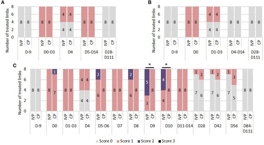
Depuydt et al.                                                                                                           tpMSC Treatment for Tendon Injuries

                                             were significantly higher in the tendon tissue of the tpMSC group, while the amount of
                                             collagen type III and smooth muscle actin was significantly lower.
                                             Conclusion: Equine allogeneic tenogenic primed mesenchymal stem cells were shown
                                             to be well-tolerated and may be effective for the management of tendon injuries.

                                             Keywords: mesenchymal stem cells, allogeneic, tenogenic primed, SDFT, peripheral blood, horse

INTRODUCTION                                                                       better tendon echogenicity and a higher collagen type I/collagen
                                                                                   type III ratio (20, 21). Although MSCs are a valuable option in
Tendon injuries are very common in both human (1, 2) and                           the treatment of tendon disease, their tenogenic properties and
equine athletes (3, 4), and often result in reinjury or early                      regenerative effects could be compromised by the inflammatory
retirement (5). In racehorses, tendon and ligament injuries are                    environment of the acute tendon injury (22). This was observed
the most common orthopedic diseases (6–8), but also dressage,                      in the study of Geburek et al. (23) where no significant reduction
eventing, show jumping, and pleasure riding horses suffer from                     in clinical signs of inflammation and no reduction of fluid
tendon pathologies (9, 10). The formation of scar tissue as a                      during ultrasonography could be detected following adipose
sequel to tendon injury hampers the natural healing process and                    tissue-derived MSC (AT-MSC) injection in surgically induced
subsequently impairs the normal tendon function (11).                              superficial digital flexor tendon (SDFT) core lesions. Although
    Healthy tendon tissue is mainly composed of type I collagen,                   not in horses, ectopic bone formation has been reported in
contributing to a remarkable tensile strength and elasticity.                      rabbits and mice after administration of bone marrow-derived
In case of an acute tendon trauma, an inflammatory phase                           MSCs (BM-MSCs) into acute tendon lesions (14, 24–26). In
is initiated that is characterized by the formation of a local                     order to avoid these disadvantageous effects, MSCs can be
hematoma and induction of an inflammatory reaction at the site                     tenogenic primed before clinical application (27). Previous work
of the tendon tear or lesion (12). Around 7 days after the injury,                 reported the safe use of equine allogeneic peripheral blood-
the proliferative phase develops, which includes angiogenesis and                  derived tenogenic primed MSCs (tpMSCs) in combination with
fibroplasia with random depositions of collagen type III (13). The                 platelet rich plasma (PRP) and positive outcome in horses with
smaller and less organized fibrils of collagen type III result in                  naturally occurring tendinopathies (28, 29). Broeckx et al. (28)
loss of original tensile strength and elasticity (13, 14). Finally,                described the successful treatment of lesions in the suspensory
6–8 weeks after injury, the remodeling phase sets in, which is                     ligament and SDFT with tpMSCs in combination with PRP in
characterized by a decrease in type III collagen and an increase                   24 out of 25 horses based on ultrasonographic and lameness
in synthesis of collagen type I. However, the repair tissue never                  evaluations 6 weeks post-treatment. Moreover, Beerts et al. (29)
reaches the tensile characteristics of healthy tendon, resulting in                reported that 85.7% of horses with a naturally occurring lesion
the formation of rigid scar tissue with increased risk of tendon                   in the SDFT returned to their previous performance level after
reinjury (12, 15). Despite major progress in the early detection                   treatment with tpMSCs in combination with PRP in a 2-year
of tendon injuries and the development of advanced therapies,                      follow-up study. These findings indicate that the use of tpMSCs
the healing process is slow with an inferior quality of the                        in combination with PRP is promising for future applications.
repaired tendon tissue (5). Therefore, there is a need for further                 However, placebo-controlled, randomized and blinded studies
research on treatments aiming to restore tendon functionality,                     to objectively investigate the safety and efficacy of allogeneic
encouraging the development of regenerative treatments such as                     tpMSCs are currently lacking.
cell-based therapies.                                                                  Therefore, the goal of this study was to assess the safety and
    Recently, promising advances have been made in the                             efficacy of a single intralesional injection of equine allogeneic
treatment of equine tendon injuries in the field of mesenchymal                    tenogenic primed MSCs in a surgical induced core lesion
stem cell (MSC) therapy. Naturally occurring superficial digital                   model in the equine SDFT. In this research, the treatment
flexor tendinopathy treated with MSCs resulted in a reduced                        effect of tpMSCs was compared to a control product (saline)
reinjury rate and better tendon echogenicity (5, 16–19).                           evaluating tendon and lameness assessment, echogenicity and
Additionally, experimental studies using MSCs as treatment for                     fiber alignment scoring, ultrasonographic tissue characterization
surgically or enzymatically induced tendon core lesions report                     and immunohistochemistry data.

Abbreviations: AAEP, American association of equine practitioners; APT,
Anterior-posterior thickness; AT-MSC(s), Adipose tissue-derived mesenchymal        MATERIALS AND METHODS
stem cell(s); BM-MSC(s), Bone marrow-derived mesenchymal stem cell(s); COL
I, Collagen type I; COL III, Collagen type III; CP, Control product; DAB,          This animal study (EC_2017_001) including the blood collection
Diaminobenzidine; EDTA, Ethylenediaminetetraacetic acid; HRP, Horseradish          from the donor horses (EC_2016_003) was approved by
peroxidase; IVP, Investigational product; MSC(s), Mesenchymal stem cell(s);        an independent ethics committee approved by the Flemish
PBS, Phosphate buffered saline; PRP, Platelet rich plasma; SDFT, Superficial
digital flexor tendon; SMA, Smooth muscle actin; tpMSC(s), Tenogenic primed
                                                                                   government (permit number: LA1700607). The study was
mesenchymal stem cell(s); UTC, Ultrasound tissue characterization; VWF, Von        conducted under GLP standards in accordance with national and
Willebrand factor.                                                                 international animal welfare regulations (Directive 2001/82/EC

Frontiers in Veterinary Science | www.frontiersin.org                          2                                      March 2021 | Volume 8 | Article 641441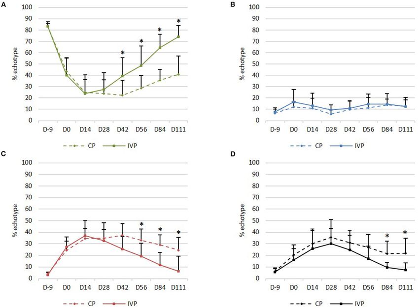
Depuydt et al.                                                                                                tpMSC Treatment for Tendon Injuries

as amended, Belgian animal welfare legislation (KB 29/05/2013),           The horses were positioned in right lateral recumbency. After
Directive 2010/63/EU and EMEA/CVMP/816/00-Final).                         circumferential clipping and aseptical preparation of the surgical
                                                                          sites, a small skin incision (1 cm) was made in the palmar midline
Study Design and Horses                                                   of the limbs just proximal to the digital flexor tendon sheath.
Eight healthy warmblood horses, four geldings and four mares              Through a small longitudinal incision into the SDFT (1 cm),
(age ranged between 3 and 12 years) were enrolled in this                 a 3.5 mm Steinmann pin was inserted in the core of the SDFT
blinded, placebo-controlled, randomized study. The horses were            under ultrasonographic guidance and a central lesion over an
free of lameness and swelling, heat or reaction to palpation              area of 8 cm was created. Next, an arthroscopic burr (∅ 3.5 mm
of the SDFT of both forelimbs at the time of enrollment. To               Razorcut blade; C9110; Linvatec, Largo, Florida, USA) was
reduce the number of horses, this study compared the effect               inserted in the defect created by the Steinmann pin and was
of the investigational product (IVP) and the control product              advanced proximally over a length of 8 cm. After activation, the
(CP) within the same horse. Each of the horses received the               burr was slowly retracted over 1 min while performing a rotating
CP in one of its forelimbs and the IVP in its contralateral               movement making sure that the medial, palmar and lateral
forelimb. The treatment allocation was randomized. Thus, one              aspect of the tendon was debrided. Local pressure on the SDFT
forelimb within a horse was randomly allocated to the IVP                 was exerted to assist in this debridement. The shaver was used
group (n = 8), while the contralateral limb was assigned to the           in forward modus at 5,000 rpm. No suction was applied. The
CP group (n = 8). All clinical, tendon, lameness, ultrasound,             incisions in the paratenon and skin were sutured using simple
and histopathological parameters were evaluated by an examiner            interrupted sutures with resorbable polyglyconate (Monosyn
blinded to the treatment.                                                 USP 3-0 B. Braun).
                                                                              A bandage was applied to the limbs for 7 days after surgery,
Investigational Product and Control                                       until treatment administration. A new bandage was placed
Product                                                                   after treatment (day 0). The bandage was removed for tendon
The IVP consisted of a proprietary formulation of equine                  assessment on day 1–3 after treatment and replaced after the
allogeneic tenogenic primed mesenchymal stem cells (tpMSCs)               assessment. From day 4 to 7 after treatment, the bandage was
prepared from peripheral blood as previously described (28).              removed at 10 a.m. ± 1 h and replaced at 4 p.m. ± 1 h right after
Briefly, blood was collected aseptically from the vena jugularis          tendon assessment. One week after surgery, the sutures and the
in sterile ethylenediaminetetraacetic acid (EDTA) tubes. After            bandage were removed permanently.
gradient centrifugation, the interphase was isolated, seeded and              Horses were walked for 6 days after surgery using a horse
cultivated in culture medium as previously described (30). At             walker (Table 2) and received an intralesional tpMSC or placebo
80% confluency, the cells were trypsinized and cultured in                treatment at 7 days post-surgery as described below. A gradually
tenogenic priming medium. At the next confluency, the cells were          increasing exercise protocol was started from day 4 after
trypsinized and resuspended in 1 mL Dulbecco’s modified Eagle             treatment onwards using a horse walker (Table 2). Care was
medium low glucose supplemented with 10% dimethyl sulfoxide.              taken to exercise in both directions equally.
The samples were cryopreserved at −80◦ C until further use. In
the present study, two different tpMSC batches of the same donor          Treatment Administration
horse that were produced on two different time points were used.          At day 0, one week post-surgery, the horses were mildly sedated
Each forelimb within one horse was randomly allocated either to           with detomidine hydrochloride (0.1 mL/100 kg body weight,
one of the two batches. The donor horse was screened for equine           Detogesic R , intravenous). The area around the injection site
pathogens as previously reported by our group (28).                       was clipped and aseptically prepared. The tendon was punctured
    The control product consisted of 1 mL sterile saline injectable       laterally and the treatments (IVP and CP) were administered by
solution (0.9% NaCl).                                                     a single intralesional injection in the SDFT core lesion under
                                                                          ultrasonographic guidance to ensure a successful administration
Surgical Procedure and Post-operative                                     to the site of the lesion. For the IVP, the tpMSCs were thawed
Treatment                                                                 in the palm of a hand and drawn into a syringe. Both IVP and
One week before treatment administration [day-7: reported                 CP treatments were administered by an assigned veterinarian
time point that fibroblast ingrowth starts (13)], a standardized          (the dispenser), who did not participate in any of the clinical
lesion in the SDFT of both forelimbs was created using a                  or laboratory examinations. This way blinding of the personnel
surgical model adapted from Schramme et al. (31) and Bosch                who assessed the efficacy and safety parameters and performed
et al. (32, 33) (Table 1). The surgery was performed under                the post-mortem evaluation was ensured.
general anesthesia and followed routine clinical procedures.
Pre-operatively, horses were sedated using romifidine (0.8                Clinical Assessment
mL/100 kg body weight; Sedivet R , intravenous). General                  A general clinical examination of the horses was conducted and
anesthesia was induced using ketamine (2.2 mL/100 kg body                 recorded daily throughout the study (from day −10 until day
weight; Ketamidor R , intravenous) and midazolam (0.1 mg/kg               112 ± 1). The body temperature, heart and respiratory rates,
body weight; Midazolam Mylan, intravenous). Anesthesia was                mucous membrane color, capillary refill time, and feed intake
maintained during the surgical procedure by administration of             were evaluated. The body weight of the horses was recorded on
isoflurane (IsoFlo R MAC∗ = 1.3%) under controlled ventilation.           day −10 and 112 ± 1.

Frontiers in Veterinary Science | www.frontiersin.org                 3                                     March 2021 | Volume 8 | Article 641441
Depuydt et al. tpMSC Treatment for Tendon Injuries
TABLE 1 | Comparison of surgically induced SDFT core lesions in the current TABLE 2 | Exercise protocol of the horses after bilateral surgical induction of a
study with the model according to Schramme et al. (31) and Bosch et al. (32, 33). SDFT core lesion.
′
Present Schramme et al. Bosch et al. Time point W = minutes walking, once per day
study (31) (32, 33) ′
T = minutes trotting, once per day
Instrument Arthroscopic Synovial resector Arthroscopic Day −7 Stall rest
burr burr Day −6 to 2.5 W
′
Change direction 2.5 W
′
Lesion 3.5 mm 3.5 mm 3.5 mm −5
diameter Day −4 to
′
5 W Change direction
′
5 W
Lesion length 8 cm 8 cm 7 cm −1
Suction No Yes No Day 0 Stall rest
Ultrasonographic Yes Yes No Day 1–3 Stall rest
guidance Day 4–14 2.5 W
′
Change direction 2.5 W
′
Bandage 7 days Limbs were 14 days Week 3
′
5 W Change direction
′
5 W
bandaged after ′ ′
Week 4 7.5 W Change direction 7.5 W
surgery, period of ′ ′
bandaging not Week 5 10 W Change direction 10 W
′ ′
reported Week 6 12.5 W Change direction 12.5 W
′ ′
Treatment tpMSCs Not applicable PRP Week 7 15 W Change direction 15 W
′ ′ ′ ′ ′ ′
Start exercise 1 week after 3 weeks after 4 weeks after Week 8–9 5 W 1 T 5 W Change direction 5 W 1 T 5 W
scheme surgery surgery surgery Week 10–11
′
5 W 2 T
′ ′
5 W Change direction
′
5 W 2 T
′ ′
5 W
Follow-up 16 weeks 2, 4, 8, and 12 24 weeks Week 12-13 ′
5 W 3 T
′ ′
5 W Change direction
′
5 W 3 T
′ ′
5 W
weeks ′ ′ ′ ′ ′ ′
Week 14–15 5 W 4 T 5 W Change direction 5 W 4 T 5 W
′ ′ ′ ′ ′ ′
Week 16 5 W 5 T 5 W Change direction 5 W 5 T 5 W
This exercise scheme was performed once a day. Care was taken to exercise equally in
′
During the study period from the day of treatment both directions and time was divided equally over both sides. ′ W = minutes walking; T
= minutes trotting.
administration (day 0) to study completion, all horses were
observed daily for adverse events, serious adverse events and
suspected adverse drug reactions and these were recorded TABLE 3 | Clinical scores used in all horses to assess local heat, swelling, and
when occurring. pain to pressure.
Parameter Score system Definition
Blood Sample Collection and Analysis
Blood was collected from the vena jugularis into EDTA tubes and Heat 0 No increased temperature sensation
serum clot activator tubes for analysis of following hematological 1 Slightly increased temperature sensation
and biochemical blood parameters in an ISO15189 certified 2 Moderately increased temperature sensation
laboratory (Vebio, France): white blood cell count, red blood 3 Severely increased temperature sensation
cell count, hemoglobin, hematocrit, mean corpuscular volume, Pain to pressure 0 No pain to pressure
mean corpuscular hemoglobin, mean corpuscular hemoglobin 1 Slight pain to pressure
concentration, neutrophils, eosinophils, lymphocytes, platelet 2 Moderate pain to pressure
count, basophils, monocytes, reticulocytes, blood urea nitrogen, 3 Severe pain to pressure
urea, creatinine, total protein, albumin, globulin, alkaline Swelling 0 No swelling
phosphatase, serum glutamic oxaloacetic transaminase, total and 1 Slight swelling
direct bilirubin, creatinine kinase, lactic dehydrogenase, and 2 Moderate swelling
gamma-glutamyl transpeptidase. 3 Severe swelling
Tendon Assessment
The tendon assessment was performed on following days: −9
(before surgical procedure), 0 (before treatment), daily on days Lameness Assessment
1–14, 28, 42, 56, 84, and 112 ± 1. Both forelimbs of each horse Lameness was assessed on following days: −9 (before the surgical
were evaluated at each time point. A score was given to the procedure), 0 (before treatment), 14, 56, and 112 ± 1 according
local heat, pain to pressure, and the swelling (Table 3). Also, the to the American Association of Equine Practitioners (AAEP)
circumference measurement (cm) of each forelimb was recorded scoring system (Table 4). The examining vet also evaluated
using a measuring tape. The tape was placed at the level of the whether the lameness was attributed to the left or right front
metacarpus just proximal to the digital flexor tendon sheath with limb or if the lameness was considered bilateral. The horses were
the limb in a weight bearing position. All measurements were examined by walking and trotting in hand, on a straight line
performed on both forelimbs and the values at each time point and with a loose line to the halter. Evaluation in a circle was
were compared to the baseline values (day −9 and day 0). performed during the revalidation exercise in the horse walker
Frontiers in Veterinary Science | www.frontiersin.org 4 March 2021 | Volume 8 | Article 641441
Depuydt et al.                                                                                                                   tpMSC Treatment for Tendon Injuries

TABLE 4 | AAEP scoring.                                                                     12-5 MHz multi-frequency ultrasound probe (12 L5; Terason
                                                                                            Ultrasound, Teratech Corporation, Burlington, MA, USA)
Grade             Degree of lameness
                                                                                            connected to a laptop computer (MacBook Pro R 17 inch, Apple,
0                 Lameness not perceptible under any circumstances                          Cupertine, CA, USA) loaded with software for data acquisition
1                 Lameness is difficult to observe and is not consistently apparent,        and analysis (UTCTM Software V.1.0.1 2010, UTC Imaging,
                  regardless of circumstances                                               Stein, The Netherlands) was used. The probe was fixed in a
2                 Lameness is difficult to observe when walking or trotting in a            motorized tracking device with built-in standoff pad (UTC-
                  straight line, but consistently apparent under certain                    TrackerTM ; UTC Imaging). Settings (depth, gain, focal zone)
                  circumstances                                                             were standardized and all examinations were performed by the
3                 Lameness is consistently observable at a trot under all                   same operator with the horse in weight bearing position, equally
                  circumstances
                                                                                            on both forelimbs. With the help of the tracking device, the probe
4                 Lameness is obvious at walk
                                                                                            moved automatically from proximal to distal parallel to the long
5                 Lameness produces minimal weight bearing in motion and/or at
                                                                                            axis of the tendon at constant speed over a distance of 12 cm.
                  rest or a complete inability to move
                                                                                            Compilation of contiguous transverse images was conducted at
                                                                                            precise steps of 0.2 mm, including the insertion point of the
TABLE 5 | Echo types and corresponding colors that were discriminated and                   arthroscopic burr (scar in the epitenon) as the reference point
determined throughout the study (34).                                                       for the analysis. A clear ultrasound image of the SDFT was
                                                                                            obtained and checked to ensure no artifacts were present due
Echo type (color code)Echo source according to histology
                                                                                            to movement or poor contact. The compiled 600 transverse
Echo type I (green)        Echo generated by intact and fully aligned fiber bundles         ultrasound images were reconstructed into a three-dimensional
Echo type II (blue)        Echo generated by discontinuous and less aligned fiber           data block of ultrasound information and stored digitally until
                           bundles                                                          the end of the examination period. The stability of the echo
Echo type III (red)        Echo generated by a mainly fibrillar matrix with                 pattern of corresponding pixels in contiguous transverse images
                           accumulation of collagen fibrils not (yet) organized into        can be analyzed (UTC2011 R Analyser V1.0.1; UTC Imaging;
                           fiber bundles                                                    window size 17). Four different pixel types were classified based
Echo type IV (black)       Echo generated by an amorphous matrix and/or fluid               on their stability and degree of persistence, resulting in four echo
                                                                                            types related to stages of ultrastructural integrity (Table 5) (34).
                                                                                            In the analysis, a 4 cm long tendon segment from 2 to 6 cm
TABLE 6 | Ultrasound assessment.
                                                                                            proximal to the scar in the epitenon was selected. In this selected
Parameter               Score system        Definition                                      segment, the percentage of echo types generated by the tendon
                                                                                            structure was evaluated.
Echogenicity                   0            Normal echogenicity
                               1            Mildly hypoechoic                               Ultrasound Assessment
                               2            Moderate hypoechoicity                          The ultrasound images obtained with the UTC device were used
                               3            Severe hypoechoicity                            for the ultrasound assessment. During this evaluation, three
Fiber alignment                0            ≥75% parallel fiber bundles in the lesion       different parameters were evaluated. First, the echogenicity of
score                          1            50–74% parallel fiber bundles in the
                                                                                            each lesion was graded according to a pre-defined scoring system
                                            lesion
                                                                                            (Table 6). Second, the fiber alignment was graded according to
                               2            25–49% parallel fiber bundles in the
                                            lesion
                                                                                            the estimated percentage of parallel fiber bundles in the lesion
                               3            ≤25% parallel fiber bundles in the lesion
                                                                                            (Table 6). Third, an increase in anterior-posterior thickness
                                                                                            (APT) in cm was recorded to monitor tendon repair.

                                                                                            Histological Sampling Procedure
on a soft surface, and on a lunge. A firm, non-slippery surface                             At day 112 ± 1 of the study, the horses were humanely
was used for trotting on a straight line and for lunging.                                   euthanized after sedation with romifidine (0.8 mL/100 kg body
                                                                                            weight; Sedivet R ) using ketamine (2.2 mL/100 kg body weight;
Ultrasound Tissue Characterization                                                          Ketamidor R ), midazolam (0.1 mg/kg body weight; Midazolam
The structural integrity of the SDFT of both forelimbs of                                   Mylan) and T61 (0.3 mL/kg body weight; Hoechst Roussell Vet,
each horse was qualified using established protocols for an                                 Frankfurt, Germany).
ultrasound tissue source characterization (UTC) (32, 34). The                                  Immediately after euthanasia, the SDFT and paratenon of
UTC assessment was performed on the following days: −9                                      both forelimbs of each horse were collected. After closing the
(before surgery), 0 (before treatment), 14, 28, 42, 84, and 112 ± 1.                        surgical wound the skin remained slightly raised, leaving the
The horses were mildly sedated with detomidine hydrochloride                                original entry point of the arthroscopic burr visible throughout
(0.1 cc/100 kg body weight, Detogesic R , intravenous) and both                             the entire study period. This remaining scar could be used as a
forelimbs were clipped, shaved and washed at the palmar aspect                              reference for the entry point of the burr. The tendon segment
of the metacarpus prior to each scan. Coupling gel was applied                              located 12 cm proximal to the entrance of the burr was obtained.
to ensure maximum contact. For the UTC examination, a                                       Next, transversal and longitudinal tendon sections were excised,

Frontiers in Veterinary Science | www.frontiersin.org                                   5                                      March 2021 | Volume 8 | Article 641441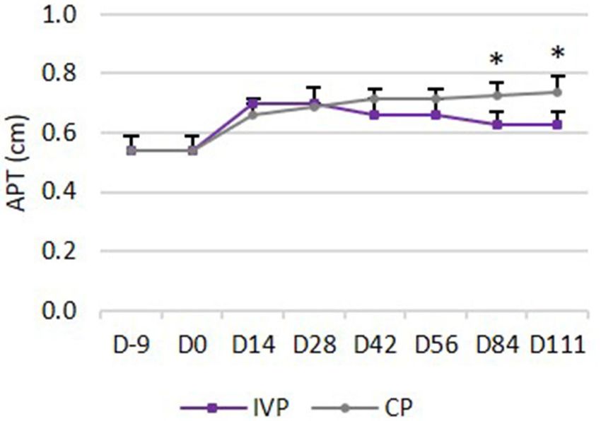
Depuydt et al.                                                                                                 tpMSC Treatment for Tendon Injuries

and a macroscopic evaluation of these sections was performed.                dehydrated and coverslips were applied. In between all steps,
Finally, digital photographs were taken from each tendon before              the sections were washed extensively and repeatedly with 0.1%
fixation in 10% natural-buffered formalin.                                   PBS-Tween 20. Only after incubation with the secondary HRP
   Samples of the cubital lymph nodes, prescapular lymph                     antibody, the slides were rinsed with PBS instead of 0.1% PBS-
nodes, spleen, liver, brain, kidneys, adrenal glands, lungs, heart           Tween 20.
(ventricles and septum), stomach, duodenum, jejunum, ileum,
colon, and cecum were fixed in 10% natural-buffered formalin                 Collagen Type III
for further histologic evaluation.                                           Antigen retrieval was performed using an EDTA buffer (10% in
                                                                             distilled water; pH 9.0) for 30 min at 95◦ C. Next, slides were
Macroscopic and Histopathologic                                              incubated with H2 O2 (S202386-2; Agilent) for 5 min at room
Examination                                                                  temperature in the dark followed by a treatment with normal
A macroscopic evaluation of tendon, paratenon, and cubital                   goat serum (30% in PBS) for 30 min at room temperature.
lymph nodes of both forelimbs of each horse was performed.                   Subsequently, incubation with the primary polyclonal rabbit
The lesion and color of the tendon were observed and                         collagen type III antibody (1:200; ab7778; Abcam) was performed
recorded (lesion visible or not, discoloration visible or not). The          for 90 min at room temperature followed by a secondary
paratenon and lymph nodes were evaluated for abnormalities                   incubation with anti-rabbit antibody (biotinylated anti-rabbit;
of the thickness and color. Normal thickness and color were                  E0432; Agilent) for 30 min at room temperature in the dark.
differentiated from any other abnormalities. Any abnormality                 Next, incubation with ABC-HRP (Vector PK-6100) took place
found was recorded.                                                          for 30 min in the dark at room temperature. Visualization was
    Histological sections were prepared from all formalin-fixed              performed in DAB as described above.
samples and stained with haematoxylin & eosin (HE). All samples
were evaluated for presence of ectopic tissue and presence                   Von Willebrand Factor
of inflammation.                                                             Slides were incubated with proteinase K (0.05M in Tris-HCL, pH
    Samples of tendon and paratenon were scored according to                 7.6; S3004; Agilent) for 15 min at room temperature for antigen
the general accepted semi-quantitative score system of Movin                 retrieval. Next, sections were incubated with H2 O2 (S202386-
et al. (35). Briefly, fiber structure, fiber arrangement, roundness of       2; Agilent) for 5 min followed by the primary polyclonal
the nuclei, regional variations in cellularity, vascularity, collagen        rabbit VWF antibody (1:3200; A0082; Agilent) incubation for
stainability, glycosaminoglycan content, and the presence of                 30 min at room temperature. Subsequently, the secondary anti-
inflammatory cells were evaluated by an experienced pathologist              rabbit antibody (EnVision HRP anti-rabbit; K4003; Agilent)
(score 0: normal; score 1: slightly abnormal; score 2: moderately            was incubated for 30 min at room temperature in the dark.
abnormal; score 3: markedly abnormal).                                       Visualization was performed as described above.

Immunohistochemistry                                                         Smooth Muscle Actin
Immunohistochemistry was performed to evaluate the                           Antigen retrieval was performed using a citrate buffer (10%
distribution percentage of collagen type I (COL I), collagen                 in distilled water; pH 6.0) for 13 min in the microwave. Next,
type III (COL III), Von Willebrand Factor (VWF) and                          sections were incubated with H2 O2 (S202386-2; Agilent) for
smooth muscle actin (SMA). For each staining, 5 µm thick                     5 min followed by incubation with the primary monoclonal
tendon sections were cut and mounted on Thermo Scientific                    mouse SMA antibody (1:200; M0851; Agilent) for 30 min at
SuperFrost PlusTM adhesion slides (Fischer Scientific, UK).                  room temperature. Then, the secondary anti-mouse antibody
Next, slides were deparaffinized and rehydrated in xylene and                (EnVision HRP anti-rabbit; K4001; Agilent) was incubated for
decreasing concentrations of alcohol in H2 O (96, 80, 70, 60,                30 min at room temperature in the dark. Visualization was
and 100% H2 O).                                                              performed as mentioned above.
                                                                                Positive stainings were confirmed on microscopy. For
Collagen Type I                                                              each section, a randomly selected area was photographed (at
Antigen retrieval was performed by incubation with pronase                   ×40 magnification). The percentage of the total stained area
(1 mg/mL) and subsequently hyaluronidase (10 mg/mL) at                       (representing COL I, COL III, SMA, or VWF content) was
37◦ C for 20 min. Sections were treated with goat serum                      calculated with software (LAS, version 4.1, Leica microsystems,
[10% in phosphate-buffered saline (PBS)] for 15 min at room                  Diegem, Belgium).
temperature, after which they were incubated with the primary
polyclonal rabbit collagen type I antibody (1:500; ab138492;                 Data Analysis
Abcam) for 60 min at room temperature. Next, the secondary                   All statistical analyses were performed using SAS R statistical
anti-rabbit antibody (EnVision horseradish peroxidase (HRP)                  analysis software version 9.3. The groups were compared to
anti-rabbit; K4001; Agilent) was incubated for 30 min at room                baseline using ANOVA for the lameness assessment, increase
temperature in the dark. Visualization was performed in                      of tendon circumference and increase of local pain to pressure.
a 3.3-diaminobenzidine solution (DAB; K3468; Agilent) for                    The treatment groups were compared using the Mantel-Haenszel
7 min at room temperature in the dark. The cell nuclei were                  test for tendon assessment (pain, heat, and swelling), lameness
counterstained with hematoxylin, after which the sections were               assessment, ultrasound assessment (echogenicity and fiber

Frontiers in Veterinary Science | www.frontiersin.org                    6                                   March 2021 | Volume 8 | Article 641441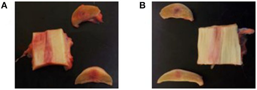
Depuydt et al.                                                                                                                       tpMSC Treatment for Tendon Injuries

 FIGURE 1 | Tendon assessment. (A) Local heat. 0 = no increase in temperature sensation; 1 = slightly increased temperature sensation; 2 = moderately increased
 temperature sensation; 3 = severely increased temperature sensation. (B) Local pain to pressure. 0 = no pain to pressure; 1 = slight pain to pressure; 2 = moderate
 pain to pressure; 3 = severe pain to pressure. (C) Local swelling. 0 = no swelling; 1 = slight swelling; 2 = moderate swelling; 3 = severe swelling. * P-values ≤ 0.05.

alignment score) and histopathology. Groups were compared                                 No serious adverse events or suspected adverse drug reactions
using Wilcoxon test for tendon circumference, anterior-posterior                        were observed throughout the study period.
thickness, UTC assessment and immunohistochemistry. Groups
were compared using the Fisher’s exact test for incidence of                            Hematological and Serum Biochemistry
adverse events, serious adverse events and suspected adverse                            Parameters
drug reactions, incidence of abnormal clinical signs and                                The analyses of the hematological and serum biochemistry
decrease of body weight. P-value ≤ 0.5 was considered                                   parameters revealed no serious deviations from the reference
statistically significant.                                                              values (Supplementary Material). The observed deviations were
                                                                                        marginally out of range and were regarded as not clinically
                                                                                        relevant by the attending veterinarian.
RESULTS
Horses                                                                                  Tendon Assessment
Due to the study design each horse received the CP in one of                            Local Heat
its forelimbs, whereas, the contralateral limb in each horse was                        There were no differences between the IVP group and the
treated with the IVP.                                                                   CP group during the study period. Prior to surgery (day −9)
                                                                                        and from day 28 onwards, none of the horses showed increase
                                                                                        in local heat at the level of the SDFT area. On days 0–14 a
Clinical Assessment                                                                     slightly increased temperature was observed in all limbs, with the
The clinical examination parameters, namely body temperature,                           exception of day 4 where half of the limbs in each treatment group
heart, and respiratory rates were all in the physiological range for                    showed no increased temperature (Figure 1A).
all horses at all time points. There was a mean increase of body
weight of 3.8 kg (± 4.05) from day −9 to day 111.                                       Pain to Pressure
    One adverse event was recorded: one horse showed mild signs                         There were no differences in pain to pressure between the IVP
of colic 38 days following treatment. The horse also had reduced                        and CP group. Prior to surgery (day −9) and from day 4 onwards,
feed intake the day after. No treatment was necessary and the                           none of the horses showed local pain to pressure. Between day 1
horse fully recovered. The condition was deemed not to be related                       and 3, no or slight pain to pressure was observed in half of the
to the study medication.                                                                limbs in each treatment group (Figure 1B).

Frontiers in Veterinary Science | www.frontiersin.org                               7                                             March 2021 | Volume 8 | Article 641441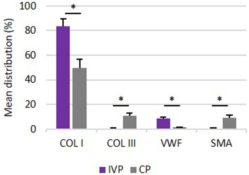
Depuydt et al.                                                                                                               tpMSC Treatment for Tendon Injuries

Local Swelling                                                                              There was no significant difference in visual lameness scores
On day −9, 84, and 111 none of the horses showed local swelling.                        between the two treatment groups (IVP vs. CP). On days −9, 56,
On all other observation days, the scores ranged between 0 and                          and 111 none of the horses showed lameness (AAEP score 0). On
2. Significant differences between groups were seen on day 9                            days 0 and 14, an AAEP lameness score of 1 was observed in both
and 10, where the percentage of moderate swelling (score 2) was                         forelimbs of all horses (Figure 2), indicating a bilateral lameness
significantly higher in the IVP group compared to the CP group                          in the horses.
(P = 0.009 and P = 0.025, respectively) (Figure 1C).
   No significant differences in tendon circumference could                             Ultrasound Assessment
be observed between groups throughout the study duration.                               Echogenicity
However, when changes of the mean tendon circumference                                  There was no difference in echogenicity between the two
were compared to the baseline values of day 0 (after surgery,                           treatment groups from day −9 until day 28. From day 28
but before treatment administration), a significant difference                          onwards, the frequency of scores indicating hypoechoicity (score
between the IVP treated limbs and the contralateral CP treated                          1–3) was significantly lower in the IVP treated limbs in
limbs could be determined on day 9 and 10 (P ≤ 0.05). The mean                          comparison to the CP treated limbs (P = 0.009 on day 28, P <
circumference of the CP group remained the same, whereas a                              0.001 from day 42 to 84, P = 0.003 on day 111) (Figure 3A).
mean increase of 0.2 cm was observed in the IVP group when
compared to baseline.                                                                   Fiber Alignment Score
                                                                                        There was no significant difference in fiber alignment score
                                                                                        between the two groups from day −9 until day 28. From day 28
                                                                                        onwards, the percentage of parallel fiber bundles was significantly
                                                                                        higher in the IVP group in comparison to the CP group (P <
                                                                                        0.001 at all time points) (Figure 3B).
                                                                                            No significant differences could be observed in anterior-
                                                                                        posterior thickness (APT) between both treatment groups from
                                                                                        day −9 until day 84. The increase in APT was significantly lower
                                                                                        of the IVP treated limbs compared to the CP treated limbs on day
                                                                                        84 (P = 0.010) and day 111 (P = 0.009) (Figure 4).

                                                                                        UTC Assessment
                                                                                        Echo type I (echo generated by intact and fully aligned fascicles,
                                                                                        green) was observed in the majority (82–84%) of the limbs in all
                                                                                        treatment groups on day −9 (before surgery). This percentage
                                                                                        dropped to 12–45% between day 0 and day 28. Significant
                                                                                        differences between groups were seen from day 42 onwards,
 FIGURE 2 | Lameness assessment. 0 = Lameness not perceptible under any
 circumstances; 1 = Lameness is difficult to observe and is not consistently            with significantly higher percentages for IVP treated limbs in
 apparent, regardless of circumstances; 2 = Lameness is difficult to observe at         comparison to the CP treated limbs (P = 0.039 on day 42, P =
 a walk, or when trotting in a straight line, but consistently apparent under           0.026 on day 56, P = 0.009 on day 84, and P = 0.007 on day 111)
 certain circumstances; 3 = Lameness is consistently observable at a trot under         (Figure 5A).
 all circumstances; 4 = Lameness is obvious at a walk 5 = Lameness produces
 minimal weight bearing in motion and/or at rest or a complete inability to move.
                                                                                           Percentage in echo type II (echo generated by discontinuous
                                                                                        and less aligned fascicles, blue) was not significantly different

 FIGURE 3 | Ultrasound assessment. (A) Echogenicity scores: 0 = normal; 1 = mildly hypoechoic; 2 = moderately hypoechoic; 3 = severely hypoechoic. (B) Fiber
 alignment scores: 0 = ≥ 75%; 1 = 50–74%; 2 = 25–49%; 3 = ≤ 24%. *P-values ≤ 0.05.

Frontiers in Veterinary Science | www.frontiersin.org                               8                                     March 2021 | Volume 8 | Article 641441
Depuydt et al. tpMSC Treatment for Tendon Injuries
between the two treatment groups at any observation day
(Figure 5B).
Echo type III (echo generated by a mainly fibrillar matrix, red)
was observed on day −9 in a relatively low percentage (3–5%)
of the limbs in all treatment groups, whereas this percentage
increased to 22–45% between day 0 and day 42. Significant
differences between groups were seen from day 56 onwards, with
lower percentages for IVP treated limbs when compared to the
CP treated limbs (P = 0.032 on day 56, P = 0.009 on day 84 and
P = 0.007 on day 111) (Figure 5C).
Echo type IV (echo generated by an amorphous matrix
and fluid, black) was observed on day −9 in a relatively low
percentage (6–7%) of the limbs in all treatment groups, whereas
this percentage increased to 11–44% between day 0 and day 56.
Significant differences between treatment groups were seen from
day 84 onwards, with lower percentages for IVP treated limbs in FIGURE 4 | Mean (+SD) anterior-posterior thickness (cm). *P-values ≤ 0.05.
comparison to the CP treated limbs (P = 0.026 on day 84 and P
= 0.021 on day 111) (Figure 5D).
On day 111, a significantly higher percentage in echo type I (P
= 0.008) and a significantly lower percentage in echo type III and Immunohistochemistry
IV (P = 0.008 and P = 0.016 respectively) was observed in the The results of percent distribution and immunohistochemical
IVP treated limbs, as compared to day 0. No significant changes stainings are depicted in Figures 7, 8, respectively.
in percentage of each echo type were observed in CP treated limbs
when compared to day 0. Collagen Type I
Moreover, no significant differences could be observed in the The percent distribution of COL I was significantly higher for all
IVP treated limbs between day −9 and day 111 for any echo IVP treated limb in comparison to CP treated limbs (P = 0.005).
type. This indicates that the tendon quality on day 111 after
Collagen Type III
intralesional tpMSC treatment is comparable with the healthy
The percent distribution of COL III was significantly lower for all
tendon quality and structure as observed on day −9. This was not
IVP treated limb in comparison to CP treated limbs (P = 0.005).
the case for the CP treated limbs, where significantly lower echo
types I (P = 0.008) and significantly higher echo types III and IV Von Willebrand Factor
(P = 0.008) were generated on day 111 compared to day −9. VWF was significantly higher for all IVP treated limbs in
comparison to CP treated limbs (P = 0.005).
Macroscopic and Histopathologic
Examination Smooth Muscle Actin
Macroscopic Evaluation The percent of distribution of SMA was significantly lower for all
The lesion site was still visible in all SDFTs collected from both IVP treated limb in comparison to CP treated limbs (P = 0.005).
treatment groups, as well as discoloration of the tendon tissue
(Figure 6). The thickness of the paratenon as well as the thickness DISCUSSION
of the curial lymph nodes was considered to be normal in all
limbs of both groups. This randomized, blinded and placebo-controlled study was
performed to evaluate the safety, tolerance, and efficacy of an
Ectopic Tissue intralesional injection with allogeneic equine peripheral blood-
No ectopic tissue was detected during histopathological derived tenogenic primed mesenchymal stem cells (tpMSCs)
examination of the tendon, paratenon, or the cubital on surgically induced SDFT core lesions. The adapted surgical
lymph nodes. procedure from Schramme et al. (31) was performed in the
SDFT of both forelimbs of each horse. This model provides
Histologic Evaluation a more standardized and localized injury in comparison
In all organ and lymph node slides ectopic tissue and with enzymatically induced lesions (36) and resembles the
inflammation were not observed. naturally occurring injuries more closely in terms of clinical,
ultrasonographic, and histological findings (33, 37). The surgical
Scoring of the Tendon Tissue procedure induced a tendon lesion in both forelimb SDFT’s,
No significant differences of fiber structure, fiber arrangement, which could be confirmed by ultrasound and UTC assessment.
roundness of the nuclei, regional variations in cellularity, Because fibroplasia generally begins around the seventh day after
vascularity, collagen stainability, glycosaminoglycan content and injury (29), the treatment was administered 1 week following
the presence of inflammatory cells could be observed between surgery, after the acute phase of the inflammatory response
treatment groups (Table 7). caused by the surgical induction of the SDFT core lesion
Frontiers in Veterinary Science | www.frontiersin.org 9 March 2021 | Volume 8 | Article 641441Depuydt et al. tpMSC Treatment for Tendon Injuries
has subsided (38, 39). One forelimb of each horse received medication since the incidence in this study (1 episode in 1
an intralesional injection with tpMSCs and the contralateral out of 8 horses over 121 days of observation) is similar to the
forelimb was treated with saline to serve as an intra-animal incidence rate of colic in horses observed in the field (3.5–
control. This design allows to minimize the variability in 10.6 cases per 100 horse years) (40). No other serious adverse
tendon quality between treatment groups, and the number of events or suspected drug reactions were observed throughout the
experimental animals used (21, 33). This study design was similar study period.
to a study by Bosch et al. (33) evaluating neovascularization No significant difference between the tpMSC and contralateral
of tendon tissue after the surgical induction of a SDFT core placebo treated limbs was observed for heat and pain to
lesion. In that study evaluating six horses, one forelimb was pressure. Swelling was observed after surgery in all treatment
treated with platelet-rich-plasma (PRP) and the contralateral groups with similar distribution of scores. A significantly
limb with placebo (saline) to serve as an intra-animal control. higher swelling in the IVP treated limbs was recorded on
Increased neovascularization in the PRP treated limbs could day 9 and 10, resulting in a higher tendon circumference in
be demonstrated at 24 weeks post-injection, pointing out the comparison to baseline values. This reaction might be related
relevance of the current study protocol and the number of to the tpMSC treatment, however no other clinical signs
horses used. were associated. The swelling was limited (2 mm), temporary
All eight treated horses were controlled for adverse events. (2 days) and decreased from day 11 onwards. Comparable
Only one adverse event, a colic episode at New Year’s Eve (38 studies also observed swelling following intra-lesional MSC
days post-treatment), was observed in one horse, and the patient treatment, reporting a decrease from week 2 or week 8 after
fully recovered the next day without the need for treatment. MSC injection onwards using autologous BM-MSCs (41) and
The adverse effect was not regarded to be related to the study AT-MSCs (23, 42).
FIGURE 5 | UTC assessment. (A) Mean (+SD) echo type I (green, fully aligned fascicles); (B) Mean (+SD) echo type II (blue, discontinuous and less aligned fascicles);
(C) Mean (+SD) echo type III (red, fibrillar matrix); (D) Mean (+SD) echo type IV (black, amorphous matrix and fluid). *P-values ≤ 0.05.
Frontiers in Veterinary Science | www.frontiersin.org 10 March 2021 | Volume 8 | Article 641441Depuydt et al. tpMSC Treatment for Tendon Injuries
FIGURE 6 | Macroscopic evaluation of the tendon tissue on day 112. (A)
Longitudinal and transverse sections of the control product treated forelimb.
(B) Longitudinal and transverse sections of the investigational product treated
forelimb.
TABLE 7 | Histological scoring of the tendon tissue.
Parameter IVP [mean (±SD)] CP [mean (±SD)] FIGURE 7 | Percentage distribution of COL I, COL III, VWF and SMA.
Comparison of mean distribution percentages (+SD) of extracellular matrix
Fiber structure 0.6 (0.9) 0.0 (0.0) proteins between investigation product treated limbs (IVP) and control product
Fiber arrangement 0.6 (0.9) 0.1 (0.4) treated limbs (CP). *P-values ≤ 0.05.
Roundness of the nuclei 1.6 (0.9) 1.3 (0.5)
Regional variations in cellularity 1.6 (0.9) 1.9 (0.8)
Vascularity 1.8 (0.5) 1.4 (0.5)
Collagen stainability 2.1 (0.8) 1.6 (0.9) from a recurrent injury of the treated limb, whereas no horse
Glycosaminoglycan content 2.8 (0.5) 2.4 (0.7) with a fiber alignment score of 0 re-injured the treated limb.
Presence of inflammatory cells 0.6 (1.1) 0.5 (1.1) This might indicate that the significantly better results of the
tpMSC treatment in the current study in comparison to the
Score 0, normal; score 1, slightly abnormal; score 2, moderately abnormal; score 3,
placebo treatment may lower the risk of re-injury in the long-
markedly abnormal.
term. However, a field study should be performed in order to
confirm this hypothesis.
On the other hand, it has been shown that conventional
Lameness (AAEP score 1) was observed on days 0 and 14 in ultrasonography is not sufficiently sensitive to accurately and
all forelimbs, but not at further lameness examinations. Within unequivocally evaluate the composition of tendon tissue (44).
the first and second week after an acute SDFT injury, mild In a human study, a lower score for hypoechoicity was not
transient lameness can occur (43). Schramme et al. (31) reported correlated with an improved clinical outcome (45). Therefore,
mild to moderate lameness in the 6-day period after surgical ultrasound tissue characterization (UTC) was included in the
induction of a bilateral SDFT core lesion. Within the following present study to improve the objective tendon characterization
2–7 days, lameness resolved rapidly. The observation of lameness by standardizing instrumental settings. The UTC assessment
on days 0 and 14 is likely related to the surgical procedure and showed significant differences between groups from day 42
is consistent with the clinical timeline observed with naturally onwards for echo type I. The tpMSC treated limbs showed a
occurring tendon lesions. significantly higher echo type I percentage in comparison to the
Ultrasonography is the standard method to assess tendon placebo group. This echo type corresponds to healthy tendon
healing over time. Significant differences in fiber alignment score structure consisting of fully aligned intact tendon structures with
and echogenicity between groups could be observed from day a high amount of collagen type I fibers (32). The percentages
28 onwards. The frequency of scores indicating hypoechoicity of echo type III and echo type IV were significantly lower in
and a relatively low percentage of parallel fiber bundles were the tpMSC treated tendons in comparison to the placebo group
significantly lower in tpMSC treated limbs in comparison to from day 56 to 84 onwards, respectively. Moreover, no significant
saline treated limbs. At the end of the study period, 7 out of differences could be observed in the IVP treated limbs between
8 tpMSC treated limbs compared to 0 out of 8 control limbs day −9 (before surgery) and day 111 for any echo type. This
showed over 75% parallel fiber bundles in the lesion (score indicates that the tendon quality on day 111 after intralesional
0). Additionally, hypoechoic structures persisted significantly tpMSC treatment is comparable with the healthy tendon quality
longer in the placebo treated limbs compared to tpMSC treaded and structure as observed on day −9.These findings demonstrate
forelimbs. This is in line with a previously performed study where that a more mature and superiorly healed tendon was present in
an improvement of the fiber alignment score was observed at the tpMSC treated group at the end of the study compared to
6 and 12 weeks after a combined tpMSC-PRP treatment (29). the placebo group (32). The UTC results of the placebo group
Dyson et al. (5) demonstrated that the fiber alignment score reported by Bosch et al. (32) at 16 weeks post-injection are in
at 16 weeks can be correlated with the re-injury rate after 2 line with the placebo data of the current results 16 weeks after
years. All horses with a fiber alignment score of 2 or 3 suffered treatment for types I and IV. This demonstrates that the model
Frontiers in Veterinary Science | www.frontiersin.org 11 March 2021 | Volume 8 | Article 641441Depuydt et al. tpMSC Treatment for Tendon Injuries
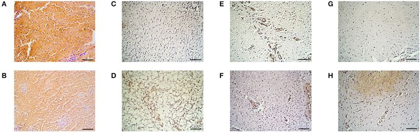
FIGURE 8 | Immunohistochemistry. Collagen type I in the (A) IVP and (B) CP treated limb, collagen type III in the (C) IVP and (D) CP treated limb, Von Willebrand
Factor in the (E) IVP and (F) CP treated limb and of smooth muscle actin in the (G) IVP and (H) CP treated limb. Scale bar = 10 µm.
as well as the UTC scans were performed in a similar way. higher percentage of VWF in the tpMSC treated limbs in
However, when looking at PRP vs. tpMSC treatment considerable the current study indicates a significantly enhanced healing
differences could be noticed at the same time point between the capacity in these limbs (33). In a similar study performed
studies. Bosch et al. used a preliminary version of the tracker, by Conze et al. (50), where nine horses received AT-MSCs
which was moved by hand along a magnetic strip (window size in a surgically created SDFT lesion in one forelimb and a
9), rather than the automatic tracker used here. The results for placebo (inactivated autologous serum) in the contralateral
PRP in that study at week 16 showed much lower values of forelimb, an increased neovascularization of the AT-MSC treated
echo type I compared to tpMSC treatment in our study (56 vs. tendons was also observed at 22 weeks post-treatment. In
74%). Approximately the same percentages for all echo types the present study no differences could be detected between
were reached at the end of the PRP study at week 24 as at week both treatment groups on histological sections, resulting in
16 in our study. This indicates a faster structural and functional an inconsistency with the UTC and immunohistochemistry
healing of the tendons treated with tpMSCs in comparison to the data. Although some studies could identify differences in
PRP treatment. standard histopathological parameters (20, 51), other studies only
The observations of the UTC assessment were confirmed by use immunohistochemistry instead of standard histopathology
the immunohistochemistry results obtained on day 112. The to obtain objectively quantified results (33). The absence of
high percentage of echo type I corresponding to healthy tendon differences between the IVP and the CP treated tendons could
structure with a high amount of collagen type I fibers are in be due to the fact that only one section of tissue was imaged for
line with the significantly higher distribution of collagen type histological evaluation.
I in the tpMSC treated limbs compared to the placebo treated Another limitation of this study was including both forelimbs
limbs. Strength and elasticity of the tendon is provided by making lameness harder to interpret, although only mild
collagen type I as the major fibrillar component. The presence lameness was observed during the study period without scoring
of a high amount of collagen type I is key for a functional, difficulties. Furthermore, the tpMSCs were not labeled so no
high-quality healing of the tendon tissue and an important conclusions could be drawn in the matter of cell survival
indicator of tendon matrix synthesis (12, 21). In the placebo and retention. Cell labeling was not performed to avoid
treated limbs, more fibrotic tissue was observed as represented alterations of the final IVP that might influence the mode of
by a higher percentage of echo type III on UTC and a higher action. There was no biomechanical testing data or return to
distribution of collagen type III and SMA and a lower amount exercise data to investigate the risk of re-injury. Additionally,
of blood vessels on immunohistochemistry. The SMA protein an experimental model was used to obtain standardized
is incorporated in myofibroblasts that play a role in early post- circumstances. However, naturally occurring tendon injuries
inflammatory events after tendon injury (11). An increased present more variability and cannot always be treated on
number of myofibroblasts results in the synthesis of abundant the right time point in the healing process. Therefore, the
amounts of COL III, resulting in the formation of persistent results of this study should be confirmed in a field trial on
scar tissue (46–48), reduced blood flow and eventually chronic a large number on horses suffering from naturally occurring
tendinopathies (49). The poor vascularization of tendons is tendon disease.
considered to be one of the reasons for their limited healing This study demonstrates that allogeneic tpMSCs are well-
potential (33). Blood vessels can be visualized by staining for tolerated and may be effective in the management of tendon
VWF, which can be found in functional endothelial cells and injuries in horses. A higher percentage of intact and fully aligned
thus acts as a marker for vascularization (50). The significantly fascicles on UTC in the tpMSC treated limbs in comparison
Frontiers in Veterinary Science | www.frontiersin.org 12 March 2021 | Volume 8 | Article 641441Depuydt et al. tpMSC Treatment for Tendon Injuries
to placebo treated limbs demonstrate the beneficial effects of of the horses were performed by SB. HVS provided the tools
tpMSCs for the treatment of tendon injuries in horses. and knowledge of the ultrasonographic tissue characterization.
Ultrasound and ultrasound tissue characterization assessments
CONCLUSION were performed by SB and ED. The surgical induction of
the core lesion was performed by AM and the treatment
The administration of tenogenic primed mesenchymal stem cells administration was done by FP. KC, LVB, and SB performed
resulted in significantly higher amounts of intact and fully aligned full necropsy and histopathology. ED wrote the first draft of
fascicles by UTC assessments and collagen type I distribution the manuscript. JS, SB, CB, LVH, AM, FP, KC, LVB, and HVS
by immunohistochemistry compared to a placebo treatment provided supervision and critical review of the manuscript.
after a single intralesional injection in surgically created SDFT All authors contributed and approved the final version of
core lesions. Additionally, significantly improved echogenicity the manuscript.
scores and fiber alignment scores were recorded. Therefore,
equine allogeneic tpMSCs are a promising therapeutic for tendon
FUNDING
injuries in horses.
This research was funded both by Global Stem cell Technology
DATA AVAILABILITY STATEMENT (GST) NV supported by a grant from the Flanders Innovation &
Entrepreneurship (Vlaio project number HBC.2018.0182).
The original contributions presented in the study are included
in the article/Supplementary Material, further inquiries can be
directed to the corresponding author. ACKNOWLEDGMENTS
The authors would like to thank Tamara Van Vooren and
ETHICS STATEMENT Sofie Perdu for the assistance during the necropsy and
The animal study was reviewed and approved by an independent Katrien Vanderperren for the ultrasonographic guidance during
ethics committee approved by the Flemish Government (permit treatment administration.
number: LA1700607).
SUPPLEMENTARY MATERIAL
AUTHOR CONTRIBUTIONS
The Supplementary Material for this article can be found
JS, SB, and LVH conceived the study and planned the design. online at: https://www.frontiersin.org/articles/10.3389/fvets.
Daily clinical assessments and periodical tendon assessments 2021.641441/full#supplementary-material
REFERENCES 8. Pinchbeck GL, Clegg PD, Proudman CJ, Stirk A, Morgan KL, French
NP. Horse injuries and racing practices in National Hunt racehorses in
1. Maffulli N, Wong J. Rupture of the Achilles and patellar tendons. Clin Sports the UK: the results of a prospective cohort study. Vet J. (2004) 167:45–
Med. (2003) 22:761–76. doi: 10.1016/S0278-5919(03)00009-7 52. doi: 10.1016/S1090-0233(03)00141-2
2. Cassel M, Baur H, Hirschmüller A, Carlsohn A, Fröhlich K, Mayer F. 9. van den Belt AJ, Dik K, Barneveld A. Ultrasonographic evaluation and long-
Prevalence of Achilles and patellar tendinopathy and their association to term follow-up of flexor tendonitis/desmitis in the metacarpal/metatarsal
intratendinous changes in adolescent athletes. Scand J Med Sci Sport. (2015) region in Dutch warmblood horses and standardbred racehorses. Vet Q.
25:e310–8. doi: 10.1111/sms.12318 (1994) 16:76–80. doi: 10.1080/01652176.1994.9694507
3. Williams RB, Harkins LS, Hammond CJ, Wood JLN. Racehorse injuries, 10. Singer ER, Barnes J, Saxby F, Murray JK. Injuries in the event horse:
clinical problems and fatalities recorded on British racecourses from flat training versus competition. Vet J. (2008) 175:76–81. doi: 10.1016/j.tvjl.2006.
racing and National Hunt racing during 1996, 1997 and 1998. Equine Vet J. 11.009
(2001) 33:478–86. doi: 10.2746/042516401776254808 11. Nichols AEC, Best KT, Loiselle AE. The cellular basis of fibrotic
4. Lam KH, Parkin TDH, Riggs CM, Morgan KL. Descriptive analysis of tendon healing: challenges and opportunities. Transl Res. (2019) 209:156–
retirement of Thoroughbred racehorses due to tendon injuries at the 68. doi: 10.1016/j.trsl.2019.02.002
Hong Kong Jockey Club (1992-2004). Equine Vet J. (2007) 39:143– 12. James R, Kesturu G, Balian G, Chhabra AB. Tendon: biology, biomechanics,
8. doi: 10.2746/042516407X159132 repair, growth factors, and evolving treatment options. J Hand Surg Am.
5. Dyson SJ. Medical management of superficial digital flexor tendonitis: a (2008) 33:102–12. doi: 10.1016/j.jhsa.2007.09.007
comparative study in 219 horses (1992-2000). Equine Vet J. (2004) 36:415– 13. Spaas JH, Guest DJ, Van de Walle GR. Tendon regeneration in human
9. doi: 10.2746/0425164044868422 and equine athletes. Sport Med. (2012) 42:871–90. doi: 10.1007/BF032
6. O’meara B, Bladon B, Parkin TDH, Fraser B, Lischer CJ. An investigation 62300
of the relationship between race performance and superficial digital flexor 14. Shojaee A, Parham A. Strategies of tenogenic differentiation of equine stem
tendonitis in the Thoroughbred racehorse. Equine Vet J. (2010) 42:322– cells for tendon repair: current status and challenges. Stem Cell Res Ther.
6. doi: 10.1111/j.2042-3306.2009.00021.x (2019) 10:1–13. doi: 10.1186/s13287-019-1291-0
7. Kasashima Y, Takahashi T, Smith RKW, Goodship AE, Kuwano A, Ueno 15. Maffulli N, Kader D. Tendinopathy of tendo achillis. J Bone Jt Surg-Ser B.
T, et al. Prevalence of superficial digital flexor tendonitis and suspensory (2002) 84:1–8. doi: 10.1302/0301-620X.84B1.0840001
desmitis in Japanese Thoroughbred flat racehorses in 1999. Equine Vet J. 16. Godwin EE, Young NJ, Dudhia J, Beamish IC, Smith RKW. Implantation
(2010) 36:346–50. doi: 10.2746/0425164044890580 of bone marrow-derived mesenchymal stem cells demonstrates improved
Frontiers in Veterinary Science | www.frontiersin.org 13 March 2021 | Volume 8 | Article 641441You can also read