Cholesterol-Ester Transfer Protein Alters M1 and M2 Macrophage Polarization and Worsens Experimental Elastase-Induced Pulmonary Emphysema - Frontiers
←
→
Page content transcription
If your browser does not render page correctly, please read the page content below
ORIGINAL RESEARCH
                                                                                                                                             published: 21 July 2021
                                                                                                                                   doi: 10.3389/fimmu.2021.684076

                                          Cholesterol-Ester Transfer Protein
                                          Alters M1 and M2 Macrophage
                                          Polarization and Worsens
                                          Experimental Elastase-Induced
                                          Pulmonary Emphysema
                                          Kelly Gomes Santana 1, Renato Fraga Righetti 2, Cristiane Naffah de Souza Breda 3,
                                          Omar Alberto Domínguez-Amorocho 3, Theresa Ramalho 4, Francisca Elda B. Dantas 1,
                                          Valéria Sutti Nunes 1, Iolanda de Fátima Lopes Calvo Tibério 2, Francisco Garcia Soriano 5,
                                          Niels O. S. Câmara 3, Eder Carlos Rocha Quintão 1 and Patrı́cia M. Cazita 1*
                                          1Laboratorio de Lipides, LIM-10, Hospital das Clinicas HCFMUSP, Faculdade de Medicina, Universidade de Sao Paulo,
                         Edited by:       Sao Paulo, Brazil, 2 Laboratório de Terapêutica Experimental I (LIM-20), Faculdade de Medicina da Universidade de São
          Christoph Thiemermann,          Paulo, Sao Paulo, Brazil, 3 Transplantation Immunobiology Lab, Department of Immunology, Institute of Biomedical Sciences,
   Queen Mary University of London,       Universidade de São Paulo, Cidade Universitária, São Paulo, Brazil, 4 Division of Infectious Diseases and Immunology,
                  United Kingdom          Department of Medicine, University of Massachusetts Medical School, Worcester, MA, United States, 5 Laboratório de
                        Reviewed by:      Emergências Clı´nicas (LIM-51), Faculdade de Medicina FMUSP, Universidade de Sao Paulo, Sao Paulo, Brazil
                  Gareth S. D. Purvis,
University of Oxford, United Kingdom
                                          Cholesterol-ester transfer protein (CETP) plays a role in atherosclerosis, the inflammatory
                      Bruno Sepodes,
        University of Lisbon, Portugal    response to endotoxemia and in experimental and human sepsis. Functional alterations in
                *Correspondence:          lipoprotein (LP) metabolism and immune cell populations, including macrophages, occur
                  Patrı´cia M. Cazita     during sepsis and may be related to comorbidities such as chronic obstructive pulmonary
              pmcazita@hotmail.com
                                          disease (COPD). Macrophages are significantly associated with pulmonary emphysema,
                   Specialty section:     and depending on the microenvironment, might exhibit an M1 or M2 phenotype.
         This article was submitted to    Macrophages derived from the peritoneum and bone marrow reveal CETP that
                          Inflammation,
               a section of the journal
                                          contributes to its plasma concentration. Here, we evaluated the role of CETP in
              Frontiers in Immunology     macrophage polarization and elastase-induced pulmonary emphysema (ELA) in human
          Received: 22 March 2021         CETP-expressing transgenic (huCETP) (line 5203, C57BL6/J background) male mice and
           Accepted: 15 June 2021
                                          compared it to their wild type littermates. We showed that bone marrow-derived
           Published: 21 July 2021
                                          macrophages from huCETP mice reduce polarization toward the M1 phenotype, but
                             Citation:
             Santana KG, Righetti RF,     with increased IL-10. Compared to WT, huCETP mice exposed to elastase showed
Breda CNdS, Domínguez-Amorocho O,         worsened lung function with an increased mean linear intercept (Lm), reflecting airspace
  Ramalho T, Dantas FEB, Nunes VS,
           Tibério IdFLC, Soriano FG,
                                          enlargement resulting from parenchymal destruction with increased expression of
      Câmara NOS, Quintão ECR and        arginase-1 and IL-10, which are M2 markers. The cytokine profile revealed increased
   Cazita PM (2021) Cholesterol-Ester
                                          IL-6 in plasma and TNF, and IL-10 in bronchoalveolar lavage (BAL), corroborating with the
   Transfer Protein Alters M1 and M2
         Macrophage Polarization and      lung immunohistochemistry in the huCETP-ELA group compared to WT-ELA. Elastase
      Worsens Experimental Elastase-      treatment in the huCETP group increased VLDL-C and reduced HDL-C. Elastase-induced
     Induced Pulmonary Emphysema.
          Front. Immunol. 12:684076.
                                          pulmonary emphysema in huCETP mice promotes lung M2-like phenotype with a
    doi: 10.3389/fimmu.2021.684076        deleterious effect in experimental COPD, corroborating the in vitro result in which CETP

Frontiers in Immunology | www.frontiersin.org                                    1                                          July 2021 | Volume 12 | Article 684076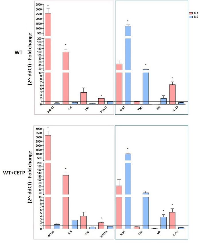
Santana et al.                                                                                        Macrophage Polarization and Pulmonary Emphysema

                                          promoted M2 macrophage polarization. Our results suggest that CETP is associated with
                                          inflammatory response and influences the role of macrophages in COPD.
                                          Keywords: cholesterol ester transfer protein, macrophage—cell, inflammation, chronic obstructive pulmonary
                                          disease, pulmonary emphysema, interleukin-10, arginase 1

INTRODUCTION                                                                   expressing human CETP, induces a rapid decrease in serum
                                                                               CETP. This suggests that CETP, which has molecular similarity
CETP is an independent risk factor for the development of                      to lipopolysaccharide-binding proteins, could modulate the
atherosclerosis. However, inhibition of CETP as a therapeutic                  lipopolysaccharide response and play a role in innate
strategy for raised HDL-C and as a protection against                          immunity and cholesterol metabolism (14).
cardiovascular disease failed to show benefits in population-                      In the present investigation, the macrophage phenotypic
based investigations. In this regard, surprisingly, a CETP                     profile was evaluated in the presence and absence of human
inhibitor (Torcetrapib) in the ILLUMINATE trial increased the                  CETP (huCETP). HuCETP expression in mice inhibits in vitro
frequency of infection cases, although this did not occur with other           macrophage polarization to the M1 phenotype and promotes M2
inhibitors (1). This undesirable effect is not attributable to a               phenotype. We further demonstrated in an experimental model
specific CETP inhibition but to secondary changes in plasma                     of pulmonary emphysema that huCETP mice exposed to elastase
lipoprotein metabolism or an elevation of plasma aldosterone (2).              have M2 phenotype macrophages in the lung tissue and that its
    In animal models, CETP inhibition protected transgenic mice                functional parameter worsens. Our results raise the possibility of
expressing human CETP (1) as well as rabbits naturally                         CETP participating in the inflammatory response and the
expressing CETP against atherosclerosis (3, 4). The CETP link                  macrophage roles in COPD.
with atherosclerosis and infection has been reviewed in humans
(5), and lower plasma CETP concentrations occurred in patients
who did not survive sepsis compared to survivors. Additionally, a
positive correlation between plasma CETP concentration and                     MATERIAL AND METHODS
sepsis survival rate was reported (6). CETP is involved in the
inflammatory response in mice expressing the human CETP                         Animal Model
gene (huCETP), which is more resistant to endotoxemia and                      The experimental protocol followed the Ethical Principles in
experimental sepsis than in wild type mice (5, 7, 8). However,                 Animal Experimentation of the Brazilian Society for Laboratory
these results diverge from more recent results concluding that                 Animal Science (SBCAL) and was approved by the University of
human CETP worsens inflammation and sepsis in mice (9), with                    São Paulo Medical School Ethics Committee the use of animals
CETP inhibition improving the survival rate in human sepsis                    (Comissão de É tica no Uso de Animais: CEUA 949/2018)
(10, 11). Although there is no explanation for these divergent                 according to the ARRIVE research guideline for the use of
results, it is possible to conjecture that the role of CETP could              Laboratory animals (16). Human CETP transgenic (Tg) mice
also depend on the animal species and pathology involved since                 (line 5203, C57BL6/J background) expressing human CETP
CETP inhibition protects against hepatic complications in                      under the control of its own promoter and other major
Schistosomiasis japonicum (12). However, it could also have                    regulatory elements were developed by Dr. Alan Tall
tissue-specific roles.                                                          (Department of Medicine, Columbia University, New York,
    Although several risk factors are present in sepsis, functional            NY, USA). A human CETP minigene with 3.4-kb (5′) and 2.2-
alterations in LP metabolism and immune cell populations,                      kb (3′) natural flanking sequences was inserted (17) and kindly
including macrophages, may worsen the disease. CETP                            provided by Professor Helena C.F. Oliveira (University of
synthesis occurs primarily in macrophage-rich tissues, such as                 Campinas, São Paulo, Brazil). Hemizygous human CETP Tg
the spleen and liver, and is secreted into the blood, with a greater           mice were cross bred to generate huCETP mice, and non-
contribution from Kupffer cells in humans and huCETP mice                      transgenic wild type (WT) mice were used as controls. The
(13). The latter study showed co-localization between CETP and                 presence of the targeted alleles, the CETP transgene, was
macrophage surface markers such as CD68. In mice, both                         determined according to the Jackson Laboratory modified
peritoneal and bone marrow-derived macrophages were                            protocol for the B6.CBA-Tg (CETP) 5203Tall/J line, number
associated with the CETP expression, with bone marrow-                         3904 [https://www.jax.org/Protocol?stockNumber=003904 (see
derived CETP being a significant contributor to the activity                    Supplementary (S) Materials Figure S1]. The CETP activity
and total concentration of CETP in plasma (14).                                and lipoprotein profile were also evaluated (Supplementary
    An important question remains about the function of CETP                   Figures S2, S3). Male CETP and WT mice, between 8 and 12
production by macrophages where activated macrophages are                      weeks of age, were housed in a conventional animal facility in a
common features of many inflammatory diseases. CETP belongs                     temperature-controlled room (22 ± 1°C), on a 12-h light/12-h
to the family of lipid transfer/lipopolysaccharide-binding                     dark cycle, with free access to water and food, and placed on a
proteins (15). Moreover, administration of lipopolysaccharide                  standard rodent chow diet (Quimtia CR1, Colombo, PR, Brazil).
to hamsters, animals that express CETP, or transgenic mice                     The plasma CETP levels measured by ELISA (µg/ml ± SD) were

Frontiers in Immunology | www.frontiersin.org                              2                                     July 2021 | Volume 12 | Article 684076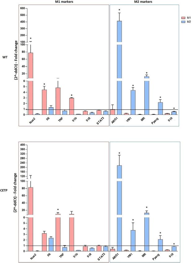
Santana et al.                                                                                 Macrophage Polarization and Pulmonary Emphysema

5.80 ± 2.07 and 0.02 ± 0.02 for CETP Tg mice and WT mice,                (Applied Biosystems). The method chosen to calculate the
respectively (8).                                                        relative quantification was the comparative Ct (cycle threshold)
                                                                         (DDCt). FAM-labeled TaqMan probe detection and the primer
Isolation of Bone-Marrow-Derived                                         assay designed for SYBR® Green (Applied Biosystems, Foster
Macrophages                                                              City, CA, USA) were used for gene expression quantitative real-
Bone marrow cells were harvested from WT or CETP Tg mice                 time PCR analysis. b-actin was the endogenous control. The
after anesthesia and exsanguination as previously described (18).        codes and sequences are listed in Supplementary Table S1.
Briefly, under aseptic conditions and after dissection of the hind
limb long bones (femurs and tibiae), the knee joint was removed          Flow Cytometry Analysis
with scissors at the proximal end. Then, a 10 ml syringe with 26G        After 24 h, cells were washed with ice-cold PBS and removed
needle containing Dulbecco’s Modified Eagle Medium (DMEM),                from the surface of the plate with 1 ml of accutase solution
high glucose, 10% fetal bovine serum (FBS) (Invitrogen), and 1%          (Sigma-Aldrich), placed gently on the plate, and maintained for
penicillin/streptomycin] was inserted to wash the bone marrow            5 min. The cell suspension was transferred to a 15 ml sterile
and obtain the gelatinous tissue that fills the medullary cavities,       conical tube and centrifuged at 300 × g (4 min) to obtain the cell
which was then placed in 50 ml Falcon® centrifuge tubes. Cell            pellet. The supernatant was completely removed by rapid
aggregates were centrifuged (6 min, 1,000 rpm) at room                   decanting, and the cell pellet was immediately resuspended in
temperature; thereafter, cells were seeded in six-well culture           200 µl of PBS containing 2% FBS. The live cells were analyzed
plates and maintained for 7 days at 37°C in a humidified                  using the Live/Dead marker (ThermoFischer L34966). Cells were
atmosphere containing 5% CO2. On day 5, the DMEM was                     incubated with fluorescence-conjugated antibodies against the
supplemented with 30% of growth supernatant of M-CSF-                    surface markers CD11b (APCCy7)/F4/80 (PercP) (BioLegend,
transduced L929 (18).                                                    San Diego, CA), in PBS containing 1% BSA for 30 min at 4°C.
                                                                         CD80 (APC) was used as a marker for M1, and CD206 (PE) was
                                                                         used for M2 macrophages (BioLegend, San Diego, CA). CD80+/
Macrophage Polarization in the Presence
                                                                         F4/80+/CD11Bc+/CD206− cells were marked as M1-positive
or Absence of Human CETP                                                 cells, while CD80−/F4/80+/CD11Bc+/CD206+ cells were marked
Heterogeneity is one of the most important macrophage
                                                                         as M2-positive cells. All samples were analyzed using a flow
characteristics. To investigate the CETP effect on macrophage
                                                                         cytometer (BD FACSDiva™ software; Biosciences, San Jose,
(M) polarization, we used huCETP Tg bone marrow cells and
                                                                         CA). A total of 100,000 events were collected for each sample,
compared then to macrophages that do not produce CETP from
                                                                         and the data were analyzed using FLOWJO (v. 8.7) software
non-transgenic littermate (WT) mice.
                                                                         (Tree Star Inc., Ashland, OR, USA) (19, 20).
   Cells were cultured in high glucose DMEM supplemented
with 10% FBS, 1% penicillin/streptomycin under stimulus                  Cytokines and CETP Protein Analysis
conditions with 5 ng/ml IFN-g (PeproTech, USA), plus 50 ng/              The protein levels of TNF, IL-6, IL-10, and CETP (plasma, cell
ml LPS (055:B5, Sigma-Aldrich) for induction of M1, or 10 ng/            supernatants, and bronchoalveolar lavage: BALF) were measured
ml IL-4 (PeproTech, USA), plus 10 ng/ml IL-13 (PeproTech,                using a commercially available enzyme-linked immunosorbent
USA) for M2 polarization at 37°C in a humidified atmosphere               assay (ELISA) kit (R&D Systems, Minneapolis, MN) and by Cell
containing 5% CO2 for 24 h. M0 macrophages were maintained               Biolabs (Cell Biolabs, Inc., San Diego, CA, USA), according to
in DMEM without polarization factors.                                    the manufacturer’s instructions.
   A second experiment was performed in which exogenous
CETP (Recombinant human CETP, Roar Biomedical, New                       Induction of Emphysema:
York, NY, USA) was added to the culture medium (1 µg/ml) in
                                                                         Elastase-Induced Model
the bone marrow-derived macrophages from WT donors. The
                                                                         WT and CETP animals were anesthetized with isofluorane, and
culture was performed in the same conditions as described above.
                                                                         the trachea was exposed after asepsis of the anterior neck region.
                                                                         Porcine pancreatic elastase (PPE) (Type 1/E1250; Sigma-Aldrich,
Gene-Expression Analysis                                                 El Camino Real Carlsbad, CA 92009, California, USA) at a single
After treatments, bone marrow-derived macrophages from WT                dose of 0.667 IU (50 µl saline solution) was administered
or CETP mice were washed with phosphate buffered saline                  intratracheally between the cartilaginous rings. The control
(PBS), removed with a scraper from the culture plate, and                group (no elastase) received only 50 µl of saline solution.
centrifuged at 1,000 rpm (4°C) to obtain the cell pellet. Total
RNA was isolated from cell lysates following the manufacturer’s          Respiratory Mechanics Analysis
instructions (RNeasy® Mini Kit; Qiagen, Hilden, Germany) and             After 28 days, animals were anesthetized, tracheostomized, and
retrotranscribed with the High Capacity cDNA reverse                     placed on a rodent mechanical ventilator (flexiVent, SCIREQ,
transcription kit (Applied Biosystems, Foster City, CA, USA).            Montreal, Canada) with a tidal volume of 10 ml/kg and a
Quantitative PCR was performed using the StepOne Plus™ Real              respiratory rate of 120 cycles/min. Respiratory mechanics were
Time PCR system (Applied Biosystems) with TaqMan Universal               performed, comprising respiratory system resistance (Rrs), tissue
Master Mix II (Thermo Fisher, Waltham, MA, USA) and SYBR                 elastance (Htis), lung tissue resistance (Gtis), respiratory system
probes plus the Master Mix Power SYBR™ Green solution                    elastance (Ers), and airway resistance (RAW), using the

Frontiers in Immunology | www.frontiersin.org                        3                                    July 2021 | Volume 12 | Article 684076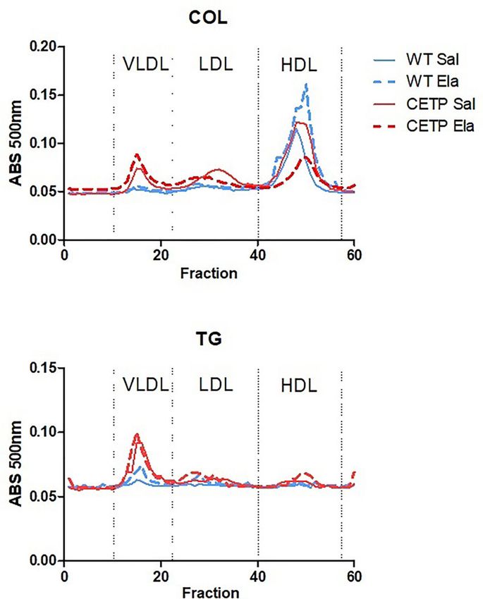
Santana et al.                                                                                     Macrophage Polarization and Pulmonary Emphysema

previously described model by (21). The exhaled nitric oxide               excluding vessels and airways. Thus, the average alveolar
(ENO) concentration was measured by collection Mylar bags                  diameter was calculated, reasoning that the number in contact
attached to the expiratory output of the ventilator for 10 min,            with the alveolar walls is also the number of intersections
and an NO filter was attached to the inspiratory breathing circuit          between lines and alveolar walls (22). The mean linear
input. Afterwards, a chemiluminescence fast-responding                     intercept (LM) was quantified by one observer in a blinded
analyzer (NOA 280; Sievers Instruments Inc., Boulder, CO,                  fashion to assess air space enlargement (21).
USA) calibrated with an NO (nitric oxide), source certified 47-
parts per billion (ppb) (White Martins, São Paulo, Brazil) and a           Statistical Analysis
zero NO filter (Sievers Instruments Inc.) was used.                         Continuous variables were tested for normality with the
                                                                           Kolmogorov–Smirnov and Shapiro Wilk tests. The values are
Bronchoalveolar Lavage                                                     expressed as median and percentiles 25 ant 75 or as the mean and
The lungs were washed with 0.9% saline (3× 0.5 ml). Volume                 standard deviation for non-parametric or parametric data
recovery of the instilled saline was over 95%, which was                   respectively. Non-parametric data were compared using the
transferred into a test tube on ice. White blood cells were                Mann–Whitney U test for two independent samples or
quantified through total and differential counting. BAL was                 Kruskal–Wallis test with original FDR method of Benjamini
centrifuged at 1,000 rpm for 10 min, and the cell pellet was               and Hochberg post-test for three or more samples. Parametric
rediluted in 0.2 ml sterile saline. The total number of viable cells       data were compared by Student’s t-test for two independent
was determined in a Neubauer hemocytometer counting                        samples or ANOVA with two-stage linear step-up procedure of
chamber (400×). The differential cell counts were performed in             Benjamini, Krieger, and Yekutieli post-test for three or more
cytocentrifuge preparations of the BAL (450 rpm for 6 min)                 samples (23–25). Graphs were prepared using GRAPHPAD
(Cytospin, Cheshire, UK) stained with Diff-Quick (Biochemical              PRISM version 9.0 (GraphPad Software, San Diego, CA, USA).
Sciences Inc., Swedesboro, NJ). Three hundred cells                        Statistical significance was set at p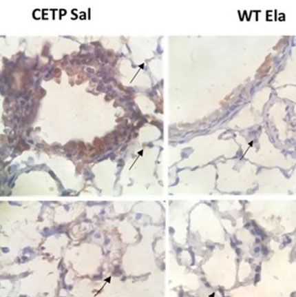
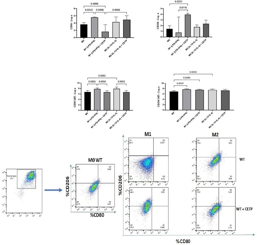
Santana et al.                                                                                           Macrophage Polarization and Pulmonary Emphysema

(IL-4 + IL-13) activation. Previous studies have reported                       mannose receptor CD206 (MR), peroxisome proliferator-
differences in gene expression between M1 and M2                                activated receptor (PPAR) gamma, and IL-10, considered
macrophages grown in vitro compared to non-activated                            markers of M2, were higher in this population although not
macrophages (M0). M1 responds by positively regulating                          differing between WT and CETP groups (Figure 2).
many pro-inflammatory genes according to the LPS stimulus,                           Considering that CETP plays an essential role in the
while induction to M2 promotes anti-inflammatory genes such                      metabolism of lipids and lipoproteins due to inflammation and
as arginase-1 (26). Here, we examined the primary genes                         lipid signaling interconnections, we analyzed mRNA expressions
representing the degree of polarization for M1 or M2 in                         of lipid metabolism genes. Increased ABCA1 expression was
macrophage CETP production (CETP endogenous) compared                           observed in M2 compared to M1 in both CETP and WT cells.
to the WT (Figure 2). Stimulation for M1, as expected,                          Furthermore, CETP-M2 cells expressed slightly increased
increased NOS2, TNF, IL-6, and IL-1b gene expression (P <                       ABCA1 than WT-M2 did and increased expression of SRB1
0.05; M1 vs. M2). Expressions of arginase (ARG1), YM1,                          (SCARB1) compared to CETP-M1 (Figure 3).

 FIGURE 2 | Gene expression of typical M1 and M2 markers from bone marrow-derived macrophages of WT and huCETP Tg mice. In vitro stimulated M1 (5 ng/ml
 IFNg + 50 ng/ml LPS) and M2 (10 ng/ml IL-13 + IL-4) for 24 h. mRNA expression was determined using RT-PCR. Results are expressed as the mean ± standard
 deviation and were compared by unpaired Student’s t-test (n = 3–5). *P < 0.05.

Frontiers in Immunology | www.frontiersin.org                               5                                        July 2021 | Volume 12 | Article 684076
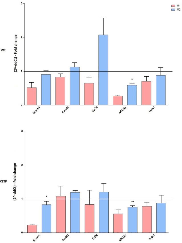
Santana et al.                                                                                            Macrophage Polarization and Pulmonary Emphysema

 FIGURE 3 | Gene expression of factors associated with lipid metabolism from bone marrow-derived macrophages of WT and huCETP Tg mice. In vitro stimulated
 M1 (5 ng/ml IFNg + 50 ng/ml LPS) and M2 (10 ng/ml IL-13 + IL-4) for 24 h. mRNA expression was determined using by RT-PCR. Results are expressed as mean ±
 standard deviation and were compared by unpaired Student’s t-test. (n = 3–5). *P < 0.05 (M1 vs. M2); **P < 0.05 (CETP M2 vs. WT M2).

   We also evaluated cell surface markers associated with M1                     Secretion of CETP in the culture medium was similar in all
(CD80) and M2 (CD206). The percentage of expression of CD80                      treatment conditions (Figure 5E). However, CETP expression
was higher in the WT-M1 than in CETP-M1 macrophages                              was downregulated by the inflammatory stimulus (Figure 5D).
(Figure 4A). CETP macrophages increased the expression of                           Next, we investigated the influence of exogenous CETP
CD206 in cells in the absence of stimulus (M0) and under                         (human recombinant CETP; rCETP) added to the culture
stimulation for M1 (IFNg + LPS) in comparison to WT-M1                           medium. Generally, gene expressions of a few M1 or M2
(Figure 4B). The mean fluorescent intensity (MFI) in flow                          markers and cytokines in supernatants collected from cell
cytometry analysis did not differ between groups (Figures 4C–E).                 culture were similar to those found in the macrophages that
   IFNg and toll-like receptor (TLR) agonists, such as LPS, are                  produce CETP (endogenous) (Figures 6 and 8). However, we
the primary stimuli generating M1 macrophages that express                       observed increased MR (CD206) gene expressions in the
inflammatory cytokines, such as TNF, whereas IL-4 and IL-13                       presence of rCETP under stimulation for M2 (Figure 6).
are mainly used to generate M2 macrophages resulting in high                     Reduction in the CD80 (Figures 7A, C) M1 marker and
production of IL-10. Analysis of the cell culture supernatants                   increased CD206 percent expression (Figure 7B) occurred in
showed higher TNF, IL-6, and IL-10 cytokines in M1 (CETP and                     the presence of rCETP compared to WT. Increased CD206 MFI
WT), confirming the inflammatory stimulus (Figures 5A–C).                          was observed in comparison to M0 (Figure 7D). Figure 7E

Frontiers in Immunology | www.frontiersin.org                                6                                        July 2021 | Volume 12 | Article 684076
Santana et al.                                                                                                 Macrophage Polarization and Pulmonary Emphysema

                               A                                                  B

                               C                                                 D

                                   E

 FIGURE 4 | Expression of cell surface markers associated with M1 (CD80) and M2 (CD206) from bone marrow-derived macrophages of WT and huCETP Tg mice.
 Expression of CD80 (A), CD206 (B), mean fluorescence intensity (MFI) of CD80 (C), MFI of CD206 (D), and representation of the gate strategy (E). Non-stimulated
 control cells (M0), stimulated M1 (5 ng/ml of IFNg + 50 ng/ml of LPS) or M2 (10 ng/ml of IL-13 + IL-4) for 24 h. Basal fluorescence was determined using unlabeled
 cells, and compensation was performed with cells labeled with the respective fluorochromes on the FACSCanto II cytometer. In total, 100,000 events were analyzed.
 Values were expressed as mean and standard deviation. Values expressed as median and percentiles 25 and 75 were analyzed by Kruskal–Wallis test with original
 FDR method of Benjamini and Hochberg post-test. The values expressed as mean and standard deviation were analyzed by ANOVA with two-stage linear step-up
 procedure of Benjamini, Krieger, and Yekutieli post-test; (n = 3), P < 0.05.

shows the gate strategy. Cell culture supernatants showed higher                      differential bronchoalveolar lavage (BAL) leukocytes, cytokine
TNF, IL-6, and IL-10 cytokines in M1 (CETP and WT)                                    production, and mouse lung histology (Figures 9–13
(Figures 8A–C). However, IL-10 production was higher in                               and Table 1).
CETP-M1 in comparison to WT-M1 (Figure 8C).                                               Elastase administration increased respiratory system resistance
                                                                                      (Rrs), tissue elastance (Htis), lung tissue resistance (Gtis),
In Vivo Studies                                                                       respiratory system elastance (Ers) in CETP mice (Figures 9A–C,
Induction of Emphysema: Elastase-Induced Model                                        E), accompanied by a reduction in airway resistance (Raw)
Considering the importance of macrophages in the development                          compared to the WT-ELA group (Figure 9D). There was no
and formation of pulmonary emphysema and its dependence on                            difference in exhaled NO analysis (Figure 9F).
the microenvironment where they may present an M1 or M2                                   Bronchoalveolar lavage (BAL) leukocyte differential and total
phenotype, we evaluated the effect of CETP on elastase-induced                        number were measured to analyze the extent of pulmonary
emphysema (ELA) in CETP Tg and WT mice. After 28 days, we                             inflammation. Exposure to ELA significantly increased
analyzed the lung mechanics, exhaled nitric oxide, total and                          macrophage cells in both groups (Figure 10D), indicating

Frontiers in Immunology | www.frontiersin.org                                    7                                          July 2021 | Volume 12 | Article 684076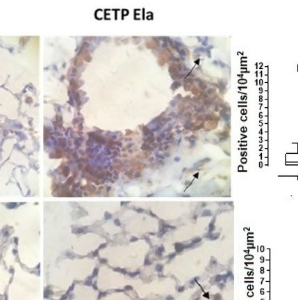
Santana et al.                                                                                                Macrophage Polarization and Pulmonary Emphysema

                          A                                            B                                       C

                            D                                          E

 FIGURE 5 | Cytokine secretion, CETP expression, and secretion in cell culture medium. Bone marrow-derived macrophages from WT and huCETP mice,
 unpolarized (M0), or stimulated to M1 (5 ng/ml IFNg + 50 ng/ml LPS) or to M2 (10 ng/ml IL-13 + IL-4) for 24 h. TNF-a (A), IL-6 (B), IL-10 (C), CETP mRNA
 expression in macrophages (D) and CETP secretion (E). Concentrations were determined by ELISA, and CETP expression was determined by RT-PCR. Values
 expressed as median and percentiles 25 and 75 were analyzed by Kruskal–Wallis test with original FDR method of Benjamini and Hochberg post-test. Values were
 expressed as mean and standard deviation analyzed by ANOVA with two-stage linear step-up procedure of Benjamini, Krieger, and Yekutieli post-test; (n = 5–8).
 P < 0.05. *M0 vs. M1 and M2.

increased pulmonary inflammation. Total cell levels from CETP-                       triggered by LPS in macrophages (5, 7, 8). However, these
ELA were more abundant than CETP-SAL (Figure 10A).                                  results diverge from recent studies claiming CETP worsens
Eosinophils and lymphocytes were higher in the WT-ELA                               inflammation and sepsis (9–11). Thus, awareness of the
group than all experimental groups (Figures 10B, E).                                environmental conditions that promote the pro- or anti-
Increased neutrophil quantity trend was observed in the ELA                         inflammatory properties of macrophages and control the
group (P < 0.07) (Figure 10C). Significantly increased BAL TNF                       plasticity of the phenotype is fundamental for understanding
and IL-10 were observed in CETP-ELA compared to all                                 the tissue-specific CETP role (14).
experimental groups (Figures 10F, H). There was no difference                           In the present study, we explored the influence of CETP on
in IL-6 (Figure 10G).                                                               the differentiation and function of macrophages as well as COPD
   Lung immunohistochemistry analysis showed increased IL-                          pathogenesis in a murine model of elastase-induced
10, ARG-1, and mean linear intercept (LM) after ELA treatment                       pulmonary emphysema.
(Figures 11B, D and 12C) and even higher in CETP-ELA                                    As expected, macrophages derived from huCETP mice
compared to all groups. These results were accompanied by                           expressed CETP (Figures 1 and 5D). Polarizing conditions
increased TNF, iNOS, and presence of CETP in the lung                               reduced mRNA CETP expression (Figure 5D), although the
(Figures 11A, C and 12A, B).                                                        CETP content in the culture medium did not differ between M1
   Plasma lipid levels were not modified on elastase infusion.                       and M2 (Figure 5E). Previous study demonstrated reduced
However, induced cholesterol distribution changed in the LP and                     expression of CETP in the presence of LPS (27).
increased CETP activity (Figure 13 and Table 1).                                        After polarization of M0 macrophages with M1 and M2
                                                                                    stimuli, M1 polarized macrophages were confirmed by NOS2,
                                                                                    TNF, IL-6, and IL-1b gene expression, and M2 polarized
DISCUSSION                                                                          macrophages increased arginase (ARG1), YM1, mannose
                                                                                    receptor CD206 (MR), peroxisome proliferator-activated
In previous studies, we demonstrated a greater survival rate in                     receptor (PPAR) gamma, and IL-10, considered markers of M2
human CETP transgenic mice than in WT mice with                                     macrophages (P < 0.05; M1 vs. M2), although it did not differ
experimental sepsis (7, 8). CETP along with lipoprotein                             between WT and CETP groups (Figure 2). However, reduced
acceptors, previously recognized key vehicles for the transport                     polarization towards M1 was observed when analyzing the
of LPS, plays a fundamental role in the initial reversible phase of                 expression of surface markers for M1 (CD80) macrophages
sepsis and in the regulation of the inflammatory response                            that produce CETP or WT in the presence of recombinant

Frontiers in Immunology | www.frontiersin.org                                   8                                         July 2021 | Volume 12 | Article 684076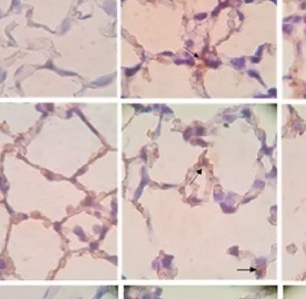
Santana et al.                                                                                               Macrophage Polarization and Pulmonary Emphysema

 FIGURE 6 | Gene expression of typical M1 and M2 markers in bone marrow-derived macrophages from WT mice stimulated in the presence of recombinant CETP
 in vitro. Macrophages were stimulated using 5 ng/ml IFNg + 50 ng/ml LPS (M1) or 10 ng/ml IL-13 + IL-4 (M2) for 24 h. mRNA expression was determined by RT-PCR.
 Results are expressed as the mean ± standard deviation and compared by unpaired Student’s t-test. (n = 3–5). *P < 0.05 for 24 h.

CETP (Figures 4A and 7A, C) whereas the expression of M2                           receptor (TLR) activation via lipopolysaccharide (LPS) or
marker mannose receptor (CD206) was higher than that of WT-                        interferon (IFN) stimulated activation and release of IL-27,
M1 (Figures 4B and 7B). Furthermore, the gene expression of                        with consequent activation of phosphorylated Jak/STAT1 and
mannose receptor (MR: CD206) increased in the presence of                          STAT3 transcription factors, leading to IL-10 production (28).
recombinant CETP, corroborating these findings (Figure 6).                          IL-10 gene transcription was greater in M1 and accompanied by
   Cytokine profiling revealed increased TNF and L-6 (M1) in                        increased STAT3 (Figure 6).
both groups as expected (Figures 5A, B and 8A, B). IL-10 has                          Anti-inflammatory cytokines increase, initiating an
been considered a hallmark of M2 polarization, mainly based on                     inflammation-controlling process and bacteria elimination.
studies in mice (28). However, recent findings demonstrated                         Additionally, anti-inflammatory IL-10 increases to hold back
greater IL-10 expression in M1 than in M2-polarized human                          the organ damage excessive inflammation. Consequently, IL-10
monocyte-derived macrophages hMDMs (19, 29). We showed                             often rises following TNF. These results reinforce that IL-10 and
significantly increased IL-10 M1 in the presence of CETP                            TNF are associated in macrophage polarization observed in the
(Figure 8C). Considering the diversity of alternatively                            CETP group (Figures 8A, C and 11A, B) (32).
activated M2 macrophages into subtypes M2a, M2b, M2c, and                             M1 and M2 phenotype polarizations represent extremes of
M2d (22, 30), we speculate that CETP alters the macrophage                         activation states. Functional distortion of mononuclear
phenotypic profile (31). This effect can be attributed to toll-like                 phagocytes occurs in vivo under physiological conditions,

Frontiers in Immunology | www.frontiersin.org                                  9                                          July 2021 | Volume 12 | Article 684076
Santana et al.                                                                                                    Macrophage Polarization and Pulmonary Emphysema

                                                      A                                          B

                                                      C                                          D

                  E

 FIGURE 7 | Expression of cell surface markers associated with M1 (CD80) and M2 (CD206) from bone marrow-derived macrophages of WT mice in the presence
 of recombinant CETP. Expression of CD80 (A), CD206 (B), mean fluorescence intensity (MFI) of CD80 (C), MFI of CD206 (D), and representation of the gate
 strategy (E). Non-stimulated control cells (M0), stimulated M1 (5 ng/ml of IFNg + 50 ng/ml of LPS) or M2 (10 ng/ml of IL-13 + IL-4) for 24 h. Basal fluorescence was
 determined using unlabeled cells, and compensation was performed with cells labeled with the respective fluorochromes on the FACSCanto II cytometer. In total,
 100,000 events were analyzed. Values were expressed as mean and standard deviation. These data were analyzed by ANOVA with two-stage linear step-up
 procedure of Benjamini, Krieger, and Yekutieli post-test; (n = 3). P < 0.05.

                   A                                   B                                     C

 FIGURE 8 | Cytokine secretion in cell culture medium. Bone marrow-derived macrophages from WT mice, unpolarized (M0), or stimulated to M1 (with 5 ng/ml IFNg
 + 50 ng/ml LPS), or to M2 (with 10 ng/ml IL-13 + IL-4) in the absence (control) or presence of recombinant human CETP (1 µg/ml) for 24 h. TNF (A), IL-6 (B) and
 IL-10 (C).

Frontiers in Immunology | www.frontiersin.org                                     10                                          July 2021 | Volume 12 | Article 684076
Santana et al. Macrophage Polarization and Pulmonary Emphysema
pregnancy, and in pathologies as allergies, chronic inflammation, production was observed in the absence of CETP (8). Another
tissue repair, infection, and cancer (33). M1 to M2 repolarization study showed that cholesterol accumulation in macrophages
exerted by distinct M2 stimuli through transcriptome-based path increased the inflammatory response mediated by toll-like 4
analysis provides a new approach to human macrophage receptors (TLR4s), exacerbating LPS-mediated secretion of pro-
phenotypes in clinically relevant disease states, such as cystic inflammatory cytokines (35). This indicates that cellular cholesterol
fibrosis and asthma (34). concentration has a direct influence on immunoregulation. Thus,
According to the hypothesis that CETP plays an important the recruitment of TLR4 to lipid-rafts is due to increased cholesterol
role in immunomodulation and favors a lower M1 macrophage in these membrane microdomains. We speculate that CETP reduces
profile, a significant increase of IL-10 in CETP compared to WT cellular cholesterol microdomains impairing TLR4 recruitment. In
furthers our understanding of the dual role of CETP in different this regard, increased ABCA1 transcription was observed in CETP-
conditions, namely: 1) its anti-inflammatory profile conferring M2 compared to WT-M2 (Figure 3). ABCA1 promoted cholesterol
longer survival in endotoxemia in CETP mice (7, 8), although efflux from macrophage and increased IL-10. Moreover, it reduces
this has been contested (9), and (2) worsening inflammation and the secretion of pro-inflammatory cytokines, activating PKA and
tissue damage in the experimental elastase-induced pulmonary contributing to the M2 type anti-inflammatory response (36, 37).
emphysema model which resulted in the widening of alveolar air Several other gene expressions that connect lipid metabolism
spaces (Lm) in CETP animals compared to WT eliciting and inflammation, like LXR, PPAR gamma, and SREBP, did not
pulmonary emphysema worsening (Figure 12C). differ between the experimental groups. However, SRB1
We demonstrated reduced LPS uptake in the presence of both (SCARB) increased after M2 polarization (Figure 3) SRB1 is a
endogenous and exogenous CETP compared to WT (7, 8). well-characterized lipoprotein receptor that binds native high-
Increased activation of NF-k beta with macrophage TNF-alpha and low-density lipoproteins (HDL and LDL), respectively (38).
A B
C D
E F
FIGURE 9 | Lung mechanics in WT and huCETP mice after elastase-induced emphysema (ELA), or saline (SAL). (A) Respiratory system resistance (Rrs); (B) Tissue
elastance (Htis); (C) Respiratory system elastance (Ers); (D) Airway resistance (RAW). (E) Lung tissue resistance (Gtis); (F) Exhaled nitric oxide. Values expressed as
median and percentiles 25 and 75 were analyzed by Kruskal–Wallis test with original FDR method of Benjamini and Hochberg post-test. Values were expressed as
mean and standard deviation analyzed by ANOVA with two-stage linear step-up procedure of Benjamini, Krieger, and Yekutieli post-test; (n = 8). P < 0.05.
Frontiers in Immunology | www.frontiersin.org 11 July 2021 | Volume 12 | Article 684076Santana et al. Macrophage Polarization and Pulmonary Emphysema
SRB1 also plays an important role in the recognition of other accordance with a study that shows increased SR-B1 mRNA
endogenous and exogenous ligands, inducing an anti- levels in THP-1 cell after M2 polarization compared to M1.
inflammatory or pro-inflammatory response depending on the It is known that the hepatic expression of CETP is regulated
nature of the ligand and the cell/tissue type (39). Our data are in by LXR (40). LXR agonists attenuate LPS-induced release of TNF
A B
C D
E F
G H
FIGURE 10 | Total and differential leukocyte count in broncho-alveolar lavage (BAL), and cytokine levels in WT and huCETP mice after elastase-induced emphysema
(ELA) or saline (SAL). Total cells (A), eosinophils (B), neutrophils (C), macrophages (D), lymphocytes (E), cytokines in BAL: TNF (F), IL-6 (G), IL-10 (H), cytokine
plasma (I). Values were expressed as mean and standard deviation. These data were analyzed by ANOVA with two-stage linear step-up procedure of Benjamini,
Krieger, and Yekutieli post-test. P < 0.01: *CETP Ela vs. all groups, (n = 8).
Frontiers in Immunology | www.frontiersin.org 12 July 2021 | Volume 12 | Article 684076Santana et al. Macrophage Polarization and Pulmonary Emphysema
alpha and prostaglandin E2 from isolated Kupffer cells (41), between the experimental groups, probably because of large
suggesting that LXRs keep the pro-inflammatory activation individual variation. However, iNOS labeling in lung tissue was
status of Kupffer cells low, which coincides with high CETP greater in CETP-ELA (Figure 11C).
expression. Reduced mRNA CETP expression was observed in Elevation of plasma IL-6 (pg/ml) (P < 0.05) and low IL-10
the generation of M1 and M2 macrophages under polarizing (P < 0.07) (Table 1) occurred in the CETP-ELA group, possibly
conditions (Figure 5D). indicating systemic inflammatory response after elastase.
In the induction of pulmonary emphysema by elastase Patients with severe COPD showed a higher IL-6/IL-10
instillation (ELA), worsening of the lung mechanical parameters ratio (44).
in the CETP ELA group (Figures 9A–C, E) was accompanied by a In contrast to plasma data, IL-10 was elevated in the CETP-
reduction in airway resistance (Raw) compared to the WT-ELA ELA in the lungs and was accompanied by a higher expression of
group (Figure 9E) These mechanical changes are consistent with Arg-1, markers of M2-type macrophages (Figures 11B, D).
the increase in the mean linear intercept, which is reflected in the Increased Arg-1 suggests an aggravation of catabolic activity
widening of the air spaces owing to the destruction of the lung but may also have implications for lung fibrosis and airway
parenchyma in the CETP ELA (Figure 12C). resistance (45). These data corroborate those of in vitro
Cytokines in BAL revealed increased IL-10 compared to WT macrophage polarization towards the M2 type in the presence
ELA, corroborating the CETP ELA lung immunohistochemistry of CETP and are potentially important for understanding the
data (Figures 10H and 11B). pathophysiology of the respiratory tract response in the presence
Eosinophils and lymphocytes were higher in WT ELA than in of CETP and the etiology of COPD.
all experimental groups (Figures 10B, E). The role of eosinophils Plasma lipid levels were not modified on elastase infusion
in the pathogenesis of COPD is still controversial, and many (Table 1). However, elastase-induced cholesterol distribution
cases occur with tissue eosinophilia without a simultaneous changes in the LP of CETP and WT mice that were distinct
increase in blood eosinophils (42). Neutrophils showed a trend according to genotype (Figure 13 and Table 1). As expected, in
to increase in ELA groups (P < 0.07) (Figure 10C). Increase huCETP mice, HDL-C decreased, and VLDL-C slightly
neutrophil levels are characteristic of acute exacerbations of increased. However, after elastase administration, substantial
COPD (43). The exhaled NO (Figure 9F) was not different HDL-C reduction occurred, which is explained by increased
A
B
C
D
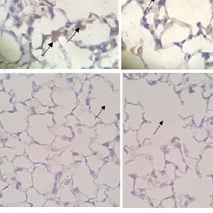
FIGURE 11 | Photomicrograph of stained slides for immunohistochemistry analysis of inflammatory markers from WT and CETP Tg mice and from huCETP mice
after elastase-induced emphysema (ELA) or saline (SAL). TNF (A), IL-10 (B), iNOS (C) and Arginase 1 (D). Representative images of micrographs at ×1,000
magnification. Values were expressed as mean and standard deviation. These data were analyzed by ANOVA with two-stage linear step-up procedure of Benjamini,
Krieger, and Yekutieli post-test. p < 0.01: (n = 8).
Frontiers in Immunology | www.frontiersin.org 13 July 2021 | Volume 12 | Article 684076Santana et al. Macrophage Polarization and Pulmonary Emphysema
A
B
C
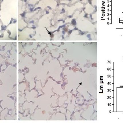
FIGURE 12 | Photomicrograph of stained slides for immunohistochemistry analysis of CETP in airways (A), lung parenchyma (B) and mean linear intercept (LM) by
dot count of airway spreads (C) from WT and huCETP mice after elastase-induced emphysema (ELA) or saline (SAL). Representative images of photomicrographs:
CETP magnification ×1,000 and (LM) ×400. Values expressed as median and percentiles 25 and 75 were analyzed by Kruskal–Wallis test with original FDR method
of Benjamini and Hochberg post-test. The values were expressed as mean and standard deviation were analyzed by ANOVA with two-stage linear step-up
procedure of Benjamini, Krieger, and Yekutieli post-test. p < 0.01, (n = 8).
CETP activity; these results were independent of plasma CETP
levels (Table 1). These results indicate that CETP expression in
the elastase group led to unfavorable modifications, with
increased apoB-LP and reduced HDL contributing to the
worsening inflammation in CETP ELA group. In humans,
there was no significant difference in the concentrations of
LDL, apolipoprotein B, or HDL between groups with mild,
moderate, and severe COPD. Triglyceride concentrations were
reduced, and apoA1 increased in the most severe cases (46).
Our results are in agreement with those of another
investigation showing alveolar macrophages with a dominant
M2 phenotype in patients with COPD (42). Another study found
increased deposition of M2 alveolar macrophages in the mouse
model of COPD and increased expression of the TGF beta/Smad
pathway in M2 macrophages, both in vitro and in vivo, indicating
that M2 macrophages contribute to COPD by altering its
phenotypic profile (26).
Although polarization to M2 cannot be attributed to CETP
alone, its presence exacerbated the effects of elastase. These
findings suggest that the dominant accumulation of M2 in the
presence of CETP in the lung should be explored in COPD (47).
Interestingly, the accumulation of M2 macrophages was observed
in adipose tissue and liver, in tissues with high expression of CETP
and the lung, as well as in the blood of patients who suffered severe
trauma, such as burns or sepsis. However, the induction of M2
FIGURE 13 | Plasma lipoprotein analysis by high-performance liquid polarization in these patients has not yet been identified (48). In this
chromatography (FPLC) in WT and huCETP mice after elastase-induced
sense, it was recently suggested that CETP inhibitors be redirected
emphysema (ELA) or saline (SAL). Average of two to three pools (n = 4).
to the treatment of sepsis (49).
Frontiers in Immunology | www.frontiersin.org 14 July 2021 | Volume 12 | Article 684076Santana et al. Macrophage Polarization and Pulmonary Emphysema
TABLE 1 | Total cholesterol (C), triglyceride (TG), lipoprotein fractions, cytokines and CETP plasma levels in WT and huCETP mice after elastase-induced emphysema
(ELA), or saline (SAL).
WT SAL WT ELA CETP SAL CETP ELA
CHOLESTEROL TOTAL 73 ± 10 64 ± 8 73 ± 9 65 ± 3
VLDL (mg/dL) 5 4 13 24
% 7 5 17 37
LDL (mg/dL) 13 7 15 12
% 18 11 21 18
HDL (mg/dL) 55 53 45 29
% 75 84 62 45
TRIGLYCERIDE 102 ± 30 123 ± 10 100 ± 20 122 ± 21
VLDL (mg/dL) 51 73 76 80
% 50 59 76 66
LDL (mg/dL) 27 37 10 19
% 26 30 10 15
HDL (mg/dL) 24 13 14 23
% 24 11 14 19
Cytokines (pg/mL)
IL-6 2,742 ± 175.20 2,561 ± 601.40 2,029 ± 330.50 3,055 ± 213.70*
IL-10 204.1 ± 220.7 189.6 ± 91.3 233.1 ± 52.7 114.5 ± 85.5
CETP
Concentration (µg/mL) 1.882 ± 0.55 1.907 ± 0.40
Activity (%) 17 ± 4 23 ± 3*
*p < 0.05: CETP ELA vs. CETP SAL; (n = 8).
Despite the limitations of animal model observations for experimental design. NC helped in the study design and data
human physiology, our results are novel, indicating that CETP interpretation EQ helped in data interpretation and in writing the
is associated with the inflammatory response and, especially, article. PC supervised the experimental design, data interpretation,
with the role of macrophages in COPD. and writing the article, and was awarded the research funding.
All authors contributed to the article and approved the
submitted version.
DATA AVAILABILITY STATEMENT
The raw data supporting the conclusions of this article will be FUNDING
made available by the authors, without undue reservation.
The authors would like to thank the financial support from
Fundação de Amparo à Pesquisa do Estado de São Paulo,
ETHICS STATEMENT FAPESP (grants # 2017/22940-6 to PC).
The animal study was reviewed and approved by Ethical
Committee of the University of São Paulo Medical School for ACKNOWLEDGMENTS
the use of animals (CEUA 949/2018).
The authors are grateful to Leandro Ianuzzi for helping with
animal care, Silvia Fukuzaki for histological analysis, Aritania S
AUTHOR CONTRIBUTIONS Santos and Chin J Lin for providing facilities for RT-qPCR, and
the Laboratorios de Investigaç ão Mé dica (LIM) for their
KS performed all the experiments and data analysis and helped to continuous support.
write the article. RR helped with animal and immunohistochemistry
experiment. CS helped with flow cytometry analyses and statistics.
OA helped with flow cytometry analyses. TR helped with data SUPPLEMENTARY MATERIAL
interpretation and molecular biology. FD helped with animal
experiments and CETP genotyping mice. VN helped with the The Supplementary Material for this article can be found online
experiments to analyse the lipid profile and chromatography. IT at: https://www.frontiersin.org/articles/10.3389/fimmu.2021.
helped with animal experimental design. FG helped in the 684076/full#supplementary-material
in Lipoprotein Metabolism and Cardiovascular Disease. Singapore: Springer-
REFERENCES Verlag Singapore Pte Ltd (2020). doi: 10.1007/978-981-15-6082-82
1. Oliveira HCF, Raposo HF. Cholesteryl Ester Transfer Protein and Lipid 2. Clark RW, Cunningham D, Cong Y, Subashi TA, Tkalcevic GT, Lloyd DB,
Metabolism and Cardiovascular Diseases. In: XC Jiang, editor. Lipid Transfer et al. Assessment of Cholesteryl Ester Transfer Protein Inhibitors for
Frontiers in Immunology | www.frontiersin.org 15 July 2021 | Volume 12 | Article 684076Santana et al. Macrophage Polarization and Pulmonary Emphysema
Interaction With Proteins Involved in the Immune Response to Infection. 21. Theodoro OA, Righetti RF, Almeida-Reis R, Martins-Oliveira BT, Oliva LV,
J Lipid Res (2010) 51(5):967–74. doi: 10.1194/jlr.M002295 Prado CM, et al. A Plant Proteinase Inhibitor From Enterolobium
3. Zhang JF, Niimi M, Yang DS, Liang JY, Xu J, Kimura T, et al. Deficiency of Contortisiliquum Attenuates Pulmonary Mechanics, Inflammation and
Cholesteryl Ester Transfer Protein Protects Against Atherosclerosis in Rabbits. Remodeling Induced by Elastase in Mice. Int J Mol Sci (2017) 18(2):403.
Arterioscler Thromb Vasc Biol (2017) 37(6):106875. doi: 10.1161/atvbaha.117.309114 doi: 10.3390/ijms18020403
4. Morehouse LA, Sugarman ED, Bourassa PA, Sand TM, Zimetti F, Gao F, et al. 22. Duluc D, Delneste Y, Tan F, Moles MP, Grimaud L, Lenoir J, et al. Tumor-
Inhibition of CETP Activity by Torcetrapib Reduces Susceptibility to Diet- Associated Leukemia Inhibitory Factor and IL-6 Skew Monocyte
Induced Atherosclerosis in New Zealand White Rabbits. J Lipid Res (2007) 48 Differentiation Into Tumor-Associated Macrophage-Like Cells. Blood (2007)
(6):1263–72. doi: 10.1194/jlr.M600332-JLR200 110(13):4319–30. doi: 10.1182/blood-2007-02-072587
5. Quintao ECR. The Controversy Over the Use of Cholesteryl Ester Transfer 23. Benjamini Y, Hochberg Y. Controlling the False Discovery Rate: A Practical
Protein Inhibitors: Is There Some Light at the End of the Tunnel? Eur J Clin and Powerful Approach to Multiple Testing. J R Stat Society Ser B
Invest (2016) 46(6):581–9. doi: 10.1111/eci.12626 (Methodological) (1995) 57:289–300. doi: 10.1111/j.2517-6161.1995.tb02031.x
6. Grion CMC, Cardoso LTQ, Perazolo TF, Garcia AS, Barbosa DS, Morimoto 24. Benjamini Y, Yekutieli D. The Control of the False Discovery Rate in Multiple
HK, et al. Lipoproteins and CETP Levels as Risk Factors for Severe Sepsis in Testing Under Dependency. Ann Statist (2001) 29(4):1165–88. doi: 10.1214/
Hospitalized Patients. Eur J Clin Invest (2010) 40(4):330–8. doi: 10.1111/ aos/1013699998
j.1365-2362.2010.02269.x 25. Benjamini Y, Krieger A, Yekutieli D. Adaptive Linear Step-Up Procedures
7. Cazita PM, Barbeiro DF, Moretti AIS, Quintao ECR, Soriano FG. Human That Control the False Discovery Rate. Biometrika (2006) 93(3):491–507. doi:
Cholesteryl Ester Transfer Protein Expression Enhances the Mouse Survival 10.1093/biomet/93.3.491
Rate in an Experimental Systemic Inflammation Model: A Novel Role for 26. Gerrick KY, Gerrick ER, Gupta A, Wheelan SJ, Yegnasubramanian S, Jaffee EM.
Cetp. Shock (2008) 30(5):590–5. doi: 10.1097/SHK.0b013e31816e30fd Transcriptional Profiling Identifies Novel Regulators of Macrophage Polarization.
8. Venancio TM, Machado RM, Castoldi A, Amano MT, Nunes VS, Quintao PloS One (2018) 13(12):e0208602. doi: 10.1371/journal.pone.0208602
ECR, et al. Cetp Lowers Tlr4 Expression Which Attenuates the Inflammatory 27. Masuccimagoulas L, Moulin P, Jiang XC, Richardson H, Walsh A, Breslow JL,
Response Induced by LPS and Polymicrobial Sepsis. Mediators Inflamm et al. Decreased Cholesteryl Ester Transfer Protein (CETP) messenger-RNA
(2016) 12:1784014. doi: 10.1155/2016/1784014 and Protein and Increased High-Density-Lipoprotein Following
9. Dusuel A, Deckert V, Barros J.-P. P. D., Dongen KV, Choubley H, Charron E, Lipopolysaccharide Administration in Human Cetp Transgenic Mice. J Clin
et al. Human CETP Lacks Lipopolysaccharide Transfer Activity, But Worsens Invest (1995) 95(4):1587–94. doi: 10.1172/jci117832
Inflammation and Sepsis Outcomes in Mice. In J Lipid Res (2020) 62:100011. 28. Iyer SS, Ghaffari AA, Cheng G. Lipopolysaccharide-Mediated IL-10 Transcriptional
doi: 10.1194/jlr.RA120000704 Regulation Requires Sequential Induction of Type I Ifns and IL-27 in Macrophages.
10. Trinder M, Genga KR, Kong HJ, Blauw LL, Lo C, Li X, et al. Cholesteryl Ester J Immunol (2010) 185(11):6599–607. doi: 10.4049/jimmunol.1002041
Transfer Protein Influences High-Density Lipoprotein Levels and Survival in 29. Tarique AA, Logan J, Thomas E, Holt PG, Sly PD, Fantino E. Phenotypic,
Sepsis. Am J Respir Crit Care Med (2019) 199(7):854–62. doi: 10.1164/ Functional, and Plasticity Features of Classical and Alternatively Activated
rccm.201806-1157OC Human Macrophages. Am J Respir Cell Mol Biol (2015) 53(5):676–88.
11. Trinder M, Wang Y, Madsen CM, Ponomarev T, Bohunek L, Daisely BA, et al. doi: 10.1165/rcmb.2015-0012OC
Inhibition of Cholesteryl Ester Transfer Protein Preserves High-Density 30. Martinez FO, Sica A, Mantovani A, Locati M. Macrophage Activation and
Lipoprotein Cholesterol and Improves Survival in Sepsis. Circulation (2021) Polarization. Front Biosci-Landmark (2008) 13:453–61. doi: 10.2741/2692
143:921–34. doi: 10.1161/CirculationAHA.120.048568 31. He S, Xie L, Lu J, Sun S. Characteristics and Potential Role of M2 Macrophages
12. Yokoyama S, Okumura-Noji K, Lu R. Prevention of Fatal Hepatic in COPD. Int J Chronic Obstructive Pulm Dis (2017) 12:3029–39. doi: 10.2147/
Complication in Schistosomiasis by Inhibition of CETP. J Biomed Res copd.s147144
(2015) 29(3):176–88. doi: 10.7555/jbr.29.20150005 32. Couper KN, Blount DG, Riley EM. Il-10: The Master Regulator of Immunity to
13. Wang Y, van der Tuin S, Tjeerdema N, van Dam AD, Rensen SS, Hendrikx T, Infection. J Immunol (2008) 180(9):5771–7. doi: 10.4049/jimmunol.180.9.577
et al. Plasma Cholesteryl Ester Transfer Protein Is Predominantly Derived 33. Sica A, Mantovani A. Macrophage Plasticity and Polarization: In Vivo Veritas.
From Kupffer Cells. Hepatology (2015) 62(6):1710–22. doi: 10.1002/hep.27985 J Clin Invest (2012) 122(3):787–95. doi: 10.1172/jci59643
14. Van Eck M, Ye D, Hildebrand RB, Kruijt JK, de Haan W, Hoekstra M, et al. 34. Gharib SA, McMahan RS, Eddy WE, Long ME, Parks WC, Aitken ML, et al.
Important Role for Bone Marrow-Derived Cholesteryl Ester Transfer Protein Transcriptional and Functional Diversity of Human Macrophage Repolarization.
in Lipoprotein Cholesterol Redistribution and Atherosclerotic Lesion J Allergy Clin Immunol (2019) 143(4):1536–48. doi: 10.1016/j.jaci.2018.10.046
Development in LDL Receptor Knockout Mice. Circ Res (2007) 100(5):678– 35. Yvan-Charvet L, Kling J, Pagler T, Li HN, Hubbard B, Fisher T, et al. Cholesterol
85. doi: 10.1161/01.RES.0000260202.79927.4f Efflux Potential and Antiinflammatory Properties of High-Density Lipoprotein
15. Bingle CD, Craven CJ. Meet the Relatives: A Family of BPI- and LBP-related After Treatment With Niacin or Anacetrapib. Arterioscler Thromb Vasc Biol
Proteins. Trends Immunol (2004) 25(2):53–5. doi: 10.1016/j.it.2003.11.007 (2010) 30(7):1430–U405. doi: 10.1161/atvbaha.110.207142
16. Kilkenny C, Browne WJ, Cuthill IC, Emerson M, Altman DG. Improving 36. Ma L, Dong FM, Zaid M, Kumar A, Zha XH. Abca1 Protein Enhances Toll-
Bioscience Research Reporting: The ARRIVE Guidelines for Reporting like Receptor 4 (TLR4)-Stimulated Interleukin-10 (Il-10) Secretion Through
Animal Research. PloS Biol (2010) 8(6):e1000412. doi: 10.1371/ Protein Kinase A (Pka) Activation. J Biol Chem (2012) 287(48):40502–12.
journal.pbio.1000412 doi: 10.1074/jbc.M112.413245
17. Jiang XC, Agellon LB, Walsh A, Breslow JL, Tall A. Dietary-Cholesterol 37. Tall AR. Cholesterol Efflux Pathways and Other Potential Mechanisms
Increases Transcription of the Human Cholesteryl Ester Transfer Protein Involved in the Athero-Protective Effect of High Density Lipoproteins.
Gene in Transgenic Mice - Dependence on Natural Flanking Sequences. J Clin J Internal Med (2008) 263(3):256–73. doi: 10.1111/j.1365-2796.2007.01898.x
Invest (1992) 90(4):1290–5. doi: 10.1172/jci115993 38. Valacchi G, Sticozzi C, Lim Y, Pecorelli A. Scavenger Receptor Class B Type I:
18. Weischenfeldt J, Porse B. Bone Marrow-Derived Macrophages (BMM): A Multifunctional Receptor. Nutr Phys Act Aging Obesity Cancer (2011) 1229:
Isolation and Applications. CSH Protoc (2008) 2008:pdb.prot5080. doi: E1–7. doi: 10.1111/j.1749-6632.2011.06205.x
10.1101/pdb.prot5080 39. Vasquez M, Simoes I, Consuegra-Fernandez M, Aranda F, Lozano F,
19. Raggi F, Pelassa S, Pierobon D, Penco F, Gattorno M, Novelli F, et al. Berraondo P. Exploiting Scavenger Receptors in Cancer Immunotherapy:
Regulation of Human Macrophage M1-M2 Polarization Balance by Lessons From CD5 and SR-B1. Eur J Immunol (2017) 47(7):1108–18.
Hypoxia and the Triggering Receptor Expressed on Myeloid Cells-1. Front doi: 10.1002/eji.201646903
Immunol (2017) 8:1097. doi: 10.3389/fimmu.2017.01097 40. Honzumi S, Shima A, Hiroshima A, Koieyama T, Ubukata N, Terasaka N. LXR
20. Henao Agudelo JS, Braga TT, Amano MT, Cenedeze MA, Cavinato RA, Alpha Regulates Human CETP Expression In Vitro and in Transgenic Mice.
Peixoto-Santos AR, et al. Mesenchymal Stromal Cell-Derived Microvesicles Atherosclerosis (2010) 212(1):139–45. doi: 10.1016/j.atherosclerosis.2010.04.025
Regulate an Internal Pro-Inflammatory Program in Activated Macrophages. 41. Wang YY, Dahle MK, Steffensen KR, Reinholt FP, Collins JL, Thiemermann C,
Front Immunol (2017) 8:881. doi: 10.3389/fimmu.2017.00881 et al. Liver X Receptor Agonist GW3965 Dose-Dependently Regulates Lps-
Frontiers in Immunology | www.frontiersin.org 16 July 2021 | Volume 12 | Article 684076Santana et al. Macrophage Polarization and Pulmonary Emphysema
Mediated Liver Injury and Modulates Posttranscriptional TNF-alpha Production induced Model. PloS One (2020) 15(1):e0228393. doi: 10.1371/journal.pone.
and P38 Mitogen-Activated Protein Kinase Activation in Liver Macrophages. 0228393
Shock (2009) 32(5):548–53. doi: 10.1097/SHK.0b013e3181a47f85 48. Xiu F, Diao L, Qi P, Catapano M, Jeschke MG. Palmitate Differentially
42. Campos LEM, Pereira LFF. Pulmonary Eosinophilia. Jornal Brasileiro Regulates the Polarization of Differentiating and Differentiated
Pneumologia (2009) 35(6):561–73. doi: 10.1590/s1806-37132009000600010 Macrophages. Immunology (2016) 147(1):82–96. doi: 10.1111/imm.12543
43. Hoenderdos K, Condliffe A. The Neutrophil in Chronic Obstructive 49. Blauw LL, Wang YN, van Dijk KW, Rensen PCN. A Novel Role for CETP as
Pulmonary Disease Too Little, Too Late or Too Much, Too Soon? Am J Immunological Gatekeeper: Raising HDL to Cure Sepsis? Trends Endocrinol
Respir Cell Mol Biol (2013) 48(5):531–9. doi: 10.1165/rcmb.2012-0492TR Metab (2020) 31(5):334–43. doi: 10.1016/j.tem.2020.01.003
44. Silva BSA, Lira FS, Ramos D, Uzeloto JS, Rossi FE, Freire APCF, et al. Severity
of COPD and its Relationship With IL-10. Cytokine (2018) 106:95–100. Conflict of Interest: The authors declare that the research was conducted in the
doi: 10.1016/j.cyto.2017.10.018 absence of any commercial or financial relationships that could be construed as a
45. Eapen MS, Hansbro PM, McAlinden K, Kim RY, Ward C, Hackett TL, et al. potential conflict of interest.
Abnormal M1/M2 Macrophage Phenotype Profiles in the Small Airway Wall
and Lumen in Smokers and Chronic Obstructive Pulmonary Disease (COPD). Copyright © 2021 Santana, Righetti, Breda, Domínguez-Amorocho, Ramalho,
Sci Rep (2017) 7:12. doi: 10.1038/s41598-017-13888-x Dantas, Nunes, Tibeŕ io, Soriano, Câmara, Quintão and Cazita. This is an open-
46. Xuan LL, Han FF, Gong LL, Lv YL, Wan ZR, Liu H, et al. Association Between access article distributed under the terms of the Creative Commons Attribution
Chronic Obstructive Pulmonary Disease and Serum Lipid Levels: A Meta- License (CC BY). The use, distribution or reproduction in other forums is permitted,
Analysis. Lipids Health Dis (2018) 17:8. doi: 10.1186/s12944-018-0904-4 provided the original author(s) and the copyright owner(s) are credited and that the
47. Moreira AR, Pereira de Castro TB, Kohler JB, Ito JT, de Franca Silva LE, original publication in this journal is cited, in accordance with accepted academic
Lourenco JD, et al. Chronic Exposure to Diesel Particles Worsened practice. No use, distribution or reproduction is permitted which does not comply with
Emphysema and Increased M2-like Phenotype Macrophages in a PPE- these terms.
Frontiers in Immunology | www.frontiersin.org 17 July 2021 | Volume 12 | Article 684076You can also read