Rapamycin rejuvenates oral health in aging mice - eLife
←
→
Page content transcription
If your browser does not render page correctly, please read the page content below
RESEARCH ARTICLE

                                       Rapamycin rejuvenates oral health in
                                       aging mice
                                       Jonathan Y An1,2, Kristopher A Kerns1,3, Andrew Ouellette4, Laura Robinson4,
                                       H Douglas Morris4, Catherine Kaczorowski4, So-Il Park2, Title Mekvanich2,
                                       Alex Kang2, Jeffrey S McLean1,5, Timothy C Cox6†, Matt Kaeberlein1,2*
                                       1
                                         Department of Oral Health Sciences, University of Washington, Seattle, United
                                       States; 2Department of Pathology, University of Washington, Seattle, United States;
                                       3
                                         Center of Excellence in Maternal and Child Health, University of Washington,
                                       Seattle, United States; 4The Jackson Laboratory, Bar Harbor, United States;
                                       5
                                         Department of Periodontics, University of Washington, Seattle, United States;
                                       6
                                         Department of Pediatrics, University of Washington, Seattle Children’s Research
                                       Institute, Seattle, United States

                                       Abstract Periodontal disease is an age-associated disorder clinically defined by periodontal
                                       bone loss, inflammation of the specialized tissues that surround and support the tooth, and
                                       microbiome dysbiosis. Currently, there is no therapy for reversing periodontal disease, and
                                       treatment is generally restricted to preventive measures or tooth extraction. The FDA-approved
                                       drug rapamycin slows aging and extends lifespan in multiple organisms, including mice. Here, we
                                       demonstrate that short-term treatment with rapamycin rejuvenates the aged oral cavity of elderly
                                       mice, including regeneration of periodontal bone, attenuation of gingival and periodontal bone
                                       inflammation, and revertive shift of the oral microbiome toward a more youthful composition. This
*For correspondence:
                                       provides a geroscience strategy to potentially rejuvenate oral health and reverse periodontal
kaeber@uw.edu
                                       disease in the elderly.
Present address: †Department
of Oral & Craniofacial Science,
School of Dentistry, University of
Missouri-Kansas City, Kansas
City, United States                    Introduction
                                       Old age is associated with failure to maintain homeostasis resulting in degradation of cellular main-
Competing interest: See
                                       tenance and repair processes (López-Otı́n et al., 2013) and is the single greatest risk factor for
page 13
                                       many human diseases including cardiovascular disorders, dementias, diabetes, and most cancers
Funding: See page 13                   (Kennedy et al., 2014). Interventions that target specific aging hallmarks have been shown to delay
Received: 10 December 2019             or prevent age-related disorders and extend lifespan in model organisms (Kaeberlein et al., 2015).
Accepted: 17 April 2020                Rapamycin, an FDA-approved drug, which directly inhibits the mechanistic target of rapamycin
Published: 28 April 2020               complex I (mTORC1), is one such intervention that extends lifespan and ameliorates a variety of age-
                                       related phenotypes (Johnson et al., 2013). In mice, rapamycin extends lifespan when administered
Reviewing editor: Veronica
Galvan, UT Health San Antonio,
                                       beginning at 9 or 20 months of age (Harrison et al., 2009), and short-term treatments ranging from
United States                          6 to 12 weeks during adulthood have been shown to increase lifespan (Bitto et al., 2016), improve
                                       cardiac function (Flynn et al., 2013; Dai et al., 2014) and restore immune function as measured by
    Copyright An et al. This article
                                       vaccine response (Chen et al., 2009). Initial indications suggest that mTORC1 inhibition may also
is distributed under the terms of
                                       reverse declines in age-related heart function in companion dogs (Urfer et al., 2017a; Urfer et al.,
the Creative Commons
Attribution License, which             2017b), and age-related immune function (Mannick et al., 2014; Mannick et al., 2018) and skin
permits unrestricted use and           aging (Chung et al., 2019) in humans.
redistribution provided that the          Periodontal disease is clinically defined by inflammation of the periodontium, the specialized tis-
original author and source are         sue surrounding and supporting the tooth structure, resulting in clinical attachment loss, alveolar
credited.                              (periodontal) bone loss and periodontal pocketing, and pathogenic changes in the oral microbiome

An et al. eLife 2020;9:e54318. DOI: https://doi.org/10.7554/eLife.54318                                                               1 of 17
Research article                                                                       Cell Biology Immunology and Inflammation

                                    eLife digest Age is the single greatest risk factor for many human diseases, including cancer,
                                    heart disease, and dementia. This is because, as the body ages, it becomes less able to repair itself.
                                    One way to prevent age-related disease and extend lifespan, at least in laboratory animals, is to use
                                    a drug called rapamycin. Mice treated with rapamycin live longer, have stronger hearts, and respond
                                    better to vaccination. But, despite these promising observations, the use of rapamycin as an anti-
                                    aging treatment is still under investigation. One open question is what age-related diseases
                                    rapamycin can help to prevent or treat.
                                       In the United States, more than 60% of adults over the age of 65 have gum disease. These
                                    people are also more likely to have other age-related diseases, like heart disease or Alzheimer’s.
                                    This association between gum problems and other age-related diseases prompted An et al. to ask
                                    whether it might be possible to treat gum disease by targeting aging.
                                       To find out whether rapamycin could improve gum health, An et al. performed three-dimensional
                                    CT scans on mice as they aged to measure the bone around the teeth. Some of mice were treated
                                    with rapamycin, while the rest received a placebo. The mice that received the placebo started to
                                    show signs of gum disease as they aged, including inflammation and loss of bone around the teeth.
                                    The types of bacteria in their mouths also changed as they aged. Treating mice with rapamycin not
                                    only delayed the onset of these symptoms, but actually reversed them. After eight-weeks of the
                                    drug, the older mice had lost less bone and showed fewer signs of inflammation. There was also a
                                    shift in their mouth bacteria, restoring the balance of species back to those found in younger mice.
                                       Rapamycin is already approved for use in people, so a clinical trial could reveal whether it has the
                                    same effects on gum health in humans as it does in mice. But there are still unanswered questions
                                    about how rapamycin affects the mouth as it ages. These include how the drug works at a molecular
                                    level, and how long the changes to gum health persist after treatment stops.

                                    (Könönen et al., 2019; Lang and Bartold, 2018). Most recent epidemiologic data in the U.S. popu-
                                    lation suggests that more than 60% of adults aged 65 years and older have periodontitis (Eke et al.,
                                    2012; Eke et al., 2015), and diagnosis with periodontal disease is associated with increased risk for
                                    other age-related conditions including heart disease, diabetes, and Alzheimer’s disease (Gil-
                                    Montoya et al., 2015; Kim and Amar, 2006; Razak et al., 2014).
                                        Given that periodontal disease shows a similar age-related risk profile as other age-associated
                                    diseases (An et al., 2018), we predicted that interventions which target biological aging could be
                                    effective at treating periodontal disease. Consistent with that hypothesis, aged mice treated with
                                    rapamycin have greater levels of periodontal bone than control animals (An et al., 2017). In order to
                                    further test this idea and to understand potential mechanisms by which mTOR activity influences oral
                                    health during aging, we carried out a longitudinal study in which we asked whether transient rapa-
                                    mycin treatment during middle age can impact three clinically defining features of periodontal dis-
                                    ease: loss of periodontal bone, inflammation of periodontal tissues, and pathogenic changes to the
                                    microbiome. Here we report that 8 weeks of treatment with rapamycin in aged mice is sufficient to
                                    regrow periodontal bone, reduce inflammation in both gingival tissue and periodontal bone, and
                                    revert the composition of the oral microbiome back toward a more youthful state.

                                    Results
                                    In order to understand potential mechanisms by which aging and mTOR activity influence oral
                                    health, we carried out two parallel longitudinal studies at two sites in which aged mice were treated
                                    with either vehicle control or rapamycin for 8 weeks. NIA-UW mice were housed at the University of
                                    Washington and JAX mice were housed at The Jackson Laboratory (see Materials and methods). We
                                    used microCT (mCT) imaging to measure the amount of periodontal bone present in the maxilla and
                                    mandible of young (6 month), adult (13 month), and old (20 month) mice from the NIA-UW cohort
                                    (Figure 1A). The amount of periodontal bone for the maxilla and mandible of each animal was calcu-
                                    lated as the distance from the cementoenamel junction (CEJ) to the alveolar bone crest (ABC) for 16
                                    landmarked sites each on the buccal aspect of the maxillary and mandibular periodontium
                                    (Figure 1B). Thus, larger values represent greater bone loss. As expected, there was a significant

An et al. eLife 2020;9:e54318. DOI: https://doi.org/10.7554/eLife.54318                                                             2 of 17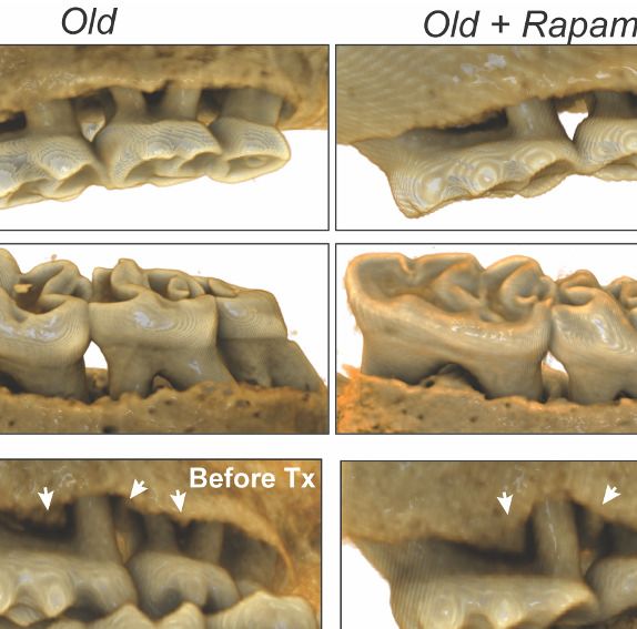
Research article Cell Biology Immunology and Inflammation
Figure 1. Cross-institution experimental design and assay for measuring periodontal bone loss. (A) The NIA-UW colonies were received directly from
the NIA Aged Rodent Colony at 4, 11, and 18 months, then acclimated for two months within the UW facilities (ARCF) until they reached 6 (Young), 13
(Adult), and 20 months (Old). The Young and Adult cohorts were harvested for oral tissues and microbiome. The Old cohorts were randomized and
either given Eudragit or 42ppm eRAPA within the food for 8 weeks. For the JAX colonies, an initial microCT image was taken prior to the 8 week
treatment and then a final microCT before harvest. All animals were harvested at the end of 8 weeks, ~22 months old. (B) Representative image of a
mandible is shown. Periodontal bone loss was measured as distance from the cementoenamel junction (CEJ, white arrows) to the alveolar bone crest
(ABC, orange arrows) on 16 predetermined landmarks on the buccal aspect of maxillary and mandibular periodontium. The CEJ-ABC distances were
totaled for each mouse.
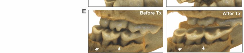
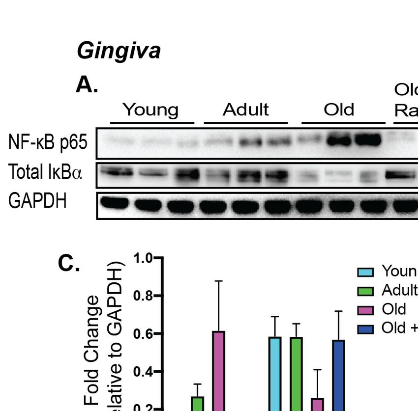
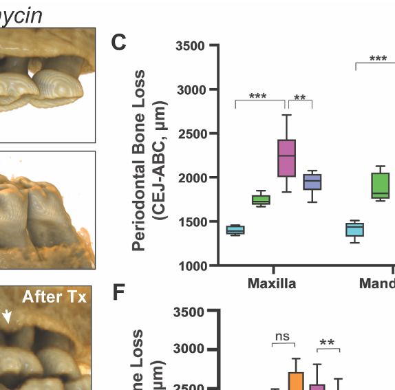
loss of periodontal bone with age in the NIA-UW cohort (Figure 2, A to C). Mice treated with rapa-
mycin for 8 weeks had significantly more bone at the end of the treatment period compared to mice
that received the control diet (eudragit) (Figure 2C). To determine whether the increase in periodon-
tal bone upon rapamycin treatment reflects attenuation of bone loss or growth of new bone, we per-
formed mCT imaging on mice before and after treatment in the JAX cohort (Figure 1A). Old mice
randomized into either the eudragit control or rapamycin treatment groups had significantly less
periodontal bone than young mice prior to the treatment period (Figure 2F). After 8 weeks, the
rapamycin treated mice had significantly more periodontal bone compared to eudragit controls and
also compared to the pre-treatment levels for the same animals (Figure 2, D to F). The presence of
new bone following rapamycin treatment can be observed by comparison of mCT images from the
same animals before and after treatment (Figure 2, D and E).
Normal bone homeostasis results from a balance between new bone growth and bone resorp-
tion, which is reflected by the ratio of RANKL (receptor-activator of nuclear factor-kB ligand) to OPG
(osteoprotegerin), and dysregulation of this balance contributes to bone loss in periodontitis (Dar-
veau, 2010). Consistent with bone loss during aging, we detected significantly greater levels of
RANKL in old animals of both cohorts compared to young animals (Figure 3A and B). OPG levels
remained relatively stable, resulting in an increase in the RANKL:OPG ratio indicative of bone resorp-
tion exceeding bone formation (Figure 3C). These age-associated defects in bone homeostasis were
suppressed by eight weeks of rapamycin treatment (Figure 3). In addition to increased RANKL:OPG
ratio, a significant increase in TRAP+ cells was also observed in periodontal bone with age
(Figure 3D and E). TRAP (tartrate-resistant acid phosphatase) is a histochemical marker of bone
resorbing osteoclasts (Hayman, 2008; Ballanti et al., 1997). Rapamycin treatment for eight weeks
also decreased TRAP+ cells. Together, our data indicate that rapamycin reverses periodontal bone
loss in the aging murine oral cavity at least in part through inhibition of bone resorption.
Along with bone loss, gingival inflammation is a defining feature of periodontal disease. Aging is
also associated with chronic accumulation of pro-inflammatory factors, a collective term referred to
as inflammaging (Chung et al., 2009; Franceschi and Ottaviani, 1997; Franceschi et al., 2000;
An et al. eLife 2020;9:e54318. DOI: https://doi.org/10.7554/eLife.54318 3 of 17
Research article                                                                                Cell Biology Immunology and Inflammation

Figure 2. Rapamycin reverses age-associated periodontal bone loss (NIA-UW and JAX). (A and B) Representative images of NIA-UW (A) maxillary and
(B) mandibular teeth of Young, Old, and Old treated with 42ppm eRAPA (rapamycin) revealing age-associated periodontal bone loss. 8 weeks of
rapamycin attenuated periodontal bone loss. (C) Box-and-whiskers plots shows median, 25th and 75th percentile with whiskers at the 5th and 95th
percentile. Statistical analysis was completed using unpaired t-test, with p-values
Research article                                                                           Cell Biology Immunology and Inflammation

Figure 3. Rapamycin attenuates age-associated increase in RANKL expression and TRAP+ cells in periodontal bone. (A and B) RANKL and OPG
expression was determined by western blot analysis of total lysates from the periodontal bone of aged animals (Young, Adult, and Old) and Old
animals treated for 8 weeks with 42ppm rapamycin (eRAPA). The periodontal bone within both the NIA-UW and JAX Colonies showed an increased
expression of RANKL while 8 weeks of rapamycin treatment ameliorated the increased RANKL expression. Each lane represents individual periodontal
bone samples. (C) Quantification of RANKL/OPG of the NIA-UW western blot analysis. (D) Representative histological sections of the alveolar bone
furcation that have undergone TRAP azo-dye staining with FastGreen counterstain. (E) Enumeration of TRAP+ cells within the periodontal bone from
two-independent observers reveals an increase number of TRAP+ cells with age and diminishes with rapamycin treatment. Statistical analysis was
completed with unpaired t-test, with significance set to p
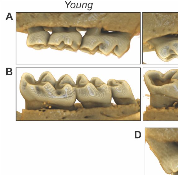
Research article                                                                              Cell Biology Immunology and Inflammation

Figure 4. Rapamycin alters increased NF-kB expression and inflammatory cytokine profiles in periodontium. NF-kB p65 and IkBa expression was
determined by western blot analysis of total lysates from the gingiva (A,C) and periodontal bone (B,D) of control animals (Young, Adult, and Old) and
Old animals treated for 8 weeks with rapamycin (42ppm eRAPA). GAPDH was used a loading control. Both in the aging gingiva and periodontal bone,
there is an overall increased expression of NF-kB p65 with corresponding alteration of IkBa or p-IkBa. 8 weeks of 42ppm eRAPA treatment attenuates
the changes seen with age. For the gingiva, each lane represents gingiva from animals co-housed (n = 2), and each lane for the periodontal bone
western blot represents individual animals. (E and F) Protein expression levels of mouse cytokines and chemokines was determined by a spotted
nitrocellulose membrane assay (Proteome Profiler Mouse, R and D Systems) by loading pooled samples from (E) gingiva and (F) periodontal bone of
Young and Old (Control, Eudragit), and Old animals treated for 8 weeks with rapamycin (42ppm eRAPA). Data are shown per manufacture’s protocol,
with fold-change relative to Young (Set to 1), expressed as mean ± SEM. All changes shown are statistically significant (p
Research article                                                                                  Cell Biology Immunology and Inflammation

Figure 5. Rapamycin shifts aged oral microbiome towards young oral microbiome. (A) Alpha diversity for all samples reveal significant differences
between young (Y) and old (O) mice without rapamycin treatment (p
Research article                                                                   Cell Biology Immunology and Inflammation

Continued

Reagent type                                                                                              Additional
(species) or resource    Designation                     Source or reference   Identifiers                information
Strain, strain           C57BL/6J                        Jackson Laboratory    RRID:IMSR_JAX:000664
background
(M. musculus)
Chemical                 Rapamycin                       Rapamycin Holdings                               Amount based upon active
compound, drug                                                                                            rapamycin content to provide
                                                                                                          42 parts per million
                                                                                                          concentration in chow.
Chemical                 Eudragit                        Rapamycin Holdings
compound, drug
Chemical                 RIPA Lysis and                  ThermoFisher          Cat#: 89901
compound, drug           Extraction Buffer               Scientific
Chemical                 HALT Protease                   ThermoFisher          Cat#: 78438
compound, drug           Inhibitor Cocktail              Scientific
Chemical                 HALT Phosphatase                ThermoFisher          Cat#: 78420
compound, drug           Inhibitor Cocktail              Scientific
Chemical                 Restore Plus                    Thermfisher           Cat#: 46430
compound,                Stripping Buffer                Scientific
drug
Antibody                 anti -NFkBp65                   Cell Signaling        Cat#: 8242                 WB (1:1000)
                         (Rabbit Monoclonal)
Antibody                 anti-phospho-IkBa               Santa Cruz            Cat#: sc8404               WB (1:1000)
                         (Mouse monoclonal)              Biotechnology
Antibody                 Anti- IkBa                      Abcam                 Cat#: 32518                WB (1:1000)
                         (Rabbit Monoclonal)
Antibody                 anti-GAPDH                      Cell Signaling        Cat#: 5174                 WB (1:1000)
                         (Rabbit monoclonal)
Antibody                 anti-RANKL                      Santa Cruz            Cat#: sc377079             WB (1:1000)
                         (Mouse monoclonal)              Biotechnology
Antibody                 anti-OPG                        R and D Systems       AF459                      WB (1:1000)
                         (Goat polyclonal)
Antibody                 anti-IgGk                       Santa Cruz            Cat#: sc516102             WB (1:10000)
                         (Mouse monoclonal)              Biotechnology
Antibody                 anti-rabbit IgG                 Thermo Fisher         Cat#: 31458                WB (1:10000)
                         (Donkey polyclonal)             Scientific
Antibody                 anti-goat IgG                   Abcam                 Cat#: ab97110              WB (1:10000)
                         (Donkey polyclonal)
Commercial               Proteome Profiler Mouse XL      R and D Systems       Cat#: ARY028
assay, kit               Cytokine Array
Commercial               Acid Phosphatase,               Millipore Sigma       Cat#: 387A-1KT
assay, kit               Leukocyte (TRAP) Kit
Commercial               QIAamp DNA                      Qiagen                Cat#: 51704
assay, kit               Microbiome Kit
Commercial               DNA Clean                       Zymo Research         Cat#: D4014
assay, kit               and
                         Concentrator Kit
Commercial               KAPA HiFi                       KAPA Biosystems       Cat#: KK2601
assay, kit               HotStart ReadyMix
Commercial               Nextera XT                      Illumina              Set A: FC-131–2001
assay, kit               Index Kit V2                                          Set D: FC-131–2004
Commercial               AMPure XP                       Agencourt             A63881
assay, kit               Beads
Commercial               SequalPrep                      Invitrogen            A1051001
assay, kit               Normalization Kit
Continued on next page

An et al. eLife 2020;9:e54318. DOI: https://doi.org/10.7554/eLife.54318                                                        8 of 17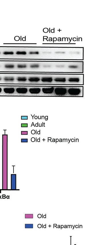
Research article                                                                          Cell Biology Immunology and Inflammation

Continued

Reagent type                                                                                                      Additional
(species) or resource     Designation                    Source or reference            Identifiers               information
Commercial                TapeStation 4200 High          Agilent Technologies           G2991AA
assay, kit                Sensitivity
                          D1000 assay
Commercial                Tapestation                    Agilent Technologies           5067–5585
assay, kit                Reagents
Commercial                High                           Agilent Technologies           5067–5584
assay, kit                Sensitivity D1000
                          ScreenTape
Commercial                Qubit High                     ThermoFisher                   Q32854
assay, kit                Sensitivity                    Scientific
                          dsDNA assay
Commercial                MiSeq                          Illumina                       Cat#: MS-102–3003
assay, kit                Reagent Kit
                          v3 (600 cycle)
Commercial                PhiX                           Illumina                       Cat#: FC-110–3001
assay, kit                Control Kit v3
Software,                 Qiime2                         https://qiime2.org/            V.2019.1
Algorithm
Software,                 DADA2 Package                  PMID:27214047
Algorithm
Software,                 Human Oral                     Homd.org                                                 v. 15.1
Algorithm                 Microbiome Database            PMID:30534599
                          (HOMD)
Software,                 R-studio                       https://rstudio.com/           RRID:SCR_000432           Version 3.5.3
Algorithm
Software,                 Phyloseq                       PMID:23630581                  RRID:SCR_013080
Algorithm
Software,                 Clustvis                       PMID:25969447                  RRID:SCR_017133
Algorithm
Software,                 Ggplot2                        https://www.springer.com/gp/   RRID:SCR_014601           https://www.springer.com/
Algorithm                                                book/9780387981413                                       gp/book/9780387981413
Software,                 Ampvis2                        http://dx.doi.org/
Algorithm                                                10.1101/299537v1
Software,                 vegan                          https://cran.r-project.org,    RRID:SCR_011950
Algorithm                                                https://github.com/
                                                         vegandevs/vegan
Software,                 Ade4                           https://www.jstatsoft.
Algorithm                                                org/article/view/v086i01
Software,                 Bioinformatic scripts and      This paper                                               https://github.com/kkerns85/
Algorithm                 microbiome data                                                                         Rapamycin_rejuvenates_
                          used in analysis                                                                        oral_health_in_aging_mice.git.
Software,                 R Markdown                     This paper                                               https://rpubs.com/kkerns85/
Algorithm                                                                                                         Rapamycin_Rmrkdown
Software,                 Graphpad Prism                 Graphpad                       RRID:SCR_002798           Version 8.4
Algorithm                                                (graphpad.com)

                                     Animal studies
                                     To enhance rigor and reproducibility, experiments were performed on two different cohorts housed
                                     at two sites: the University of Washington in Seattle, WA and the Jackson Laboratory in Bar Harbor,
                                     ME. To examine the impact of rapamycin on the periodontium during normative aging, we designed
                                     a cross institutional study between the University of Washington (UW) and the Jackson Laboratory
                                     (JAX) (Figure 1A). The UW cohorts of C57BL/6Nia (hereafter termed NIA-UW Colony) were received
                                     directly from the National Institute on Aging (NIA) Aged Rodent Colony and acclimated within the
                                     UW facilities. The JAX cohorts of C57BL/6J (hereafter termed JAX Colony) were born and raised
                                     within the JAX facilities. We then treated mice at both sites with encapsulated rapamycin (eRAPA) in

An et al. eLife 2020;9:e54318. DOI: https://doi.org/10.7554/eLife.54318                                                                  9 of 17
Research article                                                                      Cell Biology Immunology and Inflammation

                                    the diet at 42ppm, which has been shown to significantly increase lifespan of UMHET3 and C57BL6/
                                    J mice (Miller et al., 2014; Zhang et al., 2014), or control food (eudragit). All data are from female
                                    mice, which have previously been found to have greater increases in lifespan and some health met-
                                    rics compared to male mice at this dose of rapamycin (Miller et al., 2014; Zhang et al., 2014). For
                                    the NIA-UW colonies, five young, five adult, and 20 old (10 eudragit and 10 rapamycin) animals were
                                    utilized. While for the JAX colonies a total of 13 young and 26 old (13 eudragit and 13 rapamycin)
                                    animals were used. For this study, young, adult, and old mice were 6, 13, and 20 months of age,
                                    respectively.

                                    Seattle, WA
                                    Twenty NIA-UW mice (10 on eudragit, 10 on rapamycin) received assigned diet treatments at 20
                                    months of age, lasting for 8 weeks, along with five young and five adult mice as normative aging
                                    controls. Animals were housed individually in Allentown NexGen Caging (Allentown, Allentown, NJ)
                                    containing corncob bedding and nestlets. Mice were fed irradiated Picolab Rodent Diet 20 #5053
                                    (Lab Diet, St. Louis, MO). Animals were maintained in a specific pathogen free facility within a Heli-
                                    cobacter spp.-free room. Mice were housed in groups and inspected daily. National Guidelines for
                                    the Care and Use of Animals and the IACUC guidelines were followed.

                                    Bar Harbor, ME
                                    All methods are in accordance with The Jackson Laboratory Institutional Animal Care and Use Com-
                                    mittee (IACUC)-approved protocols. Animals were fed standard Lab Diet 6% 5K52 with eRapa at 42
                                    mg/kg/day or control. Animals had ad libitum access to food and water throughout the study. Ani-
                                    mals were checked daily, and once per week the food was topped off. Animals were housed at 3–5
                                    animals per cage.
                                       A cohort of mice were transferred into the JAX Center for Biometric Analysis and brought into
                                    the imaging suite in groups of 10 mice per scan group. Prior to scanning, the weight of each mouse
                                    was recorded and anesthesia induced with 2–3% isoflurane. The mice were then placed in a prone
                                    position in the CT scanner and kept anesthetized for the duration of the scan with an isoflurane level
                                    of 1.2–1.5%. A whole head scan was performed with bone mineral density phantoms included on
                                    the specimen positioning bed. After the CT scan, the mouse was placed in a warmed isolation cage
                                    and allowed to fully recover from the anesthesia. At the end of the imaging session, the cohort was
                                    returned to animal housing facility.
                                       Animal experimentation was performed in accordance with the recommendations in the Guide
                                    for the Care and Use of Laboratory Animals of the National Institutes of Health. All animals were
                                    handled according to approved institutional animal care and use committee (IACUC) protocols
                                    (#4359–01) of the University of Washington and (#06005-A24) of the Jackson Laboratory.

                                    Encapsulated rapamycin feeding
                                    Encapsulated rapamycin (eRAPA) was obtained from Rapamycin Holdings, Inc. Food pellets were
                                    ground and mixed with encapsulated rapamycin at 42ppm. 300 ml of 1% agar melted in sterile
                                    water, and 200 ml of sterile distilled water were added per kilogram of powdered chow in order to
                                    make pellets. Pellets were stored at 20˚C until use. Control food contained the same concentration
                                    of agar and encapsulated material (eudragit) without rapamycin at the concentration that matched
                                    the rapamycin chow. Eudragit is the encapsulation material used in eRAPA and is a copolymer
                                    derived from esters of acrylic and methacrylic acids. Eudragit without rapamycin was thus added to
                                    the regular chow at 42 ppm as a vehicle control.

                                    Micro-computed tomography (mCT) analysis
                                    Seattle Children’s Research Institute and Friday Harbor Lab imaging
                                    parameter and processing
                                    NIA-UW samples were scanned in a Skyscan 1076 and 1173 microCT system at the Small Animal
                                    Tomographic Analysis Facility (SANTA) at Seattle Children’s Research Institute and Friday Harbor
                                    Laboratories at the University of Washington. Resolutions were 8–18 mm with following settings: 5
                                    kV, 179mA, 360 ms exposure, 0.5 AI filter, 0.7˚ rotation step, and 3-frame averaging. Raw scan data
                                    were reconstructed with NRecon 1.6.9, and three-dimensional (3D) renderings were generated with

An et al. eLife 2020;9:e54318. DOI: https://doi.org/10.7554/eLife.54318                                                           10 of 17
Research article Cell Biology Immunology and Inflammation
Drishti 2.7 (Limaye, 2012). For periodontal bone loss, 3D rendered images were randomized and
landmarked by independent observers. Periodontal bone loss was measured as distance from the
cementoenamel junction (CEJ) to the alveolar bone crest (ABC) on 16 predetermined landmarks on
the buccal aspect of maxillary and mandibular periodontium. The CEJ-ABC distances were totaled
for each mouse through the Drishti software, and means calculated. The analysis was completed by
3–4 independent observers.
Jackson Laboratory (JAX) imaging parameter and processing
The mouse is scanned in a Perkin-Elmer Quantum GX in vivo Micro-CT tomograph. Resolutions were
17–50 microns with the following settings: 55 kV, 145 mA, 4 min exposure over 360 degrees rotation.
The native Perkin-Elmer Viewer VOX image files are converted to Drishti Volume Exploration and
Presentation Tool NetCDF format volumes using custom code specific for this study (Limaye, 2012).
Western blot and proteome profile analysis
For protein analysis by western blot, gingival tissue and alveolar bone was dissected. Total cellular
proteins were extracted in RIPA Lysis and Extraction Buffer (Thermo Scientific, MA, USA) and EDTA-
free Halt protease and phosphatase inhibitor cocktail included to prevent protein degradation dur-
ing extraction process. Gingival tissue was pooled from co-housed animals and bone samples were
from single specimens. Protein concentration was determined by Pierce BCA Protein Assay Kit
(Thermo Scientific). 10–20 mg of total protein was separated by SDS-PAGE on 10% or 12% (w/v)
polyacrylamide gel, then transferred to PVDF membrane using Trans-Blot Turbo Transfer System
(Bio-Rad, CA, USA). Antibodies to NF-kB p65 (D14E12) XP (8242, Cell Signaling Technology), phos-
pho-IkBa (B-9, Santa Cruz), IkBa (32518, Abcam), GAPDH (D16H11) XP (5174, Cell Signaling Tech-
nology), RANKL (G-1, sc377079, Santa Cruz), and Mouse OPG (R and D Systems, AF459) were used
to probe the membrane. Dependent upon the strength of the antibody-dependent signal, either the
membranes were stripped with Restore Plus Western Blot Stripping Buffer and reprobed for total
antibody, or duplicate gels were run and separate blots probed.
Analysis of the cytokine proteome was completed using a Mouse XL Cytokine Array Kit (R and D
Systems, Bio-Techne Corporation, MN, USA). Gingiva and alveolar bone samples were individually
pooled, protein concentration determined by Pierce BCA Assay Kit and 200 mg of protein lysate
loaded. Detection and imaging were performed using ChemiDoc XRS+ (Biorad, USA) and Image
Lab Software (Biorad, USA). Data analysis was completed per the manufacture’s protocol.
Histology
Tissues were fixed in Bouin’s solution, and demineralized in AFS (acetic acid, formaldehyde, sodium
chloride). Mandibles were processed and embedded in paraffin. Serial sections of 5 mm thickness
were collected in the coronal (buccal-lingual) plane. Sections were stained for tartrate-resistant acid
phosphatase (TRAP) to examine osteoclast activity and numbers (Sigma-Aldrich Kit, St. Louis, MO,
USA), and Fast Green counterstaining and examined with a Nikon Eclipse 90i Advanced Research
Scope. Representative images (40x) were taken of the alveolar bone furcation.
Microbiome analysis
DNA extraction
Mandible samples were cryogrinded and homogenized using bead-beating tubes and ceramic
beads. Bacterial genomic DNA was extracted using the QIAamp DNA Microbiome Kit (Qiagen, Hil-
den, Germany) and further purified and concentrated using DNA Clean and Concentrator Kit (Zymo
Research, Irvine, CA, USA) according to the manufacturer’s protocol, then stored at 80 ˚C until all
samples were collected.
Sequencing
The V3-V4 variable region of the 16 s ribosomal RNA gene was amplified using gene-specific primers
with Illumina adapter overhang sequences (5’-TCGTCGGCAGCGTCAGATGTGTATAAGAGA-
CAGCCTACGGGNGGCWGCAG-3’ and 5’-GTCTCGTGGGCTCGGAGATGTGTATAAGAGACAG-
GACTACHVGGGTATCTAATCC-3’). Each reaction mixture contained 2.5 ml of genomic DNA, 5 ml of
each 1 mM primer, and 12.5 ml of KAPA HiFi HotStart ReadyMix. Amplicon PCR was carried out as
An et al. eLife 2020;9:e54318. DOI: https://doi.org/10.7554/eLife.54318 11 of 17Research article Cell Biology Immunology and Inflammation
follows: denaturation at 95˚C for 3 min, 35–40 cycles at 95˚C for 30 s, 55˚C for 30 s, 72˚C for 30 s, fol-
lowed by a final extension step at 72˚C for 5 min. PCR products were verified using gel electrophore-
sis (1% agarose gel) and cleaned with AMPure XP beads (Agencourt, Beckman Coulter Inc,
Pasadena, CA, USA). Amplicons were then indexed using the Nextera XT Index Kit V2 set A and set
D (Illumina) and purified again with AMPure XP beads to remove low molecular weight primers and
primer-dimer sequences. DNA concentrations were concentration of 1–2 nM using the SequalPrep
Normalization Kit (Invitrogen). Samples were pooled into a single library which was analyzed using
the TapeStation 4200 High Sensitivity D1000 assay (Agilent Technologies, Waldbronn, Germany) and
Qubit High Sensitivity dsDNA assay (Thermo Fischer Scientific) to assess DNA quality and quantity.
The final pooled library was then loaded on to an Illumina MiSeq sequencer with 10% PhiX spike,
which served as an internal control to balance for possible low diversity and base bias present in the
16S amplicon samples, and was run for 478 cycles and generated a total of 5.68 million paired-end
reads (2 239 bp).
Bioinformatics
Raw paired-end sequences were imported in to Qiime2 (v. 2019.1) and were trimmed by 15 nt from
the 5’ end and truncated to 239 nt for the 3’ end for both the forward and reverse reads respec-
tively. The trimmed reads were then demultiplexed and denoised using the DADA2 package
(Callahan et al., 2016). Forward reads were only used in our analysis. Taxonomy was then assigned
using the feature-classifier suite trained on the Human Oral Microbiome Database (HOMD v. 15.1)
(Escapa et al., 2018). Samples were then filtered for taxonomic contaminants excluded samples
with less than 10,000 reads. Alpha and Beta diversity as well as other analysis were done in R-Studio
using the Phyloseq (McMurdie and Holmes, 2013) Clustvis (Metsalu and Vilo, 2015), ggplot2 (Wik-
ham, 2016), ampvis2 (Andersen KS et al., 2018), vegan (Oksanen J et al., 2019), ade4
(Bougeard and Dray, 2018) packages as part of the R suite.
Taxonomy filtered from samples was determined by analysis of kit controls with no template and
zymo sequencing controls of known diversity and abundance in the QIAamp DNA Microbiome Kit
(Qiagen, Hilden, Germany) and the DNA Clean and Concentrator Kit (Zymo Research). The following
taxonomic assignments were removed as part of the dada2 workflow (Callahan et al., 2016): Unas-
signed, Cyanobacteria, acidovorans, pestis, coli, flavescens, sakazakii, durans, diminuta, anthropi,
monocytogenes, parasanquinis_clade_411, otitidis, subtilis, aeruginosa, fermentum.
Statistical analysis
Results for mCT analysis, including measurements, quantitative histology, proteome analysis are
expressed as mean ± standard error of mean (SEM). Data were analyzed where appropriate using
Student’s t-test or paired t-test (comparing two groups only), or one-way analysis of variance
(ANOVA) with post-hoc Tukey test for multiple comparisons, where p-valuesResearch article Cell Biology Immunology and Inflammation
format, prior to post-processing and data analysis, have been deposited at the European Nucleotide
Archive (ENA) under study accession no. PRJEB35672.
Acknowledgements
The authors would like to thank the Karel F Liem Imaging Facility at Friday Harbor Laboratories, and
Ryan Anderson on his guidance at Seattle Children’s Research Institute. Authors would also like to
thank Ella Lamont and Archita Gadkari for their guidance and processing of the microbiome
samples.
Additional information
Competing interests
Matt Kaeberlein: Reviewing editor, eLife. The other authors declare that no competing interests
exist.
Funding
Funder Grant reference number Author
National Institute of Dental DE027254 Jonathan Y An
and Craniofacial Research
National Institute of Dental DE023810 Jonathan Y An
and Craniofacial Research Jeffrey S McLean
National Institute of Dental DE020102 Jonathan Y An
and Craniofacial Research Jeffrey S McLean
National Institute on Aging AG054180 Catherine Kaczorowski
National Institute on Aging AG038070 Catherine Kaczorowski
National Institute on Aging AG038070 Catherine Kaczorowski
National Institutes of Health TR002318 Kristopher A Kerns
National Institute on Aging AG013280 Matt Kaeberlein
The funders had no role in study design, data collection and interpretation, or the
decision to submit the work for publication.
Author contributions
Jonathan Y An, Conceptualization, Investigation, Methodology, Writing - original draft, Writing -
review and editing; Kristopher A Kerns, Formal analysis, Investigation, Methodology; Andrew Ouel-
lette, Laura Robinson, So-Il Park, Title Mekvanich, Alex Kang, Investigation; H Douglas Morris, Inves-
tigation, Methodology, Writing - review and editing; Catherine Kaczorowski, Resources, Supervision,
Methodology, Writing - review and editing; Jeffrey S McLean, Resources, Formal analysis, Supervi-
sion, Methodology, Writing - review and editing; Timothy C Cox, Resources, Software, Methodol-
ogy; Matt Kaeberlein, Conceptualization, Resources, Supervision, Visualization, Writing - original
draft, Writing - review and editing
Author ORCIDs
Jonathan Y An https://orcid.org/0000-0001-8422-8608
Kristopher A Kerns https://orcid.org/0000-0002-4380-0062
H Douglas Morris http://orcid.org/0000-0002-7942-3748
Jeffrey S McLean https://orcid.org/0000-0001-9934-5137
Matt Kaeberlein https://orcid.org/0000-0002-1311-3421
Ethics
Animal experimentation: This study was performed in strict accordance with the recommendations
in the Guide for the Care and Use of Laboratory Animals of the National Institutes of Health. All of
An et al. eLife 2020;9:e54318. DOI: https://doi.org/10.7554/eLife.54318 13 of 17Research article Cell Biology Immunology and Inflammation
the animals were handled according to approved institutional animal care and use committee
(IACUC) protocols of the University of Washington (#4359-01) and of the Jackson Laboratory
(#06005-A24).
Decision letter and Author response
Decision letter https://doi.org/10.7554/eLife.54318.sa1
Author response https://doi.org/10.7554/eLife.54318.sa2
Additional files
Supplementary files
. Transparent reporting form
Data availability
The V4-16S rDNA sequences in raw format, prior to post-processing and data analysis, have been
deposited at the European Nucleotide Archive (ENA) under study accession no. PRJEB35672. Dryad
Data link: https://doi.org/10.5061/dryad.f4qrfj6sn.
The following datasets were generated:
Database and
Author(s) Year Dataset title Dataset URL Identifier
An JY, Kerns KA, 2020 Rapamycin rejuvenates oral health http://dx.doi.org/10. Dryad Digital
Ouellette A, Ro- in aging mice 5061/dryad.f4qrfj6sn Repository, 10.5061/
binson L, Morris D, dryad.f4qrfj6sn
Kaczorowski C, Park
S, Mekvanich T,
Kang A, McLean JS,
Cox TC, Kaeberlein
M
An JY, Kerns KA, 2019 Rapamycin rejuvenates oral health https://www.ebi.ac.uk/ European Nucleotide
Ouellette A, Ro- in aging mice ena/browser/view/ Archive (ENA),
binson L, Morris D, PRJEB35672 PRJEB35672
Kaczorowski C, Park
S, Mekvanich T,
Kang A, McLean JS,
Cox TC, Kaeberlein
M
References
Abu-Amer Y. 2013. NF-kB signaling and bone resorption. Osteoporosis International 24:2377–2386.
DOI: https://doi.org/10.1007/s00198-013-2313-x, PMID: 23468073
Ambili R, Janam P. 2017. A critique on nuclear factor-kappa B and signal transducer and activator of
transcription 3: the key transcription factors in periodontal pathogenesis. Journal of Indian Society of
Periodontology 21:350–356. DOI: https://doi.org/10.4103/jisp.jisp_301_16, PMID: 29491579
An JY, Quarles EK, Mekvanich S, Kang A, Liu A, Santos D, Miller RA, Rabinovitch PS, Cox TC, Kaeberlein M.
2017. Rapamycin treatment attenuates age-associated periodontitis in mice. GeroScience 39:457–463.
DOI: https://doi.org/10.1007/s11357-017-9994-6, PMID: 28889220
An JY, Darveau R, Kaeberlein M. 2018. Oral health in geroscience: animal models and the aging oral cavity.
GeroScience 40:1–10. DOI: https://doi.org/10.1007/s11357-017-0004-9, PMID: 29282653
Andersen KS KR, Karst SM, Albertsen M. 2018. ampvis2: an R package to analyse and visualize 16S rRNA
amplicon data. bioRxiv. DOI: https://doi.org/10.1101/299537
Arabaci T, Cicek Y, Canakci V, Canakci CF, Ozgoz M, Albayrak M, Keles ON. 2010. Immunohistochemical and
stereologic analysis of NF-kappaB activation in chronic periodontitis. European Journal of Dentistry 4:454–461.
PMID: 20922166
Ballanti P, Minisola S, Pacitti MT, Scarnecchia L, Rosso R, Mazzuoli GF, Bonucci E. 1997. Tartrate-resistant acid
phosphate activity as osteoclastic marker: sensitivity of cytochemical assessment and serum assay in
comparison with standardized osteoclast histomorphometry. Osteoporosis International 7:39–43. DOI: https://
doi.org/10.1007/BF01623458, PMID: 9102061
Bitto A, Ito TK, Pineda VV, LeTexier NJ, Huang HZ, Sutlief E, Tung H, Vizzini N, Chen B, Smith K, Meza D, Yajima
M, Beyer RP, Kerr KF, Davis DJ, Gillespie CH, Snyder JM, Treuting PM, Kaeberlein M. 2016. Transient
An et al. eLife 2020;9:e54318. DOI: https://doi.org/10.7554/eLife.54318 14 of 17Research article Cell Biology Immunology and Inflammation
rapamycin treatment can increase lifespan and healthspan in middle-aged mice. eLife 5:e16351. DOI: https://
doi.org/10.7554/eLife.16351, PMID: 27549339
Bougeard S, Dray S. 2018. Supervised multiblock analysis in R with the ade4 package. Journal of Statistical
Software 86:1–17. DOI: https://doi.org/10.18637/jss.v086.i01
Callahan BJ, McMurdie PJ, Rosen MJ, Han AW, Johnson AJ, Holmes SP. 2016. DADA2: high-resolution sample
inference from Illumina amplicon data. Nature Methods 13:581–583. DOI: https://doi.org/10.1038/nmeth.3869,
PMID: 27214047
Chen C, Liu Y, Liu Y, Zheng P. 2009. mTOR regulation and therapeutic rejuvenation of aging hematopoietic stem
cells. Science Signaling 2:ra75. DOI: https://doi.org/10.1126/scisignal.2000559, PMID: 19934433
Chung HY, Cesari M, Anton S, Marzetti E, Giovannini S, Seo AY, Carter C, Yu BP, Leeuwenburgh C. 2009.
Molecular inflammation: underpinnings of aging and age-related diseases. Ageing Research Reviews 8:18–30.
DOI: https://doi.org/10.1016/j.arr.2008.07.002, PMID: 18692159
Chung CL, Lawrence I, Hoffman M, Elgindi D, Nadhan K, Potnis M, Jin A, Sershon C, Binnebose R, Lorenzini A,
Sell C. 2019. Topical rapamycin reduces markers of senescence and aging in human skin: an exploratory,
prospective, randomized trial. GeroScience 41:861–869. DOI: https://doi.org/10.1007/s11357-019-00113-y,
PMID: 31761958
Dai DF, Karunadharma PP, Chiao YA, Basisty N, Crispin D, Hsieh EJ, Chen T, Gu H, Djukovic D, Raftery D, Beyer
RP, MacCoss MJ, Rabinovitch PS. 2014. Altered proteome turnover and remodeling by short-term caloric
restriction or rapamycin rejuvenate the aging heart. Aging Cell 13:529–539. DOI: https://doi.org/10.1111/acel.
12203, PMID: 24612461
Darveau RP. 2010. Periodontitis: a polymicrobial disruption of host homeostasis. Nature Reviews Microbiology 8:
481–490. DOI: https://doi.org/10.1038/nrmicro2337, PMID: 20514045
De Martinis M, Franceschi C, Monti D, Ginaldi L. 2005. Inflamm-ageing and lifelong antigenic load as major
determinants of ageing rate and longevity. FEBS Letters 579:2035–2039. DOI: https://doi.org/10.1016/j.
febslet.2005.02.055, PMID: 15811314
Eke PI, Dye BA, Wei L, Thornton-Evans GO, Genco RJ, CDC Periodontal Disease Surveillance workgroup: James
Beck (University of North Carolina, Chapel Hill, USA), Gordon Douglass (Past President, American Academy of
Periodontology), Roy Page (University of Washin. 2012. Prevalence of periodontitis in adults in the united
states: 2009 and 2010. Journal of Dental Research 91:914–920. DOI: https://doi.org/10.1177/
0022034512457373, PMID: 22935673
Eke PI, Dye BA, Wei L, Slade GD, Thornton-Evans GO, Borgnakke WS, Taylor GW, Page RC, Beck JD, Genco RJ.
2015. Update on prevalence of periodontitis in adults in the united states: nhanes 2009 to 2012. Journal of
Periodontology 86:611–622. DOI: https://doi.org/10.1902/jop.2015.140520, PMID: 25688694
Escapa IF, Chen T, Huang Y, Gajare P, Dewhirst FE, Lemon KP. 2018. New insights into human nostril
microbiome from the expanded human oral microbiome database (eHOMD): a resource for the microbiome of
the human aerodigestive tract. mSystems 3:18. DOI: https://doi.org/10.1128/mSystems.00187-18
Flynn JM, O’Leary MN, Zambataro CA, Academia EC, Presley MP, Garrett BJ, Zykovich A, Mooney SD, Strong R,
Rosen CJ, Kapahi P, Nelson MD, Kennedy BK, Melov S. 2013. Late-life rapamycin treatment reverses age-
related heart dysfunction. Aging Cell 12:851–862. DOI: https://doi.org/10.1111/acel.12109
Franceschi C, Bonafè M, Valensin S, Olivieri F, De Luca M, Ottaviani E, De Benedictis G. 2000. Inflamm-aging. an
evolutionary perspective on immunosenescence. Annals of the New York Academy of Sciences 908:244–254.
DOI: https://doi.org/10.1111/j.1749-6632.2000.tb06651.x, PMID: 10911963
Franceschi C, Ottaviani E. 1997. Stress, inflammation and natural immunity in the aging process: a new theory.
Aging Clinical and Experimental Research 9:30–31. DOI: https://doi.org/10.1007/BF03339694
Gil-Montoya JA, de Mello AL, Barrios R, Gonzalez-Moles MA, Bravo M. 2015. Oral health in the elderly patient
and its impact on general well-being: a nonsystematic review. Clinical Interventions in Aging 10:461–467.
DOI: https://doi.org/10.2147/CIA.S54630, PMID: 25709420
Harrison DE, Strong R, Sharp ZD, Nelson JF, Astle CM, Flurkey K, Nadon NL, Wilkinson JE, Frenkel K, Carter CS,
Pahor M, Javors MA, Fernandez E, Miller RA. 2009. Rapamycin fed late in life extends lifespan in genetically
heterogeneous mice. Nature 460:392–395. DOI: https://doi.org/10.1038/nature08221, PMID: 19587680
Hayman AR. 2008. Tartrate-resistant acid phosphatase (TRAP) and the osteoclast/immune cell dichotomy.
Autoimmunity 41:218–223. DOI: https://doi.org/10.1080/08916930701694667, PMID: 18365835
Hurez V, Dao V, Liu A, Pandeswara S, Gelfond J, Sun L, Bergman M, Orihuela CJ, Galvan V, Padrón Á, Drerup J,
Liu Y, Hasty P, Sharp ZD, Curiel TJ. 2015. Chronic mTOR inhibition in mice with rapamycin alters T, B, myeloid,
and innate lymphoid cells and gut flora and prolongs life of immune-deficient mice. Aging Cell 14:945–956.
DOI: https://doi.org/10.1111/acel.12380, PMID: 26315673
Johnson SC, Rabinovitch PS, Kaeberlein M. 2013. mTOR is a key modulator of ageing and age-related disease.
Nature 493:338–345. DOI: https://doi.org/10.1038/nature11861, PMID: 23325216
Jung MJ, Lee J, Shin NR, Kim MS, Hyun DW, Yun JH, Kim PS, Whon TW, Bae JW. 2016. Chronic repression of
mTOR complex 2 induces changes in the gut Microbiota of Diet-induced obese mice. Scientific Reports 6:
30887. DOI: https://doi.org/10.1038/srep30887, PMID: 27471110
Kaeberlein M, Rabinovitch PS, Martin GM. 2015. Healthy aging: the ultimate preventative medicine. Science
350:1191–1193. DOI: https://doi.org/10.1126/science.aad3267, PMID: 26785476
Kaeberlein M. 2017. Translational geroscience: a new paradigm for 21st century medicine. Translational Medicine
of Aging 1:1–4. DOI: https://doi.org/10.1016/j.tma.2017.09.004, PMID: 32219192
An et al. eLife 2020;9:e54318. DOI: https://doi.org/10.7554/eLife.54318 15 of 17Research article Cell Biology Immunology and Inflammation
Kennedy BK, Berger SL, Brunet A, Campisi J, Cuervo AM, Epel ES, Franceschi C, Lithgow GJ, Morimoto RI,
Pessin JE, Rando TA, Richardson A, Schadt EE, Wyss-Coray T, Sierra F. 2014. Geroscience: linking aging to
chronic disease. Cell 159:709–713. DOI: https://doi.org/10.1016/j.cell.2014.10.039, PMID: 25417146
Kerns KA. 2020. Rapamycin_rejuvenates_oral_health_in_aging_mice. GitHub. 786bb8d. https://github.com/
kkerns85/Rapamycin_rejuvenates_oral_health_in_aging_mice
Kim J, Amar S. 2006. Periodontal disease and systemic conditions: a bidirectional relationship. Odontology 94:
10–21. DOI: https://doi.org/10.1007/s10266-006-0060-6, PMID: 16998613
Könönen E, Gursoy M, Gursoy UK. 2019. Periodontitis: a multifaceted disease of Tooth-Supporting tissues.
Journal of Clinical Medicine 8:1135. DOI: https://doi.org/10.3390/jcm8081135
Lang NP, Bartold PM. 2018. Periodontal health. Journal of Periodontology 89 Suppl 1:S9–S16. DOI: https://doi.
org/10.1002/JPER.16-0517, PMID: 29926938
Limaye A. 2012. Drishti: a volume exploration and presentation tool. SPIE Optical Engineering + Applications.
DOI: https://doi.org/10.1117/12.935640
Liu T, Zhang L, Joo D, Sun SC. 2017. NF-kB signaling in inflammation. Signal Transduction and Targeted Therapy
2:17023. DOI: https://doi.org/10.1038/sigtrans.2017.23, PMID: 29158945
López-Otı́n C, Blasco MA, Partridge L, Serrano M, Kroemer G. 2013. The hallmarks of aging. Cell 153:1194–
1217. DOI: https://doi.org/10.1016/j.cell.2013.05.039, PMID: 23746838
Mannick JB, Del Giudice G, Lattanzi M, Valiante NM, Praestgaard J, Huang B, Lonetto MA, Maecker HT, Kovarik
J, Carson S, Glass DJ, Klickstein LB. 2014. mTOR inhibition improves immune function in the elderly. Science
Translational Medicine 6:268ra179. DOI: https://doi.org/10.1126/scitranslmed.3009892, PMID: 25540326
Mannick JB, Morris M, Hockey HP, Roma G, Beibel M, Kulmatycki K, Watkins M, Shavlakadze T, Zhou W, Quinn
D, Glass DJ, Klickstein LB. 2018. TORC1 inhibition enhances immune function and reduces infections in the
elderly. Science Translational Medicine 10:eaaq1564. DOI: https://doi.org/10.1126/scitranslmed.aaq1564,
PMID: 29997249
McMurdie PJ, Holmes S. 2013. Phyloseq: an R package for reproducible interactive analysis and graphics of
microbiome census data. PLOS ONE 8:e61217. DOI: https://doi.org/10.1371/journal.pone.0061217,
PMID: 23630581
Metsalu T, Vilo J. 2015. ClustVis: a web tool for visualizing clustering of multivariate data using principal
component analysis and heatmap. Nucleic Acids Research 43:W566–W570. DOI: https://doi.org/10.1093/nar/
gkv468, PMID: 25969447
Miller RA, Harrison DE, Astle CM, Fernandez E, Flurkey K, Han M, Javors MA, Li X, Nadon NL, Nelson JF,
Pletcher S, Salmon AB, Sharp ZD, Van Roekel S, Winkleman L, Strong R. 2014. Rapamycin-mediated lifespan
increase in mice is dose and sex dependent and metabolically distinct from dietary restriction. Aging Cell 13:
468–477. DOI: https://doi.org/10.1111/acel.12194, PMID: 24341993
Oksanen J BF, Friendly M, Kindt R, Legendre P, McGlinn D, Minchin PR, O’Hara RB, SP, Simpson GL, Stevens
MHH, Szoecs E, Wagner H. 2019. vegan: Community Ecology Package. R Package.
Quarles E, Basisty N, Chiao YA, Merrihew G, Gu H, Sweetwyne MT, Fredrickson J, Nguyen NH, Razumova M,
Kooiker K, Moussavi-Harami F, Regnier M, Quarles C, MacCoss M, Rabinovitch PS. 2020. Rapamycin
persistently improves cardiac function in aged, male and female mice, even following cessation of treatment.
Aging Cell 19:e13086. DOI: https://doi.org/10.1111/acel.13086, PMID: 31823466
Razak PA, Richard KM, Thankachan RP, Hafiz KA, Kumar KN, Sameer KM. 2014. Geriatric oral health: a review
article. Journal of International Oral Health : JIOH 6:110–116. PMID: 25628498
Sierra F, Kohanski R. 2017. Geroscience and the trans-NIH geroscience interest group, GSIG. GeroScience 39:1–
5. DOI: https://doi.org/10.1007/s11357-016-9954-6, PMID: 28299635
Socransky SS, Haffajee AD, Cugini MA, Smith C, Kent RL. 1998. Microbial complexes in subgingival plaque.
Journal of Clinical Periodontology 25:134–144. DOI: https://doi.org/10.1111/j.1600-051X.1998.tb02419.x,
PMID: 9495612
Thomas F, Hehemann JH, Rebuffet E, Czjzek M, Michel G. 2011. Environmental and gut bacteroidetes: the food
connection. Frontiers in Microbiology 2:93. DOI: https://doi.org/10.3389/fmicb.2011.00093, PMID: 21747801
Torres PJ, Thompson J, McLean JS, Kelley ST, Edlund A. 2019. Discovery of a novel periodontal Disease-
Associated bacterium. Microbial Ecology 77:267–276. DOI: https://doi.org/10.1007/s00248-018-1200-6,
PMID: 29860637
Urfer SR, Kaeberlein TL, Mailheau S, Bergman PJ, Creevy KE, Promislow DE, Kaeberlein M. 2017a.
Asymptomatic heart valve dysfunction in healthy middle-aged companion dogs and its implications for cardiac
aging. GeroScience 39:43–50. DOI: https://doi.org/10.1007/s11357-016-9956-4, PMID: 28299636
Urfer SR, Kaeberlein TL, Mailheau S, Bergman PJ, Creevy KE, Promislow DEL, Kaeberlein M. 2017b. A
randomized controlled trial to establish effects of short-term rapamycin treatment in 24 middle-aged
companion dogs. GeroScience 39:117–127. DOI: https://doi.org/10.1007/s11357-017-9972-z, PMID: 28374166
van Winkelhoff AJ, Loos BG, van der Reijden WA, van der Velden U. 2002. Porphyromonas gingivalis,
Bacteroides forsythus and other putative periodontal pathogens in subjects with and without periodontal
destruction. Journal of Clinical Periodontology 29:1023–1028. DOI: https://doi.org/10.1034/j.1600-051X.2002.
291107.x, PMID: 12472995
Wikham H. 2016. Ggplot 2: Elegant Graphics for Data Analysis. Springer International Publishing. DOI: https://
doi.org/10.1007/978-3-319-24277-4
Zhang Y, Bokov A, Gelfond J, Soto V, Ikeno Y, Hubbard G, Diaz V, Sloane L, Maslin K, Treaster S, Réndon S, van
Remmen H, Ward W, Javors M, Richardson A, Austad SN, Fischer K. 2014. Rapamycin extends life and health in
An et al. eLife 2020;9:e54318. DOI: https://doi.org/10.7554/eLife.54318 16 of 17Research article Cell Biology Immunology and Inflammation
C57BL/6 mice. The Journals of Gerontology: Series A 69A:119–130. DOI: https://doi.org/10.1093/gerona/
glt056
An et al. eLife 2020;9:e54318. DOI: https://doi.org/10.7554/eLife.54318 17 of 17You can also read