Diverse nucleosome Site-Selectivity among histone deacetylase complexes - eLife
←
→
Page content transcription
If your browser does not render page correctly, please read the page content below
RESEARCH ADVANCE

                                      Diverse nucleosome Site-Selectivity
                                      among histone deacetylase complexes
                                      Zhipeng A Wang1,2†, Christopher J Millard3†, Chia-Liang Lin3†,
                                      Jennifer E Gurnett3, Mingxuan Wu1,2, Kwangwoon Lee1,2, Louise Fairall3,
                                      John WR Schwabe3*, Philip A Cole1,2*
                                      1
                                       Division of Genetics, Department of Medicine, Brigham and Women’s Hospital,
                                      Boston, United States; 2Department of Biological Chemistry and Molecular
                                      Pharmacology, Harvard Medical School, Boston, United States; 3Leicester Institute
                                      of Structural and Chemical Biology, Department of Molecular and Cell Biology,
                                      University of Leicester, Leicester, United Kingdom

                                      Abstract Histone acetylation regulates chromatin structure and gene expression and is removed
                                      by histone deacetylases (HDACs). HDACs are commonly found in various protein complexes to
                                      confer distinct cellular functions, but how the multi-subunit complexes influence deacetylase
                                      activities and site-selectivities in chromatin is poorly understood. Previously we reported the results
                                      of studies on the HDAC1 containing CoREST complex and acetylated nucleosome substrates which
                                      revealed a notable preference for deacetylation of histone H3 acetyl-Lys9 vs. acetyl-Lys14 (Wu
                                      et al, 2018). Here we analyze the enzymatic properties of five class I HDAC complexes: CoREST,
                                      NuRD, Sin3B, MiDAC and SMRT with site-specific acetylated nucleosome substrates. Our results
                                      demonstrate that these HDAC complexes show a wide variety of deacetylase rates in a site-
                                      selective manner. A Gly13 in the histone H3 tail is responsible for a sharp reduction in deacetylase
                                      activity of the CoREST complex for H3K14ac. These studies provide a framework for connecting
*For correspondence:                  enzymatic and biological functions of specific HDAC complexes.
john.schwabe@le.ac.uk (JWRS);
pacole@bwh.harvard.edu (PAC)
†
 These authors contributed
equally to this work                  Introduction
                                      Reversible histone acetylation (Cole, 2008) regulates a range of major fundamental biological pro-
Competing interest: See
                                      cesses including gene expression, DNA replication, cell growth, and differentiation
page 15
                                      (Jamaladdin et al., 2014). Histone acetyltransferases (HATs) catalyze Lys sidechain acetylation on
Funding: See page 15                  the N-terminal tails of each of the four core histones (Weinert et al., 2018), H2A, H2B, H3, and H4
Received: 09 April 2020               that comprise nucleosomes, the building blocks of chromatin (Helin and Dhanak, 2013; Figure 1A).
Accepted: 04 June 2020                Enzymological and cellular analysis has shown that particular HATs such as p300/CBP, PCAF/GCN5,
Published: 05 June 2020               and Myst family members display varying degrees of site selectivity in the context of histone tail
                                      peptides and/or chromatin (Shortt et al., 2017). For example, p300/CBP is best known to acetylate
Reviewing editor: Wilfred A van
                                      Lys18 and Lys27 in histone H3 (Dancy and Cole, 2015), whereas PCAF/GCN5 prefers to acetylate
der Donk, University of Illinois at
Urbana-Champaign, United
                                      Lys14 and Lys9 of histone H3 (Ali et al., 2018). Histone deacetylases (HDACs) remove acetyl-Lys
States                                modifications in chromatin and other proteins and belong to two major families in humans
                                      (Taunton et al., 1996), classical HDACs which include 11 Zn metallohydrolase enzymes and seven
   Copyright Wang et al. This
                                      sirtuins (Wang et al., 2017), that catalyze NAD-dependent Lys deacetylation (Falkenberg and John-
article is distributed under the
                                      stone, 2014). The class I HDACs, HDAC1, HDAC2, and HDAC3 are relatively similar at the amino
terms of the Creative Commons
Attribution License, which            acid sequence level and show overlapping pharmacological sensitivities to the benzamide class of
permits unrestricted use and          HDAC inhibitors represented by entinostat (Yang et al., 2019).
redistribution provided that the          HDAC1 and HDAC2 (Kelly and Cowley, 2013) are the most similar HDACs and are interchange-
original author and source are        ably found in a variety of multisubunit protein complexes including CoREST, Sin3(A/B), NuRD, and
credited.                             MiDAC whereas HDAC3 is commonly associated with the SMRT/NCoR complex (Bantscheff et al.,

Wang et al. eLife 2020;9:e57663. DOI: https://doi.org/10.7554/eLife.57663                                                             1 of 19
Research advance                                                                                       Biochemistry and Chemical Biology

                                    2011; Figure 1B). The CoREST (Humphrey et al., 2001), Sin3 (Laherty et al., 1997), NuRD
                                    (Xue et al., 1998), (Zhang et al., 1999), and SMRT (Guenther et al., 2000), (Wen et al., 2000),
                                    (Li et al., 2000) complexes are generally believed to be involved in silencing of gene expression and
                                    are recruited by various transcription factors to chromatin in a cell type and cell state dependent
                                    manner (Ooi and Wood, 2007). Less is known about the MiDAC complex although it has been asso-
                                    ciated with mitotic processes (Bantscheff et al., 2011). Recent structural studies on each of these
                                    five HDAC complexes have provided insights into their distinct molecular architectures
                                    (Millard et al., 2017) (CoREST (Song et al., 2020), NuRD (Millard et al., 2016), Sin3A (Clark et al.,
                                    2015), MiDAC (Itoh et al., 2015), and SMRT (Watson et al., 2012a) and inositol phosphates have
                                    been shown to have varying roles in regulating their stabilities and catalytic activities (Millard et al.,
                                    2013).
                                        Histone substrate site-selectivity experiments with HDAC enzymes have generally involved analyz-
                                    ing the isolated HDAC polypeptides or catalytic domains and acetyl-Lys containing histone peptide
                                    tails rather than the more physiological HDAC complexes and nucleosome substrates (Zhang et al.,

Figure 1. Histone H3 tail acetylation and HDAC complexes. (A) Different acetylation sites on H3 N-terminal tail studied in this manuscript; (B)
Components of four well- established HDAC1 complexes CoREST (LSD1, HDAC1, CoREST1), NuRD (MTA1, HDAC1, RBBP4), Sin3B (Sin3, HDAC1,
RBBP4), MiDAC (MIDEAS, HDAC1, DNTTP1), and one HDAC3 complex SMRT (GPS2-NCoR2 chimera, HDAC3, and TBL1 ).

Wang et al. eLife 2020;9:e57663. DOI: https://doi.org/10.7554/eLife.57663                                                                     2 of 19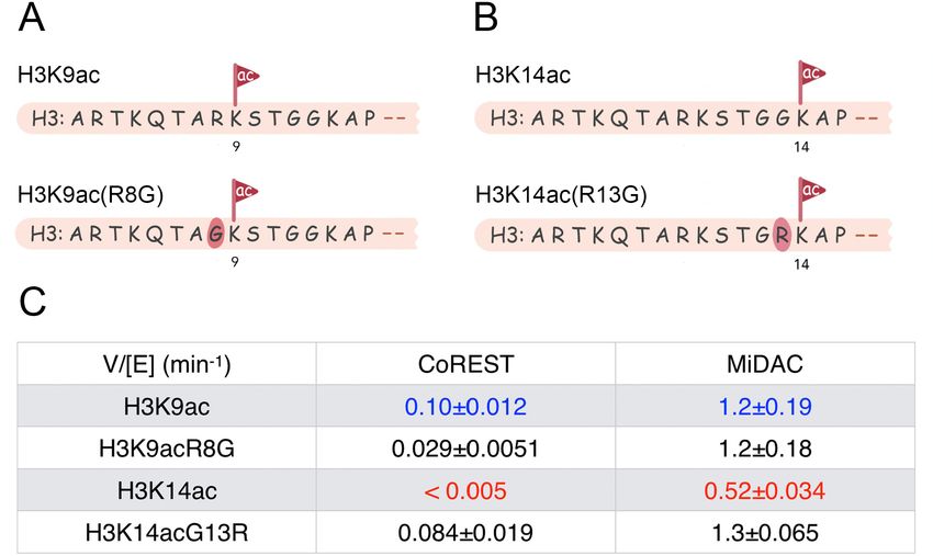
Research advance Biochemistry and Chemical Biology
2016), (Riester et al., 2007), (Gurard-Levin et al., 2009). Such studies have typically shown little to
no amino acid sequence site-selectivities for HDACs. As the physiological enzyme forms of HDAC1-3
exist in multisubunit complexes (Millard et al., 2017) and the natural substrates are chromatin com-
prised of nucleosomes, histone octamers wrapped by 146 bp of double stranded DNA, the biologi-
cal relevance of the free HDAC and histone tail substrate peptide experiments is unclear. Recent
studies on the CoREST complex and acetylated nucleosome substrates revealed a notable prefer-
ence for deacetylation of histone H3 acetyl-Lys9 vs. acetyl-Lys14 (Wu et al., 2018). This was espe-
cially interesting because the CoREST complex also includes the enzyme LSD1 which specifically
demethylates methyl-Lys4 in histone H3 and this activity is strongly inhibited when the H3 tail also
contains acetyl-Lys14 but not acetyl-Lys9 (Kalin et al., 2018). These properties suggest a biological
role in histone H3 marked by Lys14 acetylation and Lys4 methylation to protect against CoREST
complex silencing. The CoREST complex enzymatic studies also raise the possibility that different
HDAC complexes may show distinct deacetylase kinetics and nucleosome site selectivities.
In this study, we evaluate the catalytic actions of five purified class I HDAC complexes CoREST,
MiDAC, NuRD, Sin3B, and SMRT with five different histone H3 acetylated mononucleosomes as well
as the corresponding free histone H3 substrates. We find that the kinetics of the HDAC complexes
across the range of free acetylated histone H3 substrates are fairly similar, whereas there are large
differences between the complexes with respect to deacetylase rates and site-selectivity with nucle-
osome substrates. Among these complexes, the CoREST complex shows a special resistance to
H3K14ac which we find is largely driven by the preceding Gly13 residue.
Results
Production of H3 acetylated nucleosomes and HDAC complexes
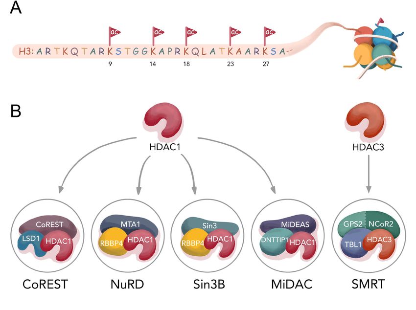
Semisynthetic X. laevis histone H3 proteins (Wang et al., 2015) mono-acetylated at positions Lys9,
Lys14, Lys18, Lys23, and Lys27 were prepared using F40 sortase (Piotukh et al., 2011). In this
approach, the N-terminal tails aa1-34 were prepared as synthetic peptides containing the acetyl-Lys
and terminating in a depsipeptide linkage between Thr and Gly and the H3 globular domain aa34-
135 was produced recombinantly. F40 sortase treatment of the H3 peptide and H3 globular domain
catalyzes transpeptidation leading to ligation of the fragments to produce pure, scarless full-length
modified histone H3s (Figure 2A–D and Figure 2—figure supplements 1–2). Western blot analysis
with the site-specific relevant acetyl-Lys antibodies demonstrated that each of the semisynthetic his-
tone H3s contained the designated marks (Figure 3—figure supplement 1 and Wu et al., 2018).
The semisynthetic acetylated H3s were incorporated into mononucleosomes containing 146 bp DNA
601 Widom sequence (Luger et al., 1997; Figure 2—figure supplement 3A–B). The HDAC com-
plexes CoREST (LSD1, HDAC1, CoREST1), NuRD (MTA1, HDAC1, RBBP4), Sin3B (Sin3, HDAC1,
RBBP4), MiDAC (MIDEAS, HDAC1, DNTTIP1), and SMRT (GPS2-NCOR2 chimera, HDAC3,
and TBL1) were expressed in HEK293F cells by transient transfection of three plasmids encoding the
relevant proteins (Figure 3—figure supplement 2A). The details of how these complexes have been
arrived at and are produced have been described previously (Song et al., 2020), (Millard et al.,
2016), (Clark et al., 2015), (Itoh et al., 2015), (Watson et al., 2012a), (Zhang et al., 2018),
(Watson et al., 2012b), (Portolano et al., 2014). In general, the two non-HDAC proteins in each
case were selected based on the following criteria: 1) A set of proteins that included both well-
established HDAC and nucleosome binding partners for a given complex, 2) Efficient transient co-
expression of soluble proteins in HEK293F that stay associated by immunoaffinity chromatography
and lead to relatively pure and concentrated complexes in peak fractions (>50% purity), 3) The abil-
ity of the core complexes to be reproducibly isolated as monodisperse peaks in appropriate stoi-
chiometries after size exclusion chromatography. The HDAC complexes employed here were shown
to be relatively pure and in the expected stoichiometries by SDS-PAGE (Figure 3—figure supple-
ment 2B).
Deacetylase assays with HDAC complexes
Each of the HDAC complexes was assayed with isolated acetylated histone H3 proteins or acetylated
nucleosomes as substrates, using Western blot to monitor the disappearance of the requisite acetyl-
Lys modification. The five HDAC complexes displayed robust catalytic activities with isolated
Wang et al. eLife 2020;9:e57663. DOI: https://doi.org/10.7554/eLife.57663 3 of 19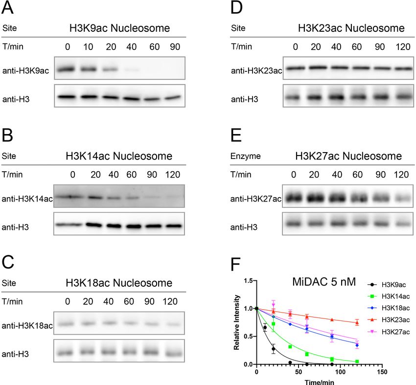
Research advance                                                                                           Biochemistry and Chemical Biology

Figure 2. The semi-synthesis of full-length histone H3 with site-specific acetylations. (A) Synthesis of H3 proteins with site-specific acetylations; gH3:
globular region of histone H3; (B) MALDI-MS for a semi-synthetic histone H3 product, using H3K27ac as an example, *: an unknown minor impurity; (C)
SDS-PAGE of all the H3 proteins with acetylations, as H3K9ac, H3K14ac, H3K18ac, H3K23ac, H3K27ac, H3K9acR8G, H3K14acG13R; (D) Native 6% TBE
gel of the nucleosome folding results with acetylated H3s, each showing 95% purity. #: minor free DNA band.
The online version of this article includes the following figure supplement(s) for figure 2:
Figure supplement 1. MALDI-TOF spectra for H3K9/14/18/23/27ac 1–34 TOG peptides.
Figure supplement 2. MALDI-TOF spectra for H3K9/14/18/23/27ac full length histone proteins.
Figure supplement 3. The assembly of H3 proteins with acetylations into corresponding nucleosomes.

                                     acetylated H3 substrates with velocities (V/E) averaging in the range of 0.5–2 min 1 and modest dif-
                                     ferences (largely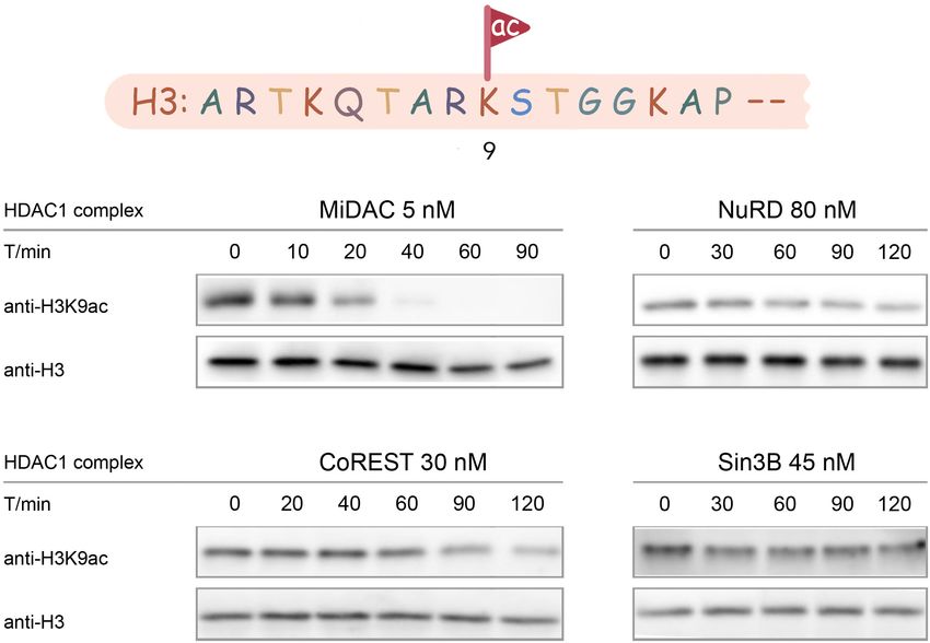
Research advance                                                                              Biochemistry and Chemical Biology

                                   not have a large impact on HDAC-catalyzed deacetylation kinetics (Table 1, Figure 4—figure sup-
                                   plements 1–6A–E,G).
                                      In contrast to the results with isolated H3 substrates, the results with acetylated nucleosome sub-
                                   strates showed dramatic differences among the HDAC complexes. The HDAC complex velocities (V/
                                   E) with acetylated nucleosomes varied over 200-fold, ranging from
Research advance Biochemistry and Chemical Biology
Figure 3. Typical results for Western-blot based deacetylation activity assay using different complexes on nucleosome with H3K9ac (n = 2 for each
assay).
The online version of this article includes the following figure supplement(s) for figure 3:
Figure supplement 1. Primary antibody specificity test for anti-H3K23ac and anti-H3K27ac.
Figure supplement 2. The expression of HDAC complexes.
less sensitivity to changes in Arg8 and Gly13. In fact, K9acR8G nucleosome deacetylation by the
MiDAC complex matched that of WT K9ac nucleosomes (V/E 1.2 min 1 for both). K14acG13R nucle-
osome deacetylation by MiDAC was accelerated by 2.5 fold relative to WT K14ac nuclesomes, indi-
cating a minor preference for Arg at the H3 13th position.
NuRD (HDAC1-MTA1-RBBP4) deacetylation of H4K16ac nucleosomes
The very slow rates of nucleosome H3 deacetylation by the RBBP4 containing NuRD and Sin3B com-
plexes prompted us to consider that the scaffold protein RBBP4 might be impeding the catalytic
activity by obstructing the H3 tails (Millard et al., 2016). We thus examined whether a nucleosome
containing acetylation on histone H4 might be an improved substrate for an RBBP4 complex. How-
ever, the deacetylation rate (V/E) for NuRD was low and similar to those of the acetylated H3 nucleo-
somes (Figure 6, Figure 6—figure supplement 1A,B). These results suggest that the low
nucleosome deacetylase activity of RBBP4-containing HDAC complexes may be unrelated to RBBP4/
H3 interaction.
Wang et al. eLife 2020;9:e57663. DOI: https://doi.org/10.7554/eLife.57663 6 of 19Research advance Biochemistry and Chemical Biology Figure 4. Typical results for Western-blot based deacetylation activity assay with 5 nM MiDAC on different nucleosomes with site-specific acetylation. (A) nucleosome with H3K9ac, n = 2, V/E = 1.2 ± 0.19 min 1; (B) nucleosome with H3K14ac, n = 3, V/E = 0.52 ± 0.034 min 1; (C) nucleosome with H3K18ac, n = 2, V/E = 0.17 ± 0.0051 min 1; (D) nucleosome with H3K23ac, n = 4, V/E = 0.048 ± 0.0071 min 1; (E) nucleosome with H3K27ac, n = 5, V/ E = 0.18 ± 0.036 min 1, and (F) the curve fitting for kinetics analysis. The online version of this article includes the following figure supplement(s) for figure 4: Figure supplement 1. CoREST (CoREST-LSD1-HDAC1) activity assay over nucleosomes or histones with acetylation at different sites. Figure supplement 2. MiDAC (MiDEAS-DNTTIP1-HDAC1) activity assay over nucleosomes or histones with acetylation at different sites. Figure supplement 3. NuRD (MTA1-RBBP4-HDAC1) activity assay over nucleosomes or histones with acetylation at different sites. Figure supplement 4. Sin3B (Sin3-HDAC1-RBBP4) activity assay over nucleosomes or histones with acetylation at different sites. Figure supplement 5. SMRT (GPS2-NCoR2 chimera-HDAC3-TBL1) activity assay over nucleosomes or histones with acetylation at different sites. Figure supplement 6. HDAC1 activity assay over nucleosomes or histones with acetylation at different sites. Wang et al. eLife 2020;9:e57663. DOI: https://doi.org/10.7554/eLife.57663 7 of 19
Research advance                                                                               Biochemistry and Chemical Biology

Table 2. Rate constants of different HDAC1 and HDAC3 complexes on various nucleosomes with site-specific acetylation on H3.
Kinetic values shown are ± S.E.M. For each enzymatic reaction on nucleosomes: the semi-synthetic H3K9/14/18/23/27ac nucleosomes
(100 nM) were incubated with different HDAC1 and HDAC3 complexes at the following concentrations (30 nM CoREST, 5 nM MiDAC,
80 nM NuRD, 45 nM Sin3B, 60 nM SMRT, and 60 nM HDAC1, n = 2–5), under a reaction buffer of 50 mM HEPES 7.5 containing 100
mM KCl, 100 mM IP6, and 0.2 mg/mL BSA at 37 ˚C. The reaction time was counted, and multiple samples were taken at different time
points generally between 0–120 min (The MiDAC complex on H3K9ac was between 0–90 min). The complexes include CoREST, NuRD,
Sin3B, MiDAC, and SMRT, together with free HDAC1 enzyme.
             1
V/[E] (min       )   HDAC1         CoREST(RCOR1)           MiDAC(MiDEAS)         NuRD(MTA1)             Sin3B(Sin3)            SMRT(NCoR2)
H3K9ac               N.D.          0.10 ± 0.012            1.2 ± 0.19            0.012 ± 0.0032         0.0050 ± 0.0043        0.046 ± 0.0045
H3K14ac              N.D.
Research advance Biochemistry and Chemical Biology
Discussion
A principal finding of this study is that distinct HDAC1 complexes can show sharply different deace-
tylase activities toward different nucleosome substrates. This would appear to suggest that the
unique biological consequences of the various HDAC1 complexes are not simply dependent on
where they are recruited to in chromatin and by which particular transcription factor (Watson et al.,
2012b). Rather, the different catalytic properties and site-selectivities of the HDAC1 complexes
likely drive different patterns of histone deacetylation and chromatin states. Although it is possible
that the closely related paralog HDAC2 replaces HDAC1 in some population of the HDAC com-
plexes analyzed here, because of the massive overexpression of HDAC1 we believe this to be a very
minor pool. Prior published structural studies of these complexes and mass spectroscopic analyses
are also consistent with this idea (Song et al., 2020), (Millard et al., 2016; Itoh et al., 2015;
Millard et al., 2013; Zhang et al., 2018), (Portolano et al., 2014). Furthermore, because HDAC1
and HDAC2 have very high sequence homology, we believe it is likely that HDAC1 and HDAC2 will
behave in similar ways in terms of the nucleosome deacetylation assays carried out here.
The data with the CoREST complex selectivity stand out for the strong resistance to one of the
five H3 tail acetylation sites, H3K14ac, investigated (Wu et al., 2018). It is likely not a coincidence
that K14ac blocks the removal of H3K4 methylation as this could allow doubly modified H3K4me1/2
and H3K14ac tails to be immune from the silencing action of the CoREST complex. H3K4 methyla-
tion is known to be commonly found in the context of enhancer elements in DNA (Hsu et al., 2018),
which is also associated with H3K14ac (Karmodiya et al., 2012), possibly insulating such K4 methyla-
tion by CoREST. Although this has not been rigorously evaluated with JMJ family demethylases, a
yeast homolog JHD2 has been shown to be more weakly able to demethylate H3K4me3 in the con-
text of H3K14ac (Maltby et al., 2012).
The Gly/Arg swaps at positions 8 and 13 in histone H3 clearly demonstrate that the presence of
Gly13 rather than an Arg residue is a principal determinant of the meager rate of CoREST-mediated
deacetylation of K14ac in nucleosome substrates. However, each of the five class I HDAC complexes
examined here were relatively ineffective at deacetylating K14ac in the context of nucleosomes pre-
sumably at least partly due to Gly13. That this K14ac deselection is not true in isolated H3 indicates
that the molecular recognition of Lys14ac by these HDAC complexes is altered when the H3 tail is
presented to the HDAC1 active site in a chromatin context. We are unsure if this represents kcat or
KM differences conferred by histone H3 Gly13 vs. Arg13 because of the challenges of making such
measurements with nucleosome substrates. We speculate that these effects may not be reflected in
overall affinities between the various HDAC complexes and particular acetylated nucleosomes, but
rather are driven by subtle local differences between the HDAC complex active sites and the nucleo-
somal substrates. The cellular relevance of these findings is supported by the fact that H3K14ac is a
relatively abundant mark at baseline (Sidoli and Garcia, 2017) and is rather unaffected by class I
HDAC inhibitor treatment compared with H3K9ac and H3K27ac which show substantial increases
with such inhibitors (Schölz et al., 2015). It is also possible that robust histone acetyltransferase tar-
geting H3K14 contributes to the elevated H3K14 acetylation levels in cells.
The MiDAC complex showed far and away the speediest average deacetylation rate across the
five acetyl-Lys marks looked at in nucleosomes. Impressively, MiDAC deacetylated K9ac nucleo-
somes (V/E 1.2 min 1) more rapidly than K9ac in free H3 histones. Unlike CoREST, NuRD, Sin3B, and
SMRT which are thought to be transcriptional silencers and recruited to chromatin by transcription
factors, MiDAC is not known to be employed in this way. Furthermore, its ground state affinity to
chromatin is thought to be quite high (Itoh et al., 2015). It is interesting that there was a modest
trend for the rate of deacetylation to be highest closest to the N-terminus of H3 in the context of
nucleosome substrates. We speculate that this may represent a greater accessibility of the extreme
N-terminus of the H3 tail in chromatin to the HDAC1 active site in this complex.
In contrast to MiDAC, the NuRD and Sin3B complexes showed the lowest deacetylation rates
with H3 acetylated nucleosomes. There are several possible reasons for this. It is plausible the NuRD
and Sin3B may have a more extreme requirement for recruitment to nucleosomes by a transcription
factor compared with the other HDAC complexes looked at here (Zhang et al., 2018),
(Walsh, 2006). This could offer the advantage of preventing gene silencing except when triggered
for by a cellular stimulus (Millard et al., 2017). In addition, there could be an additional macromolec-
ular component or post-translational modification (Wang, 2019) lacking in our system that would
Wang et al. eLife 2020;9:e57663. DOI: https://doi.org/10.7554/eLife.57663 9 of 19Research advance Biochemistry and Chemical Biology
Figure 6. Western-blot and kinetics of NuRD complex activity on nucleosome with H4K16ac. (A) Western-blot
based deacetylation activity assay with 40 nM NuRD complex (HDAC1-MTA1-RBBP4) on H4K16ac nucleosomes
blot with site-specific acetylation antibody and anti-H3 antibody to show the overall nucleosome amount
remaining unchanged (n = 3); (B) rate constant of NuRD complex deacetylation on H4K16ac nucleosome. The
reaction conditions were similar to the enzymatic reactions on H3K9/14/18/23/27ac nucleosomes described in
Table 1.
The online version of this article includes the following figure supplement(s) for figure 6:
Figure supplement 1. NuRD activity assay over H4K16ac nucleosome.
unleash the deacetylase activity or alter substrate selectivities of any of the HDAC complexes on
nucleosomes (Hudson et al., 2015). Indeed, NuRD and the other HDAC complexes investigated
here have been reported to have a variety of additional protein subunits depending on cell type and
cell state (Millard et al., 2016), (Lai and Wade, 2011), (Yang and Seto, 2008). For the NuRD com-
plex, however, it is established that the chromatin remodelling module is rather loosely associated,
and we do not expect that its absence would greatly influence HDAC activity. In addition to roles for
transcription factors and additional subunits, the NuRD or Sin3B complexes could show very high
site-selectivity on one or more sites in histones beyond the acetylated-Lys that we have examined
thus far in H3 or H4. Future studies will be needed to determine if one or more of these possibilities
is operative.
We have focused on classical HDAC acetyl-Lys selectivities in chromatin. However, there is reason
to believe that the NAD-dependent sirtuins may also show site-selectivities. Recent studies on Sirt7
have shown that this enzyme shows a strong preference for H3K36ac in the context of chromatin
(Wang et al., 2019). It will be important in future experiments to extend the analysis of deacetylase
actions across the full complement of the HDAC enzymes to create a deeper understanding of the
connections between reversible acetylation in health and disease states.
Wang et al. eLife 2020;9:e57663. DOI: https://doi.org/10.7554/eLife.57663 10 of 19Research advance Biochemistry and Chemical Biology
Materials and methods
Key resources table
Reagent type Additional
(species) or resource Designation Source or reference Identifiers information
Gene (X. laevis) Histone H2A, DOI: 10.7554/eLife.37231 Construct shared by
H2B, H4 (X. laevis) Cynthia Wolberger
(Johns Hopkins)
Gene (X. laevis) Histone gH3 (X. laevis) DOI: 10.7554/eLife.37231 Construct shared by
DOI: 10.1021/ja205630g Dirk Schwarzer
(Tübingen)
Gene (S. aureus) F40-sortase (Originally DOI: 10.7554/eLife.37231 Construct shared by
mutated from DOI: 10.1021/ja205630g Dirk Schwarzer
S. aureus sortase A) (Tübingen)
Gene (E. coli) 146 bp 601 DNA DOI: 10.7554/eLife.37231 Construct shared by
(Widom sequence for Song Tan (Penn State)
E. coliexpression)
Strain, strain BL21 Rosetta Sigma-Aldrich Cat# 70956–4 Competent cells
background (DE3) pLysS (E. coli)
(E. coli)
Cell line HEK293F Thermo RRID:CVCL_6642 Transfected with plasmids
(Homo-sapiens) (Homo-sapiens) Fisher Scientific to express different
HDAC1 complexes.
Mycoplasma tested
negative. Authenticated
by the venfor.
Not on ICLAC
misidentified cell
line registry.
Antibody Anti-H3K9ac (Rabbit) Cell Signaling Cat# 9671, WB (1:2000)
RRID:AB_331532
Antibody Anti-H3K14ac (Rabbit) EMP Millipore Cat# 07–353, WB (1:2000)
RRID:AB_310545
Antibody Anti-H3K18ac (Rabbit) EMP Millipore Cat# 07–354, WB (1:2000)
RRID:AB_ 441945
Antibody Anti-H3K23ac (Rabbit) EMP Millipore Cat# 07–355, WB (1:2000)
RRID:AB_310546
Antibody Anti-H3K27ac (Rabbit) Cell Signaling Cat# 8173, WB (1:2000)
RRID:AB_10949503
Antibody Anti-H3 (Rabbit) Abcam Cat# ab1791, WB (1:5000)
RRID:AB_302613
Antibody Anti-H4K16ac (Rabbit) Active Motif Cat# 39167, WB (1:5000)
RRID:AB_2636968
Antibody HRP conjugated Cell Signaling Technology Cat# 7074, WB (1:2000)
anti-Rabbit RRID:AB_2099233
secondary (Goat)
Peptide, HDAC1/HSP70 BPS Bioscience Cat# 50051
recombinant protein
Peptide, Nucleosome Epicypher Cat# 16–0354
recombinant protein with H4K16ac
Chemical compound 20 Fmoc-amino acids EMP Millipore Fmoc-Gly-OH as
DOI: 10.7554/eLife.37231 example: Cat# 852001
Software, algorithm ImageJ DOI: 10.7554/eLife.37231 RRID:SCR_003070
Download from
imagej.nih.gov/ij/
Software, algorithm GraphPad Prism Eight DOI: 10.7554/eLife.37231 RRID:SCR_002798
Download from
www.graphpad.com
Wang et al. eLife 2020;9:e57663. DOI: https://doi.org/10.7554/eLife.57663 11 of 19Research advance Biochemistry and Chemical Biology
Reagents
20 amino acids were purchased from EMP Millipore. Site specific primary antibodies include anti-
H3K9ac (Cell Signaling), anti-H3K14ac (EMP Millipore), anti-H3K18ac (EMP Millipore), anti-H3K23ac
(EMP Millipore), anti-H3K27ac (Cell Signaling), anti-H4K16ac (Active Motif) antibodies, as well as
anti-histone H3 (Abcam). Secondary antibody as HRP-conjugated anti-rabbit antibody from Cell Sig-
naling. H4K16ac nucleosome was purchased from Epicypher. The free enzyme HDAC1 (in the com-
plex with HSP70) is purchased from BPS Bioscience.
The synthesis of depsipeptide Fmoc-Thr(OtBu)-glycolic acid (TOG)
The depsipeptide Fmoc-Thr(OtBu)-glycolic acid was synthesized based on a reported two-step pro-
tocol (Williamson et al., 2012).
Peptide synthesis
All the histone H3 peptides 1–34 containing site-specific lysine acetylation (Kac) were synthesized by
Fmoc-SPPS by Fmoc-Gly-Wang resin. For 0.2 mmol resin, 0.8 mmol (four eq) Fmoc-amino acid, 3.2
mmol (3.75 eq) HATU and 1.6 mmol (eight eq) N-methylmorpholine (NMM) in 8 mL DMF was added
to do the peptide coupling for 90 min, followed by the deprotection of Fmoc with 20% piperidine in
dimethylformamide (DMF) for 15 min. The resin was then completely washed with DMF before
entering the next coupling cycle. The first depsipeptide TOG was coupled following a similar proto-
col. After all the coupling steps and deprotection steps, the resin was washed by DMF and dichloro-
methane (DCM) to dry. Reagent B (5% water, 5% phenol, 2.5% triisopropyl silane (TIPS), 87.5%
trifluoroacetic acid (TFA)) for 2.5 hr at room temperature. The crude peptide was concentrated
before the addition of cold diethyl ether to precipitate and dried over nitrogen gas flow. After being
dissolved in 10% acetonitrile (CH3CN)-water, the crude peptides were further purified by reversed
phase HPLC with the C18 column (Varian Dynamax Microsorb 100), with a gradient of 5% CH3CN/
0.05% TFA in H2O/0.05% TFA for 2 min and a linear gradient from 5% CH3CN/0.05% TFA to 32%
CH3CN/0.05% TFA over 25 min, and the flow rate is 10 mL/min. All the synthetic peptides were
lyophilized to powders, followed by the characterization by MALDI-TOF at the Molecular Biology
Core Facilities of Dana Farber Cancer Institute (Figure 2—figure supplement 1, Figure 5—figure
supplement 1).
H3K9ac(1-34) TOG [M + H]+ calculated as m/z 3494.9, observe at m/z 3493.0;
Sequence: ARTKQTARKS-TGGKAPRKQL-ATKAARKSAP-A-TOG-G
H3K14ac(1-34) TOG [M + H]+ calculated as m/z 3494.9, observed at m/z 3493.5;
H3K18ac(1-34) TOG [M + H]+ calculated as m/z 3494.9, observed at m/z 3492.9;
H3K23ac(1-34) TOG [M + H]+ calculated as m/z 3494.9, observed at m/z 3493.7;
H3K27ac(1-34) TOG [M + H]+ calculated as m/z 3494.9, observed at m/z 3493.1;
H3K9acR8G(1-34) TOG [M + H]+ calculated as m/z 3394.8, observed at m/z 3393.8;
H3K14acG13R(1-34) TOG [M + H]+ calculated as m/z 3594.1, observed at m/z 3591.7;
Expression and purification of F40 sortase from E. coli
F40 sortase was purified in a manner reported previously (Wu et al., 2018). The pET21-F40-sortase
plasmid was used for the transformation of the E. coli Rosetta (DE3) pLysS strain. A single colony
from the LB agar plate was inoculated into 5 mL LB media with 100 mg/L ampicillin at 37 ˚C over-
night, and subsequently inoculated into 1 L large 2YT culture at 37 ˚C until OD600 reached 0.6. F40
sortase was then induced with 0.25 mM IPTG for 4 hr at 37 ˚C. The cells were harvested by centrifu-
gation at 4,000 rpm for 15 min and the cell-pellet was resuspended in Lysis buffer containing 20 mM
Tris, 0.1% Triton X-100 and 1 mM phenylmethylsulfonyl fluoride (PMSF) at pH 7.5. The cells were
then lysed by passing through a french press cell disrupter at about 1,000 psi three times, followed
by centrifuging the lysate at 20,000 g for 40 min. The supernatant was loaded to a Ni-NTA Sephar-
ose 6 Fast Flow (GE Healthcare) resin, followed by repeated wash with Wash buffer containing 20
mM Tris, 500 mM NaCl, 20 mM imidazole and 1 mM PMSF at pH 7.5. The F40 sortase was then
eluted with Elution buffer containing 20 mM Tris, 500 mM NaCl, 400 mM imidazole and 1 mM PMSF
at pH 7.5 in different fractions. All the fractions were analyzed by SDS-PAGE gel and fractions con-
taining F40 sortase were pooled together and dialyzed against Dialysis buffer with 20 mM Tris, 150
mM NaCl, and 5 mM CaCl2 at pH 7.5. After dialysis, F40 sortase (>95% pure based on coomassie
Wang et al. eLife 2020;9:e57663. DOI: https://doi.org/10.7554/eLife.57663 12 of 19Research advance Biochemistry and Chemical Biology
stained SDS-PAGE) was concentrated using an Amicon Ultra spin column (10K MWCO, EMD Milli-
pore) to about 1 mM final concentration, and then aliquoted and stored at 80 ˚C for future usage.
Expression of WT-histone H2A, H2B, H4, and histone core region gH3
Bacterial expression and purification of X. laevis core histones H2A, H2B, globular H3 (gH3; amino
acids 34–135 with the first Met cleaved by E. coli), and H4 were performed using a previously
reported method (Piotukh et al., 2011), (Luger et al., 1997). Please note that we used gH3 contain-
ing Cys110 for K23ac and K27ac semi-synthesis and Ala110 for K9ac, K14ac, K18ac semi-synthesis
(Shimko et al., 2011) and the impact of this aa110 difference in the nucleosomes derived from these
histone H3 isoforms is assumed to be negligible (Wilkinson and Gozani, 2014). Consequently, H3
C110A has been widely employed in nucleosome chemical biology (Jani et al., 2019) and structural
studies (Gatchalian et al., 2017). The pET23-gH3 plasmid was used to transform E. coli Rosetta
(DE3) pLysS strain, and a single colony was inoculated into 1 L LB media with 100 mg/L ampicillin at
37 ˚C. The culture was let to shake and grow at 37 ˚C until OD600 reached 0.6. Then, gH3 was
induced with 0.5 mM IPTGat 37 ˚C for 3 hr. The cells were then harvested by centrifugation at 4,000
rpm for 15 min and the cell pellet was resuspended in Histone Wash Buffer (50 mM Tris-HCl at pH
7.5, 100 mM NaCl, 1 mM EDTA, 5 mM 2-mercaptoethanol (BME), 0.2 mM phenylmethylsulfonyl fluo-
ride (PMSF) with 1% Triton X-100). The cells were then lysed by passing through a french press cell
disrupter at about 1,000 psi three times, followed by centrifugation of the cell lysate at 20,000 g for
40 min. After discarding the supernatants, the pellets were washed with Histone Wash Buffer with
1% Triton X-100 once and Histone Wash Buffer without Triton X-100 twice. The pellets were dis-
solved in Histone Solubilization Buffer (7 M guanidinium hydrochloride, 20 mM Tris at pH 7.5 and 10
mM dithiothreitol). After centrifuging at 20,000 g for 15 min, the supernatant was dialyzed three
times against IEX (ion exchange) buffer (7 M urea, 10 mM Tris pH 7.8, 1 mM EDTA, 0.2 mM PMSF
and 5 mM BME). The resulting gH3 solution in IEX buffer was diluted 5-fold in IEX buffer before
being loaded onto a tandem Q-SP column (GE healthcare, HiTrap Q HP and HiTrap SP HP, 5 mL
each). Both Q and SP column were first equilibrated and washed with IEX buffer containing 100 mM
NaCl. Then, gH3 was eluted from the 100 mM - 500 mM NaCl with 100 mM increment. The fractions
containing gH3 (>90% pure by coomassie stained SDS-PAGE) were pooled after analyzing each frac-
tion by SDS-PAGE. The combined IEX solution was then dialyzed three times against pure water
containing 2 mM BME (3.5K MWCO, Spectra/Por) for 6 hr each. The solution was finally concen-
trated with an Amicon Ultra spin column (3.5 K MWCO, EMD Millipore) to 10 mL (~100 mM) and
stored at 4 ˚C until usage.
F40 sortase catalyzed histone H3 semi-synthesis
The histone H3 N-terminal peptide (aa1-34 depsipeptide, 1 mM) and gH3 (aa34-135,~70 mM) was
mixed in Sortase Reaction Buffer (50 mM HEPES at pH 7.5, 150 mM NaCl and 5 mM CaCl2). The
reaction was initiated by the addition of F40 Sortase (300 mM) and was incubated at 37 ˚C for over-
night. The reaction mixture was centrifuged to precipitate H3 with some sortase, which was then dis-
solved in 10 mL IEX buffer (7 M urea, 10 mM Tris at pH 7.8, 1 mM EDTA and 5 mM BME). The
solution was loaded onto a SP column (GE healthcare, HiTrap SP HP, 1 mL) preequilibrated with IEX
buffer containing 100 mM NaCl. After washing with IEX buffer containing 100 mM NaCl, H3 was
eluted with a gradient of IEX buffer containing increasing NaCl from 100 to 500 mM. All the fractions
were analyzed by SDS-PAGE, and the fractions with pure ligated H3 product (>90% based on coo-
massie stained SDS-PAGE gels) were pooled and dialyzed three times against pure water with 2 mM
BME (3.5K MWCO, Spectra/Por). The solution was then concentrated using an Amicon Ultra spin
column (10 K MWCO, EMD Millipore), and was lyophilized to a white powder for storage at 80 ˚C
until usage. Semi-synthesized histone H3 structures were characterized by MALDI-TOF Mass Spec-
trometry ((matrixed with CHCA) at the Molecular Biology Core Facilities of Dana Farber Cancer Insti-
tute (Figure 2—figure supplement 2, Figure 5—figure supplement 1).
H3K9ac(C110A): [M + H]+ calculated as m/z 15280.8, observed as m/z 15274.3;
H3K14ac(C110A): [M + H]+ calculated as m/z 15280.8, observed as m/z 15278.9;
H3K18ac(C110A): [M + H]+ calculated as m/z 15280.8, observed as m/z 15271.6;
H3K23ac: [M + H]+ calculated as m/z 15312.8, observed as m/z 15309.6;
H3K27ac: [M + H]+ calculated as m/z 15312.8, observed as m/z 15308.6;
Wang et al. eLife 2020;9:e57663. DOI: https://doi.org/10.7554/eLife.57663 13 of 19Research advance Biochemistry and Chemical Biology
H3K9acR8G(C110A): [M + H]+ calculated as m/z 15181.6, observed as m/z 15175.1;
H3K14acG13R(C110A): [M + H]+ calculated as m/z 15379.9, observed as m/z 15377.1.
Octamer refolding and nucleosome reconstitution
The octamer refolding and nucleosome assembly were performed following a previous report
(Dyer et al., 2004). All four histone proteins H2A, H2B, H3 and H4, each two copies, were dissolved
in Unfolding Buffer (7 M guanidine, 20 mM Tris-HCl at pH 7.5 and 10 mM DTT) and quantified by
reading A280. The mixed solution with a molar ratio at 1.1: 1.1: 1:1 one was then dialyzed three times
against high salt buffer (20 mM Tris at pH 7.5, 2.0 M NaCl, 1 mM EDTA and 5 mM BME). The
octamer was purified by size exclusion chromatography using a Superdex 200 10/300 GL column
(GE Healthcare). In addition, 146 Widom 601 DNA was prepared by previously reported methods
used for nucleosome reassembly (Luger et al., 1997). The 146 bp DNA was obtained from the
restriction digests of a specially designed DNA plasmid from E. coli and purified by precipitation
with polyethylene glycol. The histone octamer (Figure 2—figure supplement 3B, using H3K9ac and
H3K14ac as examples) and Widom DNA were mixed at a 1:1 molar ratio at high salt buffer (10 mM
Tris at pH 7.5, 2.0 M KCl, 1 mM EDTA and 1 mM DTT) with a final concentration of 7 mM DNA. Con-
tinuous-flow gradient dialysis was used to lower the salt concentration from the mixture to low salt
buffer (10 mM Tris at pH 7.5, 250 mM KCl, 1 mM EDTA and 1 mM DTT) by a Minipuls two peristaltic
pump (Gilson). The final mixture was subjected to HPLC (Waters, 1525 binary pump, 2489 UV-Vis
detector). HPLC using a TEKgel DEAE ion exchange column was employed to purify the final nucleo-
some product. The liquid phase buffers A and B were as follow: A. TES 250 (10 mM Tris 7.5, 250
mM KCl, 0.5 mM EDTA), B. TES 600 (10 mM Tris 7.5, 600 mM KCl, 0.5 mM EDTA), were used to
generate with a 0% B wash for 12 min and a linear gradient from 25% to 75% B over 30 min at 1 mL/
min flow rate. The fractions containing the nucleosome were pooled together and dialyzed against a
TCS Buffer (20 mM Tris at pH 7.5 and 1 mM DTT, no EDTA) and concentrated to about 20 mM using
an Amicon Ultra spin column (10K MWCO, EMD Millipore). The final nucleosome products were ana-
lyzed by native TBE gels and all showed over 95% purity based on the fact that free DNA band fluo-
resces 2.5-fold more brightly than the nucleosome DNA band in this setting (Shahian and Narlikar,
2012).
Expression and purification of HDAC complexes
The expression and purification of HDAC complexes were performed as described previously
(Song et al., 2020), (Millard et al., 2016), (Itoh et al., 2015), (Watson et al., 2012b),
(Portolano et al., 2014). For each complex, three plasmids corresponding to the different compo-
nents of the HDAC complex were co-transfected into HEK293F cells: LSD1 (1–852), HDAC1 (1–482),
and Flag tagged CoREST1 (86–485) for the CoREST complex; Flag tagged MTA1 (1–715), HDAC1
(1–482), and RBBP4 (1–425) for the NuRD complex; Flag tagged Sin3B (1–1162), HDAC1 (1–482),
RBBP4 (1–425) for the Sin3B complex; Flag tagged MIDEAS (628-887), HDAC1 (1–482), DNTTP1 (1–
329) for the MiDAC complex; Flag tagged GPS2-NCoR2 chimera ((1-49) - (220-480)), HDAC3 (1–
428), and TBL1 (1–526) for the SMRT complex. After cell lysis by sonication, the complexes were
purified by FLAG tag immunoaffinity chromatography and liberated from the column with TEV pro-
tease. These mixtures were further purified by gel filtration on either a Superdex 200 Increase 10/
300 GL column (MiDAC, SMRT) or a Superose 6 10/300 GL column (CoREST, NuRD, Sin3B). The
purified complexes were analyzed by SDS-PAGE stained with coomassie and concentrated to 3–5
mM (Figure 3—figure supplement 2B). After the addition of glycerol to 10%, the complexes are
stored at 80˚C until further use (Wu et al., 2018). The final HDAC complexes were analyzed by
SDS-PAGE gel stained with coomassie and each showed greater than 60% purity.
Analysis of deacetylation of acetylated nucleosomes and acetylated
histone H3s
For all the deacetylation assays, the semi-synthetic H3K9/14/18/23/27ac histone H3 proteins (1.0
mM) or the corresponding nucleosomes (100 nM) assembled in vitro were incubated with different
HDAC1 and HDAC3 complexes at different concentrations, under a reaction buffer containing 50
mM HEPES at pH 7.5, 100 mM KCl, 100 mM IP6 (Watson et al., 2016), and 0.2 mg/mL BSA at 37 ˚C.
The complexes include CoREST, NuRD, Sin3B, MiDAC, and SMRT, together with HDAC1 enzyme
Wang et al. eLife 2020;9:e57663. DOI: https://doi.org/10.7554/eLife.57663 14 of 19Research advance Biochemistry and Chemical Biology
not involved in specific complex binding from commercial source (BPS Bioscience). The reaction time
was counted, and multiple samples were taken at different time points. Typically, 10 mL aliquots of
the reaction were quenched with gel loading buffer made by mixing 2 mL 80 mM EDTA and 4 mL 4 x
Laemmli sample buffer at each time point. The samples were then boiled for 3 min at 95 ˚C and
loaded onto a 15% SDS-PAGE gel (160 Volts for 35 min). After transferring to nitrocellulose mem-
brane, site-specific antibodies for acetylated H3, such as for anti-H3K9ac, anti-H3K14ac, anti-
H3K18ac, anti-H3K23ac and anti-H3K27ac, as well as total H3 antibody, anti-H3, were used to blot
the membrane. The specificities of the primary antibodies, anti-H3K23ac and anti-H3K27ac, were val-
idated using histone H3K9ac as a negative control (Figure 3—figure supplement 1), while the spe-
cificities of the primary antibodies anti-H3K9ac, anti-H3K14ac and anti-H3K18ac have been
confirmed previously (Wu et al., 2018). After treating with the ECL substrate (Bio-Rad), the blotted
membrane was and visualized by the G:BOX mini gel imager (Syngene), and then quantified using
ImageJ. The intensity values from ImageJ were fit to a single-phase exponential decay curve soft-
ware (GraphPad Prism Eight). Then, the kinetic data V/E were calculated for nucleosome assays,
while normalized V/E were calculated for histone assays (Two H3 molecules to one nucleosome ratio
was taken into account for calculation).
Analysis of deacetylation of H4K16ac nucleosomes
For the H4 deacetylation assays, the commercial H4K16ac nucleosomes (100 nM) was incubated with
40 nM NuRD complexes in a reaction buffer containing 50 mM HEPES at pH 7.5, 100 mM KCl, 100
mM IP6, and 0.2 mg/mL BSA at 37 ˚C. Multiple samples were taken at different time points and ana-
lyzed by Western Blot with anti-H4K16ac antibody, following similar steps of the deacetylation
assays of the H3Kac nucleosomes.
Acknowledgements
The authors would like to thank the PROTEX cloning service, University of Leicester, for producing
the constructs used to make HDAC complexes. The authors thank James Lee (Molecular Biology
Core Facilities of Dana Farber Cancer Institute) for the assistance of MALDI, Yi Zheng for illustra-
tions, Ben Martin and Sam Whedon for advice, and Taylor Kay for technical support. The authors
acknowledge NIH, LLS, and the Wellcome Trust for financial support.
Additional information
Competing interests
Philip A Cole: Senior editor, eLife. The other authors declare that no competing interests exist.
Funding
Funder Grant reference number Author
NIH GM62437 Philip A Cole
Leukemia and Lymphoma So- SCOR Philip A Cole
ciety
Wellcome Trust 100237/Z/12/Z John W R Schwabe
The funders had no role in study design, data collection and interpretation, or the
decision to submit the work for publication.
Author contributions
Zhipeng A Wang, Conceptualization, Data curation, Investigation, Methodology, Writing - original
draft, Writing - review and editing; Christopher J Millard, Chia-Liang Lin, Conceptualization, Resour-
ces, Investigation, Methodology, Writing - review and editing; Jennifer E Gurnett, Conceptualization,
Resources, Investigation, Writing - review and editing; Mingxuan Wu, Conceptualization, Formal
analysis, Investigation, Methodology, Writing - review and editing; Kwangwoon Lee, Investigation,
Writing - review and editing; Louise Fairall, Conceptualization, Resources, Supervision, Project
Wang et al. eLife 2020;9:e57663. DOI: https://doi.org/10.7554/eLife.57663 15 of 19Research advance Biochemistry and Chemical Biology
administration, Writing - review and editing; John WR Schwabe, Conceptualization, Formal analysis,
Supervision, Funding acquisition, Methodology, Project administration, Writing - review and editing;
Philip A Cole, Conceptualization, Formal analysis, Supervision, Funding acquisition, Writing - original
draft, Project administration, Writing - review and editing
Author ORCIDs
Zhipeng A Wang https://orcid.org/0000-0002-5693-7359
Kwangwoon Lee https://orcid.org/0000-0002-2021-5186
John WR Schwabe https://orcid.org/0000-0003-2865-4383
Philip A Cole https://orcid.org/0000-0001-6873-7824
Decision letter and Author response
Decision letter https://doi.org/10.7554/eLife.57663.sa1
Author response https://doi.org/10.7554/eLife.57663.sa2
Additional files
Supplementary files
. Transparent reporting form
Data availability
Data has been uploaded to Dryad under the DOI: https://doi.org/10.5061/dryad.x0k6djhgc.
The following dataset was generated:
Database and
Author(s) Year Dataset title Dataset URL Identifier
Wang ZA, Millard 2020 Diverse nucleosome site-selectivity https://doi.org/10.5061/ Dryad Digital
CJ, Lin C-L, Gur- among histone deacetylase dryad.x0k6djhgc Repository, 10.5061/
nett JE, Wu M, Lee complexes dryad.x0k6djhgc
K, Fairall L,
Schwabe JW, Cole
PA
References
Ali I, Conrad RJ, Verdin E, Ott M. 2018. Lysine acetylation Goes global: from epigenetics to metabolism and
therapeutics. Chemical Reviews 118:1216–1252. DOI: https://doi.org/10.1021/acs.chemrev.7b00181, PMID: 2
9405707
Bantscheff M, Hopf C, Savitski MM, Dittmann A, Grandi P, Michon AM, Schlegl J, Abraham Y, Becher I,
Bergamini G, Boesche M, Delling M, Dümpelfeld B, Eberhard D, Huthmacher C, Mathieson T, Poeckel D,
Reader V, Strunk K, Sweetman G, et al. 2011. Chemoproteomics profiling of HDAC inhibitors reveals selective
targeting of HDAC complexes. Nature Biotechnology 29:255–265. DOI: https://doi.org/10.1038/nbt.1759,
PMID: 21258344
Clark MD, Marcum R, Graveline R, Chan CW, Xie T, Chen Z, Ding Y, Zhang Y, Mondragón A, David G,
Radhakrishnan I. 2015. Structural insights into the assembly of the histone deacetylase-associated Sin3L/Rpd3L
corepressor complex. PNAS 112:E3669–E3678. DOI: https://doi.org/10.1073/pnas.1504021112,
PMID: 26124119
Cole PA. 2008. Chemical probes for histone-modifying enzymes. Nature Chemical Biology 4:590–597.
DOI: https://doi.org/10.1038/nchembio.111, PMID: 18800048
Dancy BM, Cole PA. 2015. Protein lysine acetylation by p300/CBP. Chemical Reviews 115:2419–2452.
DOI: https://doi.org/10.1021/cr500452k, PMID: 25594381
Dyer PN, Edayathumangalam RS, White CL, Bao Y, Chakravarthy S, Muthurajan UM, Luger K. 2004.
Reconstitution of nucleosome core particles from recombinant histones and DNA. Methods in Enzymology
375:23–44. DOI: https://doi.org/10.1016/s0076-6879(03)75002-2, PMID: 14870657
Falkenberg KJ, Johnstone RW. 2014. Histone deacetylases and their inhibitors in Cancer, neurological diseases
and immune disorders. Nature Reviews Drug Discovery 13:673–691. DOI: https://doi.org/10.1038/nrd4360,
PMID: 25131830
Gatchalian J, Wang X, Ikebe J, Cox KL, Tencer AH, Zhang Y, Burge NL, Di L, Gibson MD, Musselman CA, Poirier
MG, Kono H, Hayes JJ, Kutateladze TG. 2017. Accessibility of the histone H3 tail in the nucleosome for binding
Wang et al. eLife 2020;9:e57663. DOI: https://doi.org/10.7554/eLife.57663 16 of 19Research advance Biochemistry and Chemical Biology
of paired readers. Nature Communications 8:1489. DOI: https://doi.org/10.1038/s41467-017-01598-x, PMID: 2
9138400
Guenther MG, Lane WS, Fischle W, Verdin E, Lazar MA, Shiekhattar R. 2000. A core SMRT corepressor complex
containing HDAC3 and TBL1, a WD40-Repeat protein linked to deafness. Genes & Development 14:1048–
1057. DOI: https://doi.org/10.1101/gad.14.9.1048, PMID: 10809664
Gurard-Levin ZA, Kim J, Mrksich M. 2009. Combining mass spectrometry and peptide arrays to profile the
specificities of histone deacetylases. ChemBioChem 10:2159–2161. DOI: https://doi.org/10.1002/cbic.
200900417, PMID: 19688789
Helin K, Dhanak D. 2013. Chromatin proteins and modifications as drug targets. Nature 502:480–488.
DOI: https://doi.org/10.1038/nature12751, PMID: 24153301
Hsu CC, Zhao D, Shi J, Peng D, Guan H, Li Y, Huang Y, Wen H, Li W, Li H, Shi X. 2018. Gas41 links histone
acetylation to H2A.Z deposition and maintenance of embryonic stem cell identity. Cell Discovery 4:28.
DOI: https://doi.org/10.1038/s41421-018-0027-0, PMID: 29900004
Hudson GM, Watson PJ, Fairall L, Jamieson AG, Schwabe JW. 2015. Insights into the recruitment of class IIa
histone deacetylases (HDACs) to the SMRT/NCoR transcriptional repression complex. Journal of Biological
Chemistry 290:18237–18244. DOI: https://doi.org/10.1074/jbc.M115.661058, PMID: 26055705
Humphrey GW, Wang Y, Russanova VR, Hirai T, Qin J, Nakatani Y, Howard BH. 2001. Stable histone deacetylase
complexes distinguished by the presence of SANT domain proteins CoREST/kiaa0071 and Mta-L1. Journal of
Biological Chemistry 276:6817–6824. DOI: https://doi.org/10.1074/jbc.M007372200, PMID: 11102443
Itoh T, Fairall L, Muskett FW, Milano CP, Watson PJ, Arnaudo N, Saleh A, Millard CJ, El-Mezgueldi M, Martino F,
Schwabe JW. 2015. Structural and functional characterization of a cell cycle associated HDAC1/2 complex
reveals the structural basis for complex assembly and nucleosome targeting. Nucleic Acids Research 43:2033–
2044. DOI: https://doi.org/10.1093/nar/gkv068, PMID: 25653165
Jamaladdin S, Kelly RD, O’Regan L, Dovey OM, Hodson GE, Millard CJ, Portolano N, Fry AM, Schwabe JW,
Cowley SM. 2014. Histone deacetylase (HDAC) 1 and 2 are essential for accurate cell division and the
pluripotency of embryonic stem cells. PNAS 111:9840–9845. DOI: https://doi.org/10.1073/pnas.1321330111,
PMID: 24958871
Jani KS, Jain SU, Ge EJ, Diehl KL, Lundgren SM, Müller MM, Lewis PW, Muir TW. 2019. Histone H3 tail binds a
unique sensing pocket in EZH2 to activate the PRC2 methyltransferase. PNAS 116:8295–8300. DOI: https://doi.
org/10.1073/pnas.1819029116, PMID: 30967505
Kalin JH, Wu M, Gomez AV, Song Y, Das J, Hayward D, Adejola N, Wu M, Panova I, Chung HJ, Kim E, Roberts
HJ, Roberts JM, Prusevich P, Jeliazkov JR, Roy Burman SS, Fairall L, Milano C, Eroglu A, Proby CM, et al. 2018.
Targeting the CoREST complex with dual histone deacetylase and demethylase inhibitors. Nature
Communications 9:53. DOI: https://doi.org/10.1038/s41467-017-02242-4, PMID: 29302039
Karmodiya K, Krebs AR, Oulad-Abdelghani M, Kimura H, Tora L. 2012. H3K9 and H3K14 acetylation co-occur at
many gene regulatory elements, while H3K14ac marks a subset of inactive inducible promoters in mouse
embryonic stem cells. BMC Genomics 13:424. DOI: https://doi.org/10.1186/1471-2164-13-424, PMID: 22920
947
Kelly RD, Cowley SM. 2013. The physiological roles of histone deacetylase (HDAC) 1 and 2: complex co-stars
with multiple leading parts. Biochemical Society Transactions 41:741–749. DOI: https://doi.org/10.1042/
BST20130010, PMID: 23697933
Laherty CD, Yang WM, Sun JM, Davie JR, Seto E, Eisenman RN. 1997. Histone deacetylases associated with the
mSin3 corepressor mediate mad transcriptional repression. Cell 89:349–356. DOI: https://doi.org/10.1016/
S0092-8674(00)80215-9, PMID: 9150134
Lai AY, Wade PA. 2011. Cancer biology and NuRD: a multifaceted chromatin remodelling complex. Nature
Reviews Cancer 11:588–596. DOI: https://doi.org/10.1038/nrc3091, PMID: 21734722
Li J, Wang J, Wang J, Nawaz Z, Liu JM, Qin J, Wong J. 2000. Both corepressor proteins SMRT and N-CoR exist
in large protein complexes containing HDAC3. The EMBO Journal 19:4342–4350. DOI: https://doi.org/10.
1093/emboj/19.16.4342, PMID: 10944117
Luger K, Rechsteiner TJ, Richmond TJ. 1997. Preparation of nucleosome core particle from recombinant
histones. Methods in Enzymology 304:3–19. DOI: https://doi.org/10.1016/s0076-6879(99)04003-3
Maltby VE, Martin BJ, Brind’Amour J, Chruscicki AT, McBurney KL, Schulze JM, Johnson IJ, Hills M, Hentrich T,
Kobor MS, Lorincz MC, Howe LJ. 2012. Histone H3K4 demethylation is negatively regulated by histone H3
acetylation in Saccharomyces cerevisiae. PNAS 109:18505–18510. DOI: https://doi.org/10.1073/pnas.
1202070109, PMID: 23091032
Millard CJ, Watson PJ, Celardo I, Gordiyenko Y, Cowley SM, Robinson CV, Fairall L, Schwabe JW. 2013. Class I
HDACs share a common mechanism of regulation by inositol phosphates. Molecular Cell 51:57–67.
DOI: https://doi.org/10.1016/j.molcel.2013.05.020, PMID: 23791785
Millard CJ, Varma N, Saleh A, Morris K, Watson PJ, Bottrill AR, Fairall L, Smith CJ, Schwabe JWR. 2016. The
structure of the core NuRD repression complex provides insights into its interaction with chromatin. eLife 5:
e13941. DOI: https://doi.org/10.7554/eLife.13941
Millard CJ, Watson PJ, Fairall L, Schwabe JWR. 2017. Targeting class I histone deacetylases in a "Complex"
Environment. Trends in Pharmacological Sciences 38:363–377. DOI: https://doi.org/10.1016/j.tips.2016.12.006,
PMID: 28139258
Ooi L, Wood IC. 2007. Chromatin crosstalk in development and disease: lessons from REST. Nature Reviews
Genetics 8:544–554. DOI: https://doi.org/10.1038/nrg2100, PMID: 17572692
Wang et al. eLife 2020;9:e57663. DOI: https://doi.org/10.7554/eLife.57663 17 of 19Research advance Biochemistry and Chemical Biology
Piotukh K, Geltinger B, Heinrich N, Gerth F, Beyermann M, Freund C, Schwarzer D. 2011. Directed evolution of
sortase A mutants with altered substrate selectivity profiles. Journal of the American Chemical Society 133:
17536–17539. DOI: https://doi.org/10.1021/ja205630g, PMID: 21978125
Portolano N, Watson PJ, Fairall L, Millard CJ, Milano CP, Song Y, Cowley SM, Schwabe JWR. 2014. Recombinant
protein expression for structural biology in HEK 293F suspension cells: a novel and accessible approach.
Journal of Visualized Experiments 1:e51897. DOI: https://doi.org/10.3791/51897
Riester D, Hildmann C, Grünewald S, Beckers T, Schwienhorst A. 2007. Factors affecting the substrate specificity
of histone deacetylases. Biochemical and Biophysical Research Communications 357:439–445. DOI: https://doi.
org/10.1016/j.bbrc.2007.03.158, PMID: 17428445
Schölz C, Weinert BT, Wagner SA, Beli P, Miyake Y, Qi J, Jensen LJ, Streicher W, McCarthy AR, Westwood NJ,
Lain S, Cox J, Matthias P, Mann M, Bradner JE, Choudhary C. 2015. Acetylation site specificities of lysine
deacetylase inhibitors in human cells. Nature Biotechnology 33:415–423. DOI: https://doi.org/10.1038/nbt.
3130, PMID: 25751058
Shahian T, Narlikar GJ. 2012. Analysis of changes in nucleosome conformation using fluorescence resonance
energy transfer. Methods in Molecular Biology 833:337–349. DOI: https://doi.org/10.1007/978-1-61779-477-3_
20, PMID: 22183603
Shimko JC, North JA, Bruns AN, Poirier MG, Ottesen JJ. 2011. Preparation of fully synthetic histone H3 reveals
that acetyl-lysine 56 facilitates protein binding within nucleosomes. Journal of Molecular Biology 408:187–204.
DOI: https://doi.org/10.1016/j.jmb.2011.01.003, PMID: 21310161
Shortt J, Ott CJ, Johnstone RW, Bradner JE. 2017. A chemical probe toolbox for dissecting the Cancer
epigenome. Nature Reviews Cancer 17:160–183. DOI: https://doi.org/10.1038/nrc.2016.148, PMID: 28228643
Sidoli S, Garcia BA. 2017. Middle-down proteomics: a still unexploited resource for chromatin biology. Expert
Review of Proteomics 14:617–626. DOI: https://doi.org/10.1080/14789450.2017.1345632, PMID: 28649883
Song Y, Dagil L, Fairall L, Robertson N, Wu M, Ragan TJ, Savva CG, Saleh A, Morone N, Kunze MBA, Jamieson
AG, Cole PA, Hansen DF, Schwabe JWR. 2020. Mechanism of crosstalk between the LSD1 demethylase and
HDAC1 deacetylase in the CoREST complex. Cell Reports 30:2699–2711. DOI: https://doi.org/10.1016/j.celrep.
2020.01.091, PMID: 32101746
Taunton J, Hassig CA, Schreiber SL. 1996. A mammalian histone deacetylase related to the yeast transcriptional
regulator Rpd3p. Science 272:408–411. DOI: https://doi.org/10.1126/science.272.5260.408, PMID: 8602529
Walsh CT. 2006. Posttranslational Modification of Proteins : Expanding Nature’s Inventory. Roberts and
Company Publishers.
Wang Z-P, Wang Y-H, Chu G-C, Shi J, Li Y-M. 2015. The study of the chemical synthesis and preparation of
histone with post- Translational modifications. Current Organic Synthesis 12:150–162. DOI: https://doi.org/10.
2174/1570179411666141125215343
Wang ZA, Hsu W, Liu WR. 2017. Role of SIRT1 in Epigenetics. In: Patel V, Preedy V (Eds). Handbook of Nutrition,
Diet, and Epigenetics. Springer. p. 1–19. DOI: https://doi.org/10.1007/978-3-319-55530-0_1
Wang ZA. 2019. The recent progresses in chemical synthesis of proteins with Site-Specific lysine Post-
translational modifications. Current Organic Synthesis 16:369–384. DOI: https://doi.org/10.2174/
1570179416666190328233918, PMID: 31984899
Wang WW, Angulo-Ibanez M, Lyu J, Kurra Y, Tong Z, Wu B, Zhang L, Sharma V, Zhou J, Lin H, Gao YQ, Li W,
Chua KF, Liu WR. 2019. A click chemistry approach reveals the Chromatin-Dependent histone H3K36 deacylase
nature of SIRT7. Journal of the American Chemical Society 141:2462–2473. DOI: https://doi.org/10.1021/jacs.
8b12083
Watson PJ, Fairall L, Santos GM, Schwabe JW. 2012a. Structure of HDAC3 bound to co-repressor and inositol
tetraphosphate. Nature 481:335–340. DOI: https://doi.org/10.1038/nature10728, PMID: 22230954
Watson PJ, Fairall L, Schwabe JW. 2012b. Nuclear hormone receptor co-repressors: structure and function.
Molecular and Cellular Endocrinology 348:440–449. DOI: https://doi.org/10.1016/j.mce.2011.08.033, PMID: 21
925568
Watson PJ, Millard CJ, Riley AM, Robertson NS, Wright LC, Godage HY, Cowley SM, Jamieson AG, Potter BV,
Schwabe JW. 2016. Insights into the activation mechanism of class I HDAC complexes by inositol phosphates.
Nature Communications 7:11262. DOI: https://doi.org/10.1038/ncomms11262, PMID: 27109927
Weinert BT, Narita T, Satpathy S, Srinivasan B, Hansen BK, Schölz C, Hamilton WB, Zucconi BE, Wang WW, Liu
WR, Brickman JM, Kesicki EA, Lai A, Bromberg KD, Cole PA, Choudhary C. 2018. Time-Resolved analysis
reveals rapid dynamics and broad scope of the CBP/p300 acetylome. Cell 174:231–244. DOI: https://doi.org/
10.1016/j.cell.2018.04.033, PMID: 29804834
Wen Y-D, Perissi V, Staszewski LM, Yang W-M, Krones A, Glass CK, Rosenfeld MG, Seto E, Glass CK, Rosenfeld
MG, Seto E. 2000. The histone deacetylase-3 complex contains nuclear receptor corepressors. PNAS 97:7202–
7207. DOI: https://doi.org/10.1073/pnas.97.13.7202
Wilkinson AW, Gozani O. 2014. Histone-binding domains: strategies for discovery and characterization.
Biochimica Et Biophysica Acta (BBA) - Gene Regulatory Mechanisms 1839:669–675. DOI: https://doi.org/10.
1016/j.bbagrm.2014.01.007
Williamson DJ, Fascione MA, Webb ME, Turnbull WB. 2012. Efficient N-terminal labeling of proteins by use of
sortase. Angewandte Chemie International Edition 51:9377–9380. DOI: https://doi.org/10.1002/anie.
201204538, PMID: 22890696
Wu M, Hayward D, Kalin JH, Song Y, Schwabe JW, Cole PA. 2018. Lysine-14 acetylation of histone H3 in
chromatin confers resistance to the deacetylase and demethylase activities of an epigenetic silencing complex.
eLife 7:e37231. DOI: https://doi.org/10.7554/eLife.37231, PMID: 29869982
Wang et al. eLife 2020;9:e57663. DOI: https://doi.org/10.7554/eLife.57663 18 of 19You can also read