The Placental Response to Guinea Pig Cytomegalovirus Depends Upon the Timing of Maternal Infection
←
→
Page content transcription
If your browser does not render page correctly, please read the page content below
ORIGINAL RESEARCH
                                                                                                                                                 published: 15 June 2021
                                                                                                                                        doi: 10.3389/fimmu.2021.686415

                                          The Placental Response to Guinea
                                          Pig Cytomegalovirus Depends Upon
                                          the Timing of Maternal Infection
                                          Zachary W. Berkebile 1, Dira S. Putri 1, Juan E. Abrahante 2, Davis M. Seelig 3,
                                          Mark R. Schleiss 1 and Craig J. Bierle 1*
                                          1Department of Pediatrics, Division of Pediatric Infectious Diseases, University of Minnesota, Minneapolis, MN, United States,
                                          2Informatics Institute, University of Minnesota, Minneapolis, MN, United States, 3 Department of Veterinary Clinical Sciences,
                                          University of Minnesota, Minneapolis, MN, United States

                                          Human cytomegalovirus (HCMV) infects the placenta, and these placental infections can
                                          cause fetal injury and/or demise. The timing of maternal HCMV infection during pregnancy is
                                          a determinant of fetal outcomes, but how development affects the placenta’s susceptibility
                                          to infection, the likelihood of placental injury post-infection, and the frequency of
                                          transplacental HCMV transmission remains unclear. In this study, guinea pig
                                          cytomegalovirus (GPCMV) was used to model primary maternal infection and compare
                        Edited by:        the effects of infection at two different times on the placenta. When guinea pigs were
                 Ashley L. St John,
          Duke-NUS Medical School,
                                          infected with GPCMV at either 21- or 35-days gestation (dGA), maternal and placental viral
                         Singapore        loads, as determined by droplet digital PCR, were not significantly affected by the timing of
                      Reviewed by:        maternal infection. However, when the transcriptomes of gestational age-matched
                Stephanie N. Langel,
                                          GPCMV-infected and control placentas were compared, significant infection-associated
      Duke University, United States
                      Daniel Malouli,     changes in gene expression were only observed after maternal infection at 35 dGA. Notably,
Oregon Health and Science University,     transcripts associated with immune activation (e.g. Cxcl10, Ido1, Tgtp1, and Tlr8) were
                       United States
                                          upregulated in the infected placenta. A GPCMV-specific in situ hybridization assay detected
                  *Correspondence:
                        Craig J. Bierle
                                          rare infected cells in the main placenta after maternal infection at either time, and maternal
                    cjbierle@umn.edu      infection at 35 dGA also caused large areas of GPCMV-infected cells in the junctional zone.
                                          As GPCMV infection after mid-gestation is known to cause high rates of stillbirth and/or fetal
                   Specialty section:
         This article was submitted to
                                          growth restriction, our results suggest that the placenta becomes sensitized to infection-
                      Viral Immunology,   associated injury late in gestation, conferring an increased risk of adverse pregnancy
               a section of the journal   outcomes after cytomegalovirus infection.
              Frontiers in Immunology

          Received: 26 March 2021         Keywords: cytomegalovirus, congenital infection, inflammation, placenta, fetal membranes, guinea pig
           Accepted: 25 May 2021
          Published: 15 June 2021

                            Citation:
                                          INTRODUCTION
Berkebile ZW, Putri DS, Abrahante JE,
Seelig DM, Schleiss MR and Bierle CJ
                                          Congenital cytomegalovirus infection (cCMV), a leading cause of sensorineural hearing loss and
    (2021) The Placental Response to
Guinea Pig Cytomegalovirus Depends
                                          neurocognitive disability in children, occurs in roughly 1 in 200 pregnancies (1–3). cCMV is also a
Upon the Timing of Maternal Infection.    cause of intrauterine growth restriction, preterm birth, and fetal demise (4–8). The timing of maternal
          Front. Immunol. 12:686415.      human cytomegalovirus (HCMV) infection during pregnancy affects the rate of congenital
    doi: 10.3389/fimmu.2021.686415        transmission and fetal outcomes post-infection (9–18). Neurologic sequelae are most frequently

Frontiers in Immunology | www.frontiersin.org                                      1                                            June 2021 | Volume 12 | Article 686415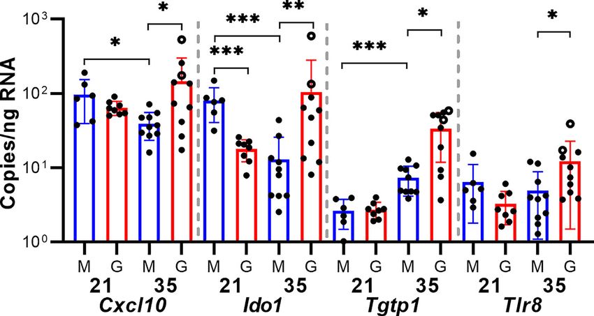
Berkebile et al.                                                                                                 The Placental Response to GPCMV

observed in congenitally infected children when maternal                       In this study, we investigated whether the susceptibility and
infection occurs in the first trimester (14–17). Maternal infection         antiviral responses of the placenta to GPCMV infection varied
late in pregnancy is associated with the highest rates of                  across gestation. Time mated guinea pigs were infected either at
intrauterine growth restriction (IUGR) and congenital infection            21 days gestation (dGA, “early”) or at 35 dGA (“late”). No
(9–13, 15, 18).                                                            significant differences in maternal or placental viral loads were
    HCMV infects the placenta and these placental infections can           noted between the two groups at 21 days post-infection (dpi).
cause fetal injury, including spontaneous abortion and neonatal            However, infection after mid-gestation caused more frequent
demise, even in the absence of detectable viral transmission to the        fetal membrane infections and significant changes in placental
fetus (4, 5, 19–22). HCMV-associated placental pathology includes          gene expression that were not observed after infection at the
chronic villitis, cytomegalic cells, and areas of necrosis and             earlier time point. Furthermore, in situ hybridization revealed
calcification (19, 22–24). HCMV was detected in 15% of fetal                that GPCMV infection primarily localized to the junctional zone
remains and/or placenta after stillbirth and infection was found to        after maternal infection at the later time. Our observations lead
be associated with fetal thrombotic vasculopathy (4). In a cohort of       us to propose that GPCMV-associated stillbirth and IUGR are
women with a prenatal diagnosis of fetal growth restriction, the           the result of developmentally regulated changes in the placenta
detection of HCMV antigen in the placenta correlated with higher           that predispose the organ to infection-associated injury late
rates of villitis and more severe growth restriction than cases of         in gestation.
IUGR without HCMV involvement (21). While HCMV appears
to cause a hypoxia-like condition in infected placenta, how HCMV
causes placental dysfunction remains poorly understood (22).               MATERIALS AND METHODS
    HCMV and other viruses may injure the placenta either
directly by causing cytopathic effects in infected cells or by             Cells and Virus
activating the maternal or fetal immune system and triggering              A minimally tissue culture adapted stock of GPCMV 22122
placental immunopathology (25). Dysmature villi were                       (ATCC VR-682), prepared as previously described by passaging
frequently observed in HCMV-infected placentas, suggesting                 guinea pig salivary gland homogenate twice on JH4 guinea pig
that infection can interfere with early placental development              lung fibroblasts (ATCC CCL-158), was used for this study (33,
(19). Trophoblast progenitor cells can be infected by HCMV,                45, 46). JH4 cells were purchased from ATCC and propagated
and HCMV infection both limits the capacity of trophoblast                 according to their specifications excepting that the growth media
progenitors to differentiate in vitro and disrupts the formation           was supplemented with sodium bicarbonate (47). GPCMV
of anchoring villi in first trimester placental explants (26–28). The       stocks were prepared as previously described and titered on
inflammatory response to viral infection during pregnancy can               JH4 cells (48). For animal studies, virus was aliquoted into single-
also cause placental dysfunction and adverse pregnancy outcomes            use units, flash-frozen, and stored at -80°C. These aliquots were
(29). For example, type I interferon signaling triggered by Zika           routinely re-titered as guinea pigs were infected to confirm that
virus can cause abnormal placental development in mice and                 the stock did not deteriorate during storage.
syncytial knot formation in villous explants (30). cCMV causes a
proinflammatory cytokine bias in the placenta and amniotic fluid,            Ethics Statement
but whether this host response disrupts normal placental function          All animal procedures were conducted in accordance with
has yet to be determined (31, 32).                                         protocols approved by the Institutional Animal Care and Use
    As the species-specificity of HCMV precludes its study in               Committee (IACUC) at the University of Minnesota, Minneapolis
animals, guinea pig cytomegalovirus (GPCMV) has become the                 (Protocol ID: 1810-36403A). Experimental protocols and
most widely used experimental model of cCMV (33, 34). Guinea               endpoints were developed in strict accordance to the National
pigs deliver precocious pups after gestational periods that                Institutes of Health Office of Laboratory Animal Welfare (Animal
average 65 days. Similarities between guinea pigs and humans               Welfare Assurance #A3456-01), Public Health Service Policy on
include hemomonochorial placentas that invade deeply into the              Humane Care and Use of Laboratory Animals, and United States
decidua and the prenatal development of major organs and the               Department of Agriculture Animal Welfare Act guidelines and
immune system (35–37). These similarities between human and                regulations (USDA Registration # 41-R-0005) with the oversight
guinea pig placentation and development make the rodent a                  and approval of the IACUC. Outbred Hartley guinea pigs were
uniquely powerful comparative model for understanding the                  initially purchased from Elm Hill Laboratories (Chelmsford, MA).
developmental origins of health and disease (35). GPCMV                    Breeding pairs of strain 2 and strain 13 guinea pigs were
infection studies have revealed that placental and fetal                   generously shared by MS and the U.S. Army Medical Research
infection occur sequentially after maternal inoculation (34, 38–           Institute of Infectious Diseases, respectively. Guinea pigs were
41). If maternal infection occurs early in pregnancy, infectious           housed in a facility maintained by the University of Minnesota
virus may not be detected in pup organs and evidence of prior              Research Animal Resources, who were accredited through the
fetal infection can be limited to the presence of infection-               Association for Assessment and Accreditation of Laboratory
associated histologic lesions in pup organs (41–43). Maternal              Animal Care, International (AAALAC). All procedures were
GPCMV infection after mid-gestation has been found to cause                conducted by trained personnel under the supervision of
high rates of stillbirth and IUGR in pups (42–44).                         veterinary staff.

Frontiers in Immunology | www.frontiersin.org                          2                                    June 2021 | Volume 12 | Article 686415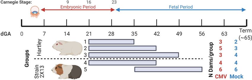
Berkebile et al.                                                                                                   The Placental Response to GPCMV

Animal Pathogenicity Study                                                   (GCF_000151735.1) as reference (52). Gene quantification was
The GPCMV serostatus of all animals received from an external                done via Cuffquant for FPKM values and Feature Counts for raw
source was tested by ELISA within one week of receipt and again              read counts (53, 54). Differentially expressed genes were identified
after one month of housing at the University of Minnesota (49).              using the edgeR (negative binomial) feature in CLCGWB (Qiagen)
Only GPCMV seronegative animals were used for infection                      using raw read counts (55). The generated list was filtered based
studies. Female guinea pigs were bred at two to three months                 on a minimum 2X Absolute Fold Change and False Discovery
of age. After the delivery of their first litter, the animals were bred       Rate corrected p < 0.05. Raw and processed RNA-Seq data have
a second time during postpartum estrus to establish timed                    been deposited in NCBI’s Gene Expression Omnibus and are
pregnancies by housing with a male for three days postpartum.                accessible through GEO Series accession number GSE169358 (56).
These second pregnancies were confirmed by progesterone                       Normalized gene expression data (FPKM) was uploaded to
ELISA (DRG International); only animals with plasma                          ClustVis for data visualization and heat map analysis (57). Gene
progesterone concentrations exceeding 15 ng/ml by 20 days                    Ontology (GO) analysis was performed by uploading a list of
postpartum were included in this study (50).                                 differentially expressed transcripts to g:Profiler (58). Results from
    At either 21 (range 18 to 23) or 35 (range 33 to 35) dGA                 this GO analysis were visualized using Cytoscape, Enrichment
guinea pigs were injected subcutaneously into the scruff of the              Map, ClusterMaker2, and WordCloud (59–63).
neck with 0.5 ml of PBS containing 2x10⁵ PFU of GPCMV or
PBS alone. Blood and plasma were collected from dams every                   Real-Time Droplet Digital PCR Analyses
seven days post-infection (dpi) until they were euthanized at 14,            RNA was extracted from placenta as described above. A two-step
21, or 28 dpi. After euthanasia, blood and plasma were collected             reverse transcriptase ddPCR (RT-ddPCR) protocol was used to
from the dams and amniotic fluid was collected from the pups.                 quantify transcript abundance (46). cDNA was synthesized from
Pup tissue samples were divided and frozen for DNA extraction,               total RNA using the Maxima™ H Minus cDNA Synthesis Master
stabilized in RNAlater (ThermoFisher) for RNA extraction,                    Mix (ThermoFisher). PCR primers targeting guinea pig transcripts
embedded in optimal cutting temperature (O.C.T.) compound                    were designed using the Primer Quest tool and were ordered from
(Fisher Scientific), or immersion-fixed using Shandon Formal-                  Integrated DNA Technologies (Table S2). ddPCR reactions were
Fixx (ThermoFisher) and embedded in paraffin.                                 prepared using the EvaGreen Digital PCR Supermix (Bio-Rad)
                                                                             using a primer concentration of 250 nM and cycled using the
Viral Load Quantification by Droplet                                          following thermal conditions: 95°C for 5 min; 40 cycles of 95°C for
Digital PCR                                                                  30 s and 60°C for 30s; 4°C for 5 min; 90°C for 5 min; hold at 4°C.
After DNA was extracted from whole blood, tissue, and amniotic               The ddPCR data were analyzed with QuantaSoft™ Analysis Pro
fluid, GPCMV genomes were quantified by droplet digital PCR                    software (Bio-Rad). Absolute quantification of gene expression
(ddPCR) using primers and probes specific to GP54 using the                   was presented as copies per nanogram of total RNA.
Bio-Rad QX200 system as previously described (46). ddPCR
results were analyzed using the QuantaSoft™ Analysis Pro                     In Situ Hybridization
software (Bio-Rad); GPCMV viral loads are presented as the                   5-mm sections of paraffin-embedded placenta were mounted onto
number of copies genome per ml of fluid or mg of tissue.                      Superfrost Plus slides (ThermoFisher). After air drying the tissue
                                                                             sections overnight, the slides were baked at 60°C for 1 hr. Tissue
RNA Sequencing                                                               was deparaffinized and pretreated using the recommended
RNA was extracted from guinea pig placentas that had been                    protocol for RNAscope® 2.5 Assays (ACD Document #322452).
stabilized in RNAlater (ThermoFisher) and stored at -20°C using              For target retrieval, samples were incubated at 99°C for 15
the RNAeasy Mini Kit (Qiagen). ~30 mg pieces of placenta were                minutes, and the slides were treated with RNAscope Protease
combined with 0.6 ml of b-mercaptoethanol-containing RLT                     Plus for 30 minutes. Slides were stained using either the RNAscope
buffer and Lysing Matrix D (MP Biomedicals) and pulsed at 6 m/               2.5 HD Detection Reagent – RED (ACD Document # 322360-
s for 30 seconds in a FastPrep 24 (MP Biomedicals). RNA was                  USM) or the RNAscope 2.5 HD Duplex Reagent (322500-USM)
extracted from 0.45 ml of the resulting homogenate using the                 and the RNAscope Probe V-CavHV-2-gp3. Stained slides were
manufacturer’s standard protocol, including the optional on-                 scanned using a Huron TissueScope LE and the number of gp3+
column DNase I digest (Qiagen), Total RNA integrity was                      foci and areas of gp3-staining were counted and calculated using
assessed by capillary electrophoresis using an Agilent                       NIS-Elements BR (Nikon).
TapeStation; all samples used for library creation had RNA
integrity numbers that exceeded 7.0.
    Dual indexed TruSeq stranded mRNA libraries were prepared                RESULTS
and sequenced using a NextSeq 550 high-output 75-bp single-end
run (mean of 22.4x10⁶ reads/sample). FastQ reads were trimmed                The Timing of Maternal GPCMV Infection
using Trimmomatic (v 0.33) enabled with the optional “-q”                    Affects the Rate of Fetal Membrane
option; 3bp sliding-window trimming from 3’ end requiring                    Infection but Not Placental Viral Loads
minimum Q30 (51). Quality control on raw sequence data for                   To study how the timing of maternal GPCMV infection affects
each sample was performed with FastQC. Read mapping was                      viral loads in the placenta, extraplacental membranes, and fetus,
performed via Hisat2 (v2.1.0) using the “Cavpor3.0” genome                   guinea pigs were time mated during postpartum estrus and

Frontiers in Immunology | www.frontiersin.org                            3                                    June 2021 | Volume 12 | Article 686415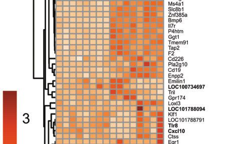
Berkebile et al.                                                                                                                  The Placental Response to GPCMV

infected at either 21 or 35 days gestation (dGA) (Figure 1).                         confirmed in all dams and no significant differences were
HCMV infection during the first trimester causes most cases of                        noted between the five groups (Table S1 and Figure S2A).
neurologic disability in congenitally infected children, and                             The placentas from the two groups of guinea pigs that were
infecting guinea pigs at 21 dGA exposes the pup and placenta                         infected at 21 dGA and euthanized at 21 dpi were compared, and
to virus during a comparable period of late embryonic/early fetal                    significantly higher viral loads were detected in the strain 2X13
development (14–17, 65). GPCMV infection after mid-gestation                         placentas than in their Hartley counterparts (Mann–Whitney U
has been found to often cause fetal growth restriction and/or                        test, P
Berkebile et al.                                                                                                                      The Placental Response to GPCMV

                                A                                                            B

    FIGURE 2 | GPCMV viral loads after maternal infection during pregnancy. Time mated guinea pigs were infected at either 21 or 35 dGA with 2X10⁵ PFU of GPCMV
    and euthanized 14, 21, or 28 days later. DNA was extracted from tissues and GPCMV viral loads were determined using a ddPCR assay specific to GP54. The limit
    of detection for this assay is indicated by the dashed line. (A) Viral load in placentas. Significantly higher viral loads (Mann-Whitney test, **** p < 0.0001) were
    observed in the placentas of strain 2X13 hybrids than in the placentas of Hartley guinea pigs. (B) Viral loads in infected fetal membranes. The number of GPCMV
    genome copies detected in the amnion and visceral yolk sac of individual fetuses was significantly correlated (Pearson r=0.5056, p < 0.001).

TABLE 1 | Summary of GPCMV viral load data.

Strain             dGA of infection, endpoint                    Placenta                         Amnion                   Yolk Sac                Fetus3          AF4

                                                        CMV+1            MVL2            CMV+1             MVL2        CMV+1        MVL2          CMV+1           CMV+1

Hartley                      21,35                       12/12          5.7x10³           0/10              N/A        0/10          N/A            9/10           0/10
                             21,42                       17/29          6.0x10³           4/29            1.5x101      4/28        2.0x101          9/29           0/29
                             21,49                       12/19          6.8x10³           6/19            1.6x10²      11/19       1.8x10²          9/19           0/18
2X13                         21,42                       18/19          2.8x10⁴           2/18            1.7x10²      2/18        1.7x101          0/18           0/18
                             35,56                       27/27          5.4x10⁴           18/24           1.8x10²      22/24       2.5x10²          4/27           4/21
1
 Samples containing detectable GPCMV DNA/total number of samples.
2
 Mean viral load (MVL) expressed as copies/ml for infected blood and amniotic fluid or copies/mg for infected tissue.
3
 Viral load in fetal brain quantified.
4
 Amniotic fluid.

infection and higher viral loads in the amnion and yolk sac                                  or 35 dGA and euthanized at 21 dpi. RNA was extracted from four
than infection at 21 dGA. However, neither the timing of                                     placentas from four GPCMV-infected dams and four placentas
maternal infection nor the experimental endpoint had a                                       from three control dams per group and sequenced (Table S1). A
significant effect on placental viral load. Higher viral loads were                           principal component analysis of RNA-Seq data revealed that the
noted in placentas from our strain 2X13 hybrid pregnancies when                              samples clustered based upon the gestational age of placenta
compared to Hartley guinea pigs, but there was no other evidence                             (Figure 3A). GPCMV- infected and control placenta from the
that indicated that infection was more severe in the inbred                                  early infection groups clustered tightly, while there was clear
animals. Presuming that there would be minimal animal-to-                                    separation between GPCMV- and mock-infected samples after
animal variation in the placental response to infection in guinea                            maternal infection at 35 dGA. Pairwise gene expression
pigs with a consistent maternal and fetal genetic background, we                             comparisons between age-matched groups of GPCMV and
focused on the strain 2X13 animals in our subsequent analysis of                             mock-infected tissues affirmed these findings. GPCMV infection
the effect of infection on placental function.                                               at 21 dGA had a limited effect on placental gene expression at 21
                                                                                             dpi: only 8 transcripts were differentially regulated (≥2 fold, p <
GPCMV Infection After Mid-Gestation                                                          0.05). In contrast, maternal infection at 35 dGA resulted in the
Significantly Alters Placental                                                                differential regulation of 126 transcripts (≥2 fold, p < 0.05) at 21
Gene Expression                                                                              dpi. A gene set enrichment analysis was performed on the
Having found that the timing of maternal GPCMV infection did                                 transcripts that were differentially expressed after GPCMV
not affect placental viral loads, we next compared how infection at                          infection late in pregnancy (58). This analysis found that several
our earlier and later time point affected placental gene expression.                         gene ontology terms related to the immune response (including
For this analysis, placentas were randomly selected from inbred                              GO:0002376, immune system process) were significantly enriched
guinea pigs that had been GPCMV- or mock-infected either at 21                               after GPCMV infection at 35 dGA (Figure S3A).

Frontiers in Immunology | www.frontiersin.org                                            5                                      June 2021 | Volume 12 | Article 686415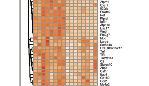
Berkebile et al. The Placental Response to GPCMV
In a pairwise gene expression analysis that compared the two
groups of mock-infected placentas, the gestational age of placentas
A
was found to have a much more significant effect on gene expression
than GPCMV infection: 1438 transcripts were differentially
regulated (≥2 fold, p < 0.05) between the two groups of control
tissue. Gene ontogeny analysis found numerous terms related to the
regulation of the mitotic cell, signaling receptor activity, and
condensed chromosome regions were enriched in this pairwise
comparison (Figure S3B). Many of the gene expression differences
between normal placenta from 42 and 56 dGA appear to be related
to the relatively higher expression of transcripts involved in cell
B division and the cell cycle by the younger tissue. While transcripts
that function as part of an immune system process were not found
to be significantly enriched in our comparison of normal placentas,
116 transcripts related to the immune response were differentially
expressed between the two groups (Figure 3B, Table S3). We
hypothesize that these normal changes in placental immunity may
cause GPCMV infection after mid-gestation to significantly affect
placental gene expression.
As only four samples of placenta per group were analyzed by
RNA-seq, sample-to-sample variation in gene expression could
be caused either by regional differences in transcription in the
relatively large guinea pig placenta or represent the unique
responses of individual fetuses to GPCMV infection. To better
elucidate host factors that regulate the placenta’s response to
GPCMV, we used reverse transcriptase droplet digital PCR (RT-
ddPCR) to measure the expression of select transcripts that
function as part of the inflammatory response in additional
placenta samples. For this experiment, RNA was extracted
from two placentas per dam (including the samples that had
been previously analyzed by RNA-Seq). RT-ddPCR confirmed
that four genes– Cxcl10, Ido1, Tgtp1, and Tlr8–were significantly
upregulated after maternal infection at 35 dGA when compared
to age-matched control placentas (Figure 4). This analysis also
found that Cxcl10 and Ido1 are normally downregulated as the
guinea pig placenta matures and that GPCMV infection at 21
dGA leads to decreased Ido1 expression relative to age-matched
normal placenta. RT-ddPCR analysis did not support the RNA-
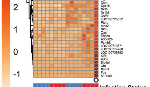
Seq finding that several other inflammatory mediators—Ccl5,
Ccl15-l, Cxcl8, Il1b, and Il36b-l—were differentially regulated by
maternal GPCMV infection at 35 dGA (Figure S4). In the case of
Cxcl8 and Il1b, high levels of cytokine transcription were noted
in the placenta of a dam that had been euthanized while
delivering stillborn pups at 56 dGA (these placentas are
represented as open circles in Figures 3, 4, and Figure S4).
The elevated transcription of these cytokines may not be specific
to GPCMV infection and instead be inflammatory markers of
preterm labor or in utero fetal demise (70, 71). Cumulatively, our
FIGURE 3 | Transcriptome profiling of GPCMV-infected placenta. Time gene expression analyses of GPCMV-infected placenta suggest
mated guinea pigs were GPCMV- or mock-infected at 21 or 35 dGA and that the immune response is dysregulated by GPCMV infection
euthanized 21 days later. RNA was extracted from placentas (N=4/group)
after mid-gestation but not after infection earlier in pregnancy.
and gene expression was quantified by Illumina RNA-Seq. (A) Principal
component analysis illustrating the similarity in gene expression between
samples. (B) Heat map illustrating the relative expression of transcripts that
The Junctional Zone Becomes Infected
function as part of an immune system processes (GO:0002376) that were by GPCMV After Maternal Infection
differentially expressed in placenta either during normal development or after After Mid-Gestation
GPCMV infection. Transcripts that were also analyzed by RT-ddPCR are
Finally, we compared the frequency and localization of GPCMV-
shown in bold.
infected cells in the placenta using in situ hybridization. For this
Frontiers in Immunology | www.frontiersin.org 6 June 2021 | Volume 12 | Article 686415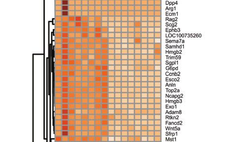
Berkebile et al. The Placental Response to GPCMV
FIGURE 4 | GPCMV infection dysregulates immune gene transcription. A two-step RT-ddPCR protocol was used to quantify the expression of select transcripts in
GPCMV- (G) and mock-infected (M) placentas after maternal infection at either 21 or 35 dGA. Data from two placentas per dam is shown. Statistically significant
differences were calculated by Mann-Whitney test (*p < 0.05, **p < 0.01, ***p < 0.001).
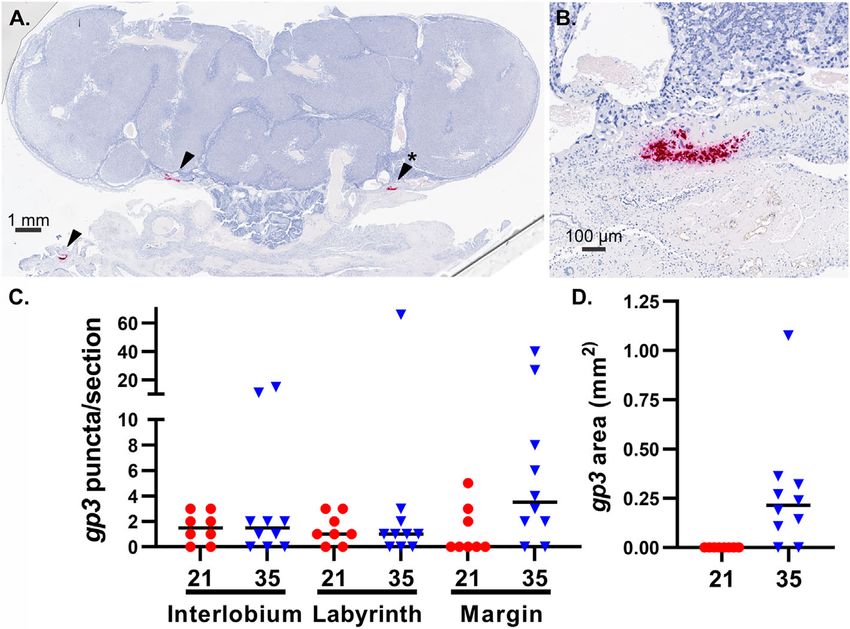
study, we designed an RNAscope probe specific to the GPCMV causes stillbirth and fetal growth restriction (42–44). Our study
transcript gp3. RNA-Seq analysis had revealed that gp3 is highly identified several possible explanations for these adverse
expressed during all phases of GPCMV replication (Figure S5) pregnancy outcomes, including an increased rate of fetal
(46). gp3+ foci, each representing either an individual infected membrane and junctional zone infection late in gestation and a
cell or a group of infected cells, were manually counted in stained transcriptional response to GPCMV that may indicate that
placenta sections (Figure 5). A small number of gp3+ foci were placental immunopathology only occurs after maternal
observed in the interlobium and labyrinth of all GPCMV- infection at later times in pregnancy.
infected placenta (Figure 5C). When the placentas of dams This study found that GPCMV infects both the amnion and
infected at 21 and 35 dGA were compared, significantly more the yolk sack, that viral loads in the two membranes are
GPCMV infected cells were noted in the margin of placentas, correlated, and that the highest rates of infection and viral
including either the parietal yolk sac or marginal syncytium, after loads in the membranes occur after maternal infection late in
infection at the later time. Large areas of gp3+ cells were detected pregnancy (46). While GPCMV joins HCMV on a short list of
in the junctional zone in eight of ten of the placentas from the viruses that have been observed to infect the fetal membranes, it
late infection group (Figures 5A, B, D). These large areas of remains unclear whether or how viral infections of the fetal
GPCMV-infected cells were often adjacent to either the membrane affect the fetus (24, 73). In contrast, ascending
subplacenta or large blood vessels and were never detected bacterial infections, a significant cause of preterm labor, have a
after maternal infection at 21 dGA. Whether GPCMV much better understood effect on fetal membrane physiology.
infection of the junctional zone reflects the recruitment of Bacterial infection, treatment with inflammatory cytokines, or
infected cells to the placenta late in pregnancy or if there is a exposure to pathogen-associated molecular patterns can all
cell type in this region that becomes permissive to GPCMV trigger preterm labor in animal models and/or cause fetal
infection as the placenta matures remains to be determined. membrane explants to biomechanically weaken ex vivo (71,
74–76). Several studies have suggested that viral infection may
sensitize the fetal membranes to later damage from bacteria (77–
DISCUSSION 79). It has also been hypothesized that fetal membrane infection
may allow viruses to circumvent the potent antiviral defense of
The timing of HCMV infection during pregnancy is a the placenta to infect the fetus (80). In this paraplacental route of
determinant of fetal outcomes, yet how placental development infection, viruses that infect the chorioamnion may be shed into
affects the course and severity of cCMV remains poorly amniotic fluid and infect the fetus. Given that this study’s
understood (25, 72). Using the GPCMV model, we assessed primary focus was on the effect of infection on the placenta,
how the placenta and fetal membranes are differentially affected future experiments that specifically analyze the effect of GPCMV
by maternal infection at two times: 21 and 35 dGA. Prior on the fetal membranes are merited.
research has found that GPCMV infection early in pregnancy Developmentally programmed changes in maternal,
can resolve prenatally while infection after mid-gestation often placental, and fetal immunity are critical for healthy
Frontiers in Immunology | www.frontiersin.org 7 June 2021 | Volume 12 | Article 686415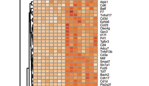
Berkebile et al.                                                                                                                       The Placental Response to GPCMV

 FIGURE 5 | GPCMV localization in infected placenta. An RNAscope probe specific to GPCMV gp3 was used to detect infected cells in sections of placenta by in
 situ hybridization. (A) A representative section of a placenta from a dam infected at 35 dGA, illustrating large areas of GPCMV infected cells in the junctional zone
 and decidua (red, indicated by arrows). (B) High-magnification image of infected cells in the junctional zone (*). (C) gp3+ puncta, representing individual infected cells
 or small groups of infected cells, were counted based on their localization in the placenta after maternal infection at 21 or 35 dGA and collection at 21 dpi. (D) The
 measured area of gp3+ stained cells in the junctional zone of placentas is shown.

pregnancy, helping to establish and maintain maternal-fetal                              are particularly relevant to the pathogenesis of cCMV. CXCL10,
tolerance, remodel the uterus, and regulate parturition (30, 71).                        a biomarker of VUE, chronic chorioamnionitis, and late preterm
Alterations in maternal immunity can increase the severity of                            birth, is found at elevated concentrations in amniotic fluid and
viral infections late in pregnancy (81). Severe, third-trimester                         maternal sera during cCMV (31, 86–89). Curiously, while the
infections caused by viruses such as influenza and hepatitis E can                        concentration of CXCL10 in amniotic fluid correlates with CMV
cause fetal injury or demise. High rates of maternal demise have                         genome abundance, the chemokine does not appear to
been reported in some GPCMV infection studies (38, 82).                                  accumulate in amniotic fluid when other viruses cause
However, HCMV infections are typically mild and there is no                              intrauterine infections (32). We and others have found that
evidence that the virus causes more severe illness in pregnant                           CXCL10 transcription is upregulated when placental cells or
individuals (83). We found that the timing of maternal GPCMV                             tissue are infected with HCMV or GPCMV in vitro (46, 84, 85).
infection did not significantly affect maternal, placental, or fetal                      Indoleamine-2,3-dioxygenase (IDO) is an enzyme that is
viral loads, and severe illness was not noted in dams that had                           encoded by two genes, IDO1 and IDO2, that catalyze the rate-
been infected at the later time. Having eliminated severe                                limiting step in tryptophan catabolism. IDO production by the
maternal illnesses as a cause of adverse pregnancy outcomes in                           placenta and the subsequent metabolism of tryptophan prevents
our experiments, the remainder of this study focused on the                              the rejection of the allogeneic fetus by suppressing T cell
placental response to GPCMV infection.                                                   proliferation and activity (90). IDO is more highly expressed
    Prior work in placental cells and explants has found that                            by the human placenta during the first-trimester than at term
gestational age affects how permissive the cells or tissue are to                        and HCMV infection suppressed IDO expression in early
viral infection and the nature of the inflammatory response that                          placenta (91). While our gene expression analysis was limited
is triggered by infection (84, 85). Our study found that maternal                        to a single time point post-infection, GPCMV may cause
infection at 35 dGA significantly altered placental gene                                  placental immunopathology but only after maternal infection
expression while infection at 21 dGA had remarkably little                               relatively late in pregnancy.
effect on placental gene expression. Infection after mid-                                    In an analysis of placental pathology after GPCMV infection,
gestation upregulated numerous transcripts that function as                              pregnant Hartley guinea pigs were inoculated with GPCMV at 30
part of an immune system process. Two transcripts that are                               dGA. Infection caused ischemic injury and necrosis associated
dysregulated by placental GPCMV infection, Cxcl10 and Ido1,                              with acute or chronic inflammation beginning at 14 dpi and

Frontiers in Immunology | www.frontiersin.org                                        8                                           June 2021 | Volume 12 | Article 686415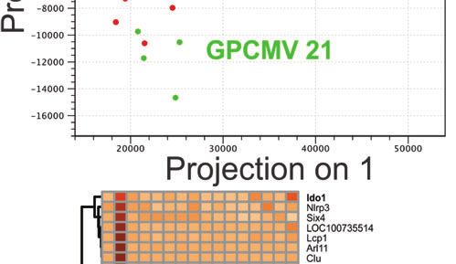
Berkebile et al.                                                                                                              The Placental Response to GPCMV

GPCMV-specific antigens and viral particles were most                              ETHICS STATEMENT
frequently observed at 28 dpi in the marginal and interlobar
transitional zones of the main placenta (39). We used in situ                     The animal study was reviewed and approved by Institutional
hybridization to compare the localization of GPCMV in the                         Animal Care and Use Committee at the University of Minnesota.
placenta 21 days after infection at 21 or 35 dGA. Like Griffith and
colleagues, we detected occasional infected cells in the main
placenta after infection at either time. However, the most
consistent pattern of infection and largest lesions localized to                  AUTHOR CONTRIBUTIONS
the junctional zone, which is situated between the main placenta
and the maternal decidua. These lesions were only found after                     CB conceived and designed the study. CB, ZB, and DP collected
infection at 35 dGA and may indicate that a ring of GPCMV-                        the data. All authors participated in data analysis and
sensitive cells develop at the base of the placenta as term                       interpretation. CB drafted the manuscript. All authors
approaches. Given how frequently we detected this pattern                         contributed to the article and approved the submitted version.
of junctional zone infection, we were surprised it had not
been previously reported. However, because our RNAscope
assay targets a viral transcript (gp3) that is highly expressed
during all phases of GPCMV’s replication, in situ hybridization                   FUNDING
may be more sensitive than electron microscopy or
                                                                                  This project was supported by grants from the Minnesota
immunohistochemistry for detecting GPCMV infected cells.
                                                                                  Masonic Charities (Masonic Early Investigator Award) and the
    Cumulatively, our data suggests the placenta becomes more
                                                                                  National Institutes of Health (R21HD087496, R01HD098866,
sensitized to GPCMV infection-associated injury late in gestation
                                                                                  and UL1TR002494).
either due to a potentially pathogenic immune response or by the
fetal membranes and junctional zone becoming more permissive to
infection. Numerous questions remain. Because GPCMV-
associated lesions had previously been reported exclusively in the
main placenta, we extracted DNA and RNA for viral load                            ACKNOWLEDGMENTS
quantification and gene expression analyses from this tissue (37,
                                                                                  Breeding pairs of strain 13 guinea pigs were generously shared by
39). A more nuanced analysis that compares the rate of GPCMV
                                                                                  the U.S. Army Medical Research Institute of Infectious Diseases.
infection and the immune response in the decidua, junctional zone,
                                                                                  Next-generation sequencing library generation and Illumina
and main placenta is justified. Our study did not investigate the
                                                                                  sequencing were completed by the University of Minnesota
effects of infection during the earliest stages of pregnancy. One                 Genomics Center. Histopathologic studies were supported by
report has noted that maternal GPCMV infection immediately
                                                                                  Colleen Forster of the University of Minnesota Histology and
prior to conception caused high rates of fetal demise, suggesting
                                                                                  Research Laboratory, which is supported by the National
that the virus may perturb early placental development and
                                                                                  Institutes of Health’s National Center for Advancing
function (40). Due to the limited availability of normal tissue
                                                                                  Translational Sciences, grant UL1TR002494, and the resources
between twenty weeks gestation and term, how viral infection
                                                                                  and staff of the University of Minnesota University Imaging
effects the function of the human placenta after mid-gestation
                                                                                  Centers (UIC, SCR_020997). Grants from the National Institutes
remains poorly characterized. Given similarities in the placental                 of Health (R21HD087496 to CB and R01HD098866 to MS) and
response of humans and guinea pigs to CMV infection and that
                                                                                  the Minnesota Masons (Masonic Early Investigator Award to
comparative gene expression analyses suggest that murine placental
                                                                                  CB) supported this work. The funders had no role in study
development largely parallels the first half of human pregnancy,
                                                                                  design, data collection, analysis, decision to publish, or
guinea pigs may be particularly well suited to model intrauterine
                                                                                  preparation of the manuscript.
infection after mid-gestation (92).

DATA AVAILABILITY STATEMENT                                                       SUPPLEMENTARY MATERIAL
The datasets presented in this study can be found in online repositories.         The Supplementary Material for this article can be found online
The names of the repository/repositories and accession number(s) can              at: https://www.frontiersin.org/articles/10.3389/fimmu.2021.
be found in the article/Supplementary Material.                                   686415/full#supplementary-material

                                                                                   2. Kenneson A, Cannon MJ. Review and Meta-Analysis of the Epidemiology of
REFERENCES                                                                            Congenital Cytomegalovirus (Cmv) Infection. Rev Med Virol (2007) 17
                                                                                      (4):253–76. doi: 10.1002/rmv.535
 1. Pereira L. Congenital Viral Infection: Traversing the Uterine-Placental        3. Mocarski ESJr., Shenk T, Griffiths PD, Pass RF. Cytomegaloviruses. In: Knipe
    Interface. Annu Rev Virol (2018) 5(1):273–99. doi: 10.1146/annurev-               DM, Howley PM, editors. Fields Virology, 6th. Philadelphia, PA: Wolters
    virology-092917-043236                                                            Kluwer/Lippincott Williams & Wilkins Health (2013). p. 1960–2014.

Frontiers in Immunology | www.frontiersin.org                                 9                                         June 2021 | Volume 12 | Article 686415
Berkebile et al.                                                                                                                           The Placental Response to GPCMV

 4. Iwasenko JM, Howard J, Arbuckle S, Graf N, Hall B, Craig ME, et al. Human                     Congenital Infection. Am J Pathol (2010) 177(3):1298–310. doi: 10.2353/
    Cytomegalovirus Infection Is Detected Frequently in Stillbirths and Is                        ajpath.2010.091210
    Associated With Fetal Thrombotic Vasculopathy. J Infect Dis (2011) 203                  23.   Sinzger C, Muntefering H, Loning T, Stoss H, Plachter B, Jahn G. Cell Types
    (11):1526–33. doi: 10.1093/infdis/jir121                                                      Infected in Human Cytomegalovirus Placentitis Identified by
 5. Pereira L, Petitt M, Fong A, Tsuge M, Tabata T, Fang-Hoover J, et al.                         Immunohistochemical Double Staining. Virchows Arch A Pathol Anat
    Intrauterine Growth Restriction Caused by Underlying Congenital                               Histopathol (1993) 423(4):249–56. doi: 10.1007/BF01606887
    Cytomegalovirus Infection. J Infect Dis (2014) 209(10):1573–84.                         24.   Uenaka M, Morizane M, Tanimura K, Deguchi M, Kanzawa M, Itoh T, et al.
    doi: 10.1093/infdis/jiu019                                                                    Histopathological Analysis of Placentas With Congenital Cytomegalovirus
 6. Panhani S, Heinonen KM. Screening for Congenital Cytomegalovirus Infection                    Infection. Placenta (2019) 75:62–7. doi: 10.1016/j.placenta.2019.01.003
    Among Preterm Infants Born Before the 34th Gestational Week in Finland.                 25.   Yockey LJ, Lucas C, Iwasaki A. Contributions of Maternal and Fetal Antiviral
    Scand J Infect Dis (1994) 26(4):375–8. doi: 10.3109/00365549409008607                         Immunity in Congenital Disease. Science (2020) 368(6491):608–12.
 7. Gibson CS, Goldwater PN, MacLennan AH, Haan EA, Priest K, Dekker GA, et al.                   doi: 10.1126/science.aaz1960
    Fetal Exposure to Herpesviruses May Be Associated With Pregnancy-Induced                26.   Tabata T, Petitt M, Zydek M, Fang-Hoover J, Larocque N, Tsuge M, et al.
    Hypertensive Disorders and Preterm Birth in a Caucasian Population. BJOG                      Human Cytomegalovirus Infection Interferes With the Maintenance and
    (2008) 115(4):492–500. doi: 10.1111/j.1471-0528.2007.01653.x                                  Differentiation of Trophoblast Progenitor Cells of the Human Placenta.
 8. Shi TL, Huang LJ, Xiong YQ, Zhong YY, Yang JJ, Fu T, et al. The Risk of                       J Virol (2015) 89(9):5134–47. doi: 10.1128/JVI.03674-14
    Herpes Simplex Virus and Human Cytomegalovirus Infection During                         27.   Tabata T, Petitt M, Fang-Hoover J, Rivera J, Nozawa N, Shiboski S, et al.
    Pregnancy Upon Adverse Pregnancy Outcomes: A Meta-Analysis. J Clin                            Cytomegalovirus Impairs Cytotrophoblast-Induced Lymphangiogenesis and
    Virol (2018) 104:48–55. doi: 10.1016/j.jcv.2018.04.016                                        Vascular Remodeling in an In Vivo Human Placentation Model. Am J Pathol
 9. Preece PM, Blount JM, Glover J, Fletcher GM, Peckham CS, Griffiths PD. The                     (2012) 181(5):1540–59. doi: 10.1016/j.ajpath.2012.08.003
    Consequences of Primary Cytomegalovirus Infection in Pregnancy. Arch Dis                28.   Zydek M, Petitt M, Fang-Hoover J, Adler B, Kauvar LM, Pereira L, et al. Hcmv
    Child (1983) 58(12):970–5. doi: 10.1136/adc.58.12.970                                         Infection of Human Trophoblast Progenitor Cells of the Placenta is Neutralized
10. Bodeus M, Hubinont C, Goubau P. Increased Risk of Cytomegalovirus                             by a Human Monoclonal Antibody to Glycoprotein B and Not by Antibodies to
    Transmission In Utero During Late Gestation. Obstet Gynecol (1999) 93(5                       the Pentamer Complex. Viruses (2014) 6(3):1346–64. doi: 10.3390/v6031346
    Pt 1):658–60. doi: 10.1097/00006250-199905000-00005                                     29.   Mor G, Aldo P, Alvero AB. The Unique Immunological and Microbial Aspects
11. Gindes L, Teperberg-Oikawa M, Sherman D, Pardo J, Rahav G. Congenital                         of Pregnancy. Nat Rev Immunol (2017) 17(8):469–82. doi: 10.1038/nri.2017.64
    Cytomegalovirus Infection Following Primary Maternal Infection in the Third             30.   Yockey LJ, Iwasaki A. Interferons and Proinflammatory Cytokines in
    Trimester. BJOG (2008) 115(7):830–5. doi: 10.1111/j.1471-0528.2007.01651.x                    Pregnancy and Fetal Development. Immunity (2018) 49(3):397–412.
12. Bodeus M, Kabamba-Mukadi B, Zech F, Hubinont C, Bernard P, Goubau P.                          doi: 10.1016/j.immuni.2018.07.017
    Human Cytomegalovirus in Utero Transmission: Follow-Up of 524 Maternal                  31.   Scott GM, Chow SS, Craig ME, Pang CN, Hall B, Wilkins MR, et al.
    Seroconversions. J Clin Virol (2010) 47(2):201–2. doi: 10.1016/j.jcv.2009.11.009              Cytomegalovirus Infection During Pregnancy With Maternofetal
13. Picone O, Vauloup-Fellous C, Cordier AG, Guitton S, Senat MV, Fuchs F, et al. A               Transmission Induces a Proinflammatory Cytokine Bias in Placenta and
    Series of 238 Cytomegalovirus Primary Infections During Pregnancy: Description                Amniotic Fluid. J Infect Dis (2012) 205(8):1305–10. doi: 10.1093/infdis/jis186
    and Outcome. Prenat Diagn (2013) 33(8):751–8. doi: 10.1002/pd.4118                      32.   Gervasi MT, Romero R, Bracalente G, Chaiworapongsa T, Erez O, Dong Z,
14. Pass RF, Fowler KB, Boppana SB, Britt WJ, Stagno S. Congenital                                et al. Viral Invasion of the Amniotic Cavity (VIAC) in the Midtrimester of
    Cytomegalovirus Infection Following First Trimester Maternal Infection:                       Pregnancy. J Matern Fetal Neonatal Med (2012) 25(10):2002–13. doi: 10.3109/
    Symptoms at Birth and Outcome. J Clin Virol (2006) 35(2):216–20.                              14767058.2012.683899
    doi: 10.1016/j.jcv.2005.09.015                                                          33.   Hartley JW, Rowe WP, Huebner RJ. Serial Propagation of the Guinea Pig
15. Faure-Bardon V, Magny JF, Parodi M, Couderc S, Garcia P, Maillotte AM,                        Salivary Gland Virus in Tissue Culture. Proc Soc Exp Biol Med (1957) 96
    et al. Sequelae of Congenital Cytomegalovirus Following Maternal Primary                      (2):281–5. doi: 10.3181/00379727-96-23455
    Infections Are Limited to Those Acquired in the First Trimester of Pregnancy.           34.   Choi YC, Hsiung GD. Cytomegalovirus Infection in Guinea Pigs. II.
    Clin Infect Dis (2019) 69(9):1526–32. doi: 10.1093/cid/ciy1128                                Transplacental and Horizontal Transmission. J Infect Dis (1978) 138
16. Lipitz S, Yinon Y, Malinger G, Yagel S, Levit L, Hoffman C, et al. Risk of                    (2):197–202. doi: 10.1093/infdis/138.2.197
    Cytomegalovirus-Associated Sequelae in Relation to Time of Infection and                35.   Morrison JL, Botting KJ, Darby JRT, David AL, Dyson RM, Gatford KL, et al.
    Findings on Prenatal Imaging. Ultrasound Obstet Gynecol (2013) 41(5):508–                     Guinea Pig Models for Translation of the Developmental Origins of Health
    14. doi: 10.1002/uog.12377                                                                    and Disease Hypothesis Into the Clinic. J Physiol (2018) 596(23):5535–69.
17. Foulon I, Naessens A, Foulon W, Casteels A, Gordts F. A 10-Year Prospective                   doi: 10.1113/JP274948
    Study of Sensorineural Hearing Loss in Children With Congenital                         36.   Mess A, Zaki N, Kadyrov M, Korr H, Kaufmann P. Caviomorph Placentation
    Cytomegalovirus Infection. J Pediatr (2008) 153(1):84–8. doi: 10.1016/                        as a Model for Trophoblast Invasion. Placenta (2007) 28(11-12):1234–8.
    j.jpeds.2007.12.049                                                                           doi: 10.1016/j.placenta.2007.08.003
18. Elkan Miller T, Weisz B, Yinon Y, Weissbach T, De Castro H, Avnet H, et al.             37.   Kaufmann P, Davidoff M. The Guinea-Pig Placenta. Adv Anat Embryol Cell
    Congenital Cytomegalovirus Infection Following Second and Third Trimester                     Biol (1977) 53(2):5–91. doi: 10.1007/978-3-642-66618-6
    Maternal Infection Is Associated With Mild Childhood Adverse Outcome Not                38.   Auerbach MR, Yan D, Vij R, Hongo JA, Nakamura G, Vernes JM, et al. A
    Predicted by Prenatal Imaging. J Pediatr Infect Dis Soc (2021) 10(5):562–8.                   Neutralizing Anti-Gh/Gl Monoclonal Antibody is Protective in the Guinea
    doi: 10.1093/jpids/piaa154                                                                    Pig Model of Congenital Cmv Infection. PloS Pathog (2014) 10(4):e1004060.
19. Garcia AG, Fonseca EF, Marques RL, Lobato YY. Placental Morphology in                         doi: 10.1371/journal.ppat.1004060
    Cytomegalovirus Infection. Placenta (1989) 10(1):1–18. doi: 10.1016/0143-               39.   Griffith BP, McCormick SR, Fong CK, Lavallee JT, Lucia HL, Goff E. The
    4004(89)90002-7                                                                               Placenta as a Site of Cytomegalovirus Infection in Guinea Pigs. J Virol (1985)
20. Muhlemann K, Miller RK, Metlay L, Menegus MA. Cytomegalovirus Infection                       55(2):402–9. doi: 10.1128/JVI.55.2.402-409.1985
    of the Human Placenta: An Immunocytochemical Study. Hum Pathol (1992)                   40.   Harrison CJ, Myers MG. Relation of Maternal Cmv Viremia and Antibody
    23(11):1234–7. doi: 10.1016/0046-8177(92)90290-j                                              Response to the Rate of Congenital Infection and Intrauterine Growth
21. Tsuge M, Hida AI, Minematsu T, Honda N, Oshiro Y, Yokoyama M, et al.                          Retardation. J Med Virol (1990) 31(3):222–8. doi: 10.1002/jmv.1890310309
    Prospective Cohort Study of Congenital Cytomegalovirus Infection During                 41.   Kumar ML, Prokay SL. Experimental Primary Cytomegalovirus Infection in
    Pregnancy With Fetal Growth Restriction: Serologic Analysis and Placental                     Pregnancy: Timing and Fetal Outcome. Am J Obstet Gynecol (1983) 145
    Pathology. J Pediatr (2019) 206:42–8 e2. doi: 10.1016/j.jpeds.2018.10.003                     (1):56–60. doi: 10.1016/0002-9378(83)90339-3
22. Maidji E, Nigro G, Tabata T, McDonagh S, Nozawa N, Shiboski S, et al.                   42.   Griffith BP, Lucia HL, Hsiung GD. Brain and Visceral Involvement During
    Antibody Treatment Promotes Compensation for Human Cytomegalovirus-                           Congenital Cytomegalovirus Infection of Guinea Pigs. Pediatr Res (1982) 16
    Induced Pathogenesis and a Hypoxia-Like Condition in Placentas With                           (6):455–9. doi: 10.1203/00006450-198206000-00010

Frontiers in Immunology | www.frontiersin.org                                          10                                            June 2021 | Volume 12 | Article 686415
Berkebile et al. The Placental Response to GPCMV
43. Griffith BP, Hsiung GD. Cytomegalovirus Infection in Guinea Pigs. IV. 63. Reimand J, Isserlin R, Voisin V, Kucera M, Tannus-Lopes C, Rostamianfar A,
Maternal Infection at Different Stages of Gestation. J Infect Dis (1980) 141 et al. Pathway Enrichment Analysis and Visualization of Omics Data Using G:
(6):787–93. doi: 10.1093/infdis/141.6.787 Profiler, GSEA, Cytoscape and Enrichmentmap. Nat Protoc (2019) 14(2):482–
44. Harrison CJ, Britt WJ, Chapman NM, Mullican J, Tracy S. Reduced 517. doi: 10.1038/s41596-018-0103-9
Congenital Cytomegalovirus (Cmv) Infection After Maternal Immunization 64. Butler H. An Atlas for Staging Mammalian and Chick Embryos. In: Juurlink
With a Guinea Pig CMV Glycoprotein Before Gestational Primary Cmv BHJ, editor. Boca Raton, Fla: CRC Press (1987).
Infection in the Guinea Pig Model. J Infect Dis (1995) 172(5):1212–20. 65. Harman MT, Dobrovolny MP. The Development of the External Form of the
doi: 10.1093/infdis/172.5.1212 Guinea-Pig (Cavia Cobaya) Between the Ages of 21 Days and 35 Days of
45. Yang D, Tamburro K, Dittmer D, Cui X, McVoy MA, Hernandez-Alvarado N, Gestation. J Morphol (1933) 54(3):493–519. doi: 10.1002/jmor.1050540306
et al. Complete Genome Sequence of Pathogenic Guinea Pig Cytomegalovirus 66. Wright S. The Effects of Inbreeding and Crossbreeding on Guinea Pigs: I.
From Salivary Gland Homogenates of Infected Animals. Genome Announc Decline in Vigor : II. Differentiation Among Inbred Families. Washington, D.C:
(2013) 1(2):e0005413. doi: 10.1128/genomeA.00054-13 U.S. Dept. of Agriculture (1922).
46. Putri DS, Berkebile ZW, Mustafa HJ, Fernandez-Alarcon C, Abrahante JE, 67. Fong CK, Lucia H, Bia FJ, Hsiung GD. Histopathologic and Ultrastructural
Schleiss MR, et al. Cytomegalovirus Infection Elicits a Conserved Chemokine Studies of Disseminated Cytomegalovirus Infection in Strain 2 Guinea Pigs.
Response From Human and Guinea Pig Amnion Cells. Virology (2020) Lab Invest (1983) 49(2):183–94.
548:93–100. doi: 10.1016/j.virol.2020.06.005 68. Nozawa N, Yamamoto Y, Fukui Y, Katano H, Tsutsui Y, Sato Y, et al.
47. Hoying JJ. Selection and Histochemical Identification of Epithelial-Like Cells Identification of a 1.6 Kb Genome Locus of Guinea Pig Cytomegalovirus
From Guinea Pig Lung. [M.s.]. Dayton, Ohio: W.S.U. Printing Service: Wright Required for Efficient Viral Growth in Animals But Not in Cell Culture.
State University (1975). Virology (2008) 379(1):45–54. doi: 10.1016/j.virol.2008.06.018
48. Britt WJ. Human Cytomegalovirus: Propagation, Quantification, and Storage. 69. Griffith BP, McCormick SR, Booss J, Hsiung GD. Inbred Guinea Pig Model of
Curr Protoc Microbiol (2010) 18:14E.3.1–3.17. doi: 10.1002/97804717 Intrauterine Infection With Cytomegalovirus. Am J Pathol (1986) 122(1):112–9.
29259.mc14e03s18 70. Goldstein JA, Gallagher K, Beck C, Kumar R, Gernand AD. Maternal-Fetal
49. Schleiss MR, Bourne N, Stroup G, Bravo FJ, Jensen NJ, Bernstein DI. Inflammation in the Placenta and the Developmental Origins of Health and
Protection Against Congenital Cytomegalovirus Infection and Disease in Disease. Front Immunol (2020) 11:531543. doi: 10.3389/fimmu.2020.531543
Guinea Pigs, Conferred by a Purified Recombinant Glycoprotein B Vaccine. 71. Romero R, Espinoza J, Goncalves LF, Kusanovic JP, Friel L, Hassan S. The
J Infect Dis (2004) 189(8):1374–81. doi: 10.1086/382751 Role of Inflammation and Infection in Preterm Birth. Semin Reprod Med
50. Bierle CJ, Fernandez-Alarcon C, Hernandez-Alvarado N, Zabeli JC, Janus BC, (2007) 25(1):21–39. doi: 10.1055/s-2006-956773
Putri DS, et al. Assessing Zika Virus Replication and the Development of Zika- 72. Chatzakis C, Ville Y, Makrydimas G, Dinas K, Zavlanos A, Sotiriadis A.
Specific Antibodies After a Mid-Gestation Viral Challenge in Guinea Pigs. Timing of Primary Maternal Cytomegalovirus Infection and Rates of Vertical
PloS One (2017) 12(11):e0187720. doi: 10.1371/journal.pone.0187720 Transmission and Fetal Consequences. Am J Obstet Gynecol (2020) 223
51. Bolger AM, Lohse M, Usadel B. Trimmomatic: A Flexible Trimmer for (6):870–83.e11. doi: 10.1016/j.ajog.2020.05.038
Illumina Sequence Data. Bioinformatics (2014) 30(15):2114–20. 73. Tabata T, Petitt M, Fang-Hoover J, Zydek M, Pereira L. Persistent
doi: 10.1093/bioinformatics/btu170 Cytomegalovirus Infection in Amniotic Membranes of the Human
52. Siren J, Valimaki N, Makinen V. Indexing Graphs for Path Queries With Placenta. Am J Pathol (2016) 186(11):2970–86. doi: 10.1016/
Applications in Genome Research. IEEE/ACM Trans Comput Biol Bioinform j.ajpath.2016.07.016
(2014) 11(2):375–88. doi: 10.1109/TCBB.2013.2297101 74. Kumar D, Moore RM, Mercer BM, Mansour JM, Redline RW, Moore JJ. The
53. Trapnell C, Roberts A, Goff L, Pertea G, Kim D, Kelley DR, et al. Differential Gene Physiology of Fetal Membrane Weakening and Rupture: Insights Gained
and Transcript Expression Analysis of RNA-seq Experiments With TopHat and From the Determination of Physical Properties Revisited. Placenta (2016)
Cufflinks. Nat Protoc (2012) 7(3):562–78. doi: 10.1038/nprot.2012.016 42:59–73. doi: 10.1016/j.placenta.2016.03.015
54. Liao Y, Smyth GK, Shi W. The Subread Aligner: Fast, Accurate and Scalable 75. Yoshimura K, Hirsch E. Effect of Stimulation and Antagonism of Interleukin-
Read Mapping by Seed-and-Vote. Nucleic Acids Res (2013) 41(10):e108. 1 Signaling on Preterm Delivery in Mice. J Soc Gynecol Investig (2005) 12
doi: 10.1093/nar/gkt214 (7):533–8. doi: 10.1016/j.jsgi.2005.06.006
55. Robinson MD, McCarthy DJ, Smyth GK. Edger: A Bioconductor Package for 76. McCarthy R, Martin-Fairey C, Sojka DK, Herzog ED, Jungheim ES, Stout MJ,
Differential Expression Analysis of Digital Gene Expression Data. et al. Mouse Models of Preterm Birth: Suggested Assessment and Reporting
Bioinformatics (2010) 26(1):139–40. doi: 10.1093/bioinformatics/btp616 Guidelines. Biol Reprod (2018) 99(5):922–37. doi: 10.1093/biolre/ioy109
56. Edgar R, Domrachev M, Lash AE. Gene Expression Omnibus: NCBI Gene 77. Potter JA, Tong M, Aldo P, Kwon JY, Pitruzzello M, Mor G, et al. Viral
Expression and Hybridization Array Data Repository. Nucleic Acids Res Infection Dampens Human Fetal Membrane Type I Interferon Responses
(2002) 30(1):207–10. doi: 10.1093/nar/30.1.207 Triggered by Bacterial Lps. J Reprod Immunol (2020) 140:103126.
57. Metsalu T, Vilo J. ClustVis: a web tool for visualizing clustering of multivariate doi: 10.1016/j.jri.2020.103126
data using Principal Component Analysis and heatmap. Nucleic Acids Res 78. Cross SN, Potter JA, Aldo P, Kwon JY, Pitruzzello M, Tong M, et al. Viral
(2015) 43(W1):W566–70. doi: 10.1093/nar/gkv468 Infection Sensitizes Human Fetal Membranes to Bacterial Lipopolysaccharide
58. Raudvere U, Kolberg L, Kuzmin I, Arak T, Adler P, Peterson H, et al. G: by MERTK Inhibition and Inflammasome Activation. J Immunol (2017) 199
Profiler: A Web Server for Functional Enrichment Analysis and Conversions (8):2885–95. doi: 10.4049/jimmunol.1700870
of Gene Lists (2019 Update). Nucleic Acids Res (2019) 47(W1):W191–W8. 79. Bakaysa SL, Potter JA, Hoang M, Han CS, Guller S, Norwitz ER, et al. Single-
doi: 10.1093/nar/gkz369 and Double-Stranded Viral RNA Generate Distinct Cytokine and Antiviral
59. Shannon P, Markiel A, Ozier O, Baliga NS, Wang JT, Ramage D, et al. Responses in Human Fetal Membranes. Mol Hum Reprod (2014) 20(7):701–8.
Cytoscape: A Software Environment for Integrated Models of Biomolecular doi: 10.1093/molehr/gau028
Interaction Networks. Genome Res (2003) 13(11):2498–504. doi: 10.1101/ 80. Tabata T, Petitt M, Puerta-Guardo H, Michlmayr D, Wang C, Fang-Hoover J,
gr.1239303 et al. Zika Virus Targets Different Primary Human Placental Cells, Suggesting
60. Merico D, Isserlin R, Stueker O, Emili A, Bader GD. Enrichment Map: A Network- Two Routes for Vertical Transmission. Cell Host Microbe (2016) 20(2):155–
Based Method for Gene-Set Enrichment Visualization and Interpretation. PloS 66. doi: 10.1016/j.chom.2016.07.002
One (2010) 5(11):e13984. doi: 10.1371/journal.pone.0013984 81. Kourtis AP, Read JS, Jamieson DJ. Pregnancy and Infection. N Engl J Med
61. Oesper L, Merico D, Isserlin R, Bader GD. Wordcloud: A Cytoscape Plugin to (2014) 370(23):2211–8. doi: 10.1056/NEJMra1213566
Create a Visual Semantic Summary of Networks. Source Code Biol Med (2011) 82. Griffith BP, Lucia HL, Tillbrook JL, Hsiung GD. Enhancement of
6:7. doi: 10.1186/1751-0473-6-7 Cytomegalovirus Infection During Pregnancy in Guinea Pig. J Infect Dis
62. Morris JH, Apeltsin L, Newman AM, Baumbach J, Wittkop T, Su G, et al. (1983) 147(6):990–8. doi: 10.1093/infdis/147.6.990
Clustermaker: A Multi-Algorithm Clustering Plugin for Cytoscape. BMC 83. Davis NL, King CC, Kourtis AP. Cytomegalovirus Infection in Pregnancy.
Bioinf (2011) 12:436. doi: 10.1186/1471-2105-12-436 Birth Defects Res (2017) 109(5):336–46. doi: 10.1002/bdra.23601
Frontiers in Immunology | www.frontiersin.org 11 June 2021 | Volume 12 | Article 686415You can also read