Autoantibody Discovery, Assay Development and Adoption: Death Valley, the Sea of Survival and Beyond
←
→
Page content transcription
If your browser does not render page correctly, please read the page content below
REVIEW
                                                                                                                                            published: 27 May 2021
                                                                                                                                   doi: 10.3389/fimmu.2021.679613

                                            Autoantibody Discovery, Assay
                                            Development and Adoption:
                                            Death Valley, the Sea of Survival
                                            and Beyond
                                            Marvin J. Fritzler 1*, May Y. Choi 1, Minoru Satoh 2 and Michael Mahler 3
                                            1 Department of Medicine, Cumming School of Medicine, Calgary, AB, Canada, 2 Department of Clinical Nursing, School of

                                            Health Sciences, University of Occupational and Environmental Health, Kitakyushu, Japan, 3 Research and Development,
                                            Inova Diagnostics, San Diego, CA, United States

                                            Dating to the discovery of the Lupus Erythematosus (LE) cell in 1948, there has been a
                                            dramatic growth in the discovery of unique autoantibodies and their cognate targets, all of
                                            which has led to the availability and use of autoantibody testing for a broad spectrum of
                            Edited by:      autoimmune diseases. Most studies of the sensitivity, specificity, commutability, and
                   Edward K. L. Chan,
                                            harmonization of autoantibody testing have focused on widely available, commercially
   University of Florida, United States
                                            developed and agency-certified autoantibody kits. However, this is only a small part of the
                        Reviewed by:
                  Gerson D. Keppeke,        spectrum of autoantibody tests that are provided through laboratories world-wide. This
Federal University of São Paulo, Brazil     manuscript will review the wider spectrum of testing by exploring the innovation pathway
            Kenneth Michael Pollard,
      The Scripps Research Institute,
                                            that begins with autoantibody discovery followed by assessment of clinical relevance,
                         United States      accuracy, validation, and then consideration of regulatory requirements as an approved
                  *Correspondence:          diagnostic test. Some tests are offered as “Research Use Only (RUO)”, some as
                       Marvin J. Fritzler
                                            “Laboratory Developed Tests (LDT)”, some enter Health Technology Assessment (HTA)
                   fritzler@ucalgary.ca
                                            pathways, while others are relegated to a “death valley” of autoantibody discovery and
                   Specialty section:       become “orphan” autoantibodies. Those that achieve regulatory approval are further
         This article was submitted to      threatened by the business world’s “Darwinian Sea of Survival”. As one example of the
                Cytokines and Soluble
               Mediators in Immunity,       trappings of autoantibody progression or failure, it is reported that more than 200 different
               a section of the journal     autoantibodies have been described in systemic lupus erythematosus (SLE), a small
              Frontiers in Immunology
                                            handful (~10%) of these have achieved regulatory approval and are widely available as
           Received: 12 March 2021
            Accepted: 04 May 2021
                                            commercial diagnostic kits, while a few others may be available as RUO or LDT assays.
            Published: 27 May 2021          However, the vast majority (90%) are orphaned and languish in an autoantibody ‘death
                             Citation:      valley’. This review proposes that it is important to keep an inventory of these “orphan
   Fritzler MJ, Choi MY, Satoh M and        autoantibodies” in ‘death valley’ because, with the increasing availability of multi-analyte
        Mahler M (2021) Autoantibody
        Discovery, Assay Development        arrays and artificial intelligence (MAAI), some can be rescued to achieve a useful role in
            and Adoption: Death Valley,     clinical diagnostic especially in light of patient stratification and precision medicine.
      the Sea of Survival and Beyond.
           Front. Immunol. 12:679613.       Keywords: autoantibodies, regulatory approval review, diagnostic testing methodologies, immunoassays,
    doi: 10.3389/fimmu.2021.679613          orphan autoantibodies

Frontiers in Immunology | www.frontiersin.org                                     1                                         May 2021 | Volume 12 | Article 679613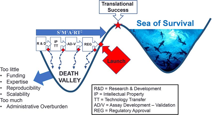
Fritzler et al. Autoantibody Discovery and Clinical Adoption
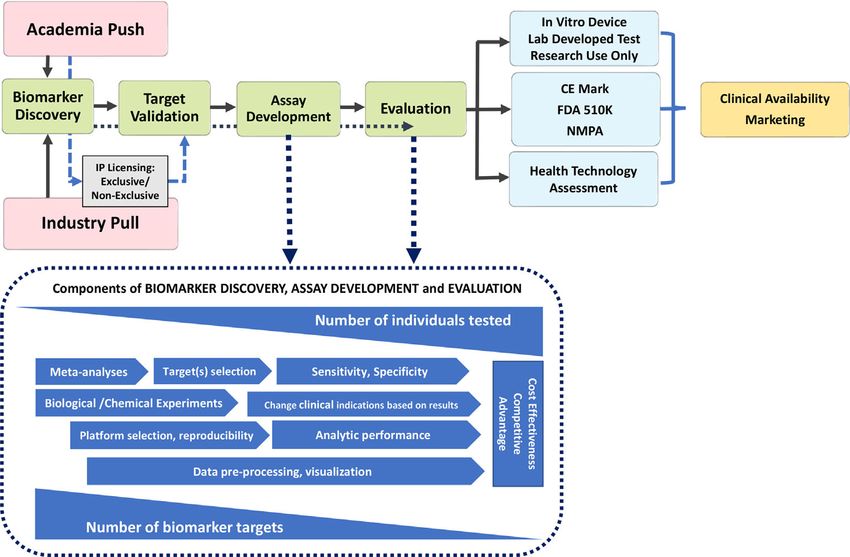
OVERVIEW THE VIRTUOUS CYCLE OF
AUTOANTIBODY DISCOVERY
The use of proteomic biomarkers has become a valuable and
effective approach to the prediction, diagnosis, and management AND ADOPTION
of individuals with a wide range autoimmune and
To understand why certain autoantibodies are in wide use while
autoinflammatory diseases (1–3). The spectrum of proteomic
others lie dormant or are in very limited use, it is important to
biomarkers used in clinical settings includes those with a long
review two main overlapping pathways of the “virtuous cycle” of
history such as C-reactive protein, those associated with the
autoantibody innovation (12, 13). The first is the pathway of
complex pathways involved in the pathogenesis of these diseases,
biomarker discovery and translation (Figure 1). Dating to the
such as anti-dsDNA and anti-citrullinated peptide antibodies
late 1970s (14), medical sciences witnessed the ‘golden age” of
(ACPA), interferons and interleukins, which reflect various
cell and molecular biology, which has in turn served as the hot-
interactions and responses of inflammatory cells.
bed for autoantibody discovery (15, 16). Historically,
To effectively utilize the huge data sets that can now be
autoantibodies were first reported in organ specific
generated through autoantibody and other biomarker analytics,
autoimmune diseases (17), then in what eventually was called
machine learning and artificial intelligence (AI) in the setting of
the anti-phospholipid syndrome (18) and in systemic lupus
precision health (PH) are major drivers for biomarker use in
erythematosus (SLE) traced to the discovery of the lupus
clinical practice (2, 4–6). For example, autoantibodies combined
erythematosus (LE) cell (19). This was followed by a
with other multi-analyte “omic” profiles are now beginning to
remarkably broad spectrum of autoantibodies in SLE, other
form the basis of predicting disease thus allowing for disease
systemic autoimmune rheumatic diseases and a growing
prevention strategies and earlier and effective personalized
spectrum of ‘new’ clinical conditions and syndromes, some
interventions for established disease (7–10). As medical
only regarded as being autoimmune for less than 10 years (20).
intervention continues to move toward disease prediction and
Again, from a historical perspective, virtually all these
a model of “intent to PREVENT” morbidity and mortality (11),
autoantibody discoveries were in academic laboratories, but
futuristic diagnostics will take into consideration symptoms and
with the realization of a significant market value of
risks, as opposed to an established disease and organ
autoantibody testing and patented biomarkers, research and
involvement approach. Closing the gaps in autoantibody
development (R&D) divisions of in-vitro diagnostic (IVD)
diagnostics will involve newer diagnostic platforms that utilize
companies have also become an important source of these
emerging megatrends such as systems medicine, consumer-
new discoveries.
driven social networks, AI and deep learning all benefiting a
Discovery of a novel autoantibody is only the first very small
paradigm shift to PH (2).
step on the pathway to adoption in clinical practice (Figure 1).
This manuscript will focus on autoantibodies and the various
While initial claims of diagnostic value (clinical relevance,
limitations and gaps that persist in their effective use in clinical
clinical phenotype, sensitivity and specificity) may be
practice. To achieve an understanding and appreciation of these
impressive, validation becomes the next critical step to ensure
limitations, the pathways leading to the discovery and adoption
the initial claims are repeatable and followed by exploration in
of some autoantibodies and the rejection of others will
more depth the potential “market value” of the autoantibody
be explored.
(e.g., does it fill a seronegative gap, does it identify an important
clinical subset, is it actionable?) Typically, at this stage of
autoantibody development, a decision may be taken to patent
the novel marker and derive a source of licensing revenues from
Abbreviations: ACPA, anti-citrullinated peptide antibodies; AI, artificial
intelligence; AIM, autoimmune inflammatory myopathies; ANA, anti-nuclear industry (another onerous process that is not part of this review)
antibody; APLA, anti-phospholipid antibodies; BICD2, bicaudal D2; BPI, and/or be entered into the publication “derby” and achieve the
bacterial permeability inhibitor; CENP, centromere protein; DFS, dense fine status of primacy (i.e., “first to publish”) and then become open
speckled; dsDNA, double-stranded deoxyribonucleic acid; FDA, Food and Drug to wider use. A critical step is to determine if the novel
Administration (USA); HMGCR, 3-hydroxy-3-methyl-glutaryl-coenzyme A
autoantibody can be detected by conventional diagnostic
reductase; IVD, in vitro diagnostic; Ku, named after the index patient serum
(Kuriowa), a dimeric 70-/80-kDA protein complex that binds to DNA double- platforms [e.g., enzyme linked immunoassay (ELISA),
strand break ends and is required for DNA repair; LDT, laboratory developed test; addressable laser bead immunoassay (ALBIA), line
MAAAA, multi-analyte arrays with analytic algorithms; MDA, melanoma immunoassay (LIA), particle-based technology (PMAT) or
differentiation-associated protein 5; MPO, myeloperoxidase; NMDAR, N- cell-based assays (CBA)] that are accessible to diagnostic
methyl-D-aspartate receptor; NOR, nucleous organizer protein; NMPA,
National Medical Products Administration; NuMA, nuclear mitotic apparatus;
laboratories and thereby achieve wide use. Unfortunately, some
NXP, nuclear matrix protein 2; PAD, protein arginine deiminase; PCNA, novel autoantibody discoveries depend on highly sophisticated
proliferating cell nuclear antigen; PDGFR, platelet derived growth factor; PH, techniques and/or protocols that are not thoroughly or clearly
precision health; PR3, proteinase 3; RA, rheumatoid arthritis; RF, rheumatoid described thereby limiting their validation by other investigators
factor; RNP, ribonucleoprotein; RUO, research use only; SaMD, software as and their potential for wide adoption. If the “first to publish”
medical device; SGNA, S- G-phase nuclear antigen; sIBM, sporadic inclusion
body myositis; SjS, Sjogren syndrome; SLE, systemic lupus erythematosus; SRP,
group does not pursue the research on the given biomarker,
signal recognition particle; SSc, systemic sclerosis; TIF, transcription intermediary follow-up studies by other investigators are met with limited
factor; TopoI, topoisomerase I; TRIM, tripartite motif. access to high impact factor journals because journal editors
Frontiers in Immunology | www.frontiersin.org 2 May 2021 | Volume 12 | Article 679613Fritzler et al. Autoantibody Discovery and Clinical Adoption FIGURE 1 | Pathway to Diagnostic Biomarker Translation. Biomarker development process from research and discovery to development and clinical use is a multi- faceted process with a wide range of timelines that undergoes several phases from discovery to clinical availability and utilization. CE marking is a certification mark that indicates conformity with health, safety, and environmental protection standards for products sold within the European Economic Area; the FDA, Food and Drug Administration (USA) and NMPA, National Medical Products Administration in China require, albeit some unique jurisdictional standards and timelines. typically prefer something new and disruptive, and furthermore, and FDA applications are being filed and adjudicated by granting councils do not see this as innovative, hypothesis- regulatory agencies, an autoantibody test is offered to clinicians generating research. Obviously, for an autoantibody discovery as a “laboratory developed test” (LDT) and with that designation, to successfully find its way through the virtuous pathway of a disclaimer is required to the same effect. Another approach to innovation, significant resources and investments are needed bridge the gap between regulatory submission and regulatory from granting councils, R&D budgets, philanthropic donations, approval is to offer the test as a “Research Use Only” (RUO) and home institutions (universities, colleges, research institutes). assay. A limitation of the LDT and RUO approaches is that, in In addition, challenges to successful navigation of the pathway some health care payer systems, reimbursement may not be come in the form of administrative overburden (“red tape”) to provided for assays having LDT or RUO status. An intermediate achieve ethical approval, material transfer agreements and approach is to proceed on a formal Health Technology intellectual property regulations imposed by academic Assessment (HTA) pathway, which is attended by clearly institutions and funders alike. If a novel autoantibody fails to defined qualifiers and qualifications. Nonetheless, the goal is to clear any of these steps, it tends to fall prey of the “valley of achieve the ‘nirvana’ of novel autoantibody innovation and that death” (21) (Figure 2). is full IVD regulatory approval status (e.g., CE mark in the Autoantibodies that pass the “acid tests” described above can European Union). From there through marketing, the assay is then proceed to the next phase of optimization wherein issues of typically widely adopted and with increasing demand by assay development like reproducibility, sensitivity, specificity, clinicians, is available for clinical use. However, even with the standardization, clinical applications, cost effectiveness and virtuous cycle hurdle having been crossed, the assay enters into a competitive advantages are rigorously evaluated, typically by a rather competitive, if not hostile, “real world” environment of IVD companies (Figure 1). Concurrently, thorough evaluation ‘dog-eat-dog’, competitive edge or what is referred to as the of the realistic market value of the autoantibody in clinical “Darwinian Sea of Survival” (22) (Figure 2). practice is needed because to proceed to IVD regulatory Returning to the ‘death valley’ of innovation, it is important to approval [European Union CE mark, Food and Drug appreciate a nuance of this metaphor because even in the “real Administration USA (FDA) approval, National Medical world” of Death Valley (California, USA), while there is Products Administration (NMPA) in China, Health Canada, or widespread evidence of death, there are remarkable evidences other regional jurisdictions] requires tremendous paperwork and of life. Even some rocks, referred to as “wandering”, “sailing” or patience coupled with attention to detail. In some cases, and “walking”, seem to be ‘alive’ (23). This is to remind that although many times as a temporary measure, while the more rigorous CE more than 90% of all autoantibodies reported in the literature Frontiers in Immunology | www.frontiersin.org 3 May 2021 | Volume 12 | Article 679613
Fritzler et al.                                                                                                         Autoantibody Discovery and Clinical Adoption

  FIGURE 2 | Death Valley of Biomarker Translation. Successful crossing ‘death valley’ is dependent on a number of factors and S3M1A2RT2 characteristics (Specific,
  Sensitive, Scalability, Measurable, Actionable, Added value, Realistic, Titratable, Temporal Timing) leading to variable timelines to Translational Success.

never achieve IVD regulatory approval (Table 1), they should                         some evolve to systemic sclerosis (SSc), Sjögren syndrome (SjS),
not be regarded as “dead” or having no value. As one example,                        rheumatoid arthritis (RA) and autoimmune inflammatory
although greater than 200 different autoantibodies have been                         myopathies (AIM) (74–78). Some predictive autoantibodies
described in SLE (24) less than 15 are typically utilized as                         and their temporal appearance, especially in RA, are known
biomarkers in clinical practice (Table 1). While an extensive                        but longitudinal studies of very early SARD/UCTD are required
catalogue of autoantibodies described to date is published (24,                      and it is here that ‘death valley’ autoantibodies may find
25) a partial list of those that may warrant re-evaluation and                       important predator/prognostic value. Recognizing this, there
rescue of these “orphan autoantibodies” (15, 26–28) from ‘death                      has been a call for more studies to identify diagnostic,
valley’ is shown in Table 1.                                                         prognostic (i.e., disease activity, remission, and outcomes) and
                                                                                     biomarkers that predict earlier autoimmune disease onset, as well
                                                                                     as biomarkers that predict effectiveness of a growing spectrum of
RESCUING AUTOANTIBODIES FROM                                                         therapeutic options [reviewed in (5, 28, 79)].
DEATH VALLEY                                                                             In addition to providing more information as predictors of
                                                                                     SARD, it is plausible that death valley of autoantibodies also hold
While some autoantibodies appear to have perished in ‘death                          important value for other key functions of autoantibodies such as
valley’ (Table 2), there are a number of reasons to “rescue” them.                   their pathogenic (80, 81), protective (26) and prognostic values
With the advent of multi-analyte arrays with algorithmic analysis                    (27, 82). For example, despite substantial advances, the high
(MAAA) as an approach to PH (2, 3, 66) the value of these                            morbidity and mortality that currently characterizes SLE can
autoantibodies may be discovered when they are combined and                          largely be attributed to a delay in diagnosis, gaps in our
permutated with other biomarkers, and hence fill seronegative                         understanding of the role of autoantibodies in early disease,
gaps such as in antinuclear antibody (ANA)-negative SLE (67,                         and limited effective therapeutic options. SSc is another SARD
68) and other systemic autoimmune rheumatic diseases (SARD)                          with heterogeneous clinical features that is extremely difficult to
(69–73). In addition, machine learning and AI approaches may                         diagnose in the early phase (83), resulting in a critical delay in
find that these autoantibodies provide value in determining                           therapy which is often begun when internal organ involvement is
subsets of disease that have a more clinically actionable basis                      already irreversible (77, 78). Older classification criteria (84–86)
(3). In addition, on future exploration, ‘death valley’                              had a remarkably low sensitivity for the early phase of disease
autoantibodies may have value predicting the evolution of very                       (87) so they were replaced by the American College of
early SARD (i.e., undifferentiated connective tissue disease,                        Rheumatology (ACR)/European League Against Rheumatism
UCTD) to confirmed, criteria-defined SARD. For example, 5-                             2013 criteria which improved the disease classification (88).
50% of UCTD patients or very early connective tissue disease                         Nevertheless, the diagnosis may be delayed for several years
evolve to fulfill diagnostic and classification criteria of a SARD.                    after the onset of Raynaud’s phenomenon (RP) or certainly after
Of the UCTD patients that do evolve to a SARD, the majority                          the first non-RP symptom. RP, ANA positivity, and puffy fingers
(80%) have been reported to develop SLE, while of the reminder,                      were recently indicated as “red flags” (by the Very Early

Frontiers in Immunology | www.frontiersin.org                                    4                                          May 2021 | Volume 12 | Article 679613
Fritzler et al. Autoantibody Discovery and Clinical Adoption
TABLE 1 | Snapshot of autoantibodies in use (survivors) for systemic autoimmune rheumatic diseases.
Regulatory Status/adoption SLE SSc AIM RA SjS Vasculitis
Widely available dsDNA CENP-A, -B Jo-1 U1-RNP RF SS-B/La PR-3
Sm TopoI/Scl-70 HMGCR ACPA SSA/Ro60 MPO
U1-RNP RNA Pol III Ro52/TRIM21 Ro52/TRIM21
Chromatin Ro52/TRIM21
Histone U1-RNP
SS-A/Ro60
SS-B/La
Ro52/TRIM21
APLA
Geographic dependent Ribosomal P Th/To ARS Ra33 Alpha fodrin Elastase
PCNA Fibrillarin SRP DFS70* BPI
Ku PM/Scl MDA-5 Lactoferrin
DFS70* NOR-90 SAE DFS70*
DFS70* TIF1y/155
NXP-2
Mi-2
PM/Scl
Ku
DFS70*
Primarily RUO/LDT NMDAR2 RuvBL1/2 NT5c1A/Mup44 CarP NuMA Lysozyme
BICD2 Azurocidin
U11/U12 (RNPC-3)
Exosome
eIF2B
*aid in the exclusion of diagnosis.
ACPA, anti-citrullinated peptide antibodies; AIM, autoimmune inflammatory myopathies; APLA, anti-phospholipid antibodies; ARS, aminoacyl tRNA synthetase; BICD2, bicaudal D2; BPI,
bacterial permeability inhibitor; CarP, carbamylated protein, CENP, centromere protein; DFS, dense fine speckled; dsDNA, double-stranded deoxyribonucleic acid; eIF2B, guanine
nucleotide exchange factor; HMGCR, 3-hydroxy-3-methyl-glutaryl-coenzyme A reductase; Ku, named after the index patient serum (Kuriowa), a dimeric 70-/80-kDA protein complex that
binds to DNA double-strand break ends and is required for DNA repair; LDT, laboratory developed test; MDA, melanoma differentiation-associated protein 5; MPO, myeloperoxidase;
NMDAR, N-methyl-D-aspartate receptor; NOR, nucleous organizer protein; NuMA, nuclear mitotic apparatus; NT5c1A, Nucleotidase 5' Cytosolic IA; NXP, nuclear matrix protein 2; PAD,
protein/peptidyl arginine deiminase; PCNA, proliferating cell nuclear antigen; PDGFR, platelet derived growth factor; PR3, proteinase 3; RF, rheumatoid factor; RNP, ribonucleoprotein;
RUO, research use only; SRP, signal recognition particle; TIF, transcription intermediary factor; TopoI, topoisomerase I; TRIM, tripartite motif.
TABLE 2 | Death Valley Autoantibodies of Interest that might address ‘Seronegative Gaps’ in Systemic Autoimmune Rheumatic Diseases.
SARD % of patients with no Death Valley Autoantibodies and Comments
identifiable autoantibody*
SLE 3-25 ASE1 anti-sense ERCC1 (nucleolar SLE) (29); cell cycle SG2NA associated with cancer (30–32); replication protein A
(RPA) complex (33), RNA helicase A, Ki/SL (33), Ago2 (34)
SSc 3-10 Bicaudal D2 (BICD2) (35); U11/U12 (RNPC-3) (36, 37); HMGs (38); B23/nucleophosmin (39), eIF2B (40)
RA 15-50 Newer biomarkers such as antibodies to PAD1, PAD2, PAD3, and PAD4, as well as to carbamylated peptides/proteins
are narrowing the seronegative gap (9, 41)
AIM 20-30 Survival of motor neuron (SMN) complex (42, 43); PUF60 (44–46); NT5c1A/Mup44 not just associated with sIBM (47–49)
SjS/Sicca 10-30 CENP-C (50–52), a-fodrin (53, 54), NA14/SSN1 (55); SS56 (56); golgins (57), TS-1 RNA; M3 receptor (58, 59), Ki/SL (33)
syndrome
Vasculitis 10-30 NA14 and CK15 (60); LAMP2 (61); EEA1 (62)
Other – p80/coilin: associated with DFS70 (63), Ge-1/GW182 (57, 64)
AIM, autoimmune inflammatory myopathies; ANA, anti-nuclear antibody; CENP, centromere protein; HMG, high mobility group proteins; NA, nuclear antigen; PAD, protein arginine
deiminase; RA, rheumatoid arthritis; SG2NA, S- G2-phase nuclear antigen; sIBM, sporadic inclusion body myositis; SjS, Sjögren syndrome; SLE, systemic lupus erythematosus; SSc,
systemic sclerosis.
*Wide ranges due to demographic and clinical variability of cohorts. In recent SLE classification criteria (65), ANA is a required criterion, hence the percent ‘seronegative’ is, by definition,
zero. However, having a positive ANA does not necessarily mean a relevant disease autoantibody will be detected.
Diagnosis Of Systemic Sclerosis (VEDOSS) study)–that is, the “window of opportunity” whereby the physician can act with
main elements for suspicion of SSc in the very early phase of the effective drugs to block or at least slow the progression of the
disease (89). Confirming the diagnosis requires further tests, disease (10, 81, 90–92). The principal challenge is to detect valid
particularly nailfold videocapillaroscopy and evaluation of predictors of disease evolution to enable treatment of patients in
disease specific autoantibodies (Table 1). In this way, patients the early stage of disease. Perhaps lying in ‘death valley’ are the
can be identified in the very early phase of disease enabling a key autoantibodies that can facilitate these goals.
Frontiers in Immunology | www.frontiersin.org 5 May 2021 | Volume 12 | Article 679613Fritzler et al. Autoantibody Discovery and Clinical Adoption
An early and accurate diagnosis of SLE and other SARD go undetected (101). And last, the VectraDA blood test, based on
through the use of autoantibody testing that has met SSSMAART measuring the concentrations of 12 biomarkers that reflect the
(specificity, sensitivity, scalability, measurable, actionable, added pathogenesis of RA, is designed to provide an objective measure
value, realistic, titres, timely) characteristics (3) (Figure 2) will of disease activity for RA by providing a score on a scale of 1 to
help improve SARD-associated clinical outcomes and healthcare 100 with high scores associated with disease progression (102).
expenditures. Clearly, not all ‘death valley’ autoantibodies should The analytical validity, clinical validity, and clinical utility of
be expected to provide value because there is compelling VectraDA have been reported and it is reputed to assist in
evidence that the vast majority of autoantibodies studied to monitoring clinical responses to disease-modifying anti-
date are “indifferent” (93) or “junk” autoantibodies (15). rheumatic drugs (102), including blockade of the CD40/CD40L
However, as a word of caution, it should be recalled that pathway (103). Based on a wide range of clinical studies on
shortly after the completion of the human genome project it VectraDA, the ACR has recently added Vectra DA as one of the
was assumed that a significant portion of the human genome was methods to assess disease activity. It has also been shown to have
“junk”, only to discover unanticipated functions of DNA were value in following the clinical progression and remission of Adult
yet to be discovered (94, 95). Accordingly, we prefer the term Onset Still’s disease (104).
‘orphan” autoantibodies over “junk” autoantibodies to categorize
those which have no known or proven function (2, 15, 26).
As briefly outlined above, it is well-established that there is an
increasing use, awareness and focus on PH and disease
AVOIDING DEATH VALLEY AND
prevention (2). PH applied to SARD will require paradigm SURVIVING THE ‘DARWINIAN SEA
shifts in the use and application of autoantibodies and other OF SURVIVAL’
biomarkers. For example, autoantibodies combined with other
multi-analyte “omic” profiles will form the basis of disease It is important to consider ways in which autoantibody discovery
prediction allowing for earlier intervention linked to disease can result in a more rapid and protective transition to an
prevention strategies, as well as earlier, effective and actionable biomarker with proven clinical value and availability
personalized interventions for established disease (2, 5). As in mainstream diagnostic testing. First, it is important to
medical intervention moves to disease prediction and a model appreciate the “push” and “pull” equation of innovation
of “intent to PREVENT,” diagnostics will include an early (Figure 1). While autoantibody discovery continues by
symptom/risk-based, as opposed to a disease-based approach. academics, supported by largely institutional investors (i.e.,
Newer diagnostic platforms that utilize emerging megatrends granting councils), this is only the “push” side of innovation.
such as AI and close the gaps in autoantibody diagnostics will For an autoantibody, or any biomarker, to succeed it needs to
benefit from paradigm shifts thereby facilitating the PH agenda. meet a need or a demand (i.e., fill a seronegative gap; identify a
disease subset with a specific actionable therapeutic choice) and,
hence, have a strong “pull” component from the diagnostic
industry, regulators, physicians, laboratory scientists, patient
TECHNOLOGY AND COOPERATION TO advocates, health care payers, and angel investors. Without a
THE RESCUE well-balanced push-pull ‘equation’, it is unlikely that an
autoantibody will make it across the ‘death valley’ of innovation.
Single autoantibody testing only provides a narrow window of Second, in the discovery research phase autoantibodies must
the clinical picture and also does not represent the ultimate be shown to be S3M1A2RT2 (Figure 2): demonstrate Specificity,
approach to an early and accurate diagnosis or following or Sensitivity and Scalability, Measurable using conventional
predicting responses to therapeutic interventions. Accordingly, technologies, Add value to clinical management, be Actionable
multi-analyte techniques for detecting multiple autoantibodies (lead to or suggest a clinical decision), Realistic (detection should
on MAAA are coming into use (28, 96). With the advent of not involve complicated processes or procedures) and address
MAAA, emerging evidence indicates that when certain the Temporal Timing during the course of the disease (i.e. is it
combinations of biomarkers, such as the interferon signature predictive or transient) (3). The latter is an important factor
and stem cell factor accompany autoantibody and ANA results, because not all autoantibodies are present at diagnosis and some
the predictive power for SLE is markedly increased (28). A few do not persist throughout the disease course (105). Early
examples of MAAA that have emerged include the SLE-Key rule attention to these factors can help assure that the autoantibody
out test, (97, 98) that uses microarray technology to identify will not only survive ‘death valley’ but also the ‘Darwinian Sea of
autoantibody patterns that discriminate SLE from healthy: Survival’ (Figure 2).
reported 94% sensitivity; 75% specificity; 93% negative The 'Darwinian Sea of Survival' in deference to Death Valley
predictive value. The Avise ® Lupus Test uses a parallel was a metaphor initially intended to describe the entire span of
approach to detect autoantibodies and cell-bound complement innovation (3) including the way it is used here as the end stage
products to distinguish SLE from other rheumatological struggle for survival in a highly competitive market where
conditions (99) and may predict disease progression in patients constantly changing medical advances, technologies, clientele
who had non-specific clinical signs (100). However, the relatively needs and expectations, and investment strategies are
low sensitivity, suggests that patients with preclinical SLE could constantly being evaluated. Despite an initial phase of triumph
Frontiers in Immunology | www.frontiersin.org 6 May 2021 | Volume 12 | Article 679613Fritzler et al. Autoantibody Discovery and Clinical Adoption
of having successfully traversed ‘death valley’, some innovations value (107), this opens additional challenges from the regulatory
simply do not survive in the ‘sea of survival’ because of perspective. Along those lines, the FDA just published an action
technological advances, economic considerations (investors, plan to outline activities and areas of focus to manage software as
managers, client’s shifting priorities) and an increasing trend medical devices (SaMD) that leverage AI [reviewed in (108)].
to central procurement where other factors that do not include
true performance of a biomarker may not be the primary factor
of interest.
SUMMARY
DIAGNOSTIC INDUSTRY CHALLENGES The discovery of novel autoantibodies is linked to an ever-
expanding spectrum of autoimmune conditions. For a number
From the industry perspective, increased regulatory burden of reasons, the vast majority of discovered autoantibodies are not
especially due to in-vitro device regulation (IVDR) is making currently used in routine clinical diagnostics and have become
the rescue of autoantibodies from the ‘death valley’ very relegated to the ‘death valley’ of innovation. With the advent of
challenging. This new regulation requires additional evidence PH and MAAA it seems plausible that some of these
about the usefulness of the biomarker beyond the clinical autoantibodies might be ‘rediscovered’ and become valuable
validity. Clinical utility studies and likely health economic predictive, prognostic and actionable biomarkers. In the
studies will be especially required for novel biomarker rescue meantime, successful innovation is a ‘real time’ partnership
and approval. In addition, the diagnostic platforms available at a with a balance of the push and pull forces of innovation.
given IVD company can also have a significant impact on the
success of a biomarker. While some autoantibodies are useful as
a standalone marker, other autoantibodies require a panel of
markers to be tested at the same time. A typical example for PATIENT-PUBLIC PARTICIPATION
biomarkers that should be measured as a multi-analyte panel are
Patients or the public were not involved in the design, or
myositis-specific antibodies (106).
conduct, or reporting, or dissemination plans of our research.
Quality control, both during the manufacturing process, and
the clinical setting, requires the availability of patient samples
that can serve as calibrators of controls (106). While this is
achievable for common markers (such as ACPA), it can AUTHOR CONTRIBUTIONS
represent a significant challenge for orphan autoantibodies
(e.g., U11/U12 RNP, RNPC-3). Human or humanized MF and MC conceived of the content. MF, MC, MS, and MM
recombinant antibodies represent a viable, but not yet cost- conducted the literature review and wrote and/or edited
intensive alternative. manuscript drafts. MC, MF, and MM provided graphical
Although AI might provide new approaches to combine content. All authors contributed to the article and approved
autoantibody results in scores that provide increased clinical the submitted version.
for Anti-Nuclear Antibodies. Clin Chem Lab Med (2018) 56:1771–7. doi:
REFERENCES 10.1515/cclm-2017-0241
1. Mahler M, Pierangeli S, Meroni PL, Fritzler MJ. Autoantibodies in Systemic 8. Eloff E, Martinsson K, Ziegelasch M, Cedergren J, Reckner Ã., Skogh T, et al.
Autoimmune Disorders. J Immunol Res (2014) 2014:263091. doi: 10.1155/ Autoantibodies are Major Predictors of Arthritis Development in Patients
2014/263091 With Anti-Citrullinated Protein Antibodies and Musculoskeletal Pain.
2. Fritzler MJ, Martinez-Prat L, Choi MY, Mahler M. The Utilization of Scand J Rheumatol (2020) Online Ahead Print. doi: 10.1080/
Autoantibodies in Approaches to Precision Health. Front Immunol (2018) 03009742.2020.1818820
9:2682. doi: 10.3389/fimmu.2018.02682 9. Lamacchia C, Courvoisier DS, Jarlborg M, Bas S, Roux-Lombard P, Möller
3. Fritzler MJ, Mahler M. Redefining Systemic Lupus Erythematosus — B, et al. Predictive Value of Anti-CarP and Anti-PAD3 Antibodies Alone or
SMAARTT Proteomics. Nat Rev Rheumatol (2018) 14:451–2. doi: in Combination With RF and ACPA on the Severity of Rheumatoid
10.1038/s41584-018-0035-3 Arthritis. Rheumatol (Oxford) Online Ahead Print (2021) Online Ahead
4. Conrad K, Shoenfeld Y, Fritzler MJ. Precision Health: A Pragmatic Print keab050. doi: 10.1093/rheumatology/keab050
Approach to Understanding and Addressing Key Factors in Autoimmune 10. Deane KD, Holers VM. Rheumatoid Arthritis: Pathogenesis, Prediction and
Diseases. Autoimmun Rev (2020) 19:102508. doi: 10.1016/j.autrev.2020 Prevention - An Emerging Paradigm Shift. Arthritis Rheumatol (2021) 73
5. Mahler M, Martinez-Prat L, Sparks JA, Deane KD. Precision Medicine in the (2):181–93. doi: 10.1002/art.41417
Care of Rheumatoid Arthritis: Focus on Prediction and Prevention of Future 11. Infantino M, Meacci F, Grossi V, Benucci M, Morozzi G, Tonutti E, et al.
Clinically-Apparent Disease. Autoimmun Rev (2020) 19(5):102506. Serological Epitope Profile of Anti-Ro52-Positive Patients With Systemic
doi: 10.1016/j.autrev.2020.102506 Autoimmune Rheumatic Diseases. Arthritis Res Ther (2015) 17:365. doi:
6. Bernatsky SM, Pfau JC, Fritzler MJ. Environmental Exposures and 10.1186/s13075-015-0871-3
Biomarkers Predictive of Rheumatoid Arthritis and the Pathway to 12. Wartman SA. Toward a Virtuous Cycle: The Changing Face of Academic
Precision Medicine. J Lab Precis Med (2017) 2:5. doi: 10.21037/ Health Centers. Acad Med (2008) 83:797–9. doi: 10.1097/ACM.
jlpm.2017.02.01 0b013e318181cf8c
7. Pãrez D, Gilburd B, Cabrera-Marante A, Martinez-Flores JA, Serrano M, 13. Pedroso JL, Dutra LA, Espay AJ, Hoftberger R, Barsottini OGP. Video
Naranjo L, et al. Predictive Autoimmunity Using Autoantibodies: Screening NeuroImages: Head Titubation in anti-mGluR1 Autoantibody-Associated
Frontiers in Immunology | www.frontiersin.org 7 May 2021 | Volume 12 | Article 679613Fritzler et al. Autoantibody Discovery and Clinical Adoption
Cerebellitis. Neurology (2018) 90:746–7. doi: 10.1212/WNL. 35. Fritzler MJ, Hudson M, Choi MY, Mahler M, Wang M, Bentow C, et al.
0000000000005338 Identification of Bicaudal D2: A Novel Autoantibody Target in Systemic
14. Lerner MR, Steitz JA. Antibodies to Small Nuclear RNAs Complexed With Sclerosis That Shares a Key Epitope With CENP-A But Has a Distinct
Proteins are Produced by Patients With Systemic Lupus Erythematosus. Clinical Phenotype. Autoimmun Rev (2018) 17:267–75. doi: 10.1016/
Proc Natl Acad Sci USA (1979) 76:5495–9. doi: 10.1073/pnas.76.11.5495 j.autrev.2018.01.006
15. Fritzler MJ. Toward a New Autoantibody Diagnostic Orthodoxy: 36. Fertig N, Domsic RT, Rodriguez-Reyna T, Kuwana M, Lucas M, Medsger
Understanding the Bad, Good and Indifferent. Autoimmun Highlights TAJr., et al. Anti-U11/U12 RNP Antibodies in Systemic Sclerosis: A New
(2012) 3:51–8. doi: 10.1007/s13317-012-0030-7 Serologic Marker Associated With Pulmonary Fibrosis. Arthritis Rheumatol
16. Fritzler MJ. Professional Insights From a Pioneer in Autoimmune Disease (2009) 61:958–65. doi: 10.1002/art.24586
Testing: The Future of Antinuclear/Anticellular Antibody Testing. J Appl 37. McMahan ZH, Domsic RT, Zhu L, Medsger TA, Casciola-Rosen L, Shah AA.
Lab Med (2019) 04:287–9. doi: 10.1373/jalm.2018.028399 Anti-RNPC-3 (U11/U12) Antibodies in Systemic Sclerosis in Patients With
17. Conrad K, Schlosser W, Hiepe F, Fritzler MJ. Autoantibodies in Organ Moderate-to-Severe Gastrointestinal Dysmotility. Arthritis Care Res
Specific Autoimmune Diseases: A Diagnostic Reference. Berlin: Pabst Science (Hoboken) (2019) 71:1164–70. doi: 10.1002/acr.23763
Publishers (2017). 38. Ayer LM, Sené cal J-L, Martin L, Dixon GH, Fritzler MJ. Antibodies to High
18. Hughes GRV, Asherson RA, Khamashta MA. Antiphospholipid Syndrome: Mobility Group (HMG) Proteins in Systemic Sclerosis. J Rheumatol (1994)
Linking Many Specialties. Ann Rheumatol Dis (1989) 48:355–6. doi: 10.1136/ 21:2071–5.
ard.48.5.355 39. Ulanet DB, Wigley FM, Gelber AC, Rosen A. Autoantibodies Against B23, a
19. Hargraves MM, Richmond H, Morton R. Presentation of Two Bone Marrow Nucleolar Phosphoprotein, Occur in Scleroderma and Are Associated With
Elements: The “Tart” Cells and the “L.E.” Cell. Mayo Clin Proc (1948) 23:25–8. Pulmonary Hypertension. Arthritis Rheumatol (2003) 49:85–92. doi:
20. Conrad K, Schlosser U, Hiepe F, Fritzler MJ. Autoantibodies in Systemic 10.1002/art.10914
Autoimmune Diseases: A Diagnostic Reference. Berlin: Pabst Scientific 40. Ceribelli A, Isailovic N, De SM, Gorlino C, Satoh M, Selmi C. Autoantibodies
Publishers (2015). as Biomarkers for Interstitial Lung Disease in Idiopathic Inflammatory
21. Song J, Schwenzer A, Wong A, Turcinov S, Rims C, RodrÃguez-MartÃnez L, Myositis and Systemic Sclerosis: The Case of Anti-eIF2B Antibodies.
et al. Shared Recognition of Citrullinated Tenascin-C Peptides by T and B J Transl Autoimmun (2020) 3:100049. doi: 10.1016/j.jtauto.2020.100049
Cells in Rheumatoid Arthritis. JCI Insight (2021) 6:e145217. doi: 10.1172/ 41. Okamato Y, Ghosh T, Okamoto T, Schuyler RP, Seifert J, Charry LL, et al.
jci.insight.145217 Subjects At-Risk for Future Development of Rheumatoid Arthritis
22. Auerswald PE, Branscomb LL. Valleys of Death and Darwinian Seas: Demonstrate a PAD4-and TLR-Dependent Enhanced Histone H3
Financing the Invention to Innovation Transition in the United States. Citrullination and Proinflammatory Cytokine Production in CD14(hi)
J Technol Transfer (2021) 28:227–39. doi: 10.1023/A:1024980525678 Monocytes. J Autoimmun (2020) 117:102581. doi: 10.1016/j.jaut.
23. Sailing Stones, Wandering Rocks. Available at: https://en.wikipedia.org/ 2020.102581
wiki/Sailing_stones, (Accesssed May 2, 2021) 42. Satoh M, Chan JY, Ross SJ, Ceribelli A, Cavazzana I, Franceschini F, et al.
24. Yaniv G, Twig G, Shor DB, Furer A, Sherer Y, Mozes O, et al. A Volcanic Autoantibodies to Survival of Motor Neuron (SMN) Complex in Patients
Explosion of Autoantibodies in Systemic Lupus Erythematosus: A Diversity With Polymyositis - Immunoprecipitation of D-E-F-G Without Other
of 180 Different Antibodies Found in SLE Patients. Autoimmun Rev (2015) Components of Small Nuclear Ribonucleoproteins. Arthritis Rheumatol
14:75–9. doi: 10.1016/j.autrev.2014.10.003 (2011) 63:1972–8. doi: 10.1002/art.30349
25. Sherer Y, Gorstein A, Fritzler MJ, Shoenfeld Y. Autoantibody Explosion in 43. Amlani A, Hazlewood GS, Hamilton L, Satoh M, Fritzler MJ. Autoantibodies
Systemic Lupus Erythematosus. Semin Arthritis Rheumatol (2004) 34:501– to the Survival of Motor Neuron Complex in a Patient With Necrotizing
37. doi: 10.1016/j.semarthrit.2004.07.002 Autoimmune Myopathy. Rheumatol (Oxford) (2018) 57:199–200. doi:
26. Meroni PL, Shoenfeld Y. Predictive, Protective, Orphan Autoantibodies: The 10.1093/rheumatology/kex392
Example of the Anti-Phospholipid Antibodies. Autoimmun Rev (2008) 44. Kobayashi S, Hoshino T, Hiwasa T, Satoh M, Rahmutulla B, Tsuchida S,
7:585–7. doi: 10.1016/j.autrev.2008.08.001 et al. Anti-FIRs (PUF60) Auto-Antibodies are Detected in the Sera of Early-
27. Fritzler MJ. Challenges to the Use of Autoantibodies as Predictors of Disease Stage Colon Cancer Patients. Oncotarget (2016) 7:82493–503. doi: 10.18632/
Onset, Diagnosis and Outcomes. Autoimmun Rev (2008) 7:616–20. doi: oncotarget.12696
10.1016/j.autrev.2008.06.007 45. Fiorentino DF, Presby M, Baer AN, Petri M, Rieger KE, Soloski M, et al.
28. Choi MY, Fritzler MJ. Autoantibodies in SLE: Prediction and the P Value PUF60: A Prominent New Target of the Autoimmune Response in
Matrix. Lupus (2019) 28:1285–91. doi: 10.1177/0961203319868531 Dermatomyositis and Sjogren’s Syndrome. Ann Rheumatol Dis (2016)
29. Edworthy S, Fritzler M, Whitehead C, Martin L, Rattner JB. ASE-1: An 75:1145–51. doi: 10.1136/annrheumdis-2015-207509
Autoantigen in Systemic Lupus Erythematosus. Lupus (2000) 9:1–7. doi: 46. Zhang YM, Yang HB, Shi JL, Chen H, Shu XM, Lu X, et al. The Prevalence
10.1191/096120300670803230 and Clinical Significance of anti-PUF60 Antibodies in Patients With
30. Zhu WG, Chan EKL, Li J, Hemmerich P, Tan EM. Transcription Activating Idiopathic Inflammatory Myopathy. Clin Rheumatol (2018) 37:1573–80.
Property of Autoantigen SG2NA and Modulating Effect of WD-40 Repeats. doi: 10.1007/s10067-018-4031-4
Exp Cell Res (2001) 269:312–21. doi: 10.1006/excr.2001.5320 47. Amlani A, Choi MY, Tarnopolsky M, Brady L, Clarke AE, Garcia-de la Torre
31. Muro Y, Chan EKL, Landberg G, Tan EM. A Cell-Cycle Nuclear I, et al. Anti-NT5c1A Autoantibodies as Biomarkers in Inclusion Body
Autoantigen Containing WD-40 Motifs Expressed Mainly in S and G2 Myositis. Front Immunol (2019) 10:745. doi: 10.3389/fimmu.2019.00745
Phase Cells. Biochem Biophys Res Commun (1995) 207:1029–37. doi: 48. Franciosi E, Blankenship K, Houk L, Rashighi M. Ovoid Palatal Patch: A
10.1006/bbrc.1995.1288 Clue to Anti-TIF1gamma Dermatomyositis. BMJ Case Rep (2020) 13:13–4-
32. Guffroy A, Dima A, Nespola B, Poindron V, Sibilia J, Herbrecht R, et al. 234111. doi: 10.1136/bcr-2019-234111
Anti-Pseudo-PCNA Type 1 (Anti-SG2NA) Pattern: Track Down Cancer, 49. Martinez-Prat L, Lucia D, Ibarra C, Mahler M, Dervieux T. Antibodies
Not SLE. Joint Bone Spine (2015) 83:330–4. doi: 10.1016/j.jbspin. Targeting Protein-Arginine Deiminase 4 (PAD4) Demonstrate Diagnostic
2015.07.002 Value in Rheumatoid Arthritis. Ann Rheumatol Dis (2019) 78:434–6. doi:
33. Yamasaki Y, Narain S, Hernandez L, Barker T, Ikeda K, Segal MS, et al. 10.1136/annrheumdis-2018-213818
Autoantibodies Against the Replication Protein A Complex in Systemic 50. Sugimoto K, Kuriyama K, Himeno M, Muro Y. Epitope Mapping of Human
Lupus Erythematosus and Other Autoimmune Diseases. Arthritis Res Ther Centromere Autoantigen Centromere Protein C (Cenp-C); Heterogeneity of
(2006) 8:R111. doi: 10.1186/ar2000 anti-CENP-C Response in Rheumatic Diseases. J Rheumatol (1998) 25:474–81.
34. Ceribelli A, Tincani A, Cavazzana I, Franceschini F, Cattaneo R, Pauley BA, 51. Pillemer SR, Casciola-Rosen L, Baum BJ, Rosen A, Gelber AC. Centromere
et al. Anti-Argonaute2 (Ago2/Su) and -Ro Antibodies Identified by Protein C is a Target of Autoantibodies in Sjogren’s Syndrome and is
Immunoprecipitation in Primary Anti-Phospholipid Syndrome (PAPS). Uniformly Associated With Antibodies to Ro and La. J Rheumatol (2004)
Autoimmunity (2011) 44:90–7. doi: 10.3109/08916934.2010.499886 31:1121–5.
Frontiers in Immunology | www.frontiersin.org 8 May 2021 | Volume 12 | Article 679613Fritzler et al. Autoantibody Discovery and Clinical Adoption
52. Gelber AC, Pillemer SR, Baum BJ, Wigley FM, Hummers LK, Morris S, et al. 71. Coffey CM, Crowson CS, Myasoedova E, Matteson EL, Davis JMIII.
Distinct Recognition of Antibodies to Centromere Proteins in Primary Evidence of Diagnostic and Treatment Delay in Seronegative Rheumatoid
Sjogren’s Syndrome Compared With Limited Scleroderma. Ann Arthritis: Missing the Window of Opportunity. Mayo Clin Proc (2019)
Rheumatol Dis (2006) 65:1028–32. doi: 10.1136/ard.2005.046003 94:2241–8. doi: 10.1016/j.mayocp.2019.05.023
53. Haneji N, Nakamura T, Takaio K, Yanagi K, Higashiyama H, Saito I, et al. 72. Yazisiz V, Aslan B, Erbasan F, Ucar I, Ogut TS, Terzioglu ME. Clinical and
Identification of a-Fodrin as a Candidate Autoantigen in Primary Sjögren’s Serological Characteristics of Seronegative Primary Sjogren’s Syndrome: A
Syndrome. Science (1997) 276:604–7. doi: 10.1126/science.276.5312.604 Comparative Study. Clin Rheumatol (2021) 40(1):221–9. doi: 10.1007/
54. Witte T, Matthias T, Arnett FC, Peter HH, Hartung K, Sachse C, et al. Iga s10067-020-05154-9
and IgG Autoantibodies Against a-Fodrin as Markers for Sjögren’s 73. Miyake M, Matsushita T, Takehara K, Hamaguchi Y. Clinical Features of
Syndrome. J Rheumatol (2000) 27:2617–20. Japanese Systemic Sclerosis (Ssc) Patients Negative for SSc-Related
55. Nozawa K, Ikeda K, Satoh M, Reeves WH, Stewart CM, Li YC, et al. Autoantibodies: A Single-Center Retrospective Study. Int J Rheumatol Dis
Autoantibody to NA14 is an Independent Marker Primarily for Sjogren’s (2020) 23(9):1219–25. doi: 10.1111/1756-185X.13908
Syndrome. Front Biosci (2009) 14:3733–9. doi: 10.2741/3484 74. Mosca M, Baldini C, Bombardieri S. Undifferentiated Connective Tissue
56. Billaut-Mulot O, Cocude C, Kolesnitchenko V, Truong MJ, Chan EKL, Diseases in 2004. Clin Exp Rheumatol (2004) 22:S14–8.
Hachula E, et al. Ss-56, a Novel Cellular Target of Autoantibody Responses 75. Mosca M, Tani C, Carli L, Bombardieri S. Undifferentiated CTD: A Wide
in Sjogren Syndrome and Systemic Lupus Erythematosus. J Clin Invest Spectrum of Autoimmune Diseases. Best Pract Res Clin Rheumatol (2012)
(2001) 108:861–9. doi: 10.1172/JCI200113469 26:73–7. doi: 10.1016/j.berh.2012.01.005
57. Stinton LM, Eystathioy T, Selak S, Chan EKL, Fritzler MJ. Autoantibodies to 76. Mosca M, Tani C, Vagnani S, Carli L, Bombardieri S. The Diagnosis and
Protein Transport and Messenger RNA Processing Pathways: Endosomes, Classification of Undifferentiated Connective Tissue Diseases. J Autoimmun
Lysosomes, Golgi Complex, Proteasomes, Assemblyosomes, Exosomes and (2014) 48:50–2. doi: 10.1016/j.jaut.2014.01.019
GW Bodies. Clin Immunol (2004) 110:30–44. doi: 10.1016/ 77. Avouac J, Fransen J, Walker UA, Riccieri V, Smith V, Muller C, et al.
j.clim.2003.10.005 Preliminary Criteria for the Very Early Diagnosis of Systemic Sclerosis:
58. Chen Y, Zheng J, Huang Q, Deng F, Huang R, Zhao W, et al. Autoantibodies Results of a Delphi Consensus Study From EULAR Scleroderma Trials and
Against the Second Extracellular Loop of M3R Do Neither Induce Nor Research Group. Ann Rheumatol Dis (2011) 70:476–81. doi: 10.1136/
Indicate Primary Sjogren’s Syndrome. PLoS One (2016) 11:e0149485. doi: ard.2010.136929
10.1371/journal.pone.0149485 78. Guiducci S, Bellando-Randone S, Matucci-Cerinic M. A New Way of
59. Sumida T, Tsuboi H, Iizuka M, Asashima H, Matsumoto I. Anti-M3 Thinking About Systemic Sclerosis: The Opportunity for a Very Early
Muscarinic Acetylcholine Receptor Antibodies in Patients With Diagnosis. Isr Med Assoc J (2016) 18:141–3.
Sjã¶Gren’s Syndrome. Mod Rheumatol (2013) 23:841–5. doi: 10.3109/ 79. Zheng Z, Mergaert AM, Fahmy LM, Bawadekar M, Holmes CL, Ong IM,
s10165-012-0788-5 et al. Disordered Antigens and Epitope Overlap Between Anti-Citrullinated
60. Kubuschok B, Preuss KD, Baier-Thoenes K, Regitz E, Thurner L, Assmann Protein Antibodies and Rheumatoid Factor in Rheumatoid Arthritis.
G, et al. Autoantibodies Against Lamin C, NA14 and CK15 in Primary Arthritis Rheumatol (2019) 10. doi: 10.1002/art.41074
Vasculitides or Autoimmune Diseases With Secondary Vasculitis. Clin Exp 80. Fritzler MJ, Choi MY. Editorial: Are Autoantibodies Involved in the
Rheumatol (2016) 34:S60–9. Pathogenesis of Systemic Sclerosis? Arthritis Rheumatol (2016) 68:2067–
61. Moran-Toro C, Fifi-Mah A, Tabassum R, Fritzler MJ. LAMP-2 a Biomarker 70. doi: 10.1002/art.39727
in Vasculitis: A Case Series of Polyarteritis Nodosa. J Rheumatol Res (2019) 81. Choi MY, Fritzler MJ. Progress in Understanding the Diagnostic and
2:70–4. Pathogenic Role of Autoantibodies Associated With Systemic Sclerosis.
62. Selak S, Schoenroth L, Sené cal J-L, Fritzler MJ. Early Endosome Antigen 1: Curr Opin Rheumatol (2016) 28:589–94. doi: 10.1097/BOR.
An Autoantigen Associated With Neurological Diseases. J Invest Med (1999) 0000000000000325
47:311–8. 82. de CC, McLaughlin K, Noseworthy TW. Criteria for Referring Patients With
63. Goto N, Sugiura K, Ogawa Y, Watanabe A, Onouchi H, Tomita Y, et al. Renal Disease for Nephrology Consultation: A Review of the Literature.
Anti-p80 Coilin Autoantibodies React With a Conserved Epitope and Are J Nephrol (2010) 23:399–407.
Associated With anti-DFS70/LEDGF Autoantibodies. J Autoimmun (2006) 83. Mahler M, Hudson M, Bentow C, Roup F, Beretta L, Simeon CP, et al.
26:42–51. doi: 10.1016/j.jaut.2005.09.001 Autoantibodies to Stratify Systemic Sclerosis Patients Into Clinically
64. Bloch DB, Rabkina D, Quertermous T, Bloch KD. The Immunoreactive Actionable Subsets. Autoimmun Rev (2020) 19(8):10258. doi: 10.1016/
Region in a Novel Autoantigen Contains a Nuclear Localization Sequence. j.autrev.2020.102583
Clin Immunol Immunopathol (1994) 72:380–9. doi: 10.1006/clin.1994.1156 84. Masi AT, Rodnan GP, Medsger TJr., Altman RD, D’Angelo WA, Fries JF,
65. Hyrich KL, Machado PM. Rheumatic Disease and COVID-19: Epidemiology et al. Preliminary Criteria for the Classification of Systemic Sclerosis
and Outcomes. Nat Rev Rheumatol (2021) 17:71–2. doi: 10.1038/s41584- (Scleroderma). Arthritis Rheumatol (1980) 23:581–90. doi: 10.1002/
020-00562-2 art.1780230510
66. Colon-Franco JM, Bossuyt PMM, Algeciras-Schimnich A, Bird C, 85. LeRoy EC, Medsger TA Jr. Criteria for the Classification of Early Systemic
Engstrom-Melnyk J, Fleisher M, et al. Current and Emerging Multianalyte Sclerosis. J Rheumatol (2001) 28:1573–6.
Assays With Algorithmic Analyses-Are Laboratories Ready for Clinical 86. Lonzetti LS, Joyal F, Raynauld JP, Roussin A, Goulet JR, Rich E, et al.
Adoption? Clin Chem (2018) 64:885–91. doi: 10.1373/clinchem.2017.275677 Updating the American College of Rheumatology Preliminary Classification
67. Pisetsky DS, Spencer DM, Lipsky PE, Rovin BH. Assay Variation in the Criteria for Systemic Sclerosis: Addition of Severe Nailfold Capillaroscopy
Detection of Antinuclear Antibodies in the Sera of Patients With Established Abnormalities Markedly Increases the Sensitivity for Limited Scleroderma.
SLE. Ann Rheumatol Dis (2018) 77:911–3. doi: 10.1136/annrheumdis-2017- Arthritis Rheumatol (2001) 44:735–6. doi: 10.1002/1529-0131(200103)
212599 44:33.0.CO;2-F
68. Choi MY, Clarke AE, St Pierre Y, Hanly JG, Urowitz MB, Romero-Diaz J, 87. Walker JG, Pope J, Baron M, Leclercq S, Hudson M, Taillefer S, et al. The
et al. Antinuclear Antibody-Negative Systemic Lupus Erythematosus in an Development of Systemic Sclerosis Classification Criteria. Clin Rheumatol
International Inception Cohort. Arthritis Care Res (Hoboken) (2018) (2007) 26:1401–9. doi: 10.1007/s10067-007-0537-x
71:893–902. 88. van den Hoogen F, Khanna D, Fransen J, Johnson S, Baron M, Tyndall A,
69. Pratt AG, Isaacs JD. Seronegative Rheumatoid Arthritis: Pathogenetic and et al. 2013 Classification Criteria for Systemic Sclerosis: An American
Therapeutic Aspects. Best Pract Res Clin Rheumatol (2014) 28:651–9. doi: College of Rheumatology/European League Against Rheumatism
10.1016/j.berh.2014.10.016 Collaborative Initiative. Arthritis Rheum (2013) 65:2737–47. doi:
70. Nayfe R, Uthman I, Aoun J, Saad AE, Merashli M, Khamashta MA. 10.1002/art.38098
Seronegative Antiphospholipid Syndrome. Rheumatol (Oxford) (2013) 89. Brodin P. Why is COVID-19 So Mild in Children? Acta Paediatr (2020)
52:1358–67. doi: 10.1093/rheumatology/ket126 109:1082–3. doi: 10.1111/apa.15271
Frontiers in Immunology | www.frontiersin.org 9 May 2021 | Volume 12 | Article 679613Fritzler et al. Autoantibody Discovery and Clinical Adoption
90. Burke E, Harkins P, Saeed M, Salama M, Ahmed I. “Dr. Google” Will See 103. Nguyen RL, Medvedeva YV, Ayyagari TE, Schmunk G, Gargus JJ.
You Now-Assessing the Quality of Information on Oesophageal Cancer on Intracellular Calcium Dysregulation in Autism Spectrum Disorder: An
the Internet. J Gastrointest Surg (2020). doi: 10.1007/s11605-019-04416-5 Analysis of Converging Organelle Signaling Pathways. Biochim Biophys
91. Choi MY, Barber MR, Barber CE, Clarke AE, Fritzler MJ. Preventing the Acta Mol Cell Res (2018) 1865:1718–32. doi: 10.1016/j.bbamcr.
Development of SLE: Identifying Risk Factors and Proposing Pathways for 2018.08.003
Clinical Care. Lupus (2016) 25:838–49. doi: 10.1177/0961203316640367 104. Gao X, Petryna O. Clinical Utility of MBDA Panel in the Management of
92. Matucci-Cerinic M, Allanore Y, Czirjak L, Tyndall A, Muller-Ladner U, Adult Onset Still’s Disease. Mediterr J Rheumatol (2017) 28:157–60. doi:
Denton C, et al. The Challenge of Early Systemic Sclerosis for the EULAR 10.31138/mjr.28.3.157
Scleroderma Trial and Research Group (EUSTAR) Community. It Is Time 105. Yamasaki Y, Narain S, Yoshida H, Hernandez L, Barker T, Hahn PC, et al.
to Cut the Gordian Knot and Develop a Orevention or Rescue Strategy. Autoantibodies to RNA Helicase a: A New Serologic Marker of Early Lupus.
Ann Rheum Dis (2009) 68:1377–80. doi: 10.1136/ard.2008.106302 Arthritis Rheumatol (2007) 56:596–604. doi: 10.1002/art.22329
93. Casadevall A, Pirofski LA. A New Synthesis for Antibody-Mediated 106. Mahler M, Vulsteke JB, Bossuyt X, De LE, Satoh M. Standardisation of
Immunity. Nat Immunol (2011) 13:21–8. doi: 10.1038/ni.2184 Myositis-Specific Antibodies: Where are We Today? Ann Rheumatol Dis
94. Doolittle WF. Is junk DNA bunk? A critique of ENCODE. Proc Natl Acad (2019) Online ahead of print. doi: 10.1136/annrheumdis-2019-216003
Sci USA (2013) 110:5294–300. doi: 10.1073/pnas.1221376110 107. Mahler M, Rossin B, Kubassova O. Augmented Versus Artificial Intelligence
95. Kim YJ, Lee J, Han K. Transposable Elements: No More ‘Junk DNA’. for Stratification of Patients With Myositis. Ann Rheumatol Dis (2019) 79:
Genomics Inform (2012) 10:226–33. doi: 10.5808/GI.2012.10.4.226 e162. doi: 10.1136/annrheumdis-2019-216000
96. Olsen NJ, Choi MY, Fritzler MJ. Emerging Technologies in Autoantibody 108. U.S. Food and Drug Administration. Artificial Intelligence and Machine
Testing for Rheumatic Diseases. Arthritis Res Ther (2017) 19:172. doi: Learning in Software as a Medical Device. (2021). Available at: https://www.
10.1186/s13075-017-1380-3 fda.gov/medical-devices/software-medical-device-samd/artificial-
97. Putterman C, Wu A, Reiner-Benaim A, Scott BD Jr., Sanz I, Oates J, et al. intelligence-and-machine-learning-software-medical-device.
SLE-Key(R) Rule-Out Serologic Test for Excluding the Diagnosis of Systemic
Lupus Erythematosus: Developing the ImmunArray Ichip(R). J Immunol
Methods (2015) 429:1–6. doi: 10.1016/j.jim.2015.12.003 Conflict of Interest: MM is an employee of Inova Diagnostics. MF is the Director
98. Putterman C, Pisetsky DS, Petri M, Caricchio R, Wu AHB, Sanz I, et al. The and MC is the Associate Director of MitogenDx. MF is a consultant for and
SLE-key Test Serological Signature: New Insights Into the Course of Lupus. received speaking honoraria from Inova Diagnostics Inc (San Diego, CA) and
Rheumatol (Oxford) (2018) 57:1632–40. doi: 10.1093/rheumatology/key149 Werfen International (Barcelona, Spain).
99. Mossell J, Goldman JA, Barken D, Alexander RV. The Avise Lupus Test and
The remaining authors declare that the research was conducted in the absence of
Cell-bound Complement Activation Products Aid the Diagnosis of Systemic
any commercial or financial relationships that could be construed as a potential
Lupus Erythematosus. Open Rheumatol J (2016) 10:71–80. doi: 10.2174/
conflict of interest.
1874312901610010071. eCollection;%2016., 71-80.
100. Liang E, Taylor M, McMahon M. Utility of the AVISE Connective Tissue The handling editor declared a past collaboration with one of the authors MF.
Disease Test in Predicting Lupus Diagnosis and Progression. Lupus Sci Med
(2020) 7:e000345. doi: 10.1136/lupus-2019-000345 Copyright © 2021 Fritzler, Choi, Satoh and Mahler. This is an open-access article
101. Olsen NJ, Karp DR. Finding Lupus in the ANA Haystack. Lupus Sci Med distributed under the terms of the Creative Commons Attribution License (CC BY).
(2020) 7:e000384. doi: 10.1136/lupus-2020-000384 The use, distribution or reproduction in other forums is permitted, provided the
102. Segurado OG, Sasso EH. Vectra DA for the Objective Measurement of original author(s) and the copyright owner(s) are credited and that the original
Disease Activity in Patients With Rheumatoid Arthritis. Clin Exp Rheumatol publication in this journal is cited, in accordance with accepted academic practice. No
(2014) 32(5 Suppl. 85):S29–34. use, distribution or reproduction is permitted which does not comply with these terms.
Frontiers in Immunology | www.frontiersin.org 10 May 2021 | Volume 12 | Article 679613You can also read