Conditional RAC1 knockout in motor neurons restores H reflex rate dependent depression after spinal cord injury
←
→
Page content transcription
If your browser does not render page correctly, please read the page content below
www.nature.com/scientificreports
OPEN Conditional RAC1 knockout
in motor neurons restores H‑reflex
rate‑dependent depression
after spinal cord injury
Curtis A. Benson1,2, Kai‑Lan Olson1,2, Siraj Patwa1,2, Marike L. Reimer1,2,
Lakshmi Bangalore1,2, Myriam Hill1,2, Stephen G. Waxman1,2 & Andrew M. Tan1,2*
A major complication with spinal cord injury (SCI) is the development of spasticity, a clinical symptom
of hyperexcitability within the spinal H-reflex pathway. We have previously demonstrated a common
structural motif of dendritic spine dysgenesis associated with hyperexcitability disorders after injury
or disease insults to the CNS. Here, we used an adeno-associated viral (AAV)-mediated Cre-Lox system
to knockout Rac1 protein expression in motor neurons after SCI. Three weeks after AAV9-Cre delivery
into the soleus/gastrocnemius of Rac1-“floxed” adult mice to retrogradely infect spinal alpha-motor
neurons, we observed significant restoration of RDD and reduced H-reflex excitability in SCI animals.
Additionally, viral-mediated Rac1 knockdown reduced presence of dendritic spine dysgenesis on
motor neurons. In control SCI animals without Rac1 knockout, we continued to observe abnormal
dendritic spine morphology associated with hyperexcitability disorder, including an increase in
mature, mushroom dendritic spines, and an increase in overall spine length and spine head size. Taken
together, our results demonstrate that viral-mediated disruption of Rac1 expression in ventral horn
motor neurons can mitigate dendritic spine morphological correlates of neuronal hyperexcitability,
and reverse hyperreflexia associated with spasticity after SCI. Finally, our findings provide evidence of
a putative mechanistic relationship between motor neuron dendritic spine dysgenesis and SCI-induced
spasticity.
Up to 80% of individuals with spinal cord injury (SCI) develop hyperreflexia and spasticity, which negatively
affects quality of life and can impede rehabilitative e fforts1,2. While currently available drugs, such as baclofen
or Botox, have good utility in providing relief in the short-term, their long-term use carries the risk for com-
plications, e.g., development of tolerance, systemic toxicity, and issues related with implanted infusion devices.
Spasticity is a clinical symptom of hyperexcitability within the spinal stretch reflex system, which presents as a
velocity-dependent increase in tonic stretch reflexes with exaggerated tendon j erks3. Several central mechanisms
may underlie pathological reflex control following injury or disease, including the loss of descending supraspi-
nal and local segmental spinal inhibition, and maladaptive increases in intrinsic motor neuron excitability4–7.
Additionally, injury-induced structural changes can powerfully affect reflex function associated with spasticity8,9
Dendritic spines are structural regulators of postsynaptic function, influencing the efficacy of synaptic trans-
mission, and can change following disease or injury, including S CI10–14. Importantly, emerging evidence suggests
that dendritic spine reorganization “locks-in” in the clinically intractable nature of SCI-induced s pasticity15–18.
Targeting molecular switches involved in dendritic spine dysgenesis may provide an avenue to alleviate spasticity.
One such target is the Rac1 signaling pathway, which controls actin depolymerization through the downstream
targets Pak1 and cofilin. Rac1 inhibits cofilin activity preventing actin depolymerization causing an increase
dendritic spine s tability19,20. In the context of SCI, disrupting Rac1 may attenuate dendritic spine dysgenesis by
reducing the formation and maturation of dendritic spines. Treatment with a pharmacological Rac1 inhibitor
can disrupt abnormal spine plasticity and partially restores spinal reflex function after SCI21. However, the use
of pharmacological inhibitors has limited empirical utility due to non-specific tissue action.
1
Department of Neurology, Yale University School of Medicine, 950 Campbell Avenue, Building 34, New Haven,
CT 06510, USA. 2Department of Veterans Affairs, Center for Neuroscience and Regeneration Research (127A),
West Haven VA Medical Center, 950 Campbell Avenue, Building 34, West Haven, CT 06516, USA. *email:
andrew.tan@yale.edu
Scientific Reports | (2021) 11:7838 | https://doi.org/10.1038/s41598-021-87476-5 1
Vol.:(0123456789)
www.nature.com/scientificreports/
Figure 1. Study design. All animals underwent baseline behavioral testing and were randomly assigned to
either SCI or sham groups. Spinal reflex excitability (EMG) and locomotor behavior was tested one-week post
SCI or Sham prior to intramuscular injection of AAV9CMVCre into the left hindlimb soleus and gastrocnemius
muscles. To assess the effect of conditional Rac1 KO (SCI Rac1−/−), locomotor behavior and spinal reflex testing
was conducted after AAV9CMVCre injection at four weeks post-SCI. After completion of EMG and behavioral
testing, tissue samples were collected for histology and dendritic spine analysis.
To assess the specific role of Rac1-regulated dendritic spine plasticity in motor neurons in spasticity after
SCI, we used a viral-mediated cre-loxp knockout system targeting Rac1. To retrogradely infect spinal alpha-
motor neurons, we delivered AAV9-Cre into the soleus/gastrocnemius of Rac1-”floxed” adult mice. Three weeks
after injection we observed a significant restoration of rate-dependent depression (RDD) and reduced H-reflex
excitability in SCI animals. Viral-mediated Rac1 knockdown also reduced dendritic spine dysgenesis on motor
neurons. In control SCI animals without Rac1 knockout, we continued to observe abnormal dendritic spine
morphology associated with hyperexcitability disorder, including an increase in mature, mushroom dendritic
spines, and an increase in overall spine length and spine head size. Collectively, these findings show that targeted
disruption of Rac1 expression in ventral horn motor neurons can reduce dendritic spine dysgenesis, and par-
tially reverse spasticity following SCI. This report provides further evidence for a novel mechanistic relationship
between abnormal dendritic spine remodeling in the spinal cord motor reflex system and spasticity following
traumatic injury.
Materials and methods
Animals. Experiments were performed in accordance with the National Institutes of Health Guidelines for
the Care and Use of Laboratory Animals and in compliance with ARRIVE guidelines. All animal protocols were
approved by the Yale University/Veterans Affairs Institutional Animal Use Committee. Animals were housed
under a 12-h light–dark cycle with food and water provided ad libitum. Eight-week old male and female mice
(c57/bl6) underwent either sham or SCI surgery. A total of 57 animals were used between sham (n = 12), SCI
control (n = 23) and SCI Rac−/− (n = 22) groups (Fig. 1). Experimental animals were conditional tdTomato
reporter Gt(ROSA)26Sortm9(CAG-tdTomato)Hze/J (Jackson labs, stock # 007909) crossed with R ac1flox (Jack-
son labs, stock #005550). Control animals were littermates with conditional tdTomato expression and wildtype
Rac1. The genotype of each animal was confirmed by PCR prior to study inclusion.
Spinal cord injury. For animals with SCI, a laminectomy was performed at the 12th thoracic vertebra (T12)
to expose the L2 spinal cord surface22. Briefly, mice were anesthetized with 1–3% isoflurane. A mild sever-
ity spinal contusion injury model was performed with an Infinite Horizons impactor (Precision Systems and
Instrumentation, LLC)23. A metal rod (diameter: 1.3 mm) was applied to the exposed dorsal surface with a
50kDyn impact force. We measured biomechanical data for actual impact force and spinal cord displacement
(μm) (see Fig. 2A, B). For Sham, animals underwent the same procedures, but without SCI. Following surgeries,
muscle, fascia, and skin were closed with 6–0 monofilament sutures. Postoperative treatments included twice
daily monitoring and post-operative injection of 0.9% saline solution (3.0 ml sc) and Baytril (0.3 ml, 3.5 mg/kg).
Behavioral assays. To validate equivalent injury across SCI animals and assess whether AAV9CMVCre
injection had an effect on overall locomotor ability, blinded investigators monitored animals using the Basso
Mouse Scale (BMS)24. Each hindlimb was scored separately and averaged together per animal. The BMS grades
locomotor function with 0 to 9 scores: 0 indicates complete paralysis of the hindlimbs; 9 indicates normal loco-
motor ability. Following baseline BMS measurements, animals with SCI were scored weekly and the average
score in each week compared across groups. To further assess function, we used the CatWalkXT (Noldus Infor-
mation Technology) system to assess kinematic function. Three runs across the walkway were averaged for each
animal and compared across groups.
Scientific Reports | (2021) 11:7838 | https://doi.org/10.1038/s41598-021-87476-5 2
Vol:.(1234567890)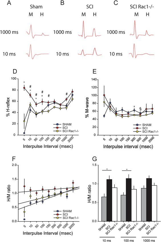
www.nature.com/scientificreports/

                                  Figure 2.  AAV9CMVCre mediated Rac1 KO does not impair recovery after SCI. (A, B) Biomechanical impact data provided by the
                                  Infinite Horizon impactor demonstrates that SCI Rac1−/− and SCI control groups received consistent injuries. (C) Blinded observers
                                  performed BMS testing at baseline, and weekly following SCI (Wk1-Wk4). Both SCI Rac1−/− and SCI control groups recovery equally
                                  over a 4-week period. (D) Representative video-still image of paw prints used for stepping pattern analysis acquired and analysis using
                                  the CatWalkXT (Noldus Information Technology). (E) Intensity plot of the left hind paw (virally injected side) acquired using the
                                  CatWalkXT (Noldus Information Technology) from an SCI animal four weeks post injury. (F) Four weeks after SCI, both SCI Rac1−/−
                                  and SCI control groups have a gait pattern (regularity index) similar to baseline testing. (G) Swing speed of the right (RH) and left
                                  (LH) hind limbs of SCI Rac1−/− and SCI group. Post SCI and viral injection there was no difference between hindlimb swing speed
                                  and compared to baseline. (* = p < 0.05). Graphs are mean ± SEM.

Scientific Reports |   (2021) 11:7838 |                   https://doi.org/10.1038/s41598-021-87476-5                                                                   3

                                                                                                                                                                 Vol.:(0123456789)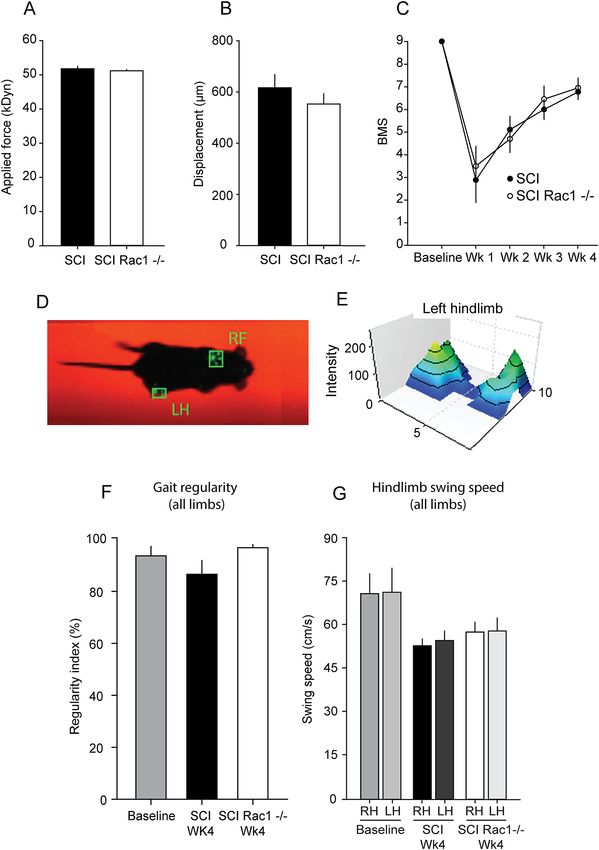
www.nature.com/scientificreports/
Adeno‑associated virus (AAV) intramuscular injection. Commercially available AAV9CMVCre
(Iowa-1066) was purchased from the Gene Transfer Vector Core (lowa University). Intramuscular injections
followed a previously described method25. Briefly, animals were anesthetized with isoflurane 1–3% and the left
soleus and gastrocnemius muscle was exposed by a skin incision. Undiluted 2.5 × 10¹³ vg/ml AAV9CMVCre was
injected into muscle tissue using a Hamilton syringe and 32-gauge 45-degree beveled needle. For optimal motor
neuron infection yield, injections were repeated once daily for 3 days (total volume of 8–10 ul at 5 locations
into the soleus/gastrocnemius tissue). Following injections, the skin incision was closed with 6–0 monofilament
sutures.
EMG H‑reflex testing. We anesthetized animals with an induction dose of ketamine and xylazine
(100/10 mg/kg, i.p.) and maintained on ketamine alone (20 mg/kg, i.p.)26,27. To record electromyogram (EMG)
data, which included the muscle response (M wave) and the monosynaptic reflex (H reflex), we used a percu-
taneous needle preparation5,28–31. This minimally invasive procedure is analogous to methods used to study
H-reflex in humans31,32. This method also permitted longitudinal study and maintained tissue integrity for his-
tology at experimental endpoint.
For stimulation, a pair of Teflon insulated stainless steel wire electrodes (0.002″ diameter; A-M Systems, Inc.,
Carlsborg, WA) were threaded into a 32 G syringe needle. The wire ends were bent into sharp barbs. The insula-
tion was removed with heat (exposed tips ~ 1 mm), and the needle/wire was then transcutaneously inserted with
the tip in close proximity to the tibial nerve. The needle was retracted and the wire remained in place. A second
electrode was inserted similarly ~ 2 mm away from the first e lectrode21,26,27. Stimulating electrode placement was
adjusted until square wave stimulating pulses (0.2 ms duration given at a rate of 1 every 3 s) elicited visible motor
twitch responses, i.e., plantar flexion28,29. To record EMG data (e.g., plantar reflex), an electrode was inserted
into the plantar muscles within the hindpaw palmar/ventral surface, proximal to the ankle. A reference electrode
was placed subcutaneously in the dorsolateral surface of the hind paw. These reflexes were chosen based on our
pilot experiments and previous work that demonstrated EMG reflex response evoked in these muscles could be
reproducibly recorded after SCI33. Moreover, plantar reflexes provide an established read out of reflexes elicited
in other hind limb muscles, i.e., tibialis anterior and gastrocnemius, innervated by L4 and L5 n erves28,29. EMG
responses were filtered, amplified, and analyzed offline using Spike 2 software (version 7.08; CED Software,
Cambridge, UK). Threshold (T) was defined as the minimum intensity required for an M-wave response ~ 50%
of the time. We used a stimulation intensity that produced consistent M- and H-wave responses (~ 1.4–1.8 T).
To measure rate-dependent depression (RDD) of the H-reflex, we applied a paired-pulse stimulation paradigm:
a control pulse and test pulse (0.2msec square) separated by a range of interpulse intervals (10–2000 ms). Three
trials (10 sweeps/trial) were recorded for each interpulse interval. We quantified the M and H wave amplitudes
from rectified and averaged waveforms8,34. For comparisons, the maximum amplitudes of the H and M response
to the test pulse were converted into a percentage of the maximum amplitude response to the control pulse (test/
control × 100). M and H waveforms were measured from baseline-to-peak amplitude. We calculated the H/M
ratio using maximum amplitude of M-wave and H-reflex responses from the test pulse. All H-reflex testing was
performed acutely (i.e., recordings lasted < 1 h per animal), and we ensured that all animals underwent similar
H-reflex testing protocols.
Immunohistochemistry and image analysis. At experimental endpoint, 4-weeks post-SCI/Sham sur-
gery, all mice were euthanized under anesthesia (100/10 mg/kg, i.p.) via intracardial perfusion with ice cold
phosphate buffered saline followed by 4% paraformaldehyde (PFA; 0.01 M PBS). Lumbar enlargement spinal
cord tissue (L4-L5) were removed and post-fixed in 4% PFA at 4 °C overnight. For cryoprotection, we submerged
tissue in 30% sucrose for ~ 48 h. 20 µm thick tissue sections were cut on a cryostat (Leica; Bannockburn, IL)
and mounted on Superfrost + slides (Fisher Scientific; Pittsburg, PA). For immunohistochemistry, sections were
blocked for 1 h at 25 °C in 4% normal donkey serum, 2% bovine serum albumin; 0.1% Triton-X100; 0.02%,
0.01 M PBS. Tissue was incubated in primary antibodies using our published protocols34,35 and included: rabbit
anti-IBA1 1:500 (Abcam, ab178846), chicken anti-GFAP 1:500 (Abcam, ab4674), and rabbit anti-ChAT 1:200
(Millpore, AB143). Secondary antibodies included donkey anti-rabbit 647 (Jackson Labs, 711-496-152) and
donkey anti-chicken 594 (Jackson Lab, 703-585-155). Immunofluorescent Z-stack images were captured with
similar acquisition settings across groups using a confocal microscope (A1R, Nikon).
Tissue image analysis was conducted by blinded investigators using Image J software (National Institutes
of Health free download: http://rsbweb.nih.gov/ij/). All AAV9CMVCre infected neurons expressed tdTomato-
reporter. IBA1 and GFAP expression was analyzed from spinal cord tissue sections with tdTomato-reporter
expressing motor neurons. Z-stack images were compiled into a single plane. Threshold-adjusted levels of IBA1
and GFAP immunoreactivity with subtracted background was compared across SCI groups as a percentage (%)
of positive-pixel area to the total measured area of the ventral horn. AAV infection of lumbar motor neurons
was confirmed by choline acetyltransferse (ChAT) i mmunolabeling36,37. To calculate the yield of viral-infected
motor neurons in the ipsilateral injected and contralateral side, we measured the number tdTomato-expressing
motor neurons co-labelled with ChAT. We report this data as a percentage (%) of the total number of ChAT-
immunopositive motor neurons within the sampled spinal cord tissue sections (n = 10 tissue sections/side).
Dendritic spine analysis. Investigators blind to treatment conditions performed all dendritic spine image
analyses. Florescent tdTomato reporter expression in viral-infected motor neurons permitted the quantification
of dendritic spines. Z-stack image volumes were acquired from tissue sections with an 0.2 microns step size
using a Andor Dragonfly spinning disc confocal microscope (Andor Technology) using Andor iXon Ultra 888
electron multiplying charge coupled camera and Plan Apo Lambda 60 × (NA, 1.4) oil objective (Nikon Instru-
Scientific Reports | (2021) 11:7838 | https://doi.org/10.1038/s41598-021-87476-5 4
Vol:.(1234567890)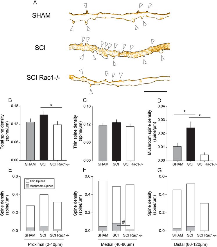
www.nature.com/scientificreports/
ments). To identify α-motor neurons, we followed a screening workflow based on data from our previous study
and others21,33,34,37. Alpha-motor neurons were identified based on their location in ventral horn Rexed lamina
IX, had soma diameters > 25 μm, and cell body cross-sectional areas > 450 μm221. As a refinement step for analy-
sis a priori, we only included α-motor neurons for analysis that had a visible cell body and with greater than one
dendritic branch. Three-dimensional (3D) traces of identified α-motor neurons were generated in Neurolucida
360 with the software’s user-guided tracing protocol (MicroBrightfield, Williston, VT). To ensure consistency,
we traced dendritic branches visibly connected to the motor neuron cell body. To determine if there were any
morphological differences across sampled neurons, we used Neuroexplorer (MicroBrightfield, Williston, VT)
to measure cell body surface area, maximum and minimum cell body diameter, and the total dendritic branch
lengths in each treatment group. We observed no differences in these morphological values across groups.
On 3D reconstructed α-motor neurons, we marked the location of dendritic spines on each dendritic branch.
As previously d escribed21,38, we defined a dendritic spine neck as the structure juxtaposed between the dendrite
branch and the spine head base, which appears as a bulb-shaped structure. Thin- and mushroom-shaped spines
were classified separately: thin spines had head diameters that were less than or equal to the length of the spine
neck, whereas mushroom spines had spine head diameters that were greater than the length of the spine neck.
Classification into only thin- and mushroom-spines allowed us to use simple, but strict rules in classifying spine
morphology. Although this prevented measurement of subtle variations in spine shape, this method permitted
collection of a large sample size. Moreover, the physiological impact of thin and mushroom spine shapes on neu-
ronal and circuit physiology is well-described10,16,39. Dendritic spine density was expressed as spine number per
μm of dendrite length. We used a Sholl analysis to assess changes in dendritic spine distribution relative to the cell
body15,21. Dendritic spine density within proximal (0–40 μm), medial (40–80 μm), and distal (80–120 μm) regions
from the cell body were averaged within each group and compared across groups. To quantify overall dendritic
spine size, we measured spine length and head width. Spine length was defined as the distance from dendrite to
the tip of the spine head. Whereas, spine head width was defined as the maximum diameter of the spine head.
Statistical analysis. All statistical tests were performed at the α-level of significance of 0.05 by two-tailed
analyses using parametric or nonparametric test, as appropriate. Normality assumptions of each data set was
determined using a Shapiro–Wilk test. We used one-way ANOVA or Kruskal–Wallis one-way ANOVA on ranks
followed by post hoc tests to correct for repeated measure error, i.e., Dunn or Bonferroni tests. Data manage-
ment, statistical analyses and graph generation were performed using Sigmaplot 13.0 (Systat) and Microsoft
Office Excel (2018). All data is described as mean ± SEM (graphs and text).
Ethics approval. Experiments were performed in accordance with the National Institutes of Health Guide-
lines for the Care and Use of Laboratory Animals and in compliance with ARRIVE guidelines. All animal proto-
cols were approved by the Yale University/Veterans Affairs Institutional Animal Use Committee.
Consent for publication. All authors consent publication.
Results
Conditional Rac1 knockout does not impair gross locomotor recovery following mild SCI. To
validate equivalent SCI severity across experimental groups (Fig. 1), we analyzed impact data from the Infinite
Horizon device, i.e., applied impact force and maximum spinal cord displacement (Fig. 2A, B). Applied impact
force predicts the amount of tissue sparing in this model, and correlates with locomotor o utcome40. In SCI
cohorts, we observed no difference in applied impact force across control SCI and experimental SCI (Rac−/−)
groups (p > 0.05; SCI vs. SCI Rac−/−; 51.8 ± 0.7 vs. 51.2 ± 0.4 kDyn; ANOVA on ranks) (Fig. 2A). Similarly, we
observed no difference in maximal spinal cord displacement (e.g., maximum depth of the impactor rod dur-
ing contusion) between groups (p > 0.05; SCI vs. SCI Rac1−/−; 616.7 ± 50.8 vs 552.7 ± 40.6; one-way ANOVA)
(Fig. 2B).
To determine whether AAV9CMVCre injection had an effect on locomotor ability, we used the BMS
open-field testing p aradigm24. Following baseline BMS measurements, SCI animals (from both wildtype or
Rac1−/− groups) were scored weekly and the average score in each week compared across groups (Fig. 2C). In
addition, the BMS score for the left and right hindlimbs were averaged for each animal, as there was no statisti-
cal difference between sides. One-week post-SCI, prior to injection of AAV9CMVCre, there was no difference
in BMS scores between SCI groups with animal displaying ankle movement and some non-weight supported
stepping (p > 0.05; SCI vs. SCI Rac1−/− 2.9 ± 1.0 vs 3.5 ± 0.9 BMS Score, Rank Sum Test) (Fig. 2C). Even after
intramuscular injection of AAV9CMVCre, we also observed no effect on gross locomotor function four-weeks
after SCI, with both groups able to perform coordinated stepping (p > 0.05 SCI vs SCI Rac1−/−; 6.8 ± 0.3 vs.
7.0 ± 0.4; one-way ANOVA). In addition, we analyzed the animals stepping pattern (Fig. 2D–G) using the Cat-
Walk system. In CatWalk analyses four-weeks after SCI (Fig. 2D–G), we observed no difference in gait pattern
and hindlimb swing speed, (p > 0.05; regularity index: baseline vs. SCI vs. SCI Rac1−/−, 94.4 ± 3.1 vs. 87.4 ± 4.8
vs. 97.6 ± 0.6; hindlimb swing speed: baseline RH vs. baseline LH vs. SCI RH vs. SCI LH vs. SCI Rac1−/− RH
vs. SCI Rac1−/− LH, 71.2 ± 6.9 vs. 71.7 ± 8.3 vs. 53.2 ± 2.4 vs. 55.0 ± 3.3 vs. 58.0 ± 3.4 vs. 58.3 ± 4.5 cm/s: p > 0.05;
one-way ANOVA with Bonferroni’s post hoc test) (Fig. 2F, G). Indicating that AAV9CMVCre did not have a
lateralized effect on gross motor function.
Intramuscular AAV injection transfects ipsilateral motor neurons. To calculate infection yield of
motor neurons, we measured the number tdTomato-expressing neurons co-labelled with ChAT within the ipsi-
or contralateral spinal cord tissue sections sampled (n = 13 animals; 10 sections/animal) (Fig. 3). Three-weeks
Scientific Reports | (2021) 11:7838 | https://doi.org/10.1038/s41598-021-87476-5 5
Vol.:(0123456789)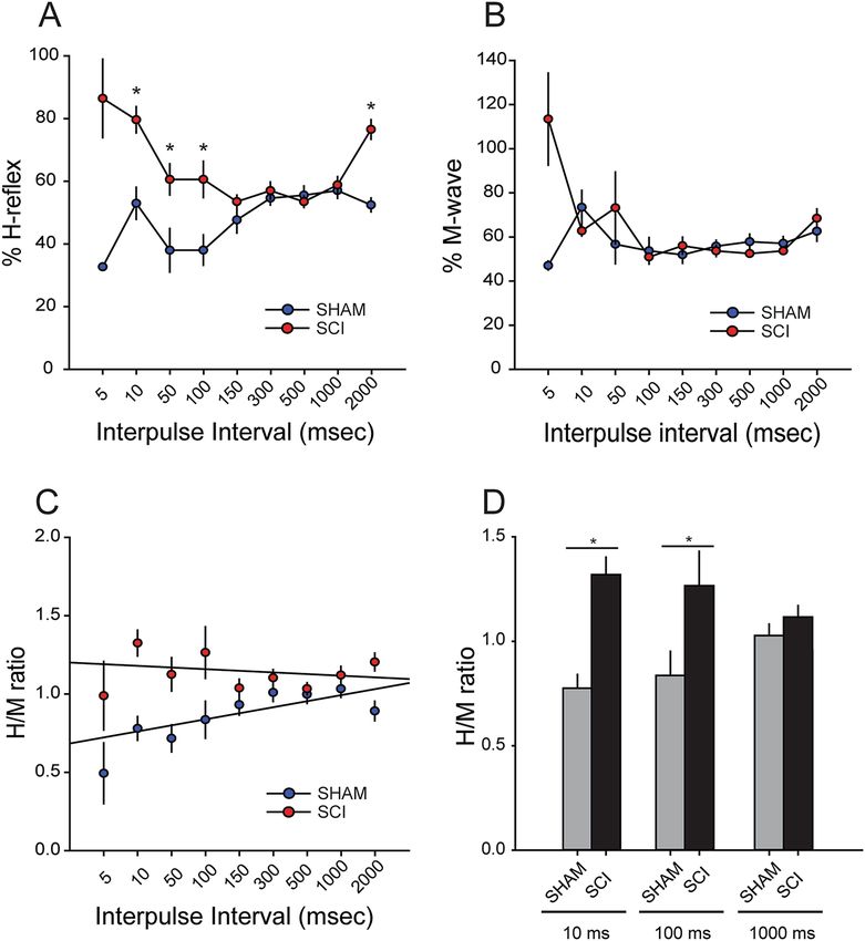
www.nature.com/scientificreports/
Figure 3. Intramuscular injection of AAV9CMVCre leads to expression of tdTomato in ventral spinal cord
motor neurons. (A) ChAT positive motor neurons within lamina IX of the spinal cord. (B) AAV9CMVCre
transfected motor neurons expressing tdTomato. (C) Merged image of (A) and (B) shows colocalization of
ChAT positive neurons and tdTomato expression (arrow shows inset *). (D) The percentage of ChAT labelled
motor neuron expressing tdTomato in the ventral horn of the spinal cord. AAV9CMVCre intramuscular
injection tranfects 51.2% of ChAT-positive neurons in lamina IX on the ipsilateral injected side (Ipsi-). On the
contralateral side (Contra-) there was no co-labelling between ChAT motor neurons and tdTomato expression.
Data shown as scatterplot with mean. Scale bar in (A) = 50 µm and applies to images in (B) and (C).
following intramuscular injections of AAV9CMVCre into the soleus/gastrocnemius tissue, we observed a trans-
duction rate of 51.2% (range 24.7–77.6%) of ChAT positive ventral horn motor neurons in lamina IX (Fig. 3D).
We did not observe any contralateral motor neurons expressing tdTomato reporter with ChAT co-labelling
(Fig. 3D).
Rac1 knockout in spinal motor neurons does not affect the glial inflammatory response. To
determine whether Rac1 KO affected microglia or astrocyte reactivity following injury, we measured expression
levels of IBA1 and GFAP immunoreactivity, e.g., microglia or astrocyte markers, respectively, in the spinal cord
ventral horn (Fig. 4A–F). We observed no difference in the percent area immunolabelled for IBA1 or GFAP
between SCI controls and SCI with conditional Rac1 knockout in motor neurons (P > 0.05; IBA1: SCI vs. SCI
Rac1−/−, 9.0 ± 0.7 vs. 9.0 ± 1.1% area, and GFAP, 15.3 ± 6.5 vs. 17.0 ± 5.6% area; one-way ANOVA) (Fig. 4C, F).
Several studies have reported that intravenously administered AAV9 can infect astrocytes due to bioavailability
through the vascular r oute41,42. However, in pilot studies we calibrated our AAV injection protocol to restrict
viral exposure through low volume intramuscular injections43. As a result, we observed close-to-none GFAP-
positive cells expressing tdTomato (data not shown). Similarly, in agreement with other work, we observed no
overlap between tdTomato expression and IBA1, demonstrating that AAV9CMVCre did not have affinity for
microglia44,45.
Scientific Reports | (2021) 11:7838 | https://doi.org/10.1038/s41598-021-87476-5 6
Vol:.(1234567890)www.nature.com/scientificreports/
Figure 4. Conditional Rac1 KO in alpha-motor neurons did not alter microglia or astrocyte reactivity in the
ventral horn. (A, B) IBA1 immunolabelling for microglia in the ventral horn of the spinal cord in SCI animals
with (A) Rac1 or (B) Rac1 KO in alpha motor neurons. (C) No difference in the percent area labelled for IBA1
between SCI and SCI Rac1−/− groups. (D, E) Image of GFAP immunolabelling for astrocytes in the ventral
horn of the spinal cord from animals with (D) Rac1 or (E) Rac1 KO in alpha motor neurons. (F) There was no
difference in the percent area labelled for GFAP between SCI and SCI Rac1−/− groups. Graphs are Mean ± SEM;
Scale bar in (A) and (D) = 50 µm and applies to images in (B), and (E).
Viral‑mediated Rac1 knockout reduces H‑reflex hyperexcitability after SCI. To determine
whether Rac1 disruption could reduce hyperreflexia after SCI, we performed longitudinal EMG recordings to
measure changes in evoked M- and H-reflex response. Under normal conditions, evoked H-reflex response
exhibits rate-dependent depression (RDD), whereby increased activity reduces the overall reflex output. In dis-
eases and injuries associated with spasticity, however, H-reflex exhibits a loss of RDD8,26,29. In SCI, reduced
H-reflex RDD (physiologically measured through %H-reflex response and H/M ratio) is an electrophysiological
and clinical diagnosis of spasticity6,46–48.
To measure H-reflex and M-wave responses, we stimulated the tibial branch of the sciatic nerve and recorded
from the plantar muscle21. To elicit the H-reflex, we used a paired-pulse stimulation protocol consisting of a
control and test pulse, separated by a range of interpulse intervals (5–2000 ms). To quantify evoked H-reflex
response, we calculated the %H reflex in each group by measured and normalized the maximum amplitude of
the H-reflex response (evoked from the test pulse) with the maximum amplitude of the H-reflex (evoked by the
control pulse) (Fig. 5D).
One week after SCI or Sham, and before AAV9CMVCre injection (see design; Fig. 1), mice underwent EMG
testing to confirm the presence of hyperreflexia, e.g., loss of RDD. As expected, SCI resulted in a loss in RDD
compared to Sham (Fig. 5). We observed an increase in %H-reflex in SCI mice at 10, 50, 100, 2000 ms interpulse
intervals compared to Sham, representing a loss of RDD and increased spinal reflex excitability (P < 0.05; sham
vs. SCI at 10 ms 53.1 ± 5.1 vs. 79.8 ± 4.2%; at 50 ms: 38.1 ± 7.0 vs. 60.8 ± 5.0%; at 100 ms: 38.2 ± 4.9 vs. 60.8 ± 5.8%;
2000 ms: 52.6 ± 2.2 vs. 76.8 ± 3.2; one-way ANOVA with Bonferroni’s post hoc test) (Fig. 5A). There was no differ-
ence in %M-wave at any interpulse interval between Sham and SCI animals 1 week after surgery (sham vs. SCI; at
5 ms: 32.8 vs. 86.7%; at 10 ms: 73.4 ± 7.7 vs. 62.8 ± 2.3; at 50 ms: 56.8 ± 8.8 vs. 73.2 ± 16.3; at 100 ms: 53.7 ± 6.0 vs.
50.9 ± 1.3; at 150 ms: 52.0 ± 3.8 vs. 56.0 ± 4.0; at 300 ms: 56.0 ± 2.7 vs. 53.7 ± 2.5; at 500 ms: 57.8 ± 3.4 vs. 52.5 ± 1.0;
at 1000 ms 57.0 ± 3.1 vs. 53.7 ± 1.3; at 2000 ms 62.6 ± 4.5 vs. 68.4 ± 4.2; one-way ANOVA with Bonferroni’s post
hoc test)(Fig. 5B). Across the range of paired interpulse intervals, H/M ratio in Sham produced a steeper slope
than compared with SCI animals (linear regression slope value, 0.04 vs. -0.01, respectively), demonstrating
an increase in post-SCI reflex excitability (Fig. 5C). Additionally, SCI animals had a statistically greater H/M
ratios at the 10 and 100 ms interpulse intervals (P < 0.05; Sham vs. SCI; 10 ms; 0.79 ± 0.08 vs. 1.32 ± 0.08; 100 ms:
Scientific Reports | (2021) 11:7838 | https://doi.org/10.1038/s41598-021-87476-5 7
Vol.:(0123456789)www.nature.com/scientificreports/
Figure 5. Enhanced H-reflex response 1-week after SCI. To confirm the development of hyperreflexia after SCI,
we measured the H-reflex response using a paired-pulse paradigm (stimulating interpulse intervals 5–2000 ms).
(A, B) % H-reflex and % M-wave are normalized values of the test pulse compared to the control pulse. (A) In
Sham animals, shorter interpulse intervals are associated with less %H-reflex, demonstrating RDD. After SCI
there was significant increase in the %H-reflex at 10, 50, 100 and 2000 ms interpulse intervals (* = p < 0.05). (B)
There was no significant difference in %M-wave response between Sham and SCI. (C) H/M ratio was calculated
by comparing peak amplitude of the test pulse evoked H- and M-wave response. H/M ratio in Sham produced a
greater linear regression slope as compared to SCI, e.g., shallow slope. (D) SCI significantly increased the H/M
ratio at 10 and 100 ms compared to Sham (* = p < 0.05). Graphs are mean ± SEM.
0.8 ± 0.1 vs. 1.3 ± 0.2; 1000 ms: 1.03 ± 0.05 vs. 1.12 ± 0.06 H/M ratio; ANOVA on ranks) (Fig. 5C, D). Importantly,
we did not observe differences in H-reflex function in SCI animals between either wild-type or “floxed” Rac1
animals (prior to AAV9CMVCre injection) (data not shown). This confirmed that transgenic “floxed” Rac1 mice
develop a similar hyperreflexia profile to wild-type animals following SCI (i.e., in the absence of cre-recombinase
expression).
To assess the effect of conditional Rac1 knockout in alpha-motor neurons, we tested H-reflex responses in the
same animals at 3 weeks after intramuscular injection of AAV9CMVCre (Fig. 6). At this post-injection timepoint,
control SCI animals continued to display an exaggerated %H-reflex at the shorter 5, 10, 50 and 100 ms interpulse
intervals compared to Sham (Fig. 6D). In contrast, 3 weeks after AAV9CMVCre injection in mice with floxed
Rac1, EMG recordings demonstrated closer-to-normal %H-reflex that were similar to Sham, indicating a par-
tial restoration of RDD and attenuated hyperreflexia (p < 0.05; Sham vs. SCI vs. SCI Rac1−/−; 5 ms: 3.4 ± 0.4 vs.
83.9 ± 5.0 vs. 24.7 ± 13.1; 10 ms: 44.5 ± 4.0 vs. 71.0 ± 4.2 vs. 53.7 ± 4.1; 50 ms: 36.4 ± 4.9 vs. 54.6 ± 4.1 vs. 38.3 ± 4.0;
100 ms: 42.1 ± 2.3 vs. 59.2 ± 5.3 vs. 41.5 ± 4.2%; 150 ms: 53.2 ± 3.7 vs. 57.9 ± 3.3 vs. 42.3 ± 2.6; 1000 ms: 52.2 ± 4.0
vs. 63.5 ± 3.0 vs. 48.5 ± 3.6 H-reflex; one-way ANOVA with Bonferroni’s post hoc) (Fig. 6D). %M-wave was
similar across groups, with only minor differences at 300 ms and 500 ms interval (p < 0.05; Sham vs. SCI vs. SCI
Scientific Reports | (2021) 11:7838 | https://doi.org/10.1038/s41598-021-87476-5 8
Vol:.(1234567890)www.nature.com/scientificreports/
Rac1−/−; 300 ms: 62.8 ± 5.1 vs. 51.0 ± 1.2 vs. 50.5 ± 1.8; 500 ms: 55.5 ± 4.4 vs. 52.4 ± 1.1 vs. 48.4 ± 1.8%M-wave;
ANOVA on ranks with Dunn’s post hoc) (Fig. 6E).
In measures of H/M ratio, we continued to observe the loss of RDD in control wildtype SCI animals 3-weeks
post-AAV9CMVCre injection, as compared with Sham (linear regression slope, 0.05 vs 0.08, respectively)
(Fig. 6F). In contrast, conditional Rac1 KO with AAV9CMVCre injection in SCI animals resulted in a sharper
H/M ratio slope, similar to Sham (linear regression slope, 0.05). Specifically, the H/M ratio at 10, 100 and 1000 ms
interpulse intervals revealed that SCI in wildtype controls resulted in a significant increase in H/M ratio that
was partly restored by conditional Rac1 KO in spinal motor neurons (p < 0.05; Sham vs. SCI vs. SCI Rac1−/−;
10 ms: 0.7 ± 0.1 vs. 1.1 ± 0.1 vs. 0.9 ± 0.1; 100 ms: 0.7 ± 0.1 vs. 1.1 ± 0.1 vs. 0.9 ± 0.1; 1000 ms: 0.9 ± 0.1 vs. 1.1 ± 0.1
vs. 1.0 ± 0.1 H/M ratio; one-way ANOVA with Bonferroni’s post hoc test) (Fig. 6F, G).
Conditional Rac1 knockout reduces dendritic spine dysgenesis in alpha‑motor neurons. Den-
dritic spine dysgenesis accompanies hyperexcitability disorders, e.g., pain and spasticity, as a result of injury
and disease17,21,49–51. To assess the development of dendritic spine dysgenesis, we profiled dendritic spines in
AAV9CMVCre-infected motor neurons expressing fluorescent tdTomato reporter. A total of 38 alpha-motor
neurons were identified and sampled using inclusion criteria and reconstructed in the Neurolucida 360 environ-
ment (see Methods). To ensure equivalent sampling across treatment groups, we compared other cellular mor-
phologies, which showed no differences across groups (p > 0.05; Sham vs. SCI vs. SCI Rac1−/−, cell body area:
580 ± 84.6 vs. 684.8 ± 70.6 vs. 544.4 ± 49.6 µm2; cell body max diameter: 39.1 ± 2.6 vs. 40.5 ± 3.0 vs. 35.8 ± 2.1 µm;
cell body min diameter: 21.4 ± 2.3 vs. 25.8 ± 1.4 vs. 22.1 ± 1.1 µm; total dendritic length: 230.3 ± 17.0 vs. 373.2 ± 55.5
vs. 216.9 ± 34.6 µm; ANOVA on ranks with Dunn’s post hoc) (Table 1). Thus, any changes in dendritic spine mor-
phology were likely not due to differences in sampling. At final endpoint, 4-weeks post-SCI or Sham surgery, a
total of 1,518 dendritic spines were measured and analyzed by blinded investigators (Sham, n = 344 spines; SCI,
n = 806; SCI Rac1−/−, n = 368 spines) (Table 1).
We analyzed the density of total-, thin- and mushroom-shape dendritic spines in alpha-motor neurons of
SHAM, SCI and SCI Rac1−/− animals (Fig. 7A). Although SCI did not significantly change total spine den-
sity as compared with Sham (p > 0.05; Sham vs. SCI; 0.1 ± 0.01 vs. 0.2 ± 0.01 spines/µm; one-way ANOVA),
viral-mediated conditional Rac1 KO in SCI animals significantly decreased total spine density in alpha-motor
neurons (P < 0.05; SCI vs. Rac1−/−; 0.2 ± 0.01 vs. 0.1 ± 0.01 total spines/µm; one-way ANOVA with Dunn’s post
hoc test) (Fig. 7B). We observed no change in thin-shaped spine density across any comparator group (p > 0.05;
sham vs. SCI vs. Rac1−/−; 0.12 ± 0.01 vs. 0.13 ± 0.01 vs. 0.11 ± 0.01 thin spines/µm; one-way ANOVA) (Fig. 7C).
Mushroom-shaped spine density significantly increased following SCI as compared with Sham or conditional
Rac1 KO SCI animals (p < 0.05; Sham vs. SCI; 0.01 ± 0.002 vs. 0.02 ± 0.004 mushroom spines/µm; SCI vs. Rac1−/−:
0.02 ± 0.004 vs. 0.004 ± 0.002 mushroom spines/µm; one-way ANOVA with Dunn’s post hoc test) (Fig. 7D).
To investigate changes in the spatial distribution of dendritic spines, we used a Sholl analysis and compared
spine densities at three distinct regional distances relative to the soma: proximal (0–40 µm), medial (40–80 µm)
and distal (80–120 µm) (Fig. 7E–G). Here, we observed no difference in the spatial distribution of total spine
densities (p > 0.05; total spine density, Sham vs. SCI vs. SCI Rac1−/−, proximal: 0.28 ± 0.05 vs. 0.40 ± 0.06 vs.
0.32 ± 0.04 total spines/µm; medial: 0.55 ± 0.07 vs. 0.48 ± 0.07 vs. 0.51 ± 0.06 total spines/µm; distal: 0.45 ± 0.1
vs. 0.52 ± 0.1 vs. 0.31 ± 0.08 total spine/µm; one-way ANOVA), or thin-shaped spine densities in the proximal,
medial, or distal regions (p > 0.05; thin spine density; Sham vs. SCI vs. SCI Rac1−/−; proximal: 0.25 ± 0.05 vs.
0.35 ± 0.06 vs. 0.30 ± 0.03 thin spines/µm; medial: 0.50 ± 0.07 vs. 0.40 ± 0.07 vs. 0.50 ± 0.06 thin spines/µm; distal:
0.4 ± 0.1 vs. 0.4 ± 0.1 vs. 0.3 ± 0.1 thin spines/µm; one-way ANOVA) (Fig. 7E–G). Interestingly, mushroom-shaped
spine densities increased in the medial region (i.e., 40–80 µm dendritic region relative to the soma) after SCI
(Fig. 7F). Within this region, AAV9CMVCre infected alpha-motor neurons had significantly decreased mush-
room spine density as compared to neurons in control SCI wildtype (p < 0.05; Sham vs. SCI vs. Rac1−/−, medial:
0.04 ± 0.01 vs. 0.08 ± 0.02 vs. 0.01 ± 0.01 mushroom spines/µm; ANOVA on ranks with Dunn’s post hoc) (Fig. 7F).
Dendritic spine size and shape regulate synaptic efficacy and can strongly influence neuronal excitability16,52,53.
To determine whether conditional Rac1 KO could reverse SCI-induced changes in dendritic spine size, we meas-
ured spine length and head width of 1,518 spines from 38 neurons across Sham, SCI, and SCI Rac1−/− groups.
As shown in Fig. 8, SCI increased the length of total- and thin-shaped spines, as compared with Sham (p < 0.05;
Sham vs. SCI vs. SCI Rac1−/−, total spines 1.07 ± 0.05 vs. 1.6 ± 0.1 vs. 1.2 ± 0.1 µm; thin spines: 1.04 ± 0.05 vs.
1.6 ± 0.1 vs. 1.2 ± 0.1 µm; ANOVA on ranks with Dunn’s post hoc) (Fig. 8A, B). Conditional Rac1 KO resulted
in no difference in the length of total- and thin-shaped spines as compared to Sham (Fig. 8A, B). On the other
hands, conditional Rac1 KO significantly reduced in the length of mushroom-shaped spines as compared to
control SCI (p < 0.05; Sham vs. SCI vs. SCI Rac1−/−, mushroom spines: 1.4 ± 0.1 vs. 1.9 ± 0.2 vs. 1.0 ± 0.2 µm; one-
way ANOVA with Bonferroni post hoc) (Fig. 8C). In control SCI, spine head width for total- and thin-shaped
spines increased compared to Sham (Fig. 8D, E). In contrast, conditional Rac1 KO in motor neurons decreased
spine head width associated with SCI for total-spines, thin-, and mushroom-shaped spines (p < 0.05; Sham vs.
SCI vs. SCI Rac1−/−, total spines: 0.64 ± 0.04 vs. 1.2 ± 0.1 vs. 0.65 ± 0.05 µm; thin spines: 0.58 ± 0.03 vs. 1.1 ± 0.1
vs. 0.64 ± 0.05 µm; mushroom spines: 1.2 ± 0.2 vs. 1.7 ± 0.1 vs. 0.7 ± 0.1; one-way ANOVA with Bonferroni post
hoc) (Fig. 8 D-F).
Discussion
Emerging evidence from our group and others has revealed a common structural motif of dendritic spine dys-
genesis associated with neuronal hyperexcitability after SCI and nerve injury14,15,21,54,55. In nociceptive dorsal
ain14,15,55–57. In the present study, we report the first
horn neurons, this motif is conserved in multiple models of p
Scientific Reports | (2021) 11:7838 | https://doi.org/10.1038/s41598-021-87476-5 9
Vol.:(0123456789)www.nature.com/scientificreports/
Figure 6. Disruption of Rac1 in alpha-motor neurons attenuates hyperreflexia after SCI. Representative test ▸
pulse traces of stim evoked H- and M-wave responses in (A) Sham, (B) SCI, and (C) SCI Rac1−/−. (A) Note
that in Sham animals, as the interpulse intervals increases (10–1000 ms) between the test and control pulse,
the amplitude of the H-response increase. (B) In contrast, SCI produces an exaggerated H-response at short
(10 ms) interpulse intervals. (C) SCI Rac1−/− restores H-response depression at the short (10 ms) interpulse
intervals. (D) After SCI there was a significant increase in %H-reflex at 5, 10, 50 and 100 ms interpulse intervals
compared to Sham, indicating a loss of RDD (* = p < 0.05). SCI Rac1−/− animals had reduced %H-reflex at 10,
50, 100, 150 and 1000 ms interpulse intervals compared to control SCI animals, suggesting a restoration of RDD
(# = p < 0.05). (E) %M-wave was mostly similar across the three groups with small, but significant differences
between Sham and SCI at 300 ms interpulse interval (* = p < 0.05) and between Sham and SCI Rac1−/− at
500 ms interpulse interval (§ = p < 0.05). (F) SCI increased H/M ratio as demonstrated as stable linear trend line.
Whereas, SCI Rac1−/− animals displayed a closer-to-normal H/M ratio, with a steeper linear trend line. G) SCI
increased the H/M ratio at 10 and 100 ms compared to sham, indicating a loss of RDD (* = p < 0.05). Graphs are
mean ± SEM.
evidence demonstrating that viral-mediated conditional Rac1 knockout reduces dendritic spine dysgenesis in
alpha-motor neurons and attenuates reflex hyperexcitability associated with spasticity after SCI.
Spasticity after SCI is a symptom of over-activity within the spinal stretch reflex circuit, e.g., H-reflex, and has
been investigated in multiple SCI m odels6,58. To investigate the relationship between dendritic spine morphology
and spasticity, we assessed the efficacy of disrupting Rac1 expression in spinal cord motor neurons after SCI.
We performed a contusion SCI at spinal segment L2, a location that may replicate human lower thoracic-level
SCI, and permits H-reflex testing of hindlimb m usculature1,6,59. All animals with SCI presented with spastic-
ity one-week after injury, exhibiting reduced RDD and increased evoked H-reflex responsiveness. Following
viral-mediated expression of Cre recombinase in Rac1 “floxed” mice, we observed a reduction of hyperreflexia
following SCI. Analysis of infected motor neurons expressing tdTomato reporter demonstrated that conditional
Rac1 knockout also reduced overall dendritic spine density, specifically mushroom spine density, attenuated
spine length and head width; and partially reversed abnormal spine distribution. Collectively, these anatomical
findings demonstrate that Rac1 in spinal cord alpha-motor neurons contribute to dendritic spine dysgenesis
associated with SCI-induced hyperreflexia.
Dendritic spine morphology is related to synaptic and neuronal activity and thus can provide a visual read-
out of neuronal f unction11,60. In healthy humans and rodents adaptive plasticity between sensory afferents and
motor neurons can influence H-reflex f unction46,61, however after spinal cord injury maladaptive plasticity can
contribute to uncontrolled H-reflex activity and s pasticity3,6. In agreement, we observed SCI-induced changes
in the density of thin and mushroom shaped dendritic spine on α-motor neurons four-weeks after injury, this
was accompanied by loss of RDD and an increase in % H-reflex at short interpulse intervals. Importantly, SCI
induced minimal changes in M-wave response, indicating that abnormal RDD and H/M ratios were largely due
to mechanisms related to the dysfunction in the motor reflex monosynaptic circuit. Our data further confirms
previous evidence in multiple disease models that dendritic spine dysgenesis can serve as a morphological cor-
relate of dysfunctional neuronal a ctivity16,21,51,55,62.
Our findings raise the question of how altered dendritic spine morphologies after SCI could directly contrib-
ute to hyperexcitable spinal reflex function. Interestingly, the monosynaptic spinal stretch reflex (i.e., H-reflex)
operates through classical operant conditioning principles, e.g., a “memory engram” that regulates spinal reflex
function63–65. In this context, the relationship between dendritic spine morphology and postsynaptic excitability
has been well-studied in the field of learning and memory66. Following long-term potentiation (LTP) induction
in hippocampal CA3–CA1 synapses, postsynaptic neurons exhibit an increase in dendritic spine number and
volume. These late-phase LTP structural changes contribute to enhancing synaptic excitability through ampli-
fied excitatory glutamatergic transmission, e.g., more or larger spines improve postsynaptic AMPA receptor
availability67,68. In the spinal cord, we observed an increase in more stable, mature spine profiles in alpha-motor
neurons through an increase in mushroom-shaped spine density, as well as an increase in spine head size. As
we and others have shown, more stable mushroom-shaped spine morphologies can have a greater impact on
neuronal excitability as compared with thin-shaped spines16,66. Additionally, larger mature spines may contrib-
ute increased input discretization by narrowing EPSP waveforms, and increase synaptic transmission fidelity at
higher rates of activity69,70. The added capability to transmit excitatory potentials at higher frequency with greater
reliability could facilitate supra- and sub-threshold temporal summation71–74. Taken together, the biophysical
properties associated with the dendritic spine changes we observed may explain the loss the H-reflex RDD and
upshift in the H/M ratio associated with spasticity after injury.
This study selectively targeted Rac1 deletion in motor neurons via an intramuscular AAV-Cre injection route,
e.g., reducing viral bioavailability to other CNS tissues. However, given that Rac1 contributes to regulating a mul-
titude of intracellular molecular signaling pathways, we cannot entirely preclude off-target effects. Notably, Rac1
function is controlled by post-translational modification, such as prenylation and SUMOylation, which affect
Rac1 localization to specific subcellular compartments75,76. In addition to actin cytoskeleton reorganization, Rac1
activity can also influence the spinal excitability through altered gene expression, production of reactive oxygen
species (ROS), e.g., which can influence synaptic transmission, and inflammatory r esponses77–79. Thus, in our
present study, Rac1 disruption could have indirectly affected reflex excitability along with structural changes to
dendritic spine morphology. This does raise a related question as to whether these cellular reactions to injury act
synergistically. For example, inhibition of Rac1 in diabetic focal cerebral ischemia led to neuroprotection, through
Scientific Reports | (2021) 11:7838 | https://doi.org/10.1038/s41598-021-87476-5 10
Vol:.(1234567890)www.nature.com/scientificreports/
Scientific Reports | (2021) 11:7838 | https://doi.org/10.1038/s41598-021-87476-5 11
Vol.:(0123456789)www.nature.com/scientificreports/
Total dendritic diameter
Cell body area (µm2) Cell body max diameter (µm) Cell body min diameter (µm) (µm)
Sham 580 ± 84.6 39.1 ± 2.6 21.4 ± 2.3 230.3 ± 17.0
SCI 684.8 ± 70.6 40.5 ± 3.0 25.8 ± 1.4 374.2 ± 55.5
SCI Rac1−/− 544.4 ± 49.6 35.8 ± 2.1 22.1 ± 1.1 216.9 ± 34.6
Table 1. Cell body dimensions and dendritic branch length of sampled tdTomato expressing spinal motor
neurons.
reduced ROS and fewer Bcl-2/Rac1 mitochondrial complexes79. Suffice it to say, our work provides a compel-
ling opportunity to further investigate the link between Rac1 activity and motor neuron excitability after SCI.
Following SCI, we observed an increase in spine size and density. These changes represent a common motif of
dendritic spine dysgenesis associated with a neuronal hyperexcitability, in this case, the symptom of s pasticity14,55.
Multiple factors contribute to spine dysgenesis after SCI: inflammation, increased glutamate, abnormal glia
response21,80,81. By targeting Rac1, we can attenuate abnormal spine remodeling regardless of the upstream mecha-
nism. Rac1 activity is well known to control the formation and maintenance of dendritic spines. For example,
in vitro experiments show that expression a dominant-negative form of Rac1 results in spine elimination, while
constitutively active Rac1 increases spine d ensity82,83. This fits with our observation that the deletion of Rac1
in motor neurons reduces the total- and mushroom spine density compared to SCI controls. In addition, Rac1
deletion reduced the size of both thin and mushroom shaped spines after SCI. This effect may be related to the
role of Rac1 in actin cytoskeleton rearrangement: Rac1 regulates the actin nucleator actin-related protein 2/3
(Arp2/3) to drive Arp2/3 through the WAVE protein complex, which is necessary for activity dependent spine
growth84,85. Finally, reduced spine size may be related to the role of Rac1 in LTP. For example, the LTP induction
leads to subsequent Rac1 activation and dendritic spine g rowth86. Rac1 KO in motor neurons thus may inhibit
maladaptive synaptic plasticity, e.g., L TP18, and abnormal spine morphology thereby reduce evoked reflex excit-
ability following SCI.
This study provides the first reported evidence demonstrating that a viral-mediated conditional knockout
model can significantly affect motor neuron dendritic spine structure in the injured spinal cord. We targeted
Rac1 in spinal cord motor neurons using a conditional cre-loxp system with transgenic “floxed” Rac1 mice and
intramuscular injection of AAV9CMVCre. Rac1 activity controls actin stability involved in the formation and
stabilization of dendritic spine structure through the Rac1/Cdc42-PAK p athway82,87–89. In our study, a minimally
invasive AAV injections into hindlimb muscle restricted infection to the ipsilateral innervating motor neurons
and did not appear to affect non-neuronal cells, e.g., astrocytes or microglia, in the ventral h orn43. This is in line
with previous studies showing that AAV vectors can selectively infect neuronal tissue without damage to the
CNS or contribute to chronic inflammation, e.g., low immunogenicity90–93. Importantly, viral-mediated Rac1
cre-loxp knockout in approximately 50% of L4 ventral horn alpha-motor neurons was sufficient to partially
restore normal evoked H-reflex response after SCI. Although it is possible that viral injection procedures led to
sensitization of sensory afferents within the spinal reflex pathway, we observed no difference across SCI groups
in CatWalk sensory-motor outcomes. Although we have shown that intrathecally administrated doses of a phar-
macological Rac1 inhibitor, NSC23766, can reduce H-reflex excitability in SCI animals with spasticity21, small
molecule inhibitors have limited empirical and clinical utility. Taken together, our findings provide a unique
basis for future studies aimed at developing a translational gene therapy by targeting the Rac1 pathway, perhaps
as others have similarly done with virally-delivered miRNA knockdown constructs driven with motor neuron
specific promoters94,95.
In summary, this study demonstrates that viral-mediated conditional Rac1 disruption in ventral horn motor
neurons reduces abnormal dendritic spine dysgenesis and attenuates hyperreflexia associated with spasticity
after SCI. Combined with our previous work17,21,49,51, these findings further support the emerging principle that
dendritic spine dysgenesis is a morphological correlate of spinal cord hyperexcitability disorder, and provides
an opportunity to explore strategies, including gene therapy approaches targeting dendritic spine dysgenesis, to
correct abnormal reflex function after spinal cord injury.
Scientific Reports | (2021) 11:7838 | https://doi.org/10.1038/s41598-021-87476-5 12
Vol:.(1234567890)www.nature.com/scientificreports/
Figure 7. Conditional Rac1 KO in alpha-motor neurons reduces abnormal dendritic spine morphology
associated with SCI and hyperreflexia. Analysis of dendritic spine profiles reveals differences in (B–D) spine
density and (E–F) distribution. (A) Reconstructed dendritic segments from tdTomato filled spinal motor
neurons showing apparent differences in dendritic spine profile between Sham, SCI and SCI Rac1−/− (arrow
denotes spine). (B) Total spine density, which includes all spines, was significantly lower in the SCI Rac1−/−
compared to control (* = p < 0.05). (C) There was no difference in the density of thin-shaped spines between
groups. (D) SCI induced a significant increase in mushroom spine density compared to Sham (* = p < 0.05). In
contrast, SCI Rac1−/− reduced mushroom spine density compared to SCI (* = p < 0.05). (E–G) Assessment of
dendritic spine density within the (E) proximal (0–40 µm), (F) medial (40–80 µm) and (G) distal (80–120 µm)
dendritic branches of tdTomato filled motor neurons. (E, G) Dendritic spine density was not different within
the proximal or distal region between Sham, SCI and SCI Rac1−/−. (F) SCI induced an increase in mushroom
spine density in the medial region, which was not observed in SCI Rac1−/− animals (# = p < 0.05). Graphs are
mean ± SEM. Scale bar in (A) = 10 µm.
Scientific Reports | (2021) 11:7838 | https://doi.org/10.1038/s41598-021-87476-5 13
Vol.:(0123456789)www.nature.com/scientificreports/
Figure 8. Conditional Rac1 KO normalizes dendritic shape on alpha-motor neurons after SCI. Analysis
if dendritic spine (A–C) length and (D–F) spine head width. (A, B) SCI increased the length of total (all
spines) and thin-shaped spine compared to Sham (* = p < 0.05). (C) Mushroom-shaped dendritic spines were
significantly shorter in SCI Rac1−/− compared to control SCI (* = p < 0.05). (D, E) SCI resulted in a significant
increase in spine head width for total and thin-shaped spines, which was not seen in the SCI Rac1−/− group
(* = p < 0.05). (F) SCI Rac1−/− decreased the width mushroom-shaped spines compared to control SCI
(* = p < 0.05). Graphs are mean ± SEM.
Data availability
Data is available upon written request to Dr. Andrew Tan.
Code availability
Not applicable.
Received: 21 December 2020; Accepted: 30 March 2021
References
1. Skold, C., Levi, R. & Seiger, A. Spasticity after traumatic spinal cord injury: nature, severity, and location. Arch. Phys. Med. Rehabil.
80, 1548–1557 (1999).
2. Walter, J. S. et al. A database of self-reported secondary medical problems among VA spinal cord injury patients: its role in clinical
care and management. J. Rehabil. Res. Dev. 39, 53–61 (2002).
3. Lance, J. W. In Spasticity: Disordered Motor Control (eds Feldman, R. G. et al.) 485–494 (Year Book Medical, 1980).
4. Bennett, D. J. et al. Spasticity in rats with sacral spinal cord injury. J. Neurotrauma 16, 69–84. https://doi.org/10.1089/neu.1999.
16.69 (1999).
5. Boulenguez, P. & Vinay, L. Strategies to restore motor functions after spinal cord injury. Curr. Opin. Neurobiol. 19, 587–600. https://
doi.org/10.1016/j.conb.2009.10.005 (2009).
6. Nielsen, J. B., Crone, C. & Hultborn, H. The spinal pathophysiology of spasticity—from a basic science point of view. Acta Physiol.
(Oxf.) 189, 171–180. https://doi.org/10.1111/j.1748-1716.2006.01652.x (2007).
7. Hunanyan, A. S., Petrosyan, H. A., Alessi, V. & Arvanian, V. L. Combination of chondroitinase ABC and AAV-NT3 promotes
neural plasticity at descending spinal pathways after thoracic contusion in rats. J. Neurophysiol. 110, 1782–1792. https://doi.org/
10.1152/jn.00427.2013 (2013).
8. Boulenguez, P. et al. Down-regulation of the potassium-chloride cotransporter KCC2 contributes to spasticity after spinal cord
injury. Nat. Med. 16, 302–307. https://doi.org/10.1038/nm.2107 (2010).
9. Fouad, K., Bennett, D. J., Vavrek, R. & Blesch, A. Long-term viral brain-derived neurotrophic factor delivery promotes spasticity
in rats with a cervical spinal cord hemisection. Front Neurol 4, 187. https://doi.org/10.3389/fneur.2013.00187 (2013).
10. Bourne, J. & Harris, K. M. Do thin spines learn to be mushroom spines that remember?. Curr. Opin. Neurobiol. 17, 381–386. https://
doi.org/10.1016/j.conb.2007.04.009 (2007).
11. Calabrese, B., Wilson, M. S. & Halpain, S. Development and regulation of dendritic spine synapses. Physiology (Bethesda) 21,
38–47. https://doi.org/10.1152/physiol.00042.2005 (2006).
12. Pongracz, F. The function of dendritic spines: a theoretical study. Neuroscience 15, 933–946 (1985).
13. Segev, I. & Rall, W. Computational study of an excitable dendritic spine. J. Neurophysiol. 60, 499–523 (1988).
14. Tan, A. M. et al. Burn injury-induced mechanical allodynia is maintained by Rac1-regulated dendritic spine dysgenesis. Exp.
Neurol. 248, 509–519. https://doi.org/10.1016/j.expneurol.2013.07.017 (2013).
Scientific Reports | (2021) 11:7838 | https://doi.org/10.1038/s41598-021-87476-5 14
Vol:.(1234567890)You can also read