Exosomes and Melatonin: Where Their Destinies Intersect - Frontiers
←
→
Page content transcription
If your browser does not render page correctly, please read the page content below
REVIEW
                                                                                                                                                  published: 11 June 2021
                                                                                                                                         doi: 10.3389/fimmu.2021.692022

                                            Exosomes and Melatonin:
                                            Where Their Destinies Intersect
                                            Adriana Alonso Novais 1, Luiz Gustavo de Almeida Chuffa 2,
                                            Débora Aparecida Pires de Campos Zuccari 3* and Russel J. Reiter 4*
                                            1 Health Sciences Institute (ICS), Mato Grosso Federal University (UFMT), Sinop, Brazil, 2 Department of Structural and

                                            Functional Biology, Institute of Biosciences, São Paulo State University (UNESP), Botucatu, Brazil, 3 Department of Molecular
                                            Biology, Cancer Molecular Research Laboratory (LIMC), São José do Rio Preto Medicine School (FAMERP), São José do
                                            Rio Preto, Brazil, 4 Department of Cell Systems and Anatomy, University of Texas (UT) Health, San Antonio, TX, United States

                                            Cell-to-cell communication is a broad and complex process associated with regular
                                            stimuli to maintain healthy cell interactions. One of the agents capable of cellular
                                            communication is known as an exosome, a subset of extracellular vesicles (EVs)
                                            released by the cell membrane. The exosome contains a wide range of functional
                           Edited by:
                                            proteins, mRNAs and miRNAs, which have the potential to interact with healthy or
                        Benjamin Frey,      diseased cells in the body. On the other hand, melatonin also acts as a cellular
University Hospital Erlangen, Germany
                                            communicator, produced and released by the pineal gland in a circadian way and also,
                        Reviewed by:
                                            non-circadian melatonin is derived from the mitochondria of all normal cells. In addition to
                          Yvette Wooff,
Australian National University, Australia   exhibiting antioxidant, anti-inflammatory, anti-tumor and anti-aging activities, melatonin
                    George Anderson,        has recently been studied by its influence on exosomes. This review summarizes the
             CRC Scotland & London,
                      United Kingdom        relationship between exosomes and melatonin in various pathological processes. There is
                  *Correspondence:          robust evidence that their combination ameliorates inflammation, ischemia-reperfusion
              Débora Aparecida Pires       injury, hepatic metabolic disturbance, cancer immunosuppression status, degenerative
                  de Campos Zuccari
                                            processes like chronic kidney disease, vascular calcification, ageing, ischemic brain injury,
            debora.zuccari@famerp.br
                       Russel J. Reiter     neurodegenerative diseases, obesity, colitis, wound healing and even embryonic
                   reiter@uthscsa.edu       development. Association of exosomes and melatonin represent a promising
                                            therapeutic tool, capable of interfering with basic molecular processes, such as
                   Specialty section:
         This article was submitted to      oxidative stress and the inflammatory cascade, which support many pathophysiological
                 Cancer Immunity and        aspects of diseases.
                       Immunotherapy,
               a section of the journal     Keywords: extracellular vesicles, melatonin, exosomes, therapeutic potential, combination, diseases
              Frontiers in Immunology

             Received: 07 April 2021
             Accepted: 24 May 2021
            Published: 11 June 2021
                                            INTRODUCTION
                            Citation:       Effective means of communication have played crucial roles in society and are relevant factors for
            Novais AA, Chuffa LGdA,
                                            the development of humanity. Likewise, communication takes place at the cellular and molecular
 Zuccari DAPdC and Reiter RJ (2021)
    Exosomes and Melatonin: Where
                                            level, making organic homeostasis possible.
            Their Destinies Intersect.         There are several mechanisms of cellular communication systematically described as being cell
         Front. Immunol. 12:692022.         signaling pathways. So, this review will focus on two entities that have attracted significant attention
   doi: 10.3389/fimmu.2021.692022           in recent years: exosomes and melatonin. This relationship is due to their numerous signaling

Frontiers in Immunology | www.frontiersin.org                                       1                                            June 2021 | Volume 12 | Article 692022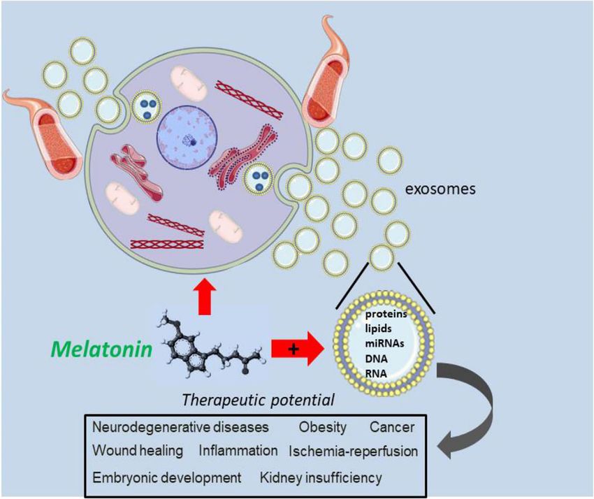
Novais et al.                                                                                                 The Exosomes and Melatonin Partnership

activities and crosstalk in eukaryotic organisms, and certainly, a         roles in the regulation of growth, metastasis and angiogenesis. Since
deeper understanding of their actions will bring valuable benefits          these processes are involved in cancer development, it is reasonable
for science.                                                               that exosomes are used as prognostic markers and as a basis for
   Recent progress on melatonin and exosome research will be               tumor graduation in patients with neoplasia (12, 13). Also, since the
outlined and the interactions between these two signaling                  exosomes carry biomacromolecules from the source cell, they can
pathways will be explored, with the potential to interfere in              represent a molecular bioprint of the original cell (14).
disease-related inflammatory, ischemic, degenerative, and
neoplastic processes. In view of the association of these
biomolecules, we propose that this partnership will benefit
possible protective molecular mechanisms against pathological
                                                                           MELATONIN: BIOGENESIS AND GENERAL
processes. Finally, the potential combined therapeutic use of              BIOLOGICAL FUNCTIONS
exosomes and melatonin, working together for a healthy
                                                                           Melatonin (N-acetyl-5-methoxytryptamine) was first described
homeostasis, will be considered.
                                                                           in 1958 when Lerner and his colleagues (15), while looking for a
                                                                           treatment for vitiligo disease, isolated the active substance from
                                                                           the bovine pineal gland extract which was capable of lightening
EXOSOMES: BIOGENESIS AND                                                   the skin of amphibians and inhibiting the melanocyte-
BIOLOGICAL FUNCTION                                                        stimulating hormone. Very soon thereafter, investigators
                                                                           observed that melatonin influenced the brain, and thereby, the
The first scientific description of exosomes dates from the 80s,             gonads and other components of the neuroendocrine systems
when Johnstone and colleagues (1) observed that sheep                      (16–18). The pineal gland produces melatonin in a circadian
reticulocytes lost their transferrin receptors during the                  way, which explains its chronobiotic influence on the body’s
maturation process to adult red blood cells; this occurred due             endocrine and non-endocrine rhythms, such as the sleep cycle:
to the release of small vesicles into the extracellular medium. In         wakefulness and reproduction (18, 19).
addition to transferrin receptors, their study provided evidence                Scientific reports have shown, through the finding of
for the loss of selective membrane proteins during in vitro                melatonin in a-proteobacteria and photosynthetic cyanobacteria,
maturation of reticulocytes.                                               that melatonin molecule evolved in bacteria that were phagocytosed
    Exosomes are a subtype of extracellular vesicles (EVs) but the         by early eukaryotes for nutritional purposes. Thereafter, the bacteria
nomenclature of the different extracellular vesicles (EVs) has             appear to have developed a symbiotic relationship with the
generated some confusion over time. However, it has been                   eukaryotic host, the a-proteobacteria evolving into mitochondria
generally accepted that EVs may be classified into three groups             while the cyanobacteria evolved into chloroplasts and these
according to their size and biogenesis, i.e., exosomes (30–                organelles continue to exist until today (20, 21).
200 nm), microvesicles (100–1000 nm) and apoptotic bodies                       The synthesis of melatonin is derived from the amino acid
(> 1000 nm) (2–5). The International Society for Extracellular             tryptophan, serotonin being an intermediate compound. Two
Vesicles recently published a position statement and update of             main enzymes control melatonin synthesis: arylalkyl N-
Minimal information for studies of extracellular vesicles (2018            acetyltransferase (AANAT) and acetylserotonin O-
MISEV guidelines) considering “extracellular vesicle” the                  methyltransferase (ASMT) (22, 23). Initially it was believed that
preferred generic term and recommending that subtypes must                 the presence of melatonin in all cells and its main effects were related
be defined by physical and biochemical characteristics and/or               to their absorption from the blood. However, it was later discovered
conditions/sources (6). In the present review, the exosome                 that many cells are probably capable of carrying out the enzymatic
terminology will be used with respect to the referenced publication.       conversion of serotonin to melatonin (19, 24–26).
    Exosomes originate from the endocytic pathway that initiate                 Although many functions of melatonin are mediated by
when the cytoplasmic membrane undergoes an invagination to                 membrane receptors, it is now known that there are functions
form an early secretory endosome. Then, intraluminal vesicles              independent of its receptors. Melatonin and its metabolites are
(ILVs) are formed inside large multivesicular bodies (MVBs) and            transported to cells via the cell oligopeptide transporter (PEPT) 1/
late endosomal maturation occurs by acidification. The last step            2 ‘, the organic anion transporter (OAT) 3 and the glucose
is the release of the ILVs as exosomes through fusion with the             transporter (27, 28). It is believed that transport via PEPT1/2
plasma membrane (5, 7).                                                    may be involved with its oncostatic effects (27). Melatonin is also
    Although initially exosomes were thought to be involved                transported into the mitochondria by means of PEPT1/2, although
merely in waste disposal (8), in 2007 Valadi (9) showed the                it is also synthesized by probably all cellular mitochondria (29–31).
use of exosomes by some cells to transfer genetic material                      The first report on the potent action of melatonin as a
between adjacent or distant cells. The transferred materials               scavenger of direct free radicals occurred 30 years ago (32). Its
include mRNAs to make proteins and microRNAs to regulate                   efficiency as a powerful antioxidant stems from its ability to
the expression of genes, secreted by cells during normal and               stimulate several antioxidant enzymes, in addition to directly
pathological conditions (10, 11).                                          neutralizing a series of free radicals and reactive oxygen and
    The exosomal content includes proteins, DNA, mRNA,                     nitrogen species (33). Melatonin interacts with the highly toxic
microRNA, long ncRNA and circular RNA, which play important                hydroxyl radical (· OH) at a constant rate equivalent to that of

Frontiers in Immunology | www.frontiersin.org                          2                                      June 2021 | Volume 12 | Article 692022
Novais et al. The Exosomes and Melatonin Partnership
other highly efficient hydroxyl radical scavengers (32). influence the apoptosis process and still others that interfere with
Additionally, there are assumptions that melatonin neutralizes immunomodulation, in different types of target cells (42). The
hydrogen peroxide, singlet oxygen, peroxynitrite anion, nitric exosomes achieve these effects mediating an autocrine and
oxide and hypochlorous acid (34). Superoxide desmutase, paracrine intercellular cross-talk that, subsequently, promotes a
glutathione peroxidase, glutathione reductase and other modification of both local and distant microenvironments (43).
antioxidant enzymes are also stimulated by melatonin (29, 35–39). Besides common core proteins that are a reflection of their
Melatonin has recently been reclassified as a multitasking biogenesis, many different proteins carried by the exosomes reflect
molecule, and not exclusively a hormone, due to the finding the original cells related to the phenotypic and physiological state,
about the existence of essential enzymes for its synthesis and the meaning that they may give important information on the
presence of melatonin receptors in many tissues and, also, the pathological processes of many medical entities (44). In turn,
discovery of its antioxidant and generalized anti-inflammatory melatonin combined with exosomes showed beneficial effects, like
properties (40, 41). suppressing oxidative stress and apoptosis in vitro and
Melatonin functions as a glycolytic molecule which inhibits inflammation, oxidative stress, DNA/mitochondrial damage, and
pathological aerobic glycolysis of diseased cells allowing them to apoptosis in vivo (45). Also, this therapeutic combination has
resume normal mitochondrial oxidative phosphorylation; this exhibited an important protection of nervous system through
change converts pathological cells to a healthier phenotype. It is the regulation of the TLR4/NF-kB signaling pathway (46). Also,
likely that the glycolytic function of melatonin explains its exosome-melatonin therapy was found to mitigate vascular
protective actions against a variety of diseases (33). calcification and aging (47) and other beneficial therapeutic
actions, that will be the scope of this review. For example, Xia and
colleagues (48) demonstrated that melatonin may induce the
EXOSOMES AND MELATONIN: polarization of macrophages by changing exosomal contents
COMPATIBLE PARTNERS released from adipocytes. Figure 1 summarizes the therapeutic
potential of melatonin in various pathological conditions.
Since exosomes express many different surface receptors, their The mitochondria play a central role in almost every activity a
influence on recipient cells differs functionally; this results in sets cell undertakes; it is the preferential site of ATP synthesis and
of exosomes that are capable to induce cell survival, others that reactive oxygen species (ROS) generation. Mitochondria can take
FIGURE 1 | The combination of melatonin and exosomes is promising therapeutic strategy. By acting directly on damage cells, melatonin is thought to intracellulary
interface with exosome trafficking, possibly by modifying their molecular contrent. The user of melatonin in combination with an exosomes may be an important
approach to alter the biology of exosomes related to dysregulated signaling pathways in several pathologies and disease conditions.
Frontiers in Immunology | www.frontiersin.org 3 June 2021 | Volume 12 | Article 692022Novais et al. The Exosomes and Melatonin Partnership
up pineal-derived or exogenously administered melatonin from important players. For patients undergoing surgeries that result in
the circulation, but they also have the ability to intrinsically prolonged ischemic intervals or liver graft transplantation, a deeper
produce it (49–51). Mitochondria are believed to have evolved understanding of liver I/R injury will reflect improvements in the
when melatonin-producing bacteria were engulfed by early clinical care of patients (55). Recently, Sun et al. (45) examined liver
eukaryotes (21, 26). When confronted with melatonin from the ischemia/reperfusion (LIR) injury and confirmed that combined
blood, the mitochondria concentrate it against a gradient (52). melatonin and exogenic adipose mesenchymal stem cell (ADMSC)-
It is believed that the transfer of information between cells can derived exosome treatment offered superior protection against I/R
be mediated by the mitochondrial genome transfer and even the injury, when compared to the isolated treatment. In vitro studies
entire mitochondria (53). The research of Guescini and used a macrophage cell line (RAW), pre-treated with
colleagues (54) provided evidence that glioblastoma and lipopolysaccharides, and hepatocytes previously incubated with
astrocyte cells release exosomes carrying mitochondrial DNA melatonin or exosomes before the hypoxia lesion. These studies
(mtDNA), which can be transferred to other cells. The role and showed that exosomes alone and in combination with melatonin,
relevance of exosomes in mitochondrial homeostasis is an respectively, cause suppression of inflammation by reducing MIF,
emerging area of research, which may clarify many details of MMP-9, IL-1b, TNF-a, COX-2 and oxidative stress markers
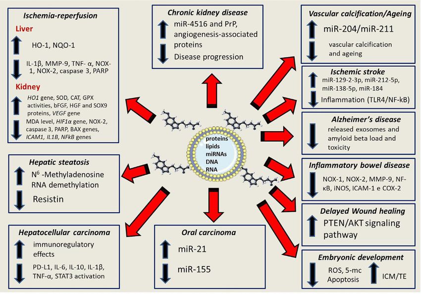
this interrelation (53, 55). Figure 2 summarizes some of the (NOX-1, NOX-2, oxidized protein), in addition to apoptotic
many molecular processes regulated by melatonin and exosomes molecules (cleaved caspase 3 and PARP). In addition, in vivo
in various pathological conditions. experiments analyzed liver specimens from male adult Sprague-
Dawley rats (n = 50) equally categorized into the following
experimental: (1) sham controls (SC), (2) LIR only, (3) LIR-
exosome (intravenous administration of 100 µg, 30-minute post-
EXOSOME-MELATONIN THERAPY LIR), (4) LIR-melatonin (intra-peritoneal administration of 20 mg/
PROTECTS AGAINST ISCHEMIA- kg, 30 minute post-LIR and 50 mg/kg at 6 and 18 hours post-LIR),
REPERFUSION INJURY and (5) LIR-exosome-melatonin groups. The authors demonstrated
that I/R animals treated with the combination of exosomes and
Hepatic ischemia-reperfusion (I/R) injury is a major complication melatonin had lower plasma AST concentrations and liver injury
of liver tissue often observed during liver surgery (e.g., liver scores, when compared to other groups. Inflammatory markers
resection, trauma, and liver trans plantation) with (ICAM-1, IL-1b, MMP-9, TNF-a, NF-kB, RANTES), cellular
pro-inflammatory components and immune cells being immunoregulatory molecules (CD3 +, CD4 +, CD8 +, CD161 +,
FIGURE 2 | The up- and down-regualtion of multiple process that involve the partnership between melatonin and exosomes; these include signaling pathways for
inflammation, oxidative stress and apoptotic events as well as well as gene. microRNAs, protein and transcription factors. An in-depth knowledge of the control of
the synergistic effects between melatonin and exosomes offers a valuable therapeutic promise for the treatment of several diseases
Frontiers in Immunology | www.frontiersin.org 4 June 2021 | Volume 12 | Article 692022Novais et al. The Exosomes and Melatonin Partnership
CD11 +, CD14 +, F4/80), expression of molecules related to IL-18, matrix metalloproteinase 9 and lipocalin neutrophils
apoptosis (cleaved caspase-3, PARP), oxidative stress (NOX-1, associated with gelatinase). The combined treatment of melatonin +
NOX-2), DNA damage (g-H2AX) and markers of mitochondrial exosomes was more effective, that is, lower scores for kidney
damage (cytosolic cytochrome- C) exhibited a similar pattern to damage, oxidative stress, inflammation and renal apoptosis
those of liver injury scores. On the other hand, after the combined parameters. The authors concluded that the combination therapy
treatment, the expression of antioxidant proteins (HO-1, NQO-1) with melatonin, mesenchymal stem cells and exosomes has
exhibited progressive increases in stem cells. The most relevant potential use in order to minimize the damage of renal I/R in rats.
findings were that the plasma AST level and liver injury score were
significantly suppressed in animals that suffered I/R injury after
exosome or melatonin therapy, and still increased significantly after
the combined treatment with exosomes and melatonin.
EXOSOME-MELATONIN THERAPY
On the other hand, renal ischemia-reperfusion injury (RIRI) and PROTECTS AGAINST HEPATIC
its dysfunction syndrome has the potential to accelerate the METABOLIC DISTURBANCE
development of chronic kidney disease (CKD). The release of free
radicals, mitochondrial dysfunction, induction of apoptosis and Fatty liver is caused by the impregnation of more than 5% of the liver
inflammation are among the main causes of RIRI (56). Hence, with fat. Although the moderate accumulation of triacylglycerol in
therapeutic agents targeting oxidative stress, apoptosis, and the liver can exert a hepatoprotective effect, when this storage of
inflammation may be beneficial in clinical approach to RIRI. In hepatic lipids becomes prolonged, it can cause metabolic
his research, Alzahrani (57) evaluated the effectiveness of exosomes dysfunction, inflammation and advanced forms of non-alcoholic
derived from mesenchymal stem cells (MSCs) preconditioned with fatty liver disease (NAFLD) (59). Metabolic stress acquired due to
melatonin. He noted that the combined treatment provided the best disorders such as abnormalities in glucose and lipid metabolism,
protection against I/R injury when compared to therapy with MSCs insulin resistance and inflammation can cause the development of
or exosomes derived from MSCs that had not been preconditioned. NAFLD. NAFLD is usually associated with metabolic comorbidities,
There was a significant improvement in I/R after all treatments such as obesity, diabetes mellitus and dyslipidemia (60).
(MSCs, Exo and Exo + Mel), but the improvement in the Exo + Mel The role of melatonin in reversing hepatic steatosis was
group was evidently greater. To assess the degree of protection studied by Rong et al. (61), who suggested that crosstalk
afforded by the therapy, they took into account some issues such as originating from adipose tissue could be a valid regulatory
histopathological score, blood levels of urea nitrogen in the blood route. Melatonin supplementation caused a significant
(BUN) and creatinine, the state of oxidative stress (MDA level, reduction in the amount of exosomal resistin derived from
HIF1a gene and NOX2 protein), the activities of antioxidant adipocytes. They later demonstrated that resistin was a
enzymes (HO1 gene and SOD, CAT, GPX activities), evidence of fundamental cytokine to suppress phosphorylation of protein
apoptosis (caspase 3 activity and PARP1, BAX genes), anti- kinase a activated by 5 ‘adenosine monophosphate, capable of
apoptotic effect (BCL-2 gene), degree of inflammation by triggering stress in the endoplasmic reticulum, which resulted in
reduction of MPO activity and low expression of the ICAM1, hepatic steatosis. Melatonin reduced the production of resistin in
IL1B, NFkB genes), intensity of regeneration (bFGF, HGF and adipocytes, through the brain and inhibition of protein 1
SOX9 proteins) and, finally, the degree of angiogenesis (VEGF transcription similar to muscle arnt. Melatonin was able to
gene). This exosome ameliorative effect was mediated, at least in improve the demethylation of the N6-methyladenosine RNA
part, by inhibition of the oxidative stress, apoptosis, and to degrade the resistin mRNA in adipocytes. In general,
inflammation, while inducing antioxidant properties, regeneration melatonin caused a reduction in the traffic of the exosomal
and angiogenesis, resulting in improved renal repair and function. resistin generated by adipocytes to hepatocytes, causing greater
The therapeutic value of melatonin (20 mg/kg body weight/day) relief of stress-induced liver steatosis in the ER. Recent scientific
given for three days, via intraperitoneal injection, in association indications, based on the study of new regulatory pathways
with administration of mesenchymal stem cells, and their mediated by melatonin, indicate that adipocyte-derived
extracellular vesicles, have also been tested in RIRI (58). Initially, exosomes are a potential new target for the treatment of obesity.
the authors established the renal I/R model and subsequently
injected mesenchymal stem cells or exosomes into both renal
arteries during reperfusion. They observed an improvement in EXOSOME-MELATONIN THERAPY
RIRI compared to different treatments, using parameters such as
histopathological score, serum levels of urea, creatinine and retinol- CHANGES THE NEOPLASTIC
binding protein, lipid peroxidation marker malondialdehyde, IMMUNOSUPPRESSION STATUS IN
superoxide dismutase and catalase activities, and degree of HEPATOCELLULAR AND ORAL
apoptosis (less damage to DNA and protein X associated with B SQUAMOUS CELL CARCINOMAS
2 cell lymphoma and higher B 2 cell lymphoma (genes/proteins).
The researchers reported a remarkable inhibition of inflammatory Hepatocellular carcinoma (HCC) is the most common
markers and kidney damage (e.g., tumor necrosis factor alpha, malignant tumor of the liver and has been linked to a high
interleukin-1b, kappa nuclear factor B, kidney injury molecule-1, rate of death in cancer patients worldwide. Liver cirrhosis is
Frontiers in Immunology | www.frontiersin.org 5 June 2021 | Volume 12 | Article 692022Novais et al. The Exosomes and Melatonin Partnership
considered the most important risk factor for the development of contributors in the pathophysiology of CKD and hypertension is
HCC, caused by chronic inflammation resulting from the intrarenal renin-angiotensin system (RAS), due to the
continuous hepatocyte damage, such as that which occurs in processes of inflammation and fibrosis. Studies using animal
cases of hepatitis B and C (62). In HCC cells, exosomes favor models of CKD have shown that the activation of intrarenal RAS
microenvironment communication, and provide a fertile and the consequent kidney injury can be mitigated by exogenous
environment for cell proliferation and metastasis; how administration of melatonin, due to its antioxidant effects (70).
miRNAs, lncRNA, and proteins are exosome-sorted in a Other studies have reported the use of isolated EVs from MSCs
specific cell type remains unclear (63). to prevent the progression of CDK in animal models and in
To assess the effects of exosomes derived from hepatocellular patients (71, 72). Recently, Yoon et al. (73) isolated exosomes
carcinoma (Exo-con) and exosomes derived from HCC cells derived from healthy melatonin-treated MSCs. Comparatively,
treated with 0.1 mM melatonin (Exo-MT) on the expression of they evaluated the biological functions of exosomes derived from
inflammatory factors and programmed death ligand 1 (PD-L1), MSCs of patients with CKD, also treated with melatonin. They
Cheng et al. (64) devised an experiment using co-culture of Exo- found that the treatment with melatonin increased the
con and Exo-MT with differentiated macrophages from THP-1 expression of cellular prion protein (PrP) in exosomes isolated
cells or RAW264.7 cells. The researchers observed that in from MSCs through the positive regulation of miR-4516. In
macrophages co-cultured with Exo-MT there was suppression addition, exosomes from melatonin-treated MSCs were able to
of the expression of PD-L1, while in those co-cultured with protect mitochondrial function, cell senescence and the
untreated exosomes (Exo-con) there was an increase in the proliferative potential of MSCs, significantly increasing the
expression of PD-L1 and levels cytokines, such as IL-6, IL-10, level of proteins associated with angiogenesis in MSCs. With
IL-1b and TNF-a. Therefore, the authors concluded that this study, the authors suggested a regenerative potential in the
exosomes treated with melatonin were able to efficiently use of MSCs treated with exosomes and melatonin in patients
attenuate the expression of PD-L1 and the secretion of with CDK through the miR-4516-PrP signaling axis.
cytokines, by decreasing the activation of STAT3, promoting
the immunoregulatory effects in HCC.
The oral cavity is the anatomical location where EXOSOME-MELATONIN
approximately 50% of the head and neck cancers (HNC) THERAPY ATTENUATES
occur. The oral squamous cell carcinoma (OSCC) and
epidermoid oral carcinoma are the most frequent malignant
VASCULAR CALCIFICATION,
cancers, representing 90–95% of cases. The challenging AGEING, ISCHEMIC BRAIN INJURY,
paradigm is due to the worse prognosis and high mortality AND NEURODEGENERATIVE DISEASES
rate, combined with the lack of better perspectives (65).
Differential microRNA (miRNA) expression profile in OSCC- Vascular calcification occurs in the elderly with comorbidities
related EVs have been documented both in vitro and in clinical such as atherosclerosis, hypertension, diabetes, macroangiopathy
experiments (66, 67). Hunsaker et al. (68) tried to identify the and chronic kidney disease. The degree of vascular calcification is
role of melatonin in oral carcinoma-associated EVs (including directly related to cardiovascular mortality and tissue
the exosomes) and impact on microRNA (miRNA) content in amputation, being an important factor in determining the
various oral cancer cell lines. They observed that melatonin health of the elderly (74).
significantly suppressed the expression of miR-155 in all The role of exosomes in angiogenesis, vascular calcification
OSCC-related extracellular vesicles. Moreover, the expression and senescence of vascular smooth muscle cells (VSMCs) has
of miR-21 was significantly increased and no significant changes been considered important in some studies (75). A study by Xu
in miR-133a expression were observed, after melatonin et al. (47) demonstrated that melatonin (10 mM) can attenuate
administration in the three isolated OSCC. These results both vascular osteogenic differentiation and VSMC senescence.
suggested a differential modulation of specific miRNAs, such as In addition, they demonstrated that exosomes isolated from
miR-21, miR-133a, and miR-155. In other words, melatonin VSMCs or calcified vascular smooth muscle cells (CVSMCs),
differentially modulates miR-expression in OSCC extracellular treated with melatonin, can be absorbed by VSMCs and
vesicles, including exosomes. attenuate osteogenic differentiation and senescence of VSMCs
or CVSMCs, respectively. In addition, they demonstrated the
mediation of the paracrine effects of exosomes secreted by
VSMCs by exosomal miR-204/miR-211, through the BMP2
EXOSOME-MELATONIN THERAPY IS gene. The authors also found that treatment with melatonin
HELPFUL FOR PATIENTS WITH CHRONIC relieved vascular calcification and aging in mice submitted to 5/6
KIDNEY DISEASE nephrectomy plus a high phosphate diet (NTP 5/6). They were
able to detect by fluorescence images that exosomes derived from
Chronic kidney disease (CKD) is a clinical syndrome that occurs VSMCs treated with melatonin were internalized in the arteries
as a consequence of kidney function and/or structure of mice, promoting a reduction in vascular calcification and
deterioration. Unfortunately, it is an irreversible process with a aging. They reported that these effects were largely abolished by
slow and progressive evolution (69). One of the most important inhibition of exosomal miR-204 or miR-211. Thus, the authors
Frontiers in Immunology | www.frontiersin.org 6 June 2021 | Volume 12 | Article 692022Novais et al. The Exosomes and Melatonin Partnership
concluded that exosomes obtained from VSMCs treated with (87) developed an in vitro Ab toxicity model to investigate the
melatonin were able to attenuate vascular calcification and possible role of melatonin treatment in exosome release and
paracrine aging through an exosomal miR-204/miR-211 exosomal tau content. The experiment demonstrated that
dependent mechanism. treatment with melatonin suppressed the number of exosomes
Ischemic stroke or cerebral ischemia is caused by the released, and consequently the amyloid beta load and toxicity,
interruption of blood flow due to the blockage in an artery that acting by blocking the secretory molecules of EVs. In addition,
supplies blood to the brain. Then, the reduction in cerebral they demonstrated that the timing of melatonin administration,
oxygenation leads to permanent neural damage or death of whether before or after the application of Ab, also affected the level
neuronal cells if the circulation cannot be quickly restored of tau carried by the exosomes. The authors concluded that the
(76). There is a need to find new neuroprotective substances, study provided a starting point for the development of AD
because although most pharmacological neuroprotectors have treatment strategies through the influence of melatonin on
been effective in experimental studies, they have failed in clinical the exosomes.
trials. Furthermore, it is important that these new substances can
act on brain repair, supporting the concept of brain plasticity (77).
In this context, Wang et al. (46) decided to investigate the
EXOSOME-MELATONIN THERAPY
therapeutic potential of plasma exosomes and melatonin in the
damage to the nervous system caused by I/R. They treated rats MAY CONTROL THE INFLAMMATORY
with melatonin and isolated plasma exosomes using a model of PROCESS ASSOCIATED WITH COLITIS
focal cerebral ischemia. They reported that inflammatory AND OBESITY AND ASSIST IN
responses induced by ischemia and inflammasome-mediated WOUND HEALING
pyroptosis were influenced (suppressed) by the treatment with
plasma exosomes. In other words, melatonin increased the Ulcerative colitis (UC) and Crohn’s disease are chronic
therapeutic effects of plasma exosomes, with a reduction in the inflammatory bowel diseases (IBD), with unknown etiology
intensity of the infarction and an improvement in recovery and partially understood pathogenesis. The process begins in
through the regulation of the TLR4/NF-kB signaling pathway. the rectum and progresses involving different extensions of the
The authors also linked the altered miRNA profile in plasma colon mucosa. Typically, clinical signs such as rectal bleeding
exosomes treated with melatonin with the regulatory and diarrhea are observed. The potential use of exosomes in IBD
mechanisms involved in neurological recovery after diagnostic and treatment strategies is based on the knowledge
ischemic injury. that they influence the main pathways related to IBD, such as
Also, organic aging is directly related to oxidative stress, immune responses, barrier functions and intestinal flora (29).
which is a condition that involves an imbalance between the Thus, Chang et al. (88) proposed to elucidate the relationship
formation of reactive oxygen species (ROS) and the cellular between melatonin and exosomes in acute inflammatory colitis
antioxidant capacity. With aging, ROS generation increases, (CIA). In a model of AIC induced by sodium dextran sulfate
but the organic antioxidant system gradually becomes (DSS) in rats, they tested the hypothesis that AIC could be
dysfunctional. Thus, aging and various pathological conditions suppressed by combining melatonin and exosomes released by
and human diseases, especially neurodegenerative diseases, are mesenchymal stem cells derived from adipose tissue. They
caused by biochemical changes in these macromolecular treated Sprague Dawley rats with doses of 50 mg/kg of
components, ultimately causing oxidative damage (78). melatonin on day 5 and 50 mM/kg of exosomes on days 5, 7
One of the most important neurodegenerative diseases is and 10, and also with the combination of melatonin with
Alzheimer’s disease (AD), characterized by severe neuronal exosomes. They observed that the number of circulating
loss in the brain, leading to cortical dementia with an intense inflammatory cells was lower in animals treated with
memory deficit (79). The pathophysiology of synaptic loss and melatonin-exosomes, when compared to those treated with
neurodegeneration involves problems in the cleavage of the melatonin or exosomes alone. The combination of melatonin
amyloid precursor protein (APP) with the production of APP and exosomes mitigated the effects of AIC induced by DSS,
beta-amyloid (Ab) fragment, together with the aggregation of the evidenced by a reduction in the expression of inflammation
hyperphosphorylated tau protein. In these individuals, the markers, oxidative stress, apoptosis and fibrosis. The combined
existence of comorbidities, metabolic, vascular and inflammatory treatment was also able to reduce colon injury scores, expression
changes, are essential factors for the disease process (80). In of inflammatory markers and DNA damage.
addition, the inducing role of exosomes in the spread of Obesity is a complex chronic disease with inflammation as a
neurodegenerative diseases such as AD, due to the spread of toxic central and reversible process. The adipose tissue undergoes
proteins has been studied (81–84). The studies have shown that the infiltration and activation of immune cells, particularly
spread of neuropathology appears to be mediated by exosomes macrophages, capable of communicating with adipocytes and
containing different forms of tau, especially the phosphorylated bringing these changes to the adipose tissue microenvironment
form of the tau protein carried by the exosomes (85, 86). However, (89, 90). In order to modulate the process of inflammation in
as the influence of melatonin on the quantity and content of obesity, Liu et al. (91) studied the effects of melatonin at a dose of
exosomes released from cells was still unknown, Ozansoy et al. 20 mg/kg/day for 14 days and reported that melatonin relieved
Frontiers in Immunology | www.frontiersin.org 7 June 2021 | Volume 12 | Article 692022Novais et al. The Exosomes and Melatonin Partnership
inflammation while raising levels of a-ketoglutarate (aKG) in influenced by oviductal EVs, which interfere in the interactions
adipose tissue of obese mice. They revealed that aKG was the between the oviduct and the embryos. The absence of EVs in cell
target for the inhibition of melatonin-mediated adipose cultures is associated with inferior embryonic development.
inflammation, by promoting mitochondrial isocitrate Coincidentally, it has been reported that melatonin is also
dehydrogenase mRNA 2 (Idh2) elevation in adipocytes, abundantly present in oviduct fluids and EVs derived from
resulting in an increased level of aKG. In addition, the oviduct fluid (95). It is also known that the addition of EVs or
researchers observed that sirtuin 1 (Sirt1) interacted physically melatonin, alone, led to a significant negative regulation of ROS
with HDI2, forming a complex to increase the circadian and 5-methylcytosine (5-mC), as well as an increase in the
amplitude of Idh2 and the content of aKG. Melatonin was proportion of embryos in the blastocyst stage. The combined
able to effectively promote the secretion of exosomes from treatment with EVs and melatonin led to the same results, but
adipocytes, increasing the level of exosomal aKG derived from there was a significant decrease in the apoptosis index and an
adipose tissue. They also reported an increase in the proportion increase in the internal cell mass index (ICM)/tropectoderm
of M2 to M1 macrophages, under the action of melatonin, (TE). In conclusion, the research provided insights into the role
transporting exosomal aKG to macrophages and promoting of EVs and melatonin in cellular communication and between
TET-mediated DNA demethylation. In addition, exosomal embryos and the oviduct.
aKG attenuated signal transducers and transduction-3
(STAT3)/NF-kB activators through its receptor 1 oxoglutarate
(OXGR1) in adipocytes. In conclusion, the authors suggested
that melatonin had the potential to relieve metabolic
CONCLUDING REMARKS AND
inflammation because it may promote an increase in aKG PERSPECTIVES
cellular and exosomal levels in adipose tissue.
Delaying disease processes or stage advances by simply activating
The healing difficulties caused by diabetes have an
or correcting specific cellular mechanisms of disease would be a
important influence on surgical results and may eventually
monumental development. Although still via unknown
develop into chronic wounds. Recent research has shown that
mechanisms, science is opening up the field of regenerative
the polarization of macrophages plays an important role in the
medicine, in which quiescent elements present during illness
healing process of diabetic wounds (92). To investigate the
can themselves be activated to reverse the processes of disease.
influence of melatonin and exosomes on the polarization of
Regenerative medicine is attractive to the medical community
macrophages, Liu et al. (93) studied whether exosomes derived
since many scientific reports provide evidence that human body
from mesenchymal stem cells (MSCs), pretreated with
is specially equipped to heal itself. Within the context of organic
melatonin (MT) (Exo-MT), could have beneficial effects on
elements capable of promoting self-healing are melatonin and
the healing of diabetic wounds, when compared to untreated
exosomes, which can significantly interfere in the main processes
exosomes. The researchers observed that MT-Exo improved
involved with cell ageing and molecular damage.
the healing of diabetic wounds by suppressing the
We explored herein the promising results of the therapeutic
inflammatory response, which was achieved due to the
use of exogenous melatonin combined with exosomes derived
increase in the M2 to M1 polarization ratio, by activating the
from several tissues (e.g., mesenchymal stem cells), and in
PTEN/AKT signaling pathway.
many pathological conditions such as ischemia-reperfusion
Also, in order to assess the potential effects of exosomes
injury, obesity, hepatocellular carcinoma, oral squamous cell
released by adipose tissue MSCs on inflammatory modulation,
carcinoma, chronic kidney disease, ischemic brain syndrome,
Heo et al. (94) examined the changes in anti-inflammatory
neurodegenerative diseases like Alzheimer’s disease, acute
genes, along with the polarization of M2 macrophages in
inflammatory colitis, diabetes-related wound healing, vascular
fibroblasts treated with pro-inflammatory cytokines and
calcification, ageing, and also in embryonic development.
THP-1 cells. They observed that exosome treatment
The evidence suggests that by changing the exosomal content
positively regulated the expression of anti-inflammatory
and load, melatonin may potentially improve the nature
mRNA associated with M2 macrophages, in an inflammatory
of exosomes associated with diseases with recognized
environment treated with gamma interferon and tumor
molecular background.
necrosis factor alpha. The increase in anti-inflammatory
Based on the above-mentioned studies, we reiterate that
modulation exerted by exosomes treated with melatonin
melatonin-exosome combinations reveal important and novel
occurred through exosomal miRNAs (for example, miR-34a,
therapeutic features possibly affecting miRNAs, proteins, and
miR-124 and miR-135b).
lipids related to degenerative, inflammatory, and neoplastic
disorders. Moreover, melatonin-exosome therapy could fight
most of these disease processes by preventing their progression,
EXOSOME-MELATONIN THERAPY not only their clinical manifestations. Finally, melatonin/exosome-
BENEFITS EMBRYONIC DEVELOPMENT based therapy presents numerous advantages over traditional
treatments, since melatonin has no adverse effects, is well-
Embryo quality and even offspring development require ideal tolerable, and possesses powerful anti-oxidant, anti-inflammatory,
growing conditions. It is known that these conditions are and anti-ageing properties.
Frontiers in Immunology | www.frontiersin.org 8 June 2021 | Volume 12 | Article 692022Novais et al. The Exosomes and Melatonin Partnership
AUTHOR CONTRIBUTIONS scope of the review. RR and DZ conceived the review and
checked all drafts and the final version of the report. All
AN compiled the literature and wrote the manuscript. LC authors contributed to the article and approved the
prepared figures for the article, guided the insertions and the submitted version.
REFERENCES 20. Manchester LC, Poeggeler B, Alvares FL, Ogden GB, Reiter RJ. Melatonin
Immunoreactivity in the Photosynthetic Prokaryote Rhodospirillum Rubrum:
1. Johnstone RM, Adam M, Hammond JR, Orr L, Turbide C. Vesicle Formation Implications for an Ancient Antioxidant System. Cell Mol Biol Res (1995)
During Reticulocyte Maturation. Association of Plasma Membrane Activities 41:391–5.
With Released Vesicles (Exosomes). J Biol Chem (1987) 262(19):9412–20. doi: 21. Zhao D, Yu Y, Shen Y, Liu Q, Zhao Z, Sharma R, et al. Melatonin Synthesis
10.1016/S0021-9258(18)48095-7 and Function: Evolutionary History in Animals and Plants. Front Endocrinol
2. Pegtel DM, Gould SJ. Exosomes. Annu Rev Biochem (2019) 88:487–514. (2019) 10:249. doi: 10.3389/fendo.2019.00249
doi: 10.1146/annurev-biochem-013118-111902 22. Champney TH, Holtorf AP, Steger RW, Reiter RJ. Concurrent Determination
3. de la Torre Gomez C, Goreham RV, Bech Serra JJ, Nann T, Kussmann M. of Enzymatic Activities and Substrate Concentrations in the Melatonin
“Exosomics”-a Review of Biophysics, Biology and Biochemistry of Exosomes Synthetic Pathway Within the Same Rat Pineal Gland. J Neurosci Res
With a Focus on Human Breast Milk. Front Genet (2018) 27:92(9). (1984) 11:59–66. doi: 10.1002/jnr.490110107
doi: 10.3389/fgene.2018.00092 23. Klein DC. Arylalkylamine N-acetyltransferase: “The Timezyme”. J Biol Chem
4. Wiklander OPB, Brennan MÁ , Lötvall J, Breakefield XO, El Andaloussi S. (2007) 282:4233–7. doi: 10.1074/jbc.R600036200
Advances in Therapeutic Applications of Extracellular Vesicles. Sci Transl 24. Stefulj J, Hörtner M, Ghosh M, Schauenstein K, Rinner I, Wölfler A, et al.
Med (2019) 11(492):eaav8521. doi: 10.1126/scitranslmed.aav8521 Gene Expression of the Key Enzymes of Melatonin Synthesis in Extrapineal
5. Gurung S, Perocheau D, Touramanidou L, Baruteau J. The Exosome Journey: Tissues of the Rat. J Pineal Res (2001) 30(4):243–7. doi: 10.1034/j.1600-
From Biogenesis to Uptake and Intracellular Signalling. Cell Communication 079x.2001.300408.x
Signaling: CCS (2021) 19(1):47. doi: 10.1186/s12964-021-00730-1 25. Venegas C, Garcı́a JA, Escames G, Ortiz F, Ló pez A, Doerrier C, et al.
6. Thé ry C, Witwer KW, Aikawa E, Alcaraz MJ, Anderson JD, Extrapineal Melatonin: Analysis of its Subcellular Distribution and Daily
Andriantsitohaina R, et al. Minimal Information for Studies of Fluctuations. J Pineal Res (2012) 52(2):217–27. doi: 10.1111/j.1600-
Extracellular Vesicles 2018 (MISEV2018): A Position Statement of the 079X.2011.00931.x
International Society for Extracellular Vesicles and Update of the 26. Tan DX, Manchester LC, Liu X, Rosales-Corral SA, Acuna-Castroviejo D,
MISEV2014 Guidelines. J Extracell Vesicles (2018) 7(1):1535750. Reiter RJ. Mitochondria and Chloroplasts as the Original Sites of Melatonin
doi: 10.1080/20013078.2018.1535750 Synthesis: A Hypothesis Related to Melatonin’s Primary Function and
7. Zhang Y, Liu Y, Liu H, Tang WH. Exosomes: Biogenesis, Biologic Function Evolution in Eukaryote. J Pineal Res (2013) 54:127–38. doi: 10.1111/jpi.12026
And Clinical Potential. Cell Biosci (2019) 9:19. doi: 10.1186/s13578-019- 27. Huo X, Wang C, Yu Z, Peng Y, Wang S, Feng S, et al. Human Transporters,
0282-2 PEPT1/2, Facilitate Melatonin Transportation Into Mitochondria of Cancer
8. Soria FN, Pampliega O, Bourdenx M, Meissner WG, Bezard E, Dehay B. Cells: An Implication of the Therapeutic Potential. J Pineal Res (2017) 62(4).
Exosomes, an Unmasked Culprit in Neurodegenerative Diseases. Front doi: 10.1111/jpi.12390.
Neurosci (2017) 11:26. doi: 10.3389/fnins.2017.00026 28. Mayo JC, Sainz RM, Gonzá lez-Mené ndez P, Hevia D, Cernuda-Cernuda R.
9. Valadi H, Ekström K, Bossios A, Sjöstrand M, Lee JJ, Lötvall JO. Exosome- Melatonin Transport Into Mitochondria. Cell Mol Life Sci (2017) 74:3927–40.
Mediated Transfer of mRNAs and microRNAs Is a Novel Mechanism of doi: 10.1007/s00018-017-2616-8
Genetic Exchange Between Cells. Nat Cell Biol (2007) 9(6):654–9. 29. Reiter RJ, Sharma R, Ma Q, Rosales-Corral S, Acuna-Castroviejo D, Escames
doi: 10.1038/ncb1596 G. Inhibition of Mitochondrial Pyruvate Dehydrogenase Kinase: A Proposed
10. Raposo G, Stoorvogel W. Extracellular Vesicles: Exosomes, Microvesicles, and Mechanism by Which Melatonin Causes Cancer Cells to Overcome Cytosolic
Friends. J Cell Biol (2013) 200(4):373–83. doi: 10.1083/jcb.201211138 Glycolysis, Reduce Tumor Biomass and Reverse Insensitivity to
11. Gurunathan S, Kang MH, Jeyaraj M, Qasim M, Kim JH. Review of the Chemotherapy. Melatonin Res (2019) 2:105–19. doi: 10.32794/mr11250033
Isolation, Characterization, Biological Function, and Multifarious Therapeutic 30. Reiter RJ, Ma Q, Sharma R. Melatonin in Mitochondria: Mitigating Clear and
Approaches of Exosomes. Cells (2019) 8(4):307. doi: 10.3390/cells8040307 Present Dangers. Physiology (2020) 35(2):86–95. doi: 10.1152/
12. Dai J, Su Y, Zhong S, Cong L, Liu B, Yang J, et al. Exosomes: Key Players in physiol.00034.2019
Cancer and Potential Therapeutic Strategy. Signal Transduct Target Ther 31. Chitimus DM, Popescu MR, Voiculescu SE, Panaitescu AM, Pavel B, Zagrean
(2020) 5(1):145. doi: 10.1038/s41392-020-00261-0 L, et al. Melatonin’s Impact on Antioxidative and Anti-Inflammatory
13. Chen Q, Li Y, Liu Y, Xu W, Zhu X. Exosomal Non-coding Rnas-Mediated Reprogramming in Homeostasis and Disease. Biomolecules (2020) 10
Crosstalk in the Tumor Microenvironment. Front Cell Dev Biol (2021) (9):1211. doi: 10.3390/biom10091211
9:646864. doi: 10.3389/fcell.2021.646864 32. Tan DX, Chen LD, Poeggeler B, Manchester LC, Reiter RJ. Melatonin: A
14. Sharma A, Khatun Z, Shiras A. Tumor Exosomes: Cellular Postmen of Cancer Potent Hydroxyl Radical Scavenger. Endocr J (1993) 1:57–60.
Diagnosis and Personalized Therapy. Nanomedicine (2016) 11(4):421–37. 33. Reiter RJ, Sharma R, Zuccari DAPC, Chuffa LGA, Rodriguez C. Melatonin
doi: 10.2217/nnm.15.210 Synthesis and Uptake by Mitochondria: Implications for Diseased Cells With
15. Lerner AB, Case JD, Takahashi Y, Lee TH, Mori W. Isolation of Melatonin, Dysfunctional Mitochondria. Future Med Chem (2021) 13(4):335–9.
the Pineal Gland Factor That Lightens Melanocytes. J Amer Chem Soc (1958) doi: 10.4155/fmc-2020-0326
80:2587. doi: 10.1021/ja01543a060 34. Galano A, Reiter RJ. Melatonin and its Metabolites vs Oxidative Stress: From
16. Wurtman RJ, Axelrod J, Chu EW. Melatonin, a Pineal Substance: Effect on Rat Individual Actions to Collective Protection. J Pineal Res (2018) 65:e12514.
Ovary. Science (1963) 141(3577):277–8. doi: 10.1126/science.141.3577.277 doi: 10.1111/jpi.12514
17. Hoffman RA, Reiter RJ. Pineal Gland: Influence on Gonads of Male Hamsters. 35. Reiter RJ, Sharma R, Rosales-Corral S. Anti-Warburg Effect of Melatonin: A
Science (1965) 148:1609–11. doi: 10.1126/science.148.3677.1609 Proposed Mechanism to Explain its Inhibition of Multiple Diseases. Int J Mol
18. Reiter RJ, Fraschini F. Endocrine Aspects of the Mammalian Pineal Gland: A Sci (2021) 22(2):764. doi: 10.3390/ijms22020764
Review. Neuroendocrinology (1969) 5(3):219–55. doi: 10.1159/000121862 36. Reiter RJ, Tan DX, Osuna C, Gitto E. Actions of Melatonin in the Reduction of
19. Acuña-Castroviejo D, Escames G, Venegas C, Dı́az-Casado ME, Lima-Cabello Oxidative Stress. A Review. J BioMed Sci (2000) 7(6):444–58. doi: 10.1007/
E, Ló pez LC, et al. Extrapineal Melatonin: Sources, Regulation, and Potential BF02253360
Functions. Cell Mol Life Sci (2014) 71(16):2997–3025. doi: 10.1007/s00018- 37. Reiter RJ, Rosales-Corral SA, Tan DX, Acuna-Castroviejo D, Qin L, Yang SF,
014-1579-2 et al. Melatonin, a Full Service Anti-Cancer Agent: Inhibition of Initiation,
Frontiers in Immunology | www.frontiersin.org 9 June 2021 | Volume 12 | Article 692022Novais et al. The Exosomes and Melatonin Partnership
Progression and Metastasis. Int J Mol Sci (2017) 18(4):843. doi: 10.3390/ 57. Alzahrani FA. Melatonin Improves Therapeutic Potential of Mesenchymal
ijms18040843 Stem Cells-Derived Exosomes Against Renal Ischemia-Reperfusion Injury in
38. Tan DX, Reiter RJ, Manchester LC, Yan MT, El-Sawi M, Sainz RM, et al. Rats. Am J Transl Res (2019) 11(5):2887–907.
Chemical and Physical Properties and Potential Mechanisms: Melatonin as a 58. Zahran R, Ghozy A, Elkholy SS, El-Taweel F, El-Magd MA. Combination
Broad Spectrum Antioxidant and Free Radical Scavenger. Cur Top Med Chem Therapy With Melatonin, Stem Cells and Extracellular Vesicles Is Effective in
(2002) 2(2):181–97. doi: 10.2174/1568026023394443 Limiting Renal Ischemia-Reperfusion Injury in a Rat Model. Int J Urol (2020)
39. Tan DX, Manchester LC, Terron MP, Flores LJ, Reiter RJ. One Molecule, 27(11):1039–49. doi: 10.1111/iju.14345
Many Derivatives: A Never-Ending Interaction of Melatonin With Reactive 59. Nassir F, Rector RS, Hammoud GM, Ibdah JA. Pathogenesis and Prevention
Oxygen and Nitrogen Species? J Pineal Res (2007) 42(1):28–42. doi: 10.1111/ of Hepatic Steatosis. Gastroenterol Hepatol (2015) 11(3):167–75.
j.1600-079X.2006.00407.x 60. Hu L, Shao X, Qiu C, Shao X, Wang X, Niu R, et al. Hepatic Steatosis Is
40. Tan DX, Manchester LC, Hardeland R, Lopez-Burillo S, Mayo JC, Sainz RM, Associated With Abnormal Hepatic Enzymes, Visceral Adiposity, Altered
et al. Melatonin: A Hormone, a Tissue Factor, an Autocoid, a Paracoid, and an Myocardial Glucose Uptake Measured By 18F-FDG Pet/Ct. BMC Endocr
Antioxidant Vitamin. J Pineal Res (2003) 34(1):75–8. doi: 10.1034/j.1600- Disord (2020) 20(1):75. doi: 10.1186/s12902-020-00556-x
079x.2003.02111.x 61. Rong B, Feng R, Liu C, Wu Q, Sun C. Reduced Delivery of Epididymal
41. Reiter RJ, Tan DX, Fuentes-Broto L. Melatonin: A Multitasking Molecule. Adipocyte-Derived Exosomal Resistin Is Essential for Melatonin Ameliorating
Prog Brain Res (2010) 181:127–51. doi: 10.1016/S0079-6123(08)81008-4 Hepatic Steatosis in Mice. J Pineal Res (2019) 66(4):e12561. doi: 10.1111/
42. Kalluri R, LeBleu VS. The Biology, Function, and Biomedical Applications of jpi.12561
Exosomes. Science (2020) 367(6478):eaau6977. doi: 10.1126/science.aau6977 62. Balogh J, Victor D, Asham EH, Burroughs SG, Boktour M, Saharia A, et al.
43. Lobb RJ, Lima LG, Möller A. Exosomes: Key Mediators of Metastasis and Pre- Hepatocellular Carcinoma: A Review. J Hepatocell Carc (2016) 3:41–53.
Metastatic Niche Formation. Semin Cell Dev Biol (2017) 67:3–10. doi: 10.2147/JHC.S61146
doi: 10.1016/j.semcdb.2017.01.004 63. Chen CC, Chen CY, Wang SH, Yeh CT, Su SC, Ueng SH, et al. Melatonin
44. Wen SW, Lima LG, Lobb RJ, Norris EL, Hastie ML, Krumeich S, et al. Breast Sensitizes Hepatocellular Carcinoma Cells to Chemotherapy Through Long
Cancer-Derived Exosomes Reflect the Cell-of-Origin Phenotype. Proteomics non-Coding RNA Rad51-As1-Mediated Suppression of DNA Repair. Cancers
(2019) 19(8):e1800180. doi: 10.1002/pmic.201800180 (Basel) (2018) 10(9):320. doi: 10.3390/cancers10090320
45. Sun CK, Chen CH, Chang CL, Chiang HJ, Sung PH, Chen KH, et al. 64. Cheng L, Liu J, Liu Q, Liu Y, Fan L, Wang F, et al. Exosomes From Melatonin
Melatonin Treatment Enhances Therapeutic Effects of Exosomes Against Treated Hepatocellularcarcinoma Cells Alter the Immunosupression Status
Acute Liver Ischemia-Reperfusion Injury. Am J Transl Res (2017) 9(4):1543– Through STAT3 Pathway in Macrophages. Int J Biol Sci (2017) 13(6):723–34.
60. doi: 10.7150/ijbs.19642
46. Wang K, Ru J, Zhang H, Chen J, Lin X, Lin Z, et al. Melatonin Enhances the 65. Capote-Moreno A, Ramos E, Egea J, Ló pez-Muñoz F, Gil-Martı́n E, Romero
Therapeutic Effect of Plasma Exosomes Against Cerebral Ischemia-Induced A. Potential of Melatonin as Adjuvant Therapy of Oral Cancer in the Era of
Pyroptosis Through the TLR4/NF-kb Pathway. Front Neurosci (2020) 14:848. Epigenomics. Cancers (2019) 11(11):1712. doi: 10.3390/cancers11111712
doi: 10.3389/fnins.2020.00848 66. Yap T, Pruthi N, Seers C, Belobrov S, McCullough M, Celentano A.
47. Xu F, Zhong JY, Lin X, Shan SK, Guo B, Zheng MH, et al. Melatonin Alleviates Extracellular Vesicles in Oral Squamous Cell Carcinoma and Oral
Vascular Calcification and Ageing Through Exosomal miR-204/miR-211 Potentially Malignant Disorders: A Systematic Review. Int J Mol Sci (2020)
Cluster in a Paracrine Manner. J Pineal Res (2020) 68(3):e12631. 21(4):1197. doi: 10.3390/ijms21041197
doi: 10.1111/jpi.12631 67. Rabinowits G, Bowden M, Flores LM, Verselis S, Vergara V, Jo VY, et al.
48. Xia Y, Chen S, Zeng S, Zhao Y, Zhu C, Deng B, et al. Melatonin in Macrophage Comparative Analysis of Microrna Expression Among Benign and Malignant
Biology: Current Understanding and Future Perspectives. J Pineal Res (2019) Tongue Tissue and Plasma of Patients With Tongue Cancer. Front Oncol
66(2):e12547. doi: 10.1111/jpi.12547 (2017) 7:191. doi: 10.3389/fonc.2017.00191
49. He C, Wang J, Zhang Z, Yang M, Li Y, Tian X, et al. Mitochondria Synthesize 68. Hunsaker M, Barba G, Kingsley K, Howard KM. Differential MicroRNA
Melatonin to Ameliorate its Function and Improve Mice Oocyte’s Quality Expression of miR-21 and miR-155 Within Oral Cancer Extracellular Vesicles
Under in Vitro Conditions. Int J Mol Sci (2016) 17:939. doi: 10.3390/ in Response to Melatonin. Dent J (Basel) (2019) 7(2):48. doi: 10.3390/
ijms17060939 dj7020048
50. Suofu Y, Li W, Jean-Alphonse FG, Jia J, Khattar NK, Li J, et al. Dual Role of 69. Ammirati AL. Chronic Kidney Disease. Rev da Assoc Mé d Bras (2020) 66
Mitochondria in Producing Melatonin and Driving GPCR Signaling to Block (Suppl. 1):s03–9. doi: 10.1590/1806-9282.66.s1.3
Cytochrome C Release. Proc Natl Acad Sci (2017) 114:E7997–8006. 70. Ohashi N, Ishigaki S, Isobe S. The Pivotal Role of Melatonin in Ameliorating
doi: 10.1073/pnas.1705768114 Chronic Kidney Disease by Suppression of the Renin–Angiotensin System in
51. Wang L, Feng C, Zheng X, Guo Y, Zhou F, Shan D, et al. Plant Mitochondria the Kidney. Hypertens Res (2019) 42:761–8. doi: 10.1038/s41440-018-0186-2
Synthesize Melatonin and Enhance the Tolerance of Plants to Drought Stress. 71. Jiang ZZ, Liu YM, Niu X, Yin JY, Hu B, Guo SC, et al. Exosomes Secreted by
J Pineal Res (2017) 63. doi: 10.1111/jpi.12429 Human Urine-Derived Stem Cells Could Prevent Kidney Complications
52. Chuffa LGA, Seiva F, Cucielo MS, Silveira HS, Reiter RJ, Lupi LA. From Type I Diabetes in Rats. Stem Cell Res Ther (2016) 7:24–37.
Mitochondrial Functions and Melatonin: A Tour of the Reproductive doi: 10.1186/s13287-016-0287-2
Cancers. Cell Mol Life Sci (2019) 76(5):837–63. doi: 10.1007/s00018-018- 72. Grange C, Skovronova R, Marabese F, Bussolati B. Stem Cell-Derived
2963-0 Extracellular Vesicles and Kidney Regeneration. Cells (2019) 8(10):1240.
53. Singh B, Modica-Napolitano JS, Singh KK. Defining the Momiome: doi: 10.3390/cells8101240
Promiscuous Information Transfer by Mobile Mitochondria and the 73. Yoon YM, Lee JH, Song K-H, Noh H, Lee SH. Melatonin-Stimulated
Mitochondrial Genome. Seminar Cancer Biol (2017) 47:1–17. doi: 10.1016/ Exosomes Enhance the Regenerative Potential of Chronic Kidney Disease-
j.semcancer.2017.05.004 Derived Mesenchymal Stem/Stromal Cells Via Cellular Prion Proteins.
54. Guescini M, Genedani S, Stocchi V, Agnati LF. Astrocytes and Glioblastoma J Pineal Res (2020) 68:e12632. doi: 10.1111/jpi.12632
Cells Release Exosomes Carrying Mtdna. J Neural Transm (Vienna) (2010) 74. Shanahan CM, Crouthamel MH, Kapustin A, Giachelli CM. Arterial Calcification
117(1):1–4. doi: 10.1007/s00702-009-0288-8 in Chronic Kidney Disease: Key Roles for Calcium and Phosphate. Circ Res (2011)
55. Konishi T, Lentsch AB. Hepatic Ischemia/Reperfusion: Mechanisms of Tissue 109(6):697–711. doi: 10.1161/CIRCRESAHA.110.234914
Injury, Repair, and Regeneration. Gene Express (2017) 17(4):Ç 277–287. 75. Li H, Sun Q, Yao Y, Yuan C, Liu G, Jing B, et al. Factors Regulating the Change
doi: 10.3727/105221617X15042750874156 of Vascular Smooth Muscle Cells in Cardiovascular Diseases: A Mini Review.
56. Zager RA, Johnson AC, Lund S. Uremia Impacts Renal Inflammatory Transl Surg (2019) 4:22–6. doi: 10.4103/ts.ts_4_19
Cytokine Gene Expression in the Setting of Experimental Acute Kidney 76. Xing C, Arai K, Lo EH, Hommel M. Pathophysiologic Cascades in Ischemic
Injury. Am J Physiol Renal Physiol (2009) 297(4):F961–70. doi: 10.1152/ Stroke. Int J stroke: Off J Int Stroke Soc (2012) 7(5):378–85. doi: 10.1111/
ajprenal.00381.2009 j.1747-4949.2012.00839
Frontiers in Immunology | www.frontiersin.org 10 June 2021 | Volume 12 | Article 692022You can also read