The Paradoxes of Viral mRNA Translation during Mammalian Orthoreovirus Infection - MDPI
←
→
Page content transcription
If your browser does not render page correctly, please read the page content below
viruses
Review
The Paradoxes of Viral mRNA Translation during Mammalian
Orthoreovirus Infection
Yingying Guo and John S. L. Parker *

                                          Baker Institute for Animal Health, College of Veterinary Medicine, Cornell University, Ithaca, New York,
                                          NY 14853, USA; yg264@cornell.edu
                                          * Correspondence: jsp7@cornell.edu

                                          Abstract: De novo viral protein synthesis following entry into host cells is essential for viral replica-
                                          tion. As a consequence, viruses have evolved mechanisms to engage the host translational machinery
                                          while at the same time avoiding or counteracting host defenses that act to repress translation. Mam-
                                          malian orthoreoviruses are dsRNA-containing viruses whose mRNAs were used as models for early
                                          investigations into the mechanisms that underpin the recognition and engagement of eukaryotic
                                          mRNAs by host cell ribosomes. However, there remain many unanswered questions and paradoxes
                                          regarding translation of reoviral mRNAs in the context of infection. This review summarizes the
                                          current state of knowledge about reovirus translation, identifies key unanswered questions, and
                                          proposes possible pathways toward a better understanding of reovirus translation.

                                          Keywords: reovirus; translation; integrated stress response; stress granules; virus factories; PKR;
                                          RNase L


                                   1. Introduction
Citation: Guo, Y.; Parker, J.S.L. The          Viruses must use the host translation machinery to synthesize viral proteins. Early in
Paradoxes of Viral mRNA Translation       infection viral mRNA transcripts must compete with a large excess of host transcripts for
during Mammalian Orthoreovirus            access to the translation machinery. Defective viral translation will severely impact viral
Infection. Viruses 2021, 13, 275.         replication. Eukaryotic translation is a complex and sophisticated cellular process which
https://doi.org/10.3390/v13020275
                                          includes four phases: initiation, elongation, termination, and recycling [1,2]. The translation
                                          of mRNA into new proteins consumes considerable energy, and cellular translation is tightly
Academic Editors: Pranav Danthi and
                                          controlled during cell proliferation and growth, especially when cells are stressed [1,3]. As a
Maya Shmulevitz
                                          consequence, host antiviral responses are integrated with suppression of host translation to
Received: 30 January 2021
                                          constrain viral replication [4–7]. Viruses have evolved a variety of mechanisms to overcome
Accepted: 9 February 2021
Published: 11 February 2021
                                          the translational constraints induced by the host antiviral response.
                                               Mammalian orthoreoviruses (reoviruses) are nonenveloped and carry a segmented
                                          double-stranded (ds)RNA genome within an icosahedral core particle. A second outer-
Publisher’s Note: MDPI stays neutral
with regard to jurisdictional claims in
                                          capsid layer that functions to mediate virus entry surrounds the core [8]. Core particles are
published maps and institutional affil-
                                          deposited in the cytoplasm following virus entry and synthesize viral mRNAs which are
iations.                                  released into the cytoplasm. The viral mRNAs are capped but are not polyadenylated [9,10].
                                          Ten species of mRNA are synthesized from the ten dsRNA genome segments. The 10 viral
                                          mRNAs encode 12 viral proteins (Table 1). Most viral mRNAs encode a single viral protein.
                                          However, the s1 mRNA encodes two proteins: the attachment protein, σ1, and a second
                                          nonstructural protein called σ1s, which is synthesized from an alternative reading frame
Copyright: © 2021 by the authors.
                                          by “leaky scanning” and initiation on a downstream methionine [11–15]. The m3 mRNA
Licensee MDPI, Basel, Switzerland.
This article is an open access article
                                          encodes the viral nonstructural protein µNS. It is believed that an N-terminally deleted form
distributed under the terms and
                                          of µNS called µNSC is synthesized by alternative initiation at a downstream methionine.
conditions of the Creative Commons        However, this has not been experimentally confirmed for mammalian orthoreoviruses.
Attribution (CC BY) license (https://
creativecommons.org/licenses/by/
4.0/).

Viruses 2021, 13, 275. https://doi.org/10.3390/v13020275                                                      https://www.mdpi.com/journal/viruses
Viruses 2021, 13, 275 2 of 17
Table 1. Reovirus genome segments, mRNAs, and proteins.
Genome Segment mRNA 50 -UTR and 30 -UTR Length Viral Proteins
L1 l1 24 nt and 35 nt λ3 (viral RNA-dependent RNA polymerase)
L2 l2 13 nt and 36 nt λ2 (core structural)
L3 l3 13 nt and 63 nt λ1 (core structural)
M1 m1 13 nt and 83 nt µ2 (polymerase cofactor; microtubule-binding)
M2 m2 29 nt and 50 nt µ1 (outer-capsid structural)
µNS (nonstructural; viral factory matrix)
M3 m3 18 nt and 60 nt
µNSC (nonstructural)
σ1 (outer-capsid structural protein; attachment)
S1 s1 12 nt and 39 nt
σ1s (nonstructural)
S2 s2 18 nt and 59 nt σ2 (core structural)
S3 s3 27 nt and 73 nt σNS (nonstructural; single-stranded RNA binding)
S4 s4 32 nt and 69 nt σ3 (outer-capsid structural; dsRNA-binding)
This review focuses on what is currently known about translation of reovirus mRNA.
Our goals are to identify outstanding questions and to highlight the implications of recent
findings regarding the compartmentalization of translation in reovirus-infected cells.
2. Reovirus mRNA Structure and Stability
Reovirus mRNAs are synthesized by viral RNA-dependent RNA polymerases (λ3
protein) within viral core particles using the negative-sense strand of each dsRNA gene
segment as a template. The viral mRNAs are capped at their 50 ends by guanylyl- and
methyl-transferase enzymes to form a eukaryotic cap 1 structure as they are extruded
into the cytoplasm [16–18]. The capping enzyme activities reside within the λ2 structural
protein, five copies of which form turret-like structures at each of the five-fold axes of
symmetry of the core [19]. Although they are capped, all reoviral mRNAs lack a poly(A)
tail. Short conserved sequences are present at the 50 (GCAT) and 30 (UCAUC) ends of each
viral mRNA.
Polyadenylation of the 30 end stabilizes mRNAs and protects them from degradation
by the cellular mRNA decay machinery, which targets mRNAs without a 30 -poly(A) tail
for decapping and degradation by 50 and 30 exonucleases [20]. Lacking a 30 -poly(A) tail,
reovirus mRNAs should be targets for the cellular mRNA decay machinery. However, the
stability of reovirus mRNAs within cells has not been examined in detail. Several positive-
strand RNA virus genomes lack a 30 poly(A), but avoid degradation by having cis-acting
stem-loop structures at their 30 ends that inhibit the normal mRNA decay pathways [21].
Early after reovirus infection, transcripts from only 4 of the 10 gene segments (L1, M3, S3,
and S4) can be detected, whereas at later times all 10 mRNA transcripts are detected [22].
These findings led to a hypothesis that early transcription differs from late transcription
and that the transcription of some genes may be suppressed by a cellular factor. However,
in vitro core transcription assays show that all gene segments are equally transcribed [23],
suggesting either that transcriptionally active core particles prepared in vitro differ from
those that are deposited into the cytosol of infected cells or that some other explanation for
the difference in transcript levels early in infection is at play. One possibility is that viral
mRNAs differ in their stability in the absence of other viral proteins; this hypothesis has
not as yet been carefully evaluated. If true, then it is possible that one or more viral proteins
synthesized from the four “early transcripts” stabilize the remaining viral transcripts. In
support of this hypothesis, recent findings have shown that the viral single-stranded RNA-
binding protein σNS, encoded by the s3 mRNA, stabilizes viral RNAs [24]. σNS interacts
with µNS, a nonstructural protein also synthesized from one of the “early transcripts” and
is responsible for forming viral factories (VFs). VFs are compartments within reovirus
infected cells that are the sites of viral replication and assembly. Viral RNAs localize and
concentrate within VFs and it is possible that sequestration of viral mRNA within these
structures may also protect them from degradation. Further evidence supporting this idea
is that reovirus core particles are rapidly embedded within VFs formed by µNS [25,26].Viruses 2021, 13, 275 3 of 17
Canonical eukaryotic mRNAs are circularized during translation by the poly(A)-
binding protein (PABP) which binds to both the 30 poly(A) and the eIF4F complex. Circu-
larization of mRNA during translation enhances translation [27]. For rotaviruses, another
member of the Reoviridae family, the NSP3 protein substitutes for PABP by specifically
binding the four-nucleotide conserved 30 sequence at the end of rotaviral mRNAs and
also interacting with eIF4F [28,29]. In contrast, reoviruses have no viral protein known to
specifically interact with the conserved 30 sequence found at the end of reovirus mRNAs.
Reoviruses express a single-stranded RNA-binding protein, σNS, that binds nonspecifi-
cally to viral RNA [24,30,31]. It is possible that circularization is not required for efficient
reoviral mRNA translation or that circularization is mediated in some other way, perhaps
by interaction with a yet-unidentified cellular factor. However, there are no conserved
RNA structures at the 30 end of reovirus mRNAs that might mediate an interaction with a
putative cellular factor.
Studies in the 1970s of the structure of reovirus mRNA 50 termini and their binding
by ribosomes contributed to our current understanding of eukaryotic cap-dependent
translation and ribosomal scanning [32–37]. Ribosome protection assays using wheat germ
extracts and reovirus mRNAs were key to uncovering the important roles of the 50 terminal
cap in stabilizing mRNA and enhancing ribosome attachment to initiate translation.
The 50 and 30 untranslated regions (UTRs) of reovirus mRNAs are short. The 50 UTRs
vary between 12 and 32 nucleotides in length, and the 30 UTRs vary between 35 and 83 nt
in length (Table 1). A ribosomal “footprint” is between 28–32 nt, with the P site for the
start codon lying approximately in the middle of this footprint (14–15 nucleotides from the
trailing edge) [38]. The very short length of the 50 UTRs of some reovirus mRNAs raises a
question of how translation of reovirus mRNAs is initiated. For example, the s1 reovirus
mRNA has a 50 UTR that is 12 nucleotides long. In order to place the initiating methionine
tRNA at the P-site would require that the footprint of the ribosome overhang the cap
structure. If the canonical eIF4F initiation complex were associated with the cap, this would
also provide a steric hindrance to proper initiation at the start codon and joining of the 60S
large ribosomal subunit. Further work to identify the specific initiation factors associated
with reovirus mRNAs, particularly those with short 50 UTRs, may help to uncover the
mechanisms behind translational initiation.
3. Challenges for Viral Translation Early in Infection
Newly synthesized reovirus mRNAs are deposited into the cytoplasm absent any
of the RNA-binding proteins that normally associate with cellular mRNAs [39]. Naked
mRNAs are not found within normal cells, are easily targeted for degradation, and can
adopt folds that contain stretches of dsRNA that can activate cellular pattern-recognition
receptors [40]. Indeed, in vitro purified s1 reovirus mRNA is a potent activator of dsRNA-
activated protein kinase R (PKR) [41]. The capped mRNAs extruded into the cytoplasm
from core particles must engage 43S preinitiation complexes to initiate translation. De novo
synthesis of viral proteins is required to establish viral replication sites or factories that
incorporate the transcribing core particles. Diffusion of viral mRNAs and proteins away
from core particles presents a challenge to organizing a replication site centered around the
incoming viral cores.
Stress granules (SGs) are dynamic structures that contain messenger ribonucleopro-
tein (mRNP) complexes and a variety of other proteins including RNA-binding proteins.
SGs are liquid–liquid phase-separation structures that form rapidly in response to stress-
activated signal-pathway-mediated stalling of translational initiation and that dissipate
upon removal of the stress [42]. Virus infection can induce translational stress by the
activation of eIF2 kinases or by the cleavage or inactivation of translation-initiation factors
leading to inhibition of translation initiation [39,43]. Stalled translation initiation leads
to the disassembly of polysomes and the release of 48S ribosome mRNP complexes. It is
thought that naked regions of mRNAs exposed upon dissociation of polysomes leads to
RNA–RNA interactions that are important drivers of SG assembly [44,45]. SubsequentViruses 2021, 13, 275 4 of 17
crosslinking of RNAs by multivalent RNA-binding proteins and their self-oligomerization
and condensation leads to the liquid–liquid-phase separation that drives SG formation [46].
Among the various RNA binding proteins found in SGs, G3BP1 plays a central role as a
molecular switch that triggers the initial phase separation that leads to SG assembly [47,48].
The carboxy terminus of G3BP1 interacts with free RNAs and 40S ribosomal subunits [49].
Three intrinsically disordered regions (IDRs) within G3BP1 regulate and tune the liquid–
liquid phase separation. Phosphorylation within the IDRs regulates the propensity of
G3BP1 to undergo phase separation and thus regulate SG dynamics [47].
SGs sequester stalled ribosomal complexes that lacked a ternary complex. SGs can
stabilize certain mRNAs that collect within them until the stress can be resolved or target
others for degradation within processing bodies [50,51]. In addition, SGs may serve as a
signaling platform for the initiation of antiviral responses. Multiple signaling molecules
involved in antiviral responses localize to SGs, including PKR, RNase L, RIG-I, MDA-5,
and others. In general, because of the translational repression accompanying SG formation
and the dependence of viruses on host translation machinery, most viruses appear to
antagonize SG formation. However, there are exceptions with some viruses tolerating the
presence of SGs or exploiting SG responses to benefit viral replication [52–55].
Reovirus infection induces SG formation as early as 2 h after infection, with peak SG
numbers seen approximately 6 h after infection. However, as the infection progresses SGs
disappear, and the infected cells become refractory to further induction of SGs [25,54]. The
initial formation of SGs correlates with and requires phosphorylation of eukaryotic transla-
tion initiation factor 2 subunit α (eIF2α) as SGs do not form in mouse embryonic fibroblasts
expressing a mutant form of eIF2α that cannot be phosphorylated. These observations
suggest that early after infection, stress kinases responsible for eIF2α phosphorylation are
activated. Removal of the viral outer-capsid proteins during virus entry is required for
the induction of SG formation, but viral transcription and translation are not required [25].
Interestingly, experiments to identify which of the four known eIF2α kinases are involved
found that mouse embryonic fibroblasts individually lacking PKR, PERK, HRI, or GCN2
still formed SGs early after reovirus infection, suggesting that more than one of these
kinases is activated early after virus entry [25]. Which kinases in addition to PKR are
activated and what viral factor or factors are responsible for their activation is an important
outstanding question.
Viral core particles localize to SGs soon after they enter the cytoplasm [25]. Unlike
the induction of SG formation, localization of viral core particles to SGs does require
active viral transcription [25]. This observation infers that viral mRNAs are responsible for
the recruitment of viral cores to SGs. It is possible that viral mRNA–mRNA interactions
drive the formation of small nascent SGs that surround the transcribing core particles.
Subsequently, these smaller core-containing SGs may be actively recruited to larger SGs in
a microtubule-dependent manner [56]. Further experimentation in this area should resolve
these questions.
Intriguingly, inducing SG formation in cells by treatment with sodium arsenite 30 min
prior to infection benefits subsequent reovirus replication [54]. Sodium arsenite activates
the heme-regulated inhibitor kinase (HRI) which phosphorylates eIF2α [57]. Cells pre-
treated with sodium arsenite showed higher levels of viral protein synthesis and higher
percentages of infected cells and viral titer in a cell-type-specific manner [54]. Given that
viral core particles localize to SGs, these observations suggest that translation of reoviral
mRNAs is not suppressed by SG formation at early times after infection. SGs contain a
higher local concentration of translation-initiation factors and other factors required for
translation. Although, it has long been assumed that active translation does not occur
within SGs, recent findings indicate that cellular mRNAs can be actively translated within
SGs, indicating that the environment within SGs is not itself inhibitory to translation [58].
SGs can also protect mRNA from degradation and serve as a reservoir of translational ma-
chinery that may be usurped by the virus for translation of its own mRNAs. One advantage
to this would be that viral transcripts and proteins are concentrated and sequestered at veryViruses 2021, 13, 275 5 of 17
early times postinfection and prior to the formation of VFs. SGs could provide a protected
environment that allows initial viral mRNA translation and prevents the diffusion of viral
proteins and mRNAs away from the core particles. It may be that SGs act as niduses for
the subsequent formation of VFs. Supporting this idea is the observation that the viral
nonstructural protein µNS, which forms the matrix of VFs, colocalizes with SG markers at
early times after infection (6 h postinfection). Amino acid residues 78 and 79 in µNS are
responsible for the localization of µNS with SGs. These residues overlap with a region of
µNS responsible for interaction with the core structural protein, λ2. µNS is itself unable to
suppress SG formation [59].
4. Reovirus and the Integrated Stress Response (ISR)
The ISR is a cellular signaling pathway that activates transcription of a set of genes
responsible for ameliorating and adapting to the initiating stress. The ISR signaling begins
with phosphorylation of eIF2α and inhibition of translational initiation. Counterintuitively,
a subset of mRNAs are translated preferentially when eIF2α is phosphorylated. This subset
includes key transcription factors (e.g., ATF4, CHOP, etc.) that act to up-regulate genes that
function to allow cells to adapt to and ameliorate the underlying stress. The mRNAs of
ISR genes often have short upstream open reading frames (uORFs) in their 50 untranslated
regions that act to inhibit translation under normal concentrations of ternary complex.
However, when translation initiation is inhibited and the concentrations of ternary complex
become limiting, ribosomes scan past the uORFs and initiate at the canonical start codon of
these mRNAs leading to synthesis of ISR proteins [60].
Reovirus infection activates and appears to benefit from phosphorylation of eIF2α
and activation of the ISR [61]. Unlike most other viruses, reoviruses replicate less ef-
ficiently in mouse embryonic fibroblast cells that lack eIF2α kinases or that express a
non-phosphorylatable mutant of eIF2α (eIF2αS51A) [54,61]. As mentioned earlier, it is
possible that reovirus infection benefits from SG formation early after virus entry. How-
ever, the exact mechanism by which reoviruses benefit from activation of the ISR remains
unknown.
Later in reovirus infection, SGs disassemble, despite the presence of high levels of
phosphorylated eIF2α [62]. Viral gene expression is required for SG disassembly, indicating
that one or more viral proteins are involved [62]. As mentioned previously, the reovirus
nonstructural protein µNS colocalizes with SGs at early times postinfection. Ectopically
expressed µNS also colocalizes with sodium arsenite-induced SGs [59]. However, ectopic
expression of µNS is not sufficient to disassemble preformed SGs [59]. The SG-associated
proteins G3BP1, Caprin1, USP10, TIAR, TIA-1, and eIF3b localize to the periphery of VFs
in a subset of infected cells. The localization of these SG proteins is dependent upon
an interaction between σNS and the C-terminus of G3BP1 and is also dependent upon
polysome dissociation [63]. The interaction between G3BP1 and σNS is important for the
relocalization of SG proteins from canonical SGs to the periphery of virus factories and may
explain the disruption of SGs that occurs later in infection. Indeed, overexpression of σNS
and µNS together disrupts the capacity of sodium arsenite to induce SGs. The findings that
SG components relocalize to the margins of VFs, together with the benefits that reovirus
replication accrues from activation of the ISR, further support the idea that SGs serve as
a reservoir of translational machinery that is used by the virus within VFs for its own
translation. Alphaviruses appear to exploit SGs in similar ways [55]. Chikungunya virus
nonstructural protein nsP3 recruits G3BP1 into cytoplasmic foci that do not contain other
canonical SG proteins [64]. The recruitment of G3BP1 and its homolog G3BP2 by nsP3 into
cytoplasmic foci benefits Chikungunya virus replication and nsP3 uses G3BP proteins as a
means to concentrate the viral replication complexes and to recruit the translation-initiation
machinery [65,66]. Our recent findings indicate that the reovirus dsRNA-binding protein
σ3 may also be involved in preventing SG formation later in infection (data not published).
How σ3 functions together with σNS and µNS remains to be investigated. Previous work
has shown that the overexpression of σ3 enhances translation of co-expressed mRNAs. TheViruses 2021, 13, 275 6 of 17
mechanism of enhancement is unclear but is hypothesized to be due to the capacity of σ3
to inhibit PKR [67].
5. Compartmentalization of Translation
Viral factories form in the cytoplasm of reovirus-infected host cells that can be seen
by immunofluorescence microscopy as early as 4 h postinfection [68–78]. Many RNA
viruses induce changes in cellular membranes or assemble inclusion-like structures that
serve as the sites of viral replication and assembly. VFs concentrate viral proteins and
viral RNAs and perhaps act to prevent their detection by innate immune sensors [79–83].
VFs may sequester components of the innate immune response to prevent downstream
signaling. Interferon regulatory factor 3 (IRF3) is sequestered within VFs as a consequence
of interactions with µNS preventing it from entering the nucleus and thus impairing
the downstream signaling events [84]. Reovirus VFs are the sites of viral replication,
transcription, and viral assembly [73,85–87]. After virus entry, transcribing core particles
become embedded within VFs and viral mRNAs accumulate within them. It was thought
that viral mRNAs were translocated out of the VFs into cytoplasm where they engaged
ribosomes to allow for the translation and synthesis of new viral proteins, which were
then somehow trafficked back into the VFs for viral assembly [73,87]. However, more
recent work has shown that reovirus translation likely occurs within or on the margins of
VFs [88–90].
Reovirus VFs are dynamic structures that show liquid–liquid phase-separated proper-
ties [88,91]. At 2–4 h postinfection, VFs appear as small, punctate, globular structures which
gradually agglomerate and get larger, moving toward the perinuclear region as the infec-
tion proceeds [68,91]. When the viral nonstructural protein µNS is expressed ectopically,
similar cytosolic globular VF-like structures (VFLs) form, suggesting that µNS nucleates
VF formation [77]. µNS acts as a hub for multiple viral protein–protein interactions that
are thought to promote viral replication and assembly [75,77,87]. The m3 mRNA, which
encodes µNS, is one of four transcripts (s3, s4, m3, and l1) that are expressed very early
in infection at high levels before other transcripts are detectable [22]. Indeed, the amount
of m3 mRNA expressed is 5 times greater than that of the other three early transcripts (l1,
s3, and s4) [22]. Reovirus VFs appear either filamentous or globular depending on the
viral strain. A small number of strains form globular VFs [92]. Strains that form globular
VFs have a serine at amino acid position 208 in the viral microtubule-associated protein,
µ2, whereas strains that form filamentous VFs have a proline at position 208 [74,77]. The
viral nonstructural protein σNS is critical for viral replication, and only small punctate
VFs form when σNS is knocked down using siRNA [93]. σNS is a single-stranded RNA
(ssRNA)-binding protein that localizes to VFs via interactions with the N-terminal 13 amino
acids of µNS [75]. When σNS is expressed ectopically, it is diffused in the cytoplasm, but in
infected cells, it localizes to VFs concentrating on their periphery [75]. The RNA-binding
domain in the N-terminus of σNS is partially required for its interaction with µNS and
localization to VFs, suggesting that RNA contributes to the interaction between µNS and
σNS. σNS binds RNA nonspecifically and does not appear to bind reovirus RNA preferen-
tially in vitro [24,30]. When bound to RNA, NS forms long, filamentous structures coating
the RNA. σNS binding protects both viral RNA and nonviral RNA from degradation [24].
Since σNS localizes to VFs, it is reasonable to hypothesize that σNS sequesters newly tran-
scribed viral mRNAs within VFs and protects them from degradation by the cytoplasmic
mRNA decay machinery. It will be interesting to test whether σNS also prevents viral RNA
from detection by host defense sensors.
Early thin-section electron microscopy studies reported that ribosomes were absent
from reovirus VFs leading to the belief that the translation of reovirus mRNAs occurred
outside of VFs [73,87]. However, we found that translational factors and ribosomal compo-
nents localize to VFs, and ribosomes and membrane fragments could be seen embedded
within VFs [88]. Using a ribopuromycylation assay (RPM) to visualize the subcellular
localization of actively translating ribosomes, we found incorporation of puromycin intoViruses 2021, 13, 275 7 of 17
ribosome-associated nascent polypeptides within VFs, suggesting that active translation
occurred within VFs [88]. However, a recent study has shown that puromycylated peptides
are released and diffused away from the sites of active translation even in the presence
of an elongation inhibitor, calling into question our original conclusion that translation
occurs within VFs [94]. However, the localization of ribosomal subunits (ribosomal P, pS6R,
L11, and rpS3) and translational factors involved in initiation, elongation, termination, and
recycling localized to VFs supports the concept that translation may actively occur within
VFs. Interestingly, we noted that components of the 43S preinitiation complex (PIC) (eIF3A
and pS6R) localize primarily to the outer margins of VFs where they colocalize with σNS.
These observations raise the possibility that the 43S PIC are recruited to the margins of
VFs and that the initiation of translation occurs there. σNS co-sediments with 40S and
60S ribosomes suggesting an active role of σNS in recruiting ribosomal and translational
factors to VFs [95]. The current evidence indicates that translation factors and ribosomes
compartmentalize within VFs and that the translation of viral mRNAs likely occurs within
or in close association with the margins of VFs.
Membranes are embedded within reovirus VFs. Studies using transmission electron
microscopy, three-dimensional image reconstructions and immunofluorescence confocal
microscopy detect ribosome-encrusted and other cellular-membrane fragments within
VFs [88–90]. The membranous fragments within VFs derive from the endoplasmic retic-
ulum (ER) and are thin, undulated, and fragmented [89,90]. Electron tomography of
Tokuyasu cryosections of reovirus-infected cells show that viral particles within VFs ap-
pear to be attached to these ER membrane fragments [90]. Based on these observations,
Tenorio et al. have suggested that the ER membranes within VFs function to physically
support viral replication and viral assembly. However, this hypothesis remains untested.
Ectopic co-expression of µNS and σNS recapitulated the ER tubulation and vesiculation
seen within infected cells [90]. The findings that rough ER membranes surround and are
embedded within VFs support the concept that the translation of viral mRNAs occurs in
close association with VFs.
Ribosomes within cells can be free in the cytoplasm or associated with the ER. Dur-
ing viral infection, mRNA dissociates from free polyribosomes and cellular translation
by ER-associated ribosomes are suppressed. However, ribosome loading on ER-bound
mRNA is substantially higher than in the cytosol [96], and ER-associated ribosomes can
maintain activity under stress that leads to inhibition of translation in the cytosol [97–99].
Cap-dependent and -independent translation following Coxsackie B virus infection contin-
ues on ER-associated polysomes, indicating that ER-associated ribosomes are relatively
protected from the effects of inhibition of cap-dependent translation [99]. Although it
was widely believed that ER-bound ribosomes function primarily to synthesize secretory
and membraned-bound proteins [100,101], it is now known that ER-bound ribosomes also
translate cytosolic proteins, suggesting a role of the ER in global protein synthesis [102]. ER-
associated ribosomes recruited to VFs may be used for the translation of viral mRNAs and
may be partially protected from the inhibitory effects of phosphorylation of eIF2α. Markers
for ER membranes colocalize with σNS at the margins of VFs, together with ribosomal
markers, pS6R and ribosomal P [88]. Taken together, these findings support a hypothesis
that the translation of reovirus mRNAs preferentially occurs on ER-associated ribosomes.
Additionally, a substantial fraction of eIF2α is bound to the ER via a ribosome-independent
mechanism [103]. This suggests that the ER may regulate initiation of translation locally.
Since ER membranes are associated with VFs, it is feasible that ER-associated translation
of viral mRNAs within VFs is able to overcome the inhibition of translational initiation
induced by eIF2α phosphorylation. Future work will be required to validate these hypothe-
ses.
By having viral translation occur within or in close proximity to VFs, viral transcription
and translation can be coupled, and the requirement for viral mRNA and viral protein
trafficking within cells is eliminated. The viral single-stranded RNA-binding protein σNS
is concentrated at the margins of VFs and likely plays a role in retaining viral mRNAsViruses 2021, 13, 275 8 of 17
within VFs [24,75]. Retention of viral RNA within VFs and association with σNS may
protect the viral RNA from degradation and detection by host innate defense sensors [24].
The reovirus dsRNA-binding protein σ3 prevents translational inhibition by inhibiting the
activation of PKR. The dsRNA-binding activity of σ3 within VFs might shelter translation
from the effects of PKR-mediated phosphorylation of eIF2α. eIF2B, the GTP exchange
factor that is inhibited by phosphorylation of eIF2α, also localizes to VFs [88]. VFs may
have higher concentrations of ternary complex that the surrounding cytosol, thus allowing
translation to proceed despite translational shutdown.
6. Role of PKR and RNase L in Translation Suppression
Viral double-stranded RNA (dsRNA) is a pathogen-associated molecular pattern
(PAMP) that is recognized by host pattern-recognition receptors (PRRs), which activate
downstream signaling pathways leading to interferon (IFN) production and induction of
antiviral effectors [104]. By compartmentalizing reovirus transcription, translation and
replication within VFs, it is possible that reovirus dsRNA is largely sequestered from host
PRRs. Although there is no evidence showing reovirus dsRNA outside of VFs, the dsRNA
in rotavirus-infected cells was detected in the cytoplasm outside of viroplasms, the site
of rotavirus replication and assembly [105]. Multiple studies have shown that reovirus
infection induces type I and type III IFN production and the up-regulation of multiple
IFN-stimulated genes [106–108]. Viral genome dsRNA or dsRNA formed by secondary
structures within viral mRNAs can stimulate a cascade of antiviral events.
The two best characterized dsRNA-triggered antiviral pathways are the dsRNA-
dependent protein-kinase (PKR) pathway and 20 –50 -oligoadenylate synthetase (OAS)/RNase
L pathway [109]. PKR is activated upon binding dsRNA leading to its dimerization and
subsequent autophosphorylation [110]. Once activated, PKR phosphorylates a variety of
substrates to execute its antiviral functions [110]. A major PKR substrate is eIF2α. Phospho-
rylated eIF2α has an increased binding affinity for the guanine nucleotide exchange factor
eIF2B and prevents the recycling of GDP for GTP. Because of limiting amounts of eIF2B,
the concentration of charged GTP-bound eIF2 decreases, leading to decreased availability
of the ternary complex of eIF2.GTP.met tRNAi and inhibition of translation initiation [60].
PKR can also phosphorylate and activate the kinases IκB kinase and NFκB-inducing kinase,
leading to phosphorylation of IκBα and IκBβ and the subsequent activation of the NF-κB
pathway [111,112]. In addition to activating PKR, dsRNA activates the 20 –50 oligoadenylate
synthetase (OAS)/RNase L pathway. Upon binding dsRNA, oligoadenylate synthetase
(OAS) synthesizes 20 –50 linked oligoadenylates, which in turn bind and activate RNase
L. RNase L cleaves both host and viral RNA substrates and inhibits cellular and viral
protein synthesis [113]. Moreover, the cellular and viral RNA cleavage products can be
recognized by RIG-I and MDA5 to further amplify the IFN-stimulated antiviral signal.
Reovirus infection activates both PKR and OAS/RNase L antiviral pathways [114].
Infection with the most reovirus strains induces host translational shutoff; the Type 2
Jones (T2J) strain or strains clone 8 (c8), clone 87 (c87), and clone 93 (c93) causes dramatic
host translational shutoff at high multiplicity [114]. In contrast, infection with the type 3
Dearing (T3D) strain causes minimal effects on host translation. The genetic determinant of
the strain-dependent difference in host translational shutoff between T2J and T3D shutoff
maps to the reovirus S4 gene segment [115]. The S4 gene encodes the reovirus outer-capsid
and dsRNA-binding protein σ3. The association of σ3 with host translational shutoff
is attributed to its dsRNA-binding capacity and its role in inhibiting PKR- and RNase
L-mediated translational shutoff. The T2J strain requires PKR to induce host translational
shutoff, as shutoff does not occur following the infection of mouse embryonic fibroblasts
lacking PKR. However, other strains show a dependency on both RNase L and PKR for
host shutoff [114]. Current evidence supports the hypothesis that dsRNA binding by σ3
acts to prevent activation of PKR. The translation inhibition induced by overexpression
of PKR can be reversed by expression of σ3, but not by σ3 mutants that are defective in
dsRNA binding [116,117]. Furthermore, in vitro activation of PKR using dsRNA can beViruses 2021, 13, 275 9 of 17
prevented by incubating with extracts prepared from reovirus-infected cells, even if they are
treated with interferon prior to collection. The PKR inhibitory activity present in reovirus-
infected cell lysates can be blocked if the cell lysate is treated with anti-σ3 serum, [118].
Finally, σ3 has been shown to rescue the replication defects of vaccinia virus [119] and
adenovirus mutants [120] that lack their PKR inhibitors, E3L or virus-associated I(VAI)
RNA, respectively. Reovirus infection induces PKR and eIF2 α phosphorylation to different
extents in a cell- and strain-specific manner [62]. Despite the different effects of reovirus
strains on host translation shutoff, all strains can translate viral proteins efficiently in the
presence of activated PKR and phosphorylated eIF2 α [62]. How reovirus mRNAs are
able to overcome the translation inhibition induced by phosphorylated eIF2α remains
unclear. One possibility is that σ3 might locally inhibit PKR and RNase L to preserve viral
translation within VFs, while permitting cellular translation to be shutoff. Differences in
the binding affinity of σ3 for its partner outer-capsid protein µ1 correlate with the degree
of host translational shutoff and the subcellular localization of σ3 to VFs, suggesting that
localization of σ3 within infected cells is important for its function [121].
Although it is believed that PKR or RNase L are antiviral factors that are antagonized
by the reovirus σ3 protein, it remains controversial to what extent PKR or RNase L exert
an antiviral effect during infection. Evidence indicating that phosphorylated PKR has an
antiviral effect on reovirus replication comes from experiments investigating the molecular
basis for the oncolytic effects of reoviruses on transformed fibroblasts. NIH-3T3 cells are
resistant to reovirus infection but become susceptible when transformed with activated Sos
or Ras [122]. The authors found that in non-transformed NIH-3T3 cells, the translation of
oncolytic T3D viral transcripts was inhibited, and this correlated with the phosphorylation
of PKR. However, T3D infection did not induce phosphorylation of PKR in NIH-3T3 cells
that were transformed with Sos or Ras. The authors suggested that signaling pathways
downstream of Sos and Ras either promoted dephosphorylation of PKR or prevented
its activation by reovirus transcripts produced early in infection of NIH-3T3 cells [122].
The authors of this study also compared the capacity of the oncolytic T3D reovirus to
synthesize viral proteins in WT and PKR-null mouse embryonic fibroblasts (MEF) and
showed that at 48 h postinfection, the PKR-null MEFs produced substantially more viral
proteins [122]. In contrast, another study investigating reovirus-induced host shutoff
using different reovirus strains found that although both PKR and RNase L contribute
to the host shutoff by reovirus infection, neither of them appeared to exert an antiviral
effect on reovirus growth [114]. Indeed, some strains (T3D in PKR null MEFs and RNase
L-null MEFs, and T2J in RNase L-null MEFs) replicated less efficiently in PKR or RNase
L-null cells than in their WT counterparts, suggesting that reovirus may benefit from PKR-
and/or RNase L-mediated antiviral signaling [114]. It is important to note differences
between the two studies that may explain the differing results. Firstly, the oncolytic strain
of T3D is genetically different to the T3D strain used by many labs and importantly has
nonsynonomous changes within the S4 gene segment that affect replication and induction
of the type I interferon response [108,123]. In addition, the study investigating reovirus-
induced host shutoff infected cells at a high multiplicity of infection (80 PFU per cell) [114],
whereas the study using the oncolytic strain of T3D used a lower multiplicity (10 PFU per
cell) that does not induce host shutoff [122].
Both PKR and RNase L participate in the formation of cytoplasmic RNP granules
in response to virus infection. Many viruses induce the formation of SGs [43,124]. Upon
treatment of a dsRNA analog poly I:C, activated RNase L promotes widespread mRNA
decay and reduces bulk translation, while preserving the translation of antiviral mRNA
like IFN- β [125]. The degradation of SG-associated mRNA by RNase L limits SG assembly
and leads to the formation of small, punctate, SG-like bodies, that are called RNase L-
dependent bodies (RLBs) [126]. RLBs are distinct from canonical SGs and have a different
protein composition and no requirement for PKR, eIF2 α phosphorylation, or G3BP1 [126].
One study using 20 –50 oligoadenylates, a more direct activator of RNase L than poly I:C,
found that RNase L promotes the formation of antiviral stress granules (avSGs), whichViruses 2021, 13, 275 10 of 17
differ again from RLBs in their protein composition and the requirement of PKR, eIF2 and
G3BP1 [127]. In addition, 20 –50 oligoadenylate is a unique ligand for RNase L, whereas poly
I:C can activate other dsRNA-binding proteins. Moreover, the level of OAS isoforms (OAS3
is the major isoform for RNase L activation) in different cell types may also contribute
to the determinant of the type of granules, either RLBs or avSGs [128]. At early times
after reovirus infection, the types of granules induced by reovirus remain incompletely
characterized. Studies to date have used one or two protein markers to label the granules,
and these markers cannot distinguish between SGs, avSGs, or RLBs, or combinations of
these ribonucleoprotein granules. Future studies are needed to characterize the different
types of RNP granules and their functions within reovirus-infected cells.
7. Viral Proteins that Associate with Ribosomes/Polysomes
Reovirus proteins σNS and σ3 associate with ribosomes or polysomes [30,31,88,95,
129,130]. σNS forms a complex that co-sediments with 40–60S ribosomes. This complex
contains reovirus RNA and can be dissociated to release free σNS by treatment with
0.5 M potassium chloride [95]. As mentioned before, σNS may play a role in recruiting
ribosomes for translation of viral mRNAs, as it can be seen to colocalize along the margins
of viral factories with eIF3a, eIF3b, components of small ribosomal subunits such as
RiboP, pS6R, and rpS3 [63,88]. In addition, antibodies against eIF3A and pS6R, components
associated with the 43S ribosome, co-immunoprecipitate σNS in cells ectopically expressing
σNS [88]. Recently, we have found that σNS cofractionates with 40S ribosomes, and, to a
lesser extent, with 60S and 80S monosomes and polysomes in infected cells as well as in
cells that are ectopically expressing σNS alone (unpublished findings). Even with RNase
A treatment, which causes dissolution of polysomes, σNS still cofractionates with 40S
ribosomes, suggesting that σNS directly interacts with small ribosomes.
The association of σ3 with ribosomes/ polysomes and its functional significance
remains unclear. Late in the infection, some reports have described the synthesis of re-
ovirus mRNAs that are uncapped yet are efficiently translated. Entering core particles
synthesize capped viral mRNAs, but newly assembled “secondary” core particles purified
from infected cells transcribe noncapped viral mRNAs [131]. It is hypothesized that the
capping enzymes within the λ2 protein turret of newly assembled secondary cores are
somehow masked. Extracts prepared from reovirus infected cells but not from uninfected
cells promote the translation of reovirus uncapped mRNAs. The key factor present in the
infected extracts was σ3, as preincubation with an anti-σ3 serum abolished the stimulatory
effect present in the infected cell extracts, and purified σ3 was also shown to stimulate the
translation of late uncapped viral mRNAs [130]. Notably, the purified σ3 protein did not
stimulate or inhibit the translation of reovirus capped mRNAs or cellular mRNAs. σ3 was
enriched in the crude initiation-factor fraction that was prepared by a high-salt wash of ri-
bosomes from late-infected cells, or by immunoprecipitation from early-infected cells [129].
The binding of σ3 to ribosomes may be essential for translation of viral late uncapped
mRNA. However, other authors did not detect uncapped reovirus mRNAs in infected cells
and suggested the phenomenon may be cell-type specific [132]. The association of σ3 with
ribosomes/polysomes and its functional significance remain unknown.
It has been proposed that σ3 may act as a translation-initiation factor that binds to
ribosomes to stimulate late uncapped viral translation. When expressed ectopically, σ3
predominantly localizes to the nucleus [133]. Similarly, early after reovirus infection, σ3
can be seen to partially localize to the nucleus before it relocates to VFs. A previous study
found that localization of σ3 to the nucleus required its dsRNA-binding activity [133].
The nucleus, specifically the nucleolus, is the main site of ribosome biogenesis [134,135].
It would be interesting to see if σ3 directly binds to ribosomal proteins or other pro-
teins involved in ribosome biogenesis. Although it was suggested that σ3 binds dsRNA
nonspecifically [95,117,136], the types of dsRNA or dsRNA regions (for example, the ribo-
somal RNA) that σ3 binds during reovirus infection have not been studied. It would beViruses 2021, 13, 275 11 of 17
worthwhile to thoroughly analyze the dsRNA sequences that are bound by σ3 to better
understand possible functions of σ3 during reovirus translation.
8. Concluding Remarks
Reovirus protein synthesis takes place in the cytoplasm and depends exclusively on
the host translational machinery. Therefore, reovirus has developed strategies to promote
efficient viral translation in a competitive cellular environment. It seems likely that trans-
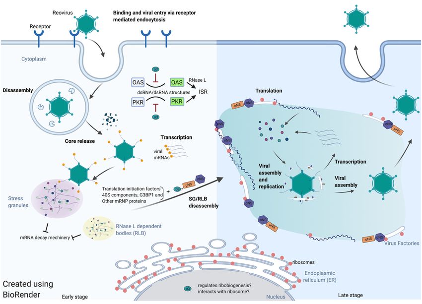
lation of reovirus mRNAs is spatiotemporally regulated (Figure 1). Evidence in support
of this hypothesis includes the observation that early after infections, reovirus induces
stress granules and the incoming viral core particles localize to SGs. Localization of enter-
ing core particles to SGs requires transcription of viral mRNA [25]. The SGs potentially
sequester and protect viral mRNA from the cellular RNA-decay machinery at the very
earliest stages of infection. As reovirus infection proceeds, reovirus viral proteins σNS,
µNS, and perhaps σ3 seem to be involved in the disruption of SGs even in the presence of
eIF2α phosphorylation. A reasonable model is that reovirus usurps SGs to provide access
to the translational machinery. The highly concentrated translation-initiation factors, 40S
ribosome components and certain mRNP regulators may be recruited to the VFs that are
formed gradually during infection. This relocation is mediated by the interaction between
σNS and SG protein G3BP1. As VFs form, active translation begins to occur on the margins
or within VFs. ER-membrane fragments are found inside the VFs that have been remodeled
by the actions of σNS and µNS. The function of the ER within VFs is as yet unknown, but
it seems reasonable to surmise that ER membranes act as scaffolds for viral core assembly
and replication. Similarly, rough ER membranes and ribosomes at the periphery
Viruses 2021, 13, x FOR PEER REVIEW 12 of 17of VFs
may function to support viral mRNA translation.
Figure
Figure 1. 1. Spatiotemporal regulation
Spatiotemporal regulation of
ofreovirus
reovirustranslation.
translation.
Funding: This research was funded by the National Institutes of Health, R01 AI121216.
Institutional Review Board Statement: Not applicable.
Informed Consent Statement: Not applicable.
Acknowledgments: We thank Shaun Cross for the careful reading and suggestions.Viruses 2021, 13, 275 12 of 17
Our growing understanding of reovirus translation leaves many questions as yet
unanswered. Some important questions to be addressed in future studies are as follows:
# What types of RNP granules or RNP granules combinations are formed at early stages
after infection, and what function do RNP granules have during viral infection?
# Are early viral mRNAs translated within RNP granules?
# What is the mechanism by which reoviruses disrupt SGs/RNP granules?
# How does σ3 regulate reovirus translation?
# What is the functional significance of the nuclear localization of σ3?
# Are uncapped viral mRNAs synthesized late in infection, and if so, how does σ3
promote their translation?
# How does the activation of the ISR and PKR or RNase L benefit viral replication?
Funding: This research was funded by the National Institutes of Health, R01 AI121216.
Institutional Review Board Statement: Not applicable.
Informed Consent Statement: Not applicable.
Acknowledgments: We thank Shaun Cross for the careful reading and suggestions.
Conflicts of Interest: The authors declare no conflict of interest.
References
1. Jackson, R.J.; Hellen, C.U.T.; Pestova, T.V. The mechanism of Eukaryotic Translation Initiation and Principles of Its Regulation.
Nat. Rev. Mol. Cell Biol. 2010, 11, 113–127. [CrossRef]
2. Schuller, A.P.; Green, R. Roadblocks and Resolutions in Eukaryotic Translation. Nat. Rev. Mol. Cell Biol. 2018, 19, 526–541.
[CrossRef]
3. Sonenberg, N.; Hinnebusch, A.G. Regulation of Translation Initiation in Eukaryotes: Mechanisms and Biological Targets. Cell
2009, 136, 731–745. [CrossRef] [PubMed]
4. Hoang, H.-D.; Neault, S.; Pelin, A.; Alain, T. Emerging Translation Strategies during Virus–Host Interaction. Wiley Interdiscip. Rev.
RNA 2021, 12, e1619. [CrossRef] [PubMed]
5. Chen, F.; Wu, P.; Deng, S.; Zhang, H.; Hou, Y.; Hu, Z.; Zhang, J.; Chen, X.; Yang, J.-R. Dissimilation of Synonymous Codon Usage
Bias in Virus–Host Coevolution Due to Translational Selection. Nat. Ecol. Evol. 2020, 4, 589–600. [CrossRef] [PubMed]
6. Quax, T.E.F.; Claassens, N.J.; Söll, D.; van der Oost, J. Codon Bias as a Means to Fine-Tune Gene Expression. Mol. Cell 2015, 59,
149–161. [CrossRef] [PubMed]
7. Mioduser, O.; Goz, E.; Tuller, T. Significant Differences in Terms of Codon Usage Bias between Bacteriophage Early and Late
Genes: A Comparative Genomics Analysis. BMC Genom. 2017, 18, 866. [CrossRef] [PubMed]
8. Fields, B.N.; Knipe, D.M.; David, M.; Howley, P.M. Fields Virology, 6th ed.; Wolters Kluwer Health/Lippincott Williams & Wilkins:
Philadelphia, PA, USA, 2013; ISBN 978-1-4511-0563-6.
9. Shatkin, A.J.; LaFiandra, A.J. Transcription by Infectious Subviral Particles of Reovirus. J. Virol. 1972, 10, 698–706. [CrossRef]
[PubMed]
10. Watanabe, Y.; Millward, S.; Graham, A.F. Regulation of Transcription of the Reovirus Genome. J. Mol. Biol. 1968, 36, 107–123.
[CrossRef]
11. Ernst, H.; Shatkin, A.J. Reovirus Hemagglutinin mRNA Codes for Two Polypeptides in Overlapping Reading Frames. Proc. Natl.
Acad. Sci. USA 1985, 82, 48–52. [CrossRef] [PubMed]
12. Belli, B.A.; Samuel, C.E. Biosynthesis of Reovirus-Specified Polypeptides: Expression of Reovirus S1-Encoded σ1 NS Protein in
Transfected and Infected Cells as Measured with Serotype Specific Polyclonal Antibody. Virology 1991, 185, 698–709. [CrossRef]
13. Sarkar, G.; Pelletier, J.; Bassel-Duby, R.; Jayasuriya, A.; Fields, B.N.; Sonenberg, N. Identification of a New Polypeptide Coded by
Reovirus Gene S1. J. Virol. 1985, 54, 720–725. [CrossRef]
14. Busch, L.K.; Rodríguez-Grille, J.; Casal, J.I.; Martínez-Costas, J.; Benavente, J. Avian and Mammalian Reoviruses Use Different
Molecular Mechanisms to Synthesize Their µNS Isoforms. J. Gen. Virol. 2011, 92, 2566–2574. [CrossRef]
15. Sagar, V.; Murray, K.E. The Mammalian Orthoreovirus Bicistronic M3 mRNA Initiates Translation Using a 50 End-Dependent,
Scanning Mechanism That Does Not Require Interaction of 50 -30 Untranslated Regions. Virus Res. 2014. [CrossRef]
16. Furuichi, Y.; Morgan, M.; Muthukrishnan, S.; Shatkin, A.J. Reovirus Messenger RNA Contains a Methylated, Blocked 50 -Terminal
Structure: M-7G(5’)Ppp(5’)G-MpCp-. Proc. Natl. Acad. Sci. USA 1975, 72, 362–366. [CrossRef] [PubMed]
17. Luongo, C.L.; Reinisch, K.M.; Harrison, S.C.; Nibert, M.L. Identification of the Guanylyltransferase Region and Active Site in
Reovirus mRNA Capping Protein Lambda2. J. Biol. Chem. 2000, 275, 2804–2810. [CrossRef] [PubMed]Viruses 2021, 13, 275 13 of 17
18. Luongo, C.L.; Contreras, C.M.; Farsetta, D.L.; Nibert, M.L. Binding Site for S-Adenosyl-L-Methionine in a Central Region of
Mammalian Reovirus Lambda2 Protein. Evidence for Activities in mRNA Cap Methylation. J. Biol. Chem. 1998, 273, 23773–23780.
[CrossRef] [PubMed]
19. Reinisch, K.M.; Nibert, M.L.; Harrison, S.C. Structure of the Reovirus Core at 3.6 Å Resolution. Nature 2000, 404, 960–967.
[CrossRef] [PubMed]
20. Narayanan, K.; Makino, S. Interplay between Viruses and Host mRNA Degradation. Biochim. Biophys. Acta Gene Regul. Mech.
2013, 1829, 732–741. [CrossRef]
21. Ng, W.C.; Soto-Acosta, R.; Bradrick, S.S.; Garcia-Blanco, M.A.; Ooi, E.E. The 50 and 30 Untranslated Regions of the Flaviviral
Genome. Viruses 2017, 9, 137. [CrossRef]
22. Nonoyama, M.; Millward, S.; Graham, A.F. Control of Transcription of the Reovirus Genome. Nucleic Acids Res. 1974, 1, 373–385.
[CrossRef]
23. Farsetta, D.L.; Chandran, K.; Nibert, M.L. Transcriptional Activities of Reovirus RNA Polymerase in Recoated Cores. Initiation
and Elongation Are Regulated by Separate Mechanisms. J. Biol. Chem. 2000, 275, 39693–39701. [CrossRef] [PubMed]
24. Zamora, P.F.; Hu, L.; Knowlton, J.J.; Lahr, R.M.; Moreno, R.A.; Berman, A.J.; Prasad, B.V.V.; Dermody, T.S. Reovirus Nonstructural
Protein σNS Acts as an RNA-Stability Factor Promoting Viral Genome Replication. J. Virol. 2018, 92, e00563-18. [CrossRef]
[PubMed]
25. Broering, T.J.; Kim, J.; Miller, C.L.; Piggott, C.D.S.; Dinoso, J.B.; Nibert, M.L.; Parker, J.S.L. Reovirus Nonstructural Protein µNS
Recruits Viral Core Surface Proteins and Entering Core Particles to Factory-like Inclusions. J. Virol. 2004, 78, 1882–1892. [CrossRef]
[PubMed]
26. Qin, Q.; Hastings, C.; Miller, C.L. Mammalian Orthoreovirus Particles Induce and Are Recruited into Stress Granules at Early
Times Postinfection. J. Virol. 2009, 83, 11090–11101. [CrossRef] [PubMed]
27. Wells, S.E.; Hillner, P.E.; Vale, R.D.; Sachs, A.B. Circularization of mRNA by Eukaryotic Translation Initiation Factors. Mol. Cell
1998, 2, 135–140. [CrossRef]
28. Groft, C.M.; Burley, S.K. Recognition of EIF4G by Rotavirus NSP3 Reveals a Basis for mRNA Circularization. Mol. Cell 2002, 9,
1273–1283. [CrossRef]
29. Deo, R.C.; Groft, C.M.; Rajashankar, K.R.; Burley, S.K. Recognition of the Rotavirus mRNA 3’ Consensus by an Asymmetric NSP3
Homodimer. Cell 2002, 108, 71–81. [CrossRef]
30. Gillian, A.L.; Schmechel, S.C.; Livny, J.; Schiff, L.A.; Nibert, M.L. Reovirus Protein σNS Binds in Multiple Copies to Single-
Stranded RNA and Shares Properties with Single-Stranded DNA Binding Proteins. J. Virol. 2000, 74, 5939–5948. [CrossRef]
[PubMed]
31. Gillian, A.L.; Nibert, M.L. Amino Terminus of Reovirus Nonstructural Protein σNS Is Important for ssRNA Binding and
Nucleoprotein Complex Formation. Virology 1998, 240, 1–11. [CrossRef]
32. Kozak, M.; Shatkin, A.J. Characterization of Ribosome-Protected Fragments from Reovirus Messenger RNA. J. Biol. Chem. 1976,
251, 4259–4266. [CrossRef]
33. Kozak, M. Binding of Wheat Germ Ribosomes to Fragmented Viral mRNA. J. Virol. 1980, 35, 748–756. [CrossRef] [PubMed]
34. Kozak, M. Binding of Wheat Germ Ribosomes to Bisulfite-Modified Reovirus Messenger RNA: Evidence for a Scanning
Mechanism. J. Mol. Biol. 1980, 144, 291–304. [CrossRef]
35. Kozak, M. Analysis of Ribosome Binding Sites from the S1 Message of Reovirus. Initiation at the First and Second AUG Codons.
J. Mol. Biol. 1982, 156, 807–820. [CrossRef]
36. Shatkin, A.J. Methylated Messenger RNA Synthesis In Vitro by Purified Reovirus. Proc. Natl. Acad. Sci. USA 1974, 71, 3204–3207.
[CrossRef] [PubMed]
37. Furuichi, Y.; Muthukrishnan, S.; Shatkin, A.J. 5’-Terminal m-7G(5’)Ppp(5’)G-m-p in Vivo: Identification in Reovirus Genome
RNA. Proc. Natl. Acad. Sci. USA 1975, 72, 742–745. [CrossRef] [PubMed]
38. Ingolia, N.T. Ribosome Profiling: New Views of Translation, from Single Codons to Genome Scale. Nat. Rev. Genet. 2014.
[CrossRef] [PubMed]
39. Castelló, A.; Fischer, B.; Eichelbaum, K.; Horos, R.; Beckmann, B.M.; Strein, C.; Davey, N.E.; Humphreys, D.T.; Preiss, T.; Steinmetz,
L.M.; et al. Insights into RNA Biology from an Atlas of Mammalian mRNA-Binding Proteins. Cell 2012, 149, 1393–1406. [CrossRef]
[PubMed]
40. Shi, Z.; Barna, M. Translating the Genome in Time and Space: Specialized Ribosomes, RNA Regulons, and RNA-Binding Proteins.
Annu. Rev. Cell Dev. Biol. 2015, 31, 31–54. [CrossRef]
41. Bischoff, J.R.; Samuel, C.E. Mechanism of Interferon Action. Activation of the Human P1/EIF-2 Alpha Protein Kinase by
Individual Reovirus s-Class mRNAs: S1 mRNA Is a Potent Activator Relative to S4 mRNA. Virology 1989, 172, 106–115. [CrossRef]
42. Ivanov, P.; Kedersha, N.; Anderson, P. Stress Granules and Processing Bodies in Translational Control. Cold Spring Harb. Perspect.
Biol. 2019, 11, a032813. [CrossRef]
43. Tsai, W.-C.; Lloyd, R.E. Cytoplasmic RNA Granules and Viral Infection. Annu. Rev. Virol. 2014, 1, 147–170. [CrossRef] [PubMed]
44. Treeck, B.V.; Protter, D.S.W.; Matheny, T.; Khong, A.; Link, C.D.; Parker, R. RNA Self-Assembly Contributes to Stress Granule
Formation and Defining the Stress Granule Transcriptome. Proc. Natl. Acad. Sci. USA 2018, 115, 2734–2739. [CrossRef] [PubMed]
45. Van Treeck, B.; Parker, R. Emerging Roles for Intermolecular RNA-RNA Interactions in RNP Assemblies. Cell 2018, 174, 791–802.
[CrossRef]You can also read