The NHL domain of BRAT is an RNA-binding domain that directly contacts the hunchback mRNA for regulation
←
→
Page content transcription
If your browser does not render page correctly, please read the page content below
Downloaded from genesdev.cshlp.org on October 25, 2015 - Published by Cold Spring Harbor Laboratory Press

The NHL domain of BRAT is an RNA-
binding domain that directly contacts
the hunchback mRNA for regulation
Inga Loedige,1,8,9 Mathias Stotz,1,8 Saadia Qamar,2,3 Katharina Kramer,2,3 Janosch Hennig,4,5
Thomas Schubert,6 Patrick Löffler,7 Gernot Längst,6 Rainer Merkl,7 Henning Urlaub,2,3
and Gunter Meister1,9
1
 Biochemistry Center Regensburg (BZR), Laboratory for RNA Biology, University of Regensburg, 93053 Regensburg, Germany;
2
 Bioanalytical Mass Spectrometry, Max Planck Institute for Biophysical Chemistry, 37077 Göttingen, Germany; 3Bioanalytics,
Institute for Clinical Chemistry, University Medical Center Göttingen, 37075 Göttingen, Germany; 4Group Biomolecular NMR,
Institute of Structural Biology, Helmholtz Zentrum München-German Research Center for Environmental Health, 85764
Neuherberg, Germany; 5Center for Integrated Protein Science Munich, Biomolecular NMR, Department Chemie, Technische
Universität München, 85748 Garching, Germany; 6Institute for Biochemistry III, University of Regensburg, 93053 Regensburg,
Germany; 7Institute of Biophysics and Physical Biochemistry, University of Regensburg, 93053 Regensburg, Germany

The Drosophila protein brain tumor (Brat) forms a complex with Pumilio (Pum) and Nanos (Nos) to repress
hunchback (hb) mRNA translation at the posterior pole during early embryonic development. It is currently
thought that complex formation is initiated by Pum, which directly binds the hb mRNA and subsequently
recruits Nos and Brat. Here we report that, in addition to Pum, Brat also directly interacts with the hb mRNA.
We identify Brat-binding sites distinct from the Pum consensus motif and show that RNA binding and
translational repression by Brat do not require Pum, suggesting so far unrecognized Pum-independent Brat
functions. Using various biochemical and biophysical methods, we also demonstrate that the NHL (NCL-1,
HT2A, and LIN-41) domain of Brat, a domain previously believed to mediate protein–protein interactions, is
a novel, sequence-specific ssRNA-binding domain. The Brat-NHL domain folds into a six-bladed b propeller, and
we identify its positively charged top surface as the RNA-binding site. Brat belongs to the functional diverse
TRIM (tripartite motif)-NHL protein family. Using structural homology modeling, we predict that the NHL
domains of all TRIM-NHL proteins have the potential to bind RNA, indicating that Brat is part of a conserved
family of RNA-binding proteins.
[Keywords: BRAT; RNA binding; TRIM-NHL; gene regulation; hunchback; translational repression]
Supplemental material is available for this article.
Received December 16, 2013; revised version accepted February 20, 2014.

Post-transcriptional gene regulation plays an impor-                           protein at the posterior pole, on the contrary, disrupts
tant role during early development of many organisms.                          abdominal segmentation.
In Drosophila embryos, the genome is transcriptionally                           Inhibition of hb translation at the posterior pole re-
silent, and the differential localization, stabilization, and                  quires a repressive complex containing the proteins Pumilio
translation of maternally provided mRNAs generate pro-                         (Pum), Nanos (Nos), and brain tumor (Brat) and two
tein gradients that specify the body axes (Nusslein-                           regulatory elements, referred to as the Nanos response
Volhard et al. 1987; Curtis et al. 1995). Translation of                       elements (NREs), located in the 39 untranslated region
the maternally provided and, in the embryo, uniformly                          (UTR) of the hb mRNA (Wharton and Struhl 1991;
distributed hunchback (hb) mRNA, for example, is re-                           Murata and Wharton 1995; Sonoda and Wharton 2001).
stricted to the anterior pole of the embryo, where it                          The NREs are high-affinity binding sites for the sequence-
directs head and thorax formation (Tautz and Pfeifle                           specific RNA-binding protein Pum (Murata and Wharton
1989; Hulskamp et al. 1990). Aberrant expression of Hb

                                                                               Ó 2014 Loedige et al. This article is distributed exclusively by Cold
8
 These authors contributed equally to this work.                               Spring Harbor Laboratory Press for the first six months after the full-issue
9
 Corresponding authors                                                         publication date (see http://genesdev.cshlp.org/site/misc/terms.xhtml).
E-mail gunter.meister@vkl.uni-regensburg.de                                    After six months, it is available under a Creative Commons License
E-mail inga.loedige@vkl.uni-regensburg.de                                      (Attribution-NonCommercial 4.0 International), as described at http://
Article is online at http://www.genesdev.org/cgi/doi/10.1101/gad.236513.113.   creativecommons.org/licenses/by-nc/4.0/.

        GENES & DEVELOPMENT 28:749–764 Published by Cold Spring Harbor Laboratory Press; ISSN 0890-9369/14; www.genesdev.org                           749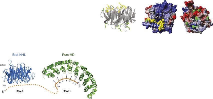
Downloaded from genesdev.cshlp.org on October 25, 2015 - Published by Cold Spring Harbor Laboratory Press

Loedige et al.

1995; Zamore et al. 1997), which further recruits Nos and            regulation of myc has been reported in this context
Brat to the complex (Sonoda and Wharton 1999, 2001).                 (Betschinger et al. 2006). Strikingly, the same mutations
While Nos determines the site and extent of hb repression            that impair Brat’s interaction with Pum and therefore
(by a mechanism that is not yet fully understood), Brat              disrupt Brat-mediated translational repression are also
interacts with the cap-binding protein d4EHP to inhibit              the cause of the overproliferation phenotype. A role for
translational initiation (Cho et al. 2006).                          Pum in this process, however, has not been described.
   Apart from the regulation of the hb mRNA during                      Our previous work indicated that the mammalian
early embryogenesis, several additional examples of                  TRIM-NHL protein TRIM71/LIN-41, which shares a sim-
translational regulation by a repressive complex of Brat             ilar domain architecture with Brat, acts as a translational
and Pum or Brat, Pum, and Nos in various developmen-                 repressor, and we identified its NHL domain as necessary
tal contexts have been reported. For example, Brat and               and sufficient for RNA targeting (Loedige et al. 2013).
Pum repress myc and mad translation to promote differ-               A role for the mammalian Pum proteins PUM1 and PUM2
entiation during oogenesis (Harris et al. 2011). Further-            in mediating RNA binding of TRIM71, however, was not
more, Brat, Pum, and Nos inhibit the paralytic mRNA to               evident. In the meantime, a more global UV-cross-linking
control excitability in motorneurons (Muraro et al. 2008),           approach reported an RNA-binding activity of the TRIM71-
and, finally, in neuromuscular synapses, repression of               NHL domain (Kwon et al. 2013).
mad mRNA by Brat regulates synaptic size (Shi et al.                    Using various biochemical and biophysical assays, we
2013).                                                               demonstrated that the NHL domain of Brat directly binds
   Brat belongs to the conserved family of tripartite motif          RNA with high affinity. We identified the BoxA motif of
(TRIM)-NHL (NCL-1, HT2A, and LIN-41), which are                      the NRE as the Brat-binding site and demonstrated that
characterized by their N-terminal TRIM and a C-termi-                RNA binding and translational repression by Brat are
nal NHL domain (Sardiello et al. 2008). The TRIM motif               independent of Pum. Using UV-cross-linking followed by
consists of a RING domain, conferring ubiquitin or                   mass spectrometry (MS), we identified the top surface of
ubiquitin-like ligase activity, followed by one or two               the NHL domain as the RNA-binding surface. Mutations
B-box motifs and a coiled coil region. Brat, however, is             of single residues on the top surface abrogate Brat-NHL
only an ‘‘incomplete’’ TRIM, as it lacks the RING domain             RNA binding in vitro and impair translational repression
(Fig. 1A). The NHL domain, which is named after the                  by Brat in vivo.
three proteins NCL-1, HT2A (TRIM32), and LIN-41 in
which it was first identified (Slack and Ruvkun 1998), is
of critical importance for Brat function: Flies carrying             Results
a deletion of or single point mutations within the NHL
                                                                     The NHL domain of Drosophila Brat directly
domain are characterized by various mutant phenotypes,
                                                                     binds RNA
including abdominal segmentation defects that can be
rescued by re-expression of the wild-type NHL domain                 An ;100-nucleotide (nt)-long fragment from the hb 39
alone (Arama et al. 2000; Sonoda and Wharton 2001).                  UTR that contains the two NRE’s (termed hb RNA
   The crystal structure of the Brat-NHL domain revealed             hereafter) (Fig. 1B) is required for translational repression
a six-bladed b propeller, a fold resembling that of WD40             of the hb mRNA in vivo (Wharton and Struhl 1991). The
domains (Edwards et al. 2003). Owing to their large                  current model suggests that upon RNA binding, Pum
surfaces, b-propeller structures often provide platforms             serves as a platform and recruits Nos and Brat to the RNA
for multiple protein–protein interactions and often serve            by protein–protein interactions (Sonoda and Wharton
as hubs in cellular protein interaction networks (Stirnimann         1999, 2001). To gain insight into a putative RNA-binding
et al. 2010). Consistent with this idea, mutagenesis studies         activity of Brat, we performed electrophoretic mobility
in flies showed that the top surface of the Brat-NHL                 shift assays (EMSAs or band shift assays) using in vitro
domain interacts with Pum and is thus essential for the              transcribed hb RNA as a substrate and the recombinantly
recruitment of Brat to RNA (Sonoda and Wharton 2001;                 expressed and purified NHL domain of Brat (encompass-
Edwards et al. 2003), while its bottom surface contacts              ing amino acids 756–1037) (Supplemental Fig. 1A).
d4EHP (Cho et al. 2006).                                               We incubated increasing amounts of recombinant Brat-
   In addition to its role as a translational repressor, Brat        NHL with the radiolabeled hb RNA and analyzed the
has been recognized as a powerful growth suppressor and              resulting protein–RNA complexes by native gel electro-
differentiation factor (Frank et al. 2002; Betschinger et al.        phoresis (Fig. 1C). Indeed, the Brat-NHL domain readily
2006; Lee et al. 2006; Harris et al. 2011). During asym-             bound RNA with high affinity: At a protein concentration
metric cell divisions (a characteristic hallmark of stem             of ;100–150 nM, half of the free RNA was shifted into
cells), Brat is confined to the differentiating daughter cell,       a slower-migrating RNA–protein complex (Fig. 1C, lanes
where it restricts proliferation and promotes differentia-           6,7; Supplemental Fig. 2B, indicated by a black arrow).
tion. Lack of Brat function causes tumorous overgrowth               With increasing protein concentrations, a second, less
of the larval brain that is due to the failure of neuronal           well-defined band appeared (Fig. 1C, lanes 9–13, Supple-
progenitor cells to exit proliferation (Betschinger et al.           mental Fig. 2B, marked by an asterisk). RNA binding of
2006; Lee et al. 2006). It is currently unclear whether the          the Brat-NHL domain was sequence-specific, as the labeled
brain tumor phenotype is linked to Brat’s function as                RNA–protein complexes could be chased by an excess of
a translational repressor, but post-transcriptional down-            unlabeled hb RNA but not by an excess of tRNA that was

750       GENES & DEVELOPMENT
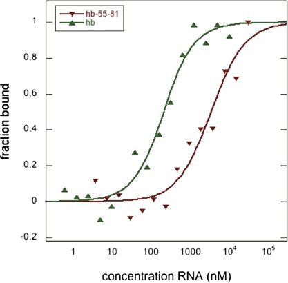
Downloaded from genesdev.cshlp.org on October 25, 2015 - Published by Cold Spring Harbor Laboratory Press

Figure 1. Direct RNA binding of the Brat-NHL domain. (A) Schematic representation of Drosophila melanogaster Brat domain
organization. The two B-boxes (B1 and B2) are shown in orange, the coiled coil (CC) domain is in yellow, and the NHL domain, composed
of six NHL repeats, is in green. Also indicated and colored in gray are serine-rich (S), glutamine-rich (Q), and histidine-rich (H) stretches. The
numbers below indicate domain boundaries. (B) Schematic presentation of the maternally derived hb mRNA (NM_169234) and hb RNA
fragments used in this study. The ;100-nt-long hb RNA fragment (for simplicity referred to here as hb RNA) contains two NREs, each
composed of one BoxA and one BoxB motif. Nucleotides mutated in hb 55–81 are indicated by red letters. (C) Native gel analysis to probe for
RNA binding of the Brat-NHL domain. Increasing amounts of recombinant Brat-NHL (amino acids 756–1037) were incubated with 500 pM
32
  P-labeled hb RNA, and complexes were analyzed by native gel electrophoresis. (D) Brat-NHL binds sequences that contain the NRE. Short,
27-nt, 32P-labeled RNA probes that span the hb RNA (as depicted in A) were incubated with the indicated amounts of recombinant Brat-
NHL, and complexes were analyzed by native gel electrophoresis. (E) MST measurements. (Top) Representative binding curves for the
interaction of Brat-NHL with hb RNA (green triangles) or hb fragment 55–81 (red triangles). (Bottom) Summary of independent MST
measurements. Brat-NHL binds the hb RNA with a Kd of 137.4 nM 6 36.8 nM (six independent repeats); the short hb (fragment 55–81) is
bound with lower affinity and a Kd of 2.0 mM 6 0.8 mM (five independent repeats). No binding was detected for hb mutants that lack the
Brat-binding motif (NRE2BoxA or NRE1+2BoxA) (see Fig. 2). (F) The Brat-NHL domain is a sequence-specific, ssRNA-binding domain.
Complex formation of recombinant Brat-NHL with ssRNA (lanes 1–3), ssRNA of antisense sequence (lanes 4–6), dsRNA (lanes 7–9), or
ssDNA (lanes 10–12) was analyzed by native gel electrophoresis. In all cases, nucleotide sequences corresponding to hb fragment 55–81 were
used. DNA oligonucleotides contained dT instead of dU. (G) Mutations in BoxA but not in BoxB abrogate Brat-NHL binding to the NRE2.
Native gel analysis to test Brat-NHL binding to hb 55–81 or the indicated mutants. Free RNA or RNA–protein complexes are indicated by
black arrows. Asterisk (*) denotes a less well-defined Brat-NHL–hb RNA complex appearing at high protein concentrations.
Downloaded from genesdev.cshlp.org on October 25, 2015 - Published by Cold Spring Harbor Laboratory Press

Loedige et al.

present in all reactions (Fig. 1C; Supplemental Fig. 2A).            regulation in vivo (Murata and Wharton 1995). While
Our data therefore demonstrate that the NHL domain of                the BoxB motif constitutes the high-affinity binding site
Brat is able to bind to the hb RNA directly and in                   for Pum (Zamore et al. 1997; Wang et al. 2002), the exact
a sequence-specific manner.                                          contribution of the BoxA motif to hb regulation is un-
                                                                     clear. To further define which sequence element within
The Brat-NHL domain binds the NRE                                    the NRE in the hb mRNA is responsible for Brat-NHL
To determine which RNA sequences within the hb                       recruitment, we introduced mutations into either the
mRNA are responsible for Brat recruitment, we divided                short 27-nt hb RNA fragment encompassing NRE2 (hb
the 100-nt-long hb RNA used above into seven over-                   55–81) (Fig. 1G) or the long 100-nt hb RNA (Fig. 2) and
lapping fragments of 27 nt in length (Fig. 1B) and tested            determined their effect on Brat-NHL binding in band shift
them in EMSAs (Fig. 1D). The Brat-NHL domain inter-                  assays. Mutation of four uracils into adenines within or
acted with fragments that contained either NRE1 (frag-               preceding the BoxA motif (hb 55–81 mut BoxA) abolished
ment 1–27) or NRE2 (fragment 55–81) (Fig. 1D, lanes 2,10,            Brat-NHL binding to the short hb 55–81 fragment
respectively). Of note, binding of the Brat-NHL domain to            (Fig. 1G, lanes 6–8), but Brat-NHL still bound this
these short fragments was much weaker than observed                  fragment when the BoxB motif was mutated (hb 55–81
for the longer hb RNA, and a concentration of 2 mM                   mut BoxB) (Fig. 1G, lanes 10–12). Similarly, Brat-NHL
protein was needed to shift half of the short hb RNA                 still bound the long hb RNA with high affinity when both
(fragment 55–81) into a slower-migrating protein–RNA                 BoxB sites were mutated (Fig. 2B, lanes 5,6), but mutation
complex (Supplemental Fig. 2C).                                      of both BoxA sites greatly impaired Brat-NHL binding
   To confirm our observations by an independent ap-                 (Fig. 2B, lanes 8,9), indicating that the Brat-NHL domain
proach, we determined binding affinities using micro-                binds to the BoxA motif of the hb NREs. Notably, while
scale thermophoresis (MST) (Fig. 1E), a technique that is            mutating both BoxB sites did not affect the affinity of
based on the movement of molecules in temperature                    Brat-NHL for the hb RNA, it did abolish the appearance of
gradients (Jerabek-Willemsen et al. 2011; Zillner et al.             the second, less well-defined Brat-RNA complex that
2012). The thermophoretic mobility of fluorescently                  appeared at high protein concentrations (Fig. 2B, marked
labeled Brat-NHL was recorded at different RNA concen-               by an asterisk).
trations ranging from 0 to 10 mM (long hb RNA) or 30 mM                 To complement our binding studies, we also performed
(short hb RNA), and the data were used to calculate                  band shift assays with the purified RNA-binding domain
binding affinities (Fig. 1E). The determined binding affin-          of Pum (amino acids 1093–1426, Pum homology domain
ities from independent thermophoresis experiments,                   [Pum-HD]) (Supplemental Fig. 3; Zamore et al. 1997). As
each performed with independently labeled Brat-NHL,                  shown previously, the Pum-HD binds to hb mRNA with
closely matched the affinities determined in our gel shift           high affinity. Under our assay conditions, half of the free
assays: The Brat-NHL domain bound to the long hb RNA                 RNA was shifted into a protein–RNA complex at a con-
with a Kd of 137.4 nM 6 36.8 nM, while the shorter                   centration of ;20 nM Pum-HD (Supplemental Fig. 3B). In
fragment encompassing NRE2 (hb 55–81) interacted with                contrast to Brat-NHL and as shown previously (Zamore
a Kd of 2 mM 6 0.8 mM (Fig. 1E). Hb RNA mutants that                 et al. 1997), binding of the Pum-HD to hb RNA was
lack the Brat-binding motif, which is described in more              abrogated by mutation of both BoxB sites but not by
detail below (Figs. 1G, 2), showed no detectable binding             mutation of both BoxA sites (Supplemental Fig. 3C, lanes
(Fig. 1E), confirming the validity of the assay.                     5,6 and 8,9, respectively). Thus, our data clearly indicate
                                                                     that the BoxB motif of the NRE recruits Pum, and the
The Brat-NHL domain binds ssRNA                                      BoxA motif is responsible for Brat binding.

To determine the substrate preference of the Brat-NHL
domain, we analyzed Brat-NHL binding to ssRNA (hb 55–                Two Brat-binding sites are necessary for
81), ssRNA containing the antisense sequence, dsRNA,                 high-affinity binding
or ssDNA (Fig. 1F). All oligonucleotides were 27 nt long             The Pum-HD binds independently and with equal affinity
and were based on the hb 55–81 fragment shown in                     to each of the two NREs, and no cooperative binding of
Figure 1D. Brat-NHL bound to the single-stranded sense               two Pum-HD molecules occupying adjacent NREs was
(Fig. 1D, lanes 2,3) but not the single-stranded antisense           observed (Zamore et al. 1999). Consistently, mutation of
(Fig. 1D, lanes 5,6) sequence, again confirming sequence-            either NRE1BoxB or NRE2BoxB did not abolish binding of
specific binding. No binding was observed to ssDNA (Fig.             the PUM-HD to hb RNA but simply eliminated the
1D, lanes 11,12), and only weak binding to dsRNA was                 possibility of recruiting two PUM-HD molecules onto
detectable (Fig. 1D, lanes 8,9). It is conceivable that this         one hb RNA (Supplemental Fig. 3D).
weak binding is due to incomplete annealing of the two                 In stark contrast, mutation of either NRE1BoxA or
single strands and would thus reflect binding of the Brat-           NRE2BoxA greatly impaired detectable binding of Brat-
NHL domain to ssRNA.                                                 NHL to the hb RNA (Fig. 2C, lanes 8,9 and 11,12,
                                                                     respectively), indicating that two intact binding sites
The Brat-NHL domain binds to BoxA of the hb NRE
                                                                     are necessary for the recruitment of the Brat-NHL do-
Each of the two NREs is composed of one BoxA and one                 main in vitro. The requirement for two adjacent Brat-
BoxB motif, both of which contribute to hb mRNA                      binding sites on one RNA molecule might explain our

752       GENES & DEVELOPMENT
Downloaded from genesdev.cshlp.org on October 25, 2015 - Published by Cold Spring Harbor Laboratory Press
The BRAT-NHL domain directly binds hb RNA
Figure 2. Mutations in BoxA, but not BoxB, abrogate binding of Brat-NHL to hb RNA. (A) Sequence of the hb RNA and its mutants
used in binding assays. Mutated nucleotides are indicated by red letters. (B–E) Recombinant Brat-NHL was incubated with 32P-labeled
hb RNA or mutant RNAs as indicated and analyzed by native gel electrophoresis. Mutation of either BoxA site is sufficient to greatly
impair RNA binding of Brat-NHL. Free RNA or RNA–protein complexes are indicated by black arrows. Asterisk (*) denotes a less well-
defined Brat-NHL–hb RNA complex appearing at high protein concentrations.
observation that the shorter hb fragments (hb 1–27 and hb NRE1BoxB or NRE2BoxB individually (Fig. 2D) as well as
55–81) that contain only one Brat binding site show additional mutations in NRE2BoxA (Fig. 2E) on Brat-NHL
a much lower affinity to Brat-NHL (Fig. 1D) compared binding. The two NRE2BoxA mutants (NRE2BoxA mut1 and
with the long hb RNA containing two sites. NRE2BoxA mut2) revealed that exchanging only two uracils
To complement our binding studies and further narrow within or preceding the BoxA motif of NRE2 suffice to
down the Brat-binding motif, we also tested mutations in abrogate Brat-NHL binding to the hb RNA in vitro (Fig. 2E,
GENES & DEVELOPMENT 753Downloaded from genesdev.cshlp.org on October 25, 2015 - Published by Cold Spring Harbor Laboratory Press
Loedige et al.
lanes 8,9 and 11,12, respectively), while mutation of complex only in the presence of RNA (Supplemental
NRE1BoxB (Fig. 2D, lanes 8,9) or the exchange of a uracil Fig. 5). Since the NHL domain was described as a putative
preceding the BoxA motif in NRE2 into an adenine protein–protein interaction domain (Slack and Ruvkun
(NRE2BoxA mut3) (Fig. 2E, lanes 14,15) prevented the 1998), these findings had previously been interpreted as
appearance of the less well-defined second Brat-NHL–hb a direct protein–protein interaction between RNA-bound
RNA complex, an observation that we cannot currently Pum and Brat (Sonoda and Wharton 2001; Edwards et al.
explain but that might indicate a potential contribution 2003). Given the close proximity of the BoxA and BoxB
of RNA folding to the accessibility of the Brat-binding motifs, we asked whether the Pum-HD and Brat-NHL
sites. might influence each other’s binding to the hb RNA.
To answer this question, we performed band shift
Stoichiometry of the Brat-NHL–hb RNA complex assays with increasing amounts of Brat-NHL and either
free hb RNA (Fig. 3A, lanes 1–7) or hb RNA that was
Although we observed only one high-affinity Brat-NHL–
prebound to the Pum-HD (Fig. 3A, lanes 8–14). While
hb RNA complex in our band shift assays (Fig. 1C) and the
Brat-NHL bound the free RNA with a Kd of ;100 nM
binding curves determined by MST did not indicate
(Figs. 1C, 3A; Supplemental Fig. 2B), a concentration of
cooperative or independent binding of two Brat-NHL
only 10 nM Brat-NHL was sufficient to shift half of the
molecules (Fig. 1E), the presence of two adjacent Brat-
Pum-HD-bound hb RNA into a slower-migrating com-
binding sites and their requirement for high-affinity
plex containing Brat-NHL, the Pum-HD, and hb RNA
binding (Fig. 2C) suggested that two Brat-NHL moieties
(Fig. 3A, lane 11; Supplemental Fig. 3). At a concentration
(possibly as a dimer) might bind to one hb RNA. To test
of 50 nM Brat-NHL, all of the Pum-HD-bound RNA was
this, static light scattering was used to determine the
shifted into the slower-migrating complex (Fig. 3A, lane
molecular weight of fractions eluted from an analytical
12). Thus, Brat-NHL binds to a Pum-HD–hb RNA com-
gel filtration column. As shown in Supplemental Figure 4,
plex with higher affinity than to free RNA, explaining
only peaks corresponding to unbound Brat-NHL and
previous findings (Edwards et al. 2003). The presence of
a Brat-NHL:hb RNA 1:1 complex could be detected even
the Pum-HD, however, could not overcome the require-
when Brat-NHL was added at a 5.3-fold excess over hb
ment for two Brat-binding sites, as no detectable binding
RNA. Thus, in vitro, only one Brat-NHL domain binds to
of the Brat-NHL domain to hb RNA occurred when the
the hb RNA containing two binding sites.
BoxA motif of either NRE2 (Fig. 3B) or of NRE1 (data not
shown) was mutated even if the RNA was prebound to
Pum-HD enhances Brat-NHL binding to the hb RNA
the Pum-HD. We also performed the reciprocal experi-
An RNA-dependent interaction between Pum and Brat ment, incubating increasing amounts of the Pum-HD
had previously been observed in yeast four-hybrid and in with either free hb RNA or hb RNA that was prebound to
vitro pull-down assays (Sonoda and Wharton 2001), and Brat-NHL (Supplemental Fig. 6C). Correspondingly, the
gel filtration experiments performed by us similarly Pum-HD preferentially bound hb RNA that was pre-
indicated that Brat-NHL and the Pum-HD formed a stable bound by Brat-NHL. Thus, binding of the Pum-HD or
Figure 3. Binding of the Pum-HD to hb RNA
facilitates Brat-NHL binding but does not over-
come the requirement for two Brat-binding sites.
(A) Binding of the Pum-HD and Brat-NHL to the hb
RNA is not mutually exclusive, and preincubation
of hb RNA with the Pum-HD facilitates Brat-NHL
binding. Indicated amounts of Brat-NHL were
mixed with 32P labeled hb RNA alone (lanes 1–7)
or hb RNA preincubated with 10 nM Pum-HD
(lanes 8–14). Complexes were separated by native
gel electrophoresis. (B) Binding of the Pum-HD to
hb RNA does not overcome the requirement for
two Brat-binding sites. Experiment was done as
described in A except that the hb mutant NRE2BoxA
was used as a substrate for complex formation.
754 GENES & DEVELOPMENTDownloaded from genesdev.cshlp.org on October 25, 2015 - Published by Cold Spring Harbor Laboratory Press
The BRAT-NHL domain directly binds hb RNA
Brat-NHL to RNA facilitates the recruitment of the other, porter generally showed elevated expression in all sam-
an observation that could be explained by a stabilizing ples when compared with the other hb reporter con-
effect due to weak protein–protein interactions between structs, indicative of repression relief due to mutation
the Pum-HD and Brat-NHL or by an altered, more of the Pum-binding sites (see raw data in Supplemental
accessible RNA structure that would facilitate binding Fig. 7A). For reasons that we cannot yet explain, knock-
of the second protein. It is currently unknown whether down of Brat did not relieve repression of any reporter
a stronger protein–protein interaction between Brat construct tested (Fig. 4D), although Brat was expressed in
and Pum might occur in the context of the full-length Dmel2 cells and was efficiently depleted by the knock-
proteins. down approach (Fig. 4C, lanes 5,6), demonstrating a lack
of repression by endogenous Brat in Dmel2 cells. Possibly,
the endogenous expression level of Brat in Dmel2 cells is
Repression of hb by Brat is independent of Pum
not sufficient to repress the transfected reporter plasmids.
To test whether our in vitro binding studies have impli- Under Pum knockdown conditions (Fig. 4E), the extent
cations for the repression of the hb mRNA by Brat and of Brat-mediated hb repression was comparable with that
Pum in vivo, we performed reporter gene assays in Dmel2 in untreated cells, again demonstrating that repression by
cells, a cell line derived from Drosophila embryonic Brat is independent of Pum.
Schneider cells (Fig. 4). The 100-nt hb 39 UTR fragment To analyze other known Brat and Pum targets as well
used above or respective mutants were fused to the (Harris et al. 2011; Shi et al. 2013), we tested repression of
coding sequence of firefly luciferase (FL) (Fig. 4A) and myc and mad 39 UTRs by HA-Brat and HA-Pum over-
coexpressed with either full-length HA-tagged Pum or expression (Fig. 4F). While the myc 39 UTR reporter was
HA-tagged Brat and a Renilla luciferase (RL)-containing repressed ;1.5-fold by expression of HA-Brat or HA-Pum,
control vector. repression of the mad 39 UTR reporter was observed only
Expression of HA-Brat or HA-Pum, but not of HA- under HA-Brat but not HA-Pum overexpression condi-
Gawky (HA-GW) (a translational repressor not implicated tions. Similarly, depletion of endogenous Pum did not
in the regulation of hb), led to a dose-dependent repres- cause repression relief of the mad 39 UTR reporter, while
sion of the hb reporter (Fig. 4B; Supplemental Fig. 7B). The the expression of the myc and hb reporters was up-
hb NRE1+2BoxA mutant, which did not bind Brat-NHL in regulated (Fig. 4G). Our data therefore suggest that mad
our in vitro binding assays, was not repressed by HA-Brat, might be a Pum-independent Brat target.
and the hb NRE1+2BoxB mutant, which failed to recruit
the Pum-HD (Fig. 4B), showed no repression by HA-Pum,
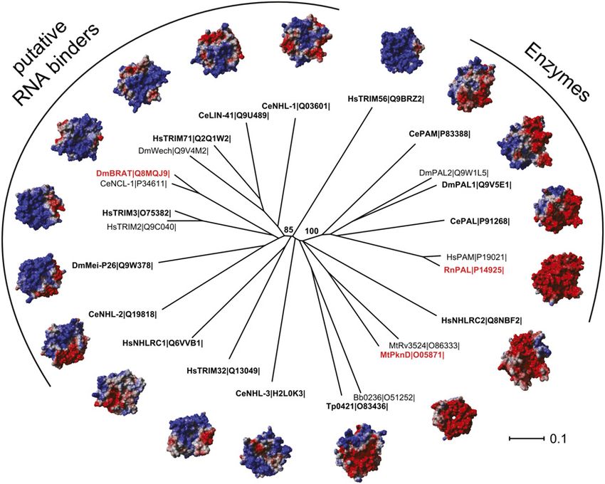
The electropositive top surface of the NHL domain
clearly demonstrating that the BoxA motif is responsible
contacts RNA
for recruitment of and repression by Brat, while the BoxB
motif recruits and confers repression by Pum. Addition- The six NHL repeats of the Brat-NHL domain fold into
ally, these experiments demonstrate that repression by a six-bladed b propeller (Fig. 5A,B; Edwards et al. 2003),
Brat and Pum can occur independently of each other, as a structure that serves as a platform for diverse molecular
the mutant that did not recruit Pum (NRE1+2BoxB) was interactions (Stirnimann et al. 2010). Electrostatic calcu-
still repressed by Brat, and the mutant that did not bind lations reveal an overall positive charge of the top surface
Brat (NRE1+2BoxA) was still repressed by Pum. (Fig. 5C; Edwards et al. 2003), and many positively charged
Contrary to our in vitro binding studies, mutation of and aromatic residues ideally suited for the interaction
each BoxA motif individually did not fully abrogate with RNA protrude from this surface. Strikingly, all
repression by Brat: Mutation of NRE2BoxA impaired re- mutations that cause a Brat mutant phenotype or that
pression by Brat, but mutation of NRE1BoxA had no abrogate Brat function affect this surface (Arama et al.
appreciable effect on Brat-mediated repression, suggest- 2000; Sonoda and Wharton 2001; Harris et al. 2011).
ing that one Brat-binding site is sufficient for Brat re- Additionally, this surface had been proposed to mediate
cruitment in vivo. These data also suggest that Brat the interaction between Brat and Pum (Sonoda and
exhibits lower affinity toward the BoxA motif of NRE1 Wharton 2001; Edwards et al. 2003). Our finding of a
than to that of NRE2, which is in agreement with our in direct RNA-binding activity of the Brat-NHL domain
vitro binding studies (Fig. 1D; data not shown). suggests that the described RNA-induced interaction be-
Since Dmel2 cells express endogenous Brat and Pum tween Brat and Pum (Sonoda and Wharton 2001; Edwards
(Cherbas et al. 2011; Harris et al. 2011; Weidmann and et al. 2003) might actually be an RNA-mediated interac-
Goldstrohm 2012), we also performed knockdown exper- tion, and mutations affecting Pum binding might in fact be
iments. As we lacked an antibody against endogenous RNA-binding mutants.
Pum, we confirmed knockdown efficiency by the down- To test this hypothesis, we performed band shift assays
regulation of coexpressed HA-tagged Pum (Fig. 4C, lanes with recombinant Brat-NHL (Supplemental Fig. 1B) car-
1,2). Knockdown of Pum relieved repression of the hb rying point mutations on the top (reported to affect
reporter (Fig. 4D), demonstrating its repression by endog- interaction with Pum) or the bottom (shown to have no
enous Pum. Repression was dependent on two intact effect on Pum binding) surface (Fig. 5D,E). While all top
BoxB motifs, as the mutant hb reporter construct surface mutations greatly impaired (Y829A and R847A)
NRE1+2BoxB was not affected or was only slightly affected (Fig. 5D, lanes 6,7 and 8,9, respectively) or completely
by Pum knockdown. Notably, the NRE1+2BoxB hb re- abrogated (H802L and R875A) (Fig. 5D, lanes 4,5 and
GENES & DEVELOPMENT 755Downloaded from genesdev.cshlp.org on October 25, 2015 - Published by Cold Spring Harbor Laboratory Press Figure 4. Repression by Brat is independent of Pum. (A) Schematic representation of the FL reporter constructs used in this study. The reporter constructs contain the FL coding sequence fused to a 39 UTR of interest. In B and D–G, the 100-nt hb 39 UTR fragment and its various mutants (as depicted in Fig. 2A) were analyzed. In F and G, the 39 UTRs of myc and mad were studied. (B) Dmel2 cells were cotransfected with plasmids expressing the indicated FL 39 UTR reporter constructs, a RL control, and the indicated HA fusion proteins. HA-GW, not known to regulate hb translation, served as an additional control. Values represent means of three independent experiments, each performed in triplicate, and error bars show standard error of mean. A representative experiment, including raw FL and RL values and normalization steps, is shown in Supplemental Figure 5. Expression of HA-Brat or HA-Pum, but not of HA-GW, led to an ;1.5-fold repression of the hb 39 UTR reporter. Mutations in BoxA abrogated repression by HA-Brat but not by HA-Pum, while mutations in BoxB impaired repression by HA-Pum but not by HA-Brat. (C) Efficacy of dsRNA treatment was assayed by Western blotting. Due to the lack of an antibody against endogenous Pum, Pum depletion was assayed by cotransfected HA-Pum. (D) Knockdown of endogenous Pum but not Brat relieves hb repression. Dmel2 cells were treated with dsRNA to Pum and Brat as detailed in the Supplemental Material. The indicated FL reporter constructs were cotransfected with a RL control vector, and reporter gene expression was analyzed as described in B. (E) Knockdown of endogenous Pum does not affect repression by Brat. Reporter gene assay was performed as described in D except that plasmids expressing the indicated HA fusion proteins were cotransfected along with the luciferase constructs. (F) Repression of myc and mad 39 UTRs by HA-Pum and HA-Brat. Experiment was performed as described in B. Expression of Brat leads to repression of both the myc and the mad 39 UTR reporter, while expression of Pum results in repression of myc but not of mad. (G) Knockdown of endogenous Pum relieves repression of hb and myc but not of mad. Experiment was performed as described in D.
Downloaded from genesdev.cshlp.org on October 25, 2015 - Published by Cold Spring Harbor Laboratory Press

                                                                                                  The BRAT-NHL domain directly binds hb RNA

Figure 5. Residues on the top, electropositive surface of the NHL domain contact RNA. (A) Structure-based sequence alignment of the
six NHL repeats that form the Brat-NHL domain (based on the crystal structure of Brat [PDB ID 1Q7F]) (Edwards et al. 2003). Secondary
structure elements are depicted above the alignment. Residues that make up the b strands are shown in bold and accentuated in
yellow, pink, orange, and purple for b strands ba, bb, bc, and bd, respectively. (B) Structure of the Brat-NHL domain looking from the
top (left) or the side (right). Each blade of the six-bladed b propeller is composed of four anti-parallel b strands (termed ba to bd) that are
connected by flexible loop regions. By definition, the loops that connect bb with bc and bd with ba form the top surface of the
molecule, while loops connecting ba with bb and bc with bd make up the bottom surface. b Strands of blade V are colored according to
the sequence alignment shown in A. (C) Electrostatic calculations reveal an electropositive top surface and an electronegative bottom
surface. Negative surface potential is shown in red, and positive surface potential is shown in blue. (D) Mutation of the top surface
residues greatly impairs BRAT-NHL RNA binding. Recombinant Brat-NHL or the indicated point mutants were incubated with
32
   P-labeled hb RNA and analyzed by native gel electrophoresis. (E) Electrostatic surface potential, with the residues tested in D
indicated in yellow. (F–H) Summary of in vitro cross-linking experiments. Brat-NHL–hb RNA complexes were UV-cross-linked, and,
following isolation, peptide–oligonucleotide cross-links were analyzed by liquid chromatography (LC)/MS. Identified peptides are
shown in purple, while residues sequenced as RNA adducts are highlighted in yellow. Five out of six peptides span the top surface.

10,11, respectively) binding of the Brat-NHL domain to                    In vitro formed Brat-NHL–hb RNA complexes were UV-
hb RNA, mutations on the bottom surface had no effect.                    cross-linked, and, following their enrichment by tita-
A more thorough titration (Supplemental Fig. 8) con-                      niumdioxid chromatography, peptide–oligonucleotide
firmed that the R875A mutant completely lost RNA                          cross-links were analyzed by liquid chromatography
binding, while the R874A mutant lost high-affinity bind-                  (LC)/MS. As summarized in Figure 5, F–H, five out of
ing but still showed the appearance of the second, less                   six peptides that were identified (highlighted in pink)
well-defined band at high protein concentrations (Sup-                    span the top surface of the NHL domain. Within these
plemental Fig. 8, marked by an asterisk). MST experi-                     peptides, six residues (Fig. 5F–H, highlighted in yellow)
ments confirmed the lack of RNA binding for the R875A                     were identified as nucleotide adducts, indicating their
mutant (data not shown).                                                  direct contact with or very close proximity to RNA. Three
   In addition, we also took an unbiased approach using                   of these residues (including Y829, whose mutation lead to
UV-cross-linking followed by MS to identify protein–                      greatly impaired hb RNA binding in our in vitro binding
RNA contact sites (Luo et al. 2008; Kramer et al. 2011).                  assay) (Fig. 5D) are located on the top surface, while two

                                                                                                           GENES & DEVELOPMENT             757
Downloaded from genesdev.cshlp.org on October 25, 2015 - Published by Cold Spring Harbor Laboratory Press
Loedige et al.
others (K865 and F866) lie in a positively charged patch at proteins show largely positively charged top surface
the circumference (Fig. 5H). areas, indicative of their potential to bind negatively
charged molecules such as nucleic acids. In agreement
Individual mutation of top surface residues abrogates with recent large-scale mRNA interactome studies (Baltz
Brat-mediated repression et al. 2012; Castello et al. 2012; Kwon et al. 2013), the
TRIM-NHL proteins may therefore be direct RNA-bind-
Next, we tested the effect of single NHL domain point
ing proteins, and RNA binding is likely to be mediated by
mutations on Brat-mediated repression using the pre-
their NHL domains.
viously introduced luciferase reporter assay. Here, we
also included residues that we identified in our MS
approach (Fig. 6). While repression was unaffected by Discussion
mutation of residues that are located on the bottom
In this study, we identify the NHL domain of Drosophila
surface of the NHL domain, almost all top surface
Brat as a novel, sequence-specific ssRNA-binding do-
mutations (except C820A and Y829A) impaired or abro-
main. We show that the positively charged top surface
gated Brat-mediated repression (Fig. 6A, top). Repression
of the NHL domain contacts RNA, and mutations of
relief was due to a defect in RNA binding, as the same
single residues on this surface abrogate Brat-NHL binding
mutations showed no effect on Brat-mediated repression
to the hb mRNA in vitro and impair Brat-mediated trans-
when the protein was artificially tethered to the RNA
lational repression in vivo. Notably, all mutations known to
(Fig. 6B). As summarized in Figure 6, C–E, eight out of
date that cause a Brat mutant phenotype affect the NHL
nine top surface mutants tested either compromised Brat-
domain. The strongest alleles are NHL domain truncations,
mediated reporter gene repression (Fig. 6A) or impaired
while weaker alleles carry point mutations of top surface
Brat-NHL–hb RNA binding in in vitro binding assays (Fig.
residues (Arama et al. 2000; Sonoda and Wharton 2001),
5D), while none of the bottom surface mutants showed an
strongly suggesting that RNA binding is key to Brat function.
effect in either of the two assays, clearly demonstrating
The RNA-dependent interaction between the Brat-
that the top surface of the Brat-NHL domain is the RNA-
NHL domain and the RNA-binding domain of Pum,
binding platform. Notably, the bottom surface mutant
which had been observed in yeast four-hybrid assays
R837D, which was reported to disrupt the interaction to
and in vitro binding studies (Supplemental Fig. 6; Sonoda
the cap-binding protein 4EHP (Cho et al. 2006), showed
and Wharton 2001; Edwards et al. 2003), had previously
no effect in our reporter assays, possibly due to the
been interpreted as a protein–protein interaction between
limitations of this assay.
RNA-bound Pum and Brat (Sonoda and Wharton 2001;
Edwards et al. 2003). RNA requirement for the interac-
A positively charged top surface distinguishes the
tion was explained by an assumed RNA-induced confor-
NHL domain of TRIM-NHL proteins from other
mational change of the Pum-HD that would allow sub-
NHL domain-containing proteins
sequent Brat binding (Sonoda and Wharton 2001; Edwards
The NHL repeat sequence (Fig. 5A) is found in a variety of et al. 2003). Our data provide the basis for a modification
different proteins from eukaryotic as well as prokaryotic of the current model of the hb mRNA regulation by the
organisms (Slack and Ruvkun 1998), with the number of Pum–Nos–Brat complex: We show that Brat and Pum
readily identifiable repeats varying between two and six. contact the RNA independently of each other, and, in
In addition to Brat, the crystal structures of the six-bladed addition, translational repression by Brat can occur inde-
NHL domains of the receptor serine/threonine protein pendently of Pum (Fig. 6F). Residues previously thought
kinase PknD of Mycobacterium tuberculosis (Good et al. to mediate the direct interaction between Brat and Pum
2004) and of the peptidyl-a-hydroxyglycine-a-amidating (Edwards et al. 2003) are in fact important for RNA
lyase (PAL) of Rattus norvegicus (Chufan et al. 2009) have binding, indicating that the RNA-dependent interaction
been solved. While the NHL domain of PknD serves as an between Brat and Pum is RNA-mediated. While a com-
extracellular sensor domain for a so far unidentified plex of Pum, Nos, and Brat seems to be required for
ligand (Good et al. 2004), the NHL domain of PAL catalyzes correct abdominal segmentation in Drosophila mela-
the second and last steps in the amidation of neuropetides, nogaster, the dispensability of Pum for Brat’s RNA-
and its top surface harbors the active site residues (Chufan binding activity and Brat-mediated translational repres-
et al. 2009). Evidently, different NHL domains have evolved sion strongly suggests that Pum-independent Brat targets
to accommodate very distinct binding partners. Calcula- exist. Mad, which we found to be repressed by over-
tion of the electrostatic surface potential revealed that, in expression of Brat—but not by Pum—in Dmel2 cells,
contrast to the overall positively charged top surface of might be such a target.
the RNA-binding NHL domain of Brat, the top surface of Apart from its role during embryogenesis, Brat also
the NHL domains of PknD and PAL show an overall functions in other developmental contexts; e.g., during
negative surface charge (Fig. 7). the development of the larval brain. The differences be-
To gain more insight, we performed structural homol- tween Brat and Pum mutant phenotypes might indicate
ogy modeling for several six-bladed NHL domains, in- Pum-independent Brat functions. Whether these func-
cluding all human, fly, and worm TRIM-NHL proteins, tions require the RNA-binding activity of Brat or Brat
and calculated their electrostatic surface potential. As cooperates with RNA-binding proteins other than Pum in
shown in Figure 7, the NHL domains of all TRIM-NHL these conditions needs to be determined.
758 GENES & DEVELOPMENTDownloaded from genesdev.cshlp.org on October 25, 2015 - Published by Cold Spring Harbor Laboratory Press
Figure 6. Mutation of the top surface residues abrogates Brat-mediated repression. (A) Repression of the hb 39 UTR by HA-Brat or the
indicated HA-Brat point mutants in Dmel2 cells. (Top) Dmel2 cells were cotransfected with plasmids expressing the FL hb 39 UTR reporter,
the indicated HA fusion proteins, and a RL control plasmid. FL was normalized to RL, and values of normalized FL produced in the presence of
an empty control vector were set to 1. (Bottom) Protein expression was analyzed by Western blotting. (B) Tethering experiment. Mutations
that impair Brat-mediated hb repression have no effect when Brat is artificially tethered to the RNA via fusion to the l phage N-peptide (N),
targeting the fusion protein to hairpin structures in the 39 UTR of the reporter (see the inset). Dmel2 cells were cotransfected with plasmids
expressing FL-5boxB, the indicated NHA or HA fusion proteins, and a RL control plasmid. FL was normalized to RL, and values of normalized
FL produced in the presence of an empty control vector were set to 1. Tethering of GW served as a positive control. Values represent means of
three independent experiments, each performed in triplicate, and error bars show standard error of the mean. (C,D) Summary of mutagenesis
studies. Mutations that affect Brat RNA binding are shown in yellow, while mutations that have no effect on Brat RNA binding are depicted
in green. All mutations that lie on the top surface of the molecule, except C820A, either impair Brat-mediated hb repression (A) or in vitro
binding of the Brat-NHL domain to hb RNA (Fig. 5D). (F) Model of the Brat:Pum:RNA repressor complex. The Brat-NHL and the Pum-HD
contact the RNA directly. A ribbon representation of the Brat-NHL domain (Protein Data Bank [PDB] ID: 1Q7F, chain A) is shown colored in
blue, and a ribbon representation of the Pum-HD (PDB ID: 3H3D, chain X) domain is shown colored in green. A ribbon representation of the
NRE1 RNA is shown in orange and purple. Binding of the D. melanogaster Pum-HD to the NRE1 sequence was modeled by superposition of
the domain on the structure of the Homo sapiens Pum-HD of Pumilio1 bound to the NRE1 sequence (PDB ID: 1M8X, chain A) using the align
algorithm implemented in Pymol. Protein domains and ribbon representations of the RNA are drawn to scale. The dotted orange line
indicates the 59 region of the NRE1 RNA containing BoxA and, in the absence of structural information, is not drawn to scale.
GENES & DEVELOPMENT 759Downloaded from genesdev.cshlp.org on October 25, 2015 - Published by Cold Spring Harbor Laboratory Press
Loedige et al.
Figure 7. Phylogeny of six-bladed NHL
domains. The neighbor-joining tree was de-
rived from a structure-based multiple se-
quence alignment as described in the
Materials and Methods. Characteristic pro-
teins are represented by their known three-
dimensional structure (name in red) or by
homology models (name in bold and black).
The subfamilies of putative RNA binders
and of enzymes are separated from the rest
by a bootstrap value of 85% or 100%, re-
spectively. For each structure, Particle Mesh
Ewald long-range electrostatic calculations
were performed in YASARA and used to
color-code the solvent-accessible surface:
A negative charge is indicated by a red surface,
and a positive charge is indicated by a blue
surface. Abbreviations for protein names
and species are given next to the Uniprot ID.
(Bb) Borrelia burgdorferi; (Ce) Caenorhabditis
elegans; (Dm) Drosophila melanogaster; (Hs)
Homo sapiens; (Mt) Mycobacterium tubercu-
losis; (Rn) Rattus norvegicus; (Tp) Treponema
pallidum. The length of the horizontal bar
corresponds to 0.1 substitutions per site.
Pum proteins bind to the well-defined consensus se- as RNA contact sites have previously been shown to
quence UGUA(N)AUA (Gerber et al. 2006). The bipartite mediate the interaction between Brat and Mira (Lee
NRE, composed of a BoxA and a BoxB motif, has been et al. 2006). It will thus be interesting to test the RNA
considered a specialized Pum-binding site, characterized requirement of this interaction and identify the RNAs
so far only for Drosophila (Wang et al. 2002). However, potentially involved.
only the BoxB motif, which conforms to the Pum con- The NHL domain folds into a six-bladed b propeller,
sensus, binds Pum with high affinity (Zamore et al. 1997; a structure that cursorily resembles that of WD40 do-
Wang et al. 2002). Here we show that the BoxA motif is mains (Edwards et al. 2003). Initially recognized as
a high-affinity binding site for Brat. In addition to the hb versatile protein-binding domains, recent evidence sug-
39 UTR, NREs are found in the 39 UTRs of bicoid and gests that b-propeller structures also evolved as platforms
cyclin B (Wharton and Struhl 1991; Asaoka-Taguchi et al. for nucleic acid binding (Stirnimann et al. 2010). The
1999). It is tempting to speculate that NREs generally recent crystal structure of the DNA damage-binding
recruit Brat in addition to Pum. The close proximity of protein 1 (DDB1)/DDB2 heterodimer, a protein complex
the Brat- and Pum-binding sites and the occurrence of involved in DNA repair, revealed that the top surface of
the NREs in different 39 UTRs might indicate a close the seven-bladed WD40 domain of DDB2 directly binds
relationship between Brat and Pum. Our in vitro binding to DNA in a sequence-unspecific manner (Scrima et al.
assays, carried out with the isolated RNA-binding do- 2008). Many basic residues are located on this surface and
mains, show that Brat and Pum facilitate each other’s are either in direct contact with or line the path of the
binding to the hb RNA. Counter to our expectations, we DNA phosphate backbone along the propeller. Another
did not detect an additive or even cooperative effect of example is the WD40 domain of Gemin5 that sequence-
Brat and Pum in reporter gene assays (Supplemental specifically binds to snRNAs (Lau et al. 2009). In this
Fig. 7B), possibly due to the limitations of this assay. case, the 13-repeat-containing WD40 domain is predicted
One prominent feature of Brat is its role as a growth to form a tandem b-propeller structure, and basic and
suppressor and differentiation factor (Frank et al. 2002; aromatic residues of one of the propellers’ top surfaces
Betschinger et al. 2006; Lee et al. 2006; Harris et al. 2011) contact the RNA. Additionally, the WD40 domain was
that becomes most evident as a tumorous overprolifera- suggested as a potential new RNA-binding domain in
tion of the larval brain in flies that lack functional Brat recent mRNA interactome studies, which were carried
(Arama et al. 2000). During asymmetric neuroblast di- out in HEK293, HeLa, and mouse embryonic stem (mES)
visions, Brat is confined to the differentiating daughter cells (Baltz et al. 2012; Castello et al. 2012; Kwon et al.
cell, where it is needed to stop proliferation and promote 2013). Using UV-cross-linking followed by MS, these
differentiation (Betschinger et al. 2006; Lee et al. 2006). studies identified direct RNA-binding proteins globally
A critical Brat-interacting partner in this process is and extended the number of putative RNA-binding pro-
the asymmetrically segregating cell fate determinant teins, many of which do not harbor any classical RNA-
Miranda (Mira) (Betschinger et al. 2006; Lee et al. binding motif. The 28 WD40 domain containing proteins
2006). Notably, the same residues that we identified that were identified in this study are characterized by an
760 GENES & DEVELOPMENTDownloaded from genesdev.cshlp.org on October 25, 2015 - Published by Cold Spring Harbor Laboratory Press
The BRAT-NHL domain directly binds hb RNA
enrichment of positively charged and aromatic residues amplified from Drosophila embryonic lysate cDNA. The coding
(Castello et al. 2012; Kwon et al. 2013). sequence of full-length Pum or the Pum-HD were amplified from
To provide a global overview over the nucleic acid- the Drosophila Genomics Resource Center clone number
binding potential of NHL domains, we used the electro- SD07661.
The PUM-HD (for, CTCCGCGGTGGTTCTCGCCTTCTCG
static surface potential of structural homology models as
AAGATTTCCGC; rev, TTATTAGAATTCTTACTTCTCCAAC
well as solved structures as an indication of the probabil- TTGGCATTGAT) and the Brat-NHL domain (for, CTCCG
ity of an NHL domain binding to RNA (Fig. 7). Strikingly, CGGTGGTAAGTCGCAGATCAAGCGACAGA; rev, CTACT
while the NHL domains of all TRIM-NHL proteins are AGTCGACTTACATACCCACTGGCGCCA) were cloned into
predicted to contain a positively charged top surface or at the pHUE expression vector using SacII/EcoRI or SacII/SalI sites,
least contain large, positively charged patches suitable for respectively. The 101-nt-long fragment of the hb 39 UTR that
accommodating RNA, the top surfaces of other NHL contains both NREs was amplified with the following primers:
domains—for example, those with known enzymatic activ- for, TAATACGACTCACTATAGGGAGACCTAGCCTCATAT
ities (e.g., RnPAL)—display an overall negative charge. AATCGTTGTCCAGAATTGTATA; and rev, AGAATTAGCGG
The family of TRIM-NHL proteins comprises three CTTAATTGGCTTA. The forward primer contains a T7 poly-
merase promoter sequence for subsequent in vitro transcription
members in flies, four members in Caenorhabditis ele-
and an 11-nt-long adapter sequence that would allow pull-down
gans, and five members in mammals, and many of them of the RNA but was not used in this study. To permit site-
have been linked to RNA metabolism. For example, directed mutagenesis, the PCR product was cloned into the
several TRIM-NHL proteins (including Drosophila Brat, pGEMTeasy vector (Promega). Reporter plasmids pAC-NHA-
Mei-P26, and Wech/Dappled; C. elegans NHL-2 and GW, pAC-HA-GW, and pAC-FL-5boxB were a kind gift of
LIN-41; or mammalian TRIM2, TRIM3, TRIM32, and M. Chekulaeva and have been described (Chekulaeva et al. 2009).
TRIM71) were found associated with RNA–protein com- The pAC-RL control vector was kindly provided by J. Medenbach.
plexes (RNPs) (Kanai et al. 2004; Duchaine et al. 2006; NHA-Brat, HA-Brat, and HA-Pum were generated by replacing
Neumuller et al. 2008; Hammell et al. 2009; Rybak et al. the GW insert in pAC-NHA-GW or pAC-HA-GW with the
2009; Schwamborn et al. 2009; Chang et al. 2012; Li et al. respective Brat or Pum coding sequence using SbfI/NotI or
SbfI/EcoRI sites, respectively. Full-length Brat was amplified
2012, 2013; Loedige et al. 2013), and, in some cases, these
with the following primers: for, ATATATCCTGCAGGCA
interactions were shown to be dependent on either the TGGCGTCCTCACCGACACCATCTCTGGACTC; and rev,
RNA (Hammell et al. 2009; Chang et al. 2012; Li et al. ATATATGCGGCCGCTTACATACCCACTGGCGCCAGTTG
2012) or the respective NHL domain within the RNP GACATAGC. Full-length Pum was amplified with the following
(Neumuller et al. 2008; Schwamborn et al. 2009; Chang primers: for, ATATATCCTGCAGGCATGAAGTTTTTGGGTG
et al. 2012; Loedige et al. 2013). In addition to Brat, Mei- GTAACGATGATC; and rev, ATATATGAATTCAATTTGTTA
P26 and TRIM71 were also recently shown to repress TTTCCTTTACAGCACAACGTTG. The FL-39UTR constructs
translation (Chang et al. 2012; Li et al. 2012, 2013; Loedige were generated by replacing the 5boxB sequence from pAC-FL-
et al. 2013), and C. elegans LIN-41 and human TRIM71/ 5boxB with myc (for, ATATATGCTAGCGCGCTCGGTTAGTG
LIN-41 regulate expression of the transcription factors GATAGT; rev, ATATATCTCGAGTGTTTCGTTTCTCCGCT
AGG), mad (for, ATATATGCTAGCCCTCAATGGAGACGGAA
LIN-29 and EGR1, respectively, possibly at the level of
GAG; rev, ATATATCTCGAGAAGGCAATTTTCTCGTGGTC),
translation (Slack et al. 2000; Worringer et al. 2014). In all or hb (for, ATATATGCTAGCCATATAATCGTTGTCCAGAA;
cases, repression depends on the NHL domain (Slack et al. rev, ATATATCTCGAGAGAATTAGCGGCTTAATTGG) 39 UTRs
2000; Chang et al. 2012; Li et al. 2012; Loedige et al. 2013). or 39 UTR fragments using NheI/Xho sites. Point mutations were
In support of a direct RNA-binding activity, the aforemen- introduced into pGEMT-hb or pHUE-BRAT-NHL by site-directed
tioned mRNA interactome studies identified TRIM71 and mutagenesis (Zheng et al. 2004) with the primers listed in
TRIM56 as putative novel RNA-binding proteins (Baltz Supplemental Table 2. In the case of full-length Brat, all point
et al. 2012; Castello et al. 2012; Kwon et al. 2013), and, in mutations were first introduced into pHUE-Brat-NHL and fur-
case of TRIM71, direct RNA-binding was verified and ther subcloned into NHA-Brat or HA-Brat using EcoRI/NotI
mapped to the NHL domain (Kwon et al. 2013). The cell sites. The correctness of all plasmids was verified by sequencing.
type-specific expression of other TRIM-NHL proteins has
presumably precluded their identification in these stud- RNA
ies, as neither TRIM2, TRIM3, nor TRIM32 are expressed Small RNAs (# 27 base pairs [bp]) were ordered chemically
in HEK293, HeLa, or mES cells to significant amounts synthesized (Biomers). The long hb 39 UTR fragment and its
(Reymond et al. 2001; Baltz et al. 2012; Kwon et al. 2013). point mutants were in vitro transcribed from 2 mg/mL PCR-
Although RNA binding appears to be common to all amplified DNA templates using 0.1 mg/mL T7 polymerase in 30
TRIM-NHL proteins, their diverse biological roles sug- mM Tris (pH 8), 25 mM MgCl2, 0.01% Triton-X100, 1 mM DTT,
gest that they have distinct sets of RNA-binding partners. 10 mM each NTP, 2 U/mL pyrophosphatase (New England
Our data suggest that this sequence-specific RNA binding Biolabs), and 2 mM spermidin for 4 h at 37°C. In vitro transcribed
is mediated by their NHL domains. RNA was gel-purified on a 15% polyacrylamide gel containing
7.5 M urea (SequaGel systems, National Diagnostics).
Materials and methods
Protein expression and purification
DNA constructs
All purification steps were performed at 4°C, and protein
The coding sequence of full-length Brat, the Brat-NHL domain, concentration was determined spectrophotometrically at 280
and the 39 UTRs or 39 UTR fragments of hb, myc, and mad, were nm. Proteins were produced as His6-ubiquitin fusion using the
GENES & DEVELOPMENT 761You can also read