Global regulatory features of alternative splicing across tissues and within the nervous system of C. elegans
←
→
Page content transcription
If your browser does not render page correctly, please read the page content below

Downloaded from genome.cshlp.org on December 7, 2020 - Published by Cold Spring Harbor Laboratory Press

Global regulatory features of alternative splicing across
  tissues and within the nervous system of C. elegans

Bina Koterniak1,*, Pallavi P. Pilaka1,*, Xicotencatl Gracida2,*, Lisa-Marie Schneider1,3,
                Iva Pritišanac1,4, Yun Zhang2, and John A. Calarco1,#

      1. Department of Cell and Systems Biology, University of Toronto, Canada
  2. Department of Organismal and Evolutionary Biology, Harvard University, USA
            3. Department of Chemistry, University of Bayreuth, Germany
 4. Program in Molecular Medicine, The Hospital for Sick Children, Toronto, Canada

                     *These authors contributed equally to this work
                  #Address correspondence to: john.calarco@utoronto.ca

                                                                                                            1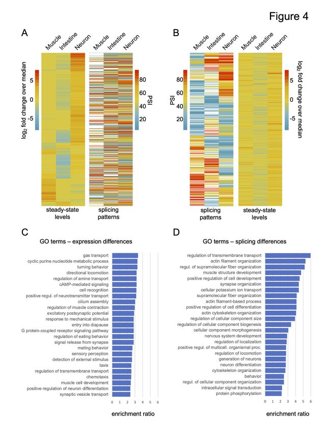
Downloaded from genome.cshlp.org on December 7, 2020 - Published by Cold Spring Harbor Laboratory Press

Abstract

Alternative splicing plays a major role in shaping tissue-specific transcriptomes. Among the
broad tissue types present in metazoans, the central nervous system contains some of the highest
levels of alternative splicing. While many documented examples of splicing differences between
broad tissue-types exist, there remains much to be understood about the splicing factors and
the cis sequence elements controlling tissue and neuron subtype-specific splicing patterns. Using
Translating Ribosome Affinity Purification coupled with deep-sequencing (TRAP-seq) in C.
elegans, we have obtained high coverage profiles of ribosome-associated mRNA for three broad
tissue classes (nervous system, muscle, and intestine) and two neuronal subtypes (dopaminergic
and serotonergic neurons). We have identified hundreds of splice junctions that exhibit distinct
splicing patterns between tissue types or within the nervous system. Alternative splicing events
differentially regulated between tissues are more often frame-preserving, conserved across
Caenorhabditis species and enriched in specific cis regulatory motifs. Utilizing this information,
we have identified a likely mechanism of splicing repression by the RNA-binding protein UNC-
75/CELF via interactions with cis elements that overlap a 5 splice site. Alternatively spliced
                                                                            ′

exons also overlap more frequently with intrinsically disordered peptide regions than constitutive
exons. Moreover, regulated exons are often shorter than constitutive exons but are flanked by
longer intron sequences. Among these tissue-regulated exons are several highly conserved
microexons less than 27 nucleotides in length. Collectively, our results indicate a rich layer of
tissue-specific gene regulation at the level of alternative splicing in C. elegans that parallel the
evolutionary forces and constraints observed across metazoa.

                                                                                                                 2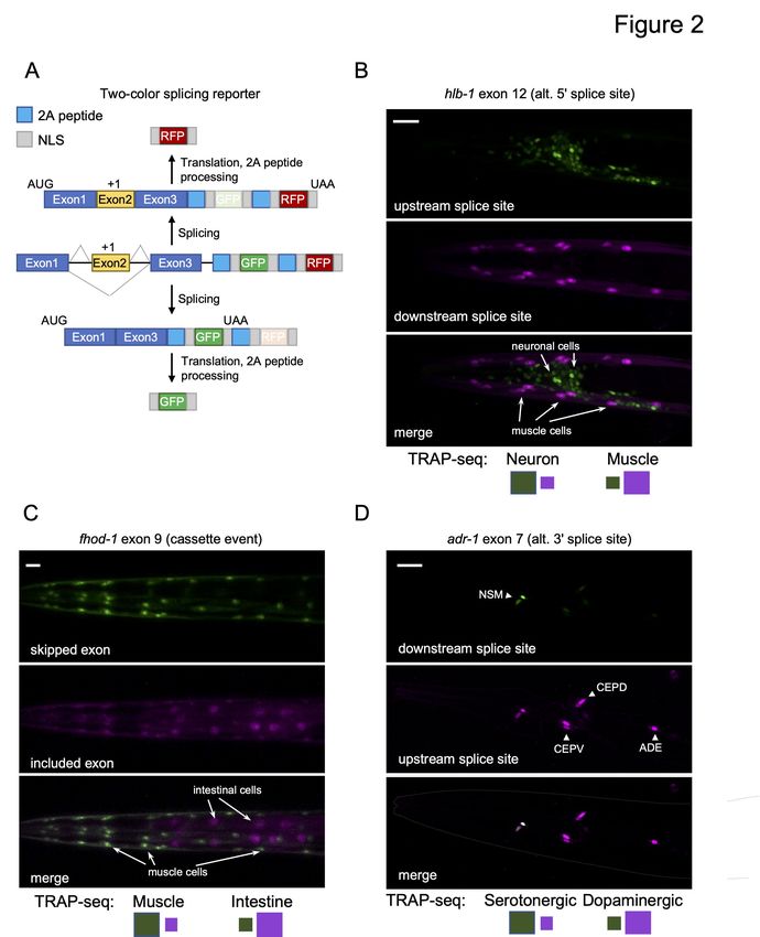
Downloaded from genome.cshlp.org on December 7, 2020 - Published by Cold Spring Harbor Laboratory Press

Introduction

Alternative precursor mRNA (pre-mRNA) splicing is a critical layer of gene expression in

multicellular animals and plays a key role in increasing the functional diversity of proteins within

an organism. Indeed, a significant fraction of multi-exon genes undergo alternative splicing, with

higher frequencies reported across vertebrates (Barbosa-Morais et al. 2012; Merkin et al. 2012;

Pan et al. 2008; Wang et al. 2008) in comparison to invertebrate model organisms such as the

nematode Caenorhabditis elegans and the fruit fly Drosophila melanogaster (Gerstein et al.

2010; Brown et al. 2014). Splice isoforms are often differentially expressed across tissues and/or

during development (for select examples, see Pan et al. 2008; Wang et al. 2008; Gerstein et al.

2010; Brown et al. 2014; Ramani et al. 2011). As such, it remains an important goal to identify

the repertoire of splice variants that help define cell and tissue-specific functions in multi-cellular

animals.

The relevance of alternative splicing in establishing proteomic diversity is especially reflected in

the nervous system. Given its cellular complexity, the nervous system displays elevated levels of

tissue-specific splicing that are also conserved throughout vertebrate evolution (Yeo et al. 2004;

Xu et al. 2002; Barbosa-Morais et al. 2012; Merkin et al. 2012). More recently, transcriptome

profiling in specific neuronal subtypes has revealed an even greater degree of transcript diversity

generated by alternative splicing (Furlanis et al. 2019; Wang et al. 2018; Wamsley et al. 2018;

Saito et al. 2019). Alternative splicing in the nervous system regulates an array of physiological

events and properties, such as neurogenesis, neuronal migration, synaptic plasticity and ion

channel kinetics, and contributes to the diversity needed to fine tune the exact activity of neurons

(Ule and Darnell 2006; Raj and Blencowe 2015; Norris and Calarco 2012; Vuong et al. 2016).

                                                                                                                  3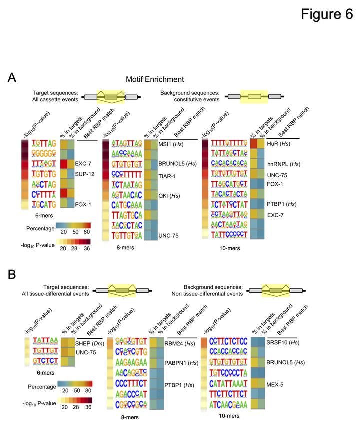
Downloaded from genome.cshlp.org on December 7, 2020 - Published by Cold Spring Harbor Laboratory Press

Alternative splicing must be tightly regulated, especially in coding sequences, to give rise to

functional proteins. Splice site selection and tissue/developmental specificity are established by

auxiliary trans-acting proteins and cis acting sequence elements, which are primarily RNA-

binding proteins (RBPs) and their cognate motifs, respectively (Lee and Rio 2015). Collectively,

these elements direct the assembly and activity of the spliceosome and are often referred to as the

‘splicing code’ (Wang and Burge 2008).

Although significant progress has been made in understanding the combinations of features

enabling the accurate prediction of splicing outcomes (Xiong et al. 2015; Barash et al. 2010;

Zhang et al. 2010; Jaganathan et al. 2019; Baeza-Centurion et al. 2019) our understanding of the

splicing code remains incomplete. This fact is not surprising given that alternative splicing is

regulated in a combinatorial manner, with multiple cis elements and/or multiple RBPs

cooperating or antagonizing each other to generate splicing outcomes (Fu and Ares 2014).

Additionally, the inclusion of tissue, cell and developmental contexts is required in order to

accurately predict splicing outcomes, as a number of RBPs vary correspondingly in expression

patterns and the majority of alternative splicing events are tissue- and developmentally-regulated

(Gerstberger et al. 2014).

The nematode worm Caenorhabditis elegans possesses multiple tissue types, including a well-

differentiated nervous system estimated to contain at least 118 distinct neuronal subtypes

including the major neurotransmitter-releasing classes found in vertebrates (Hobert 2010; White

et al. 1986). Moreover, C. elegans has served as an excellent model system to study the

mechanisms governing alternative splicing regulation (Wani and Kuroyanagi 2017; Zahler 2012;

                                                                                                                 4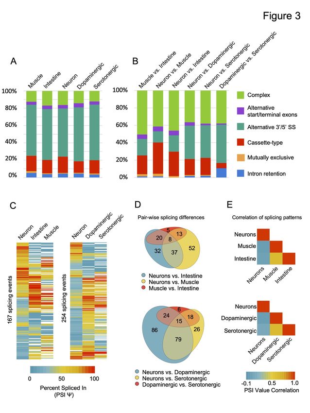
Downloaded from genome.cshlp.org on December 7, 2020 - Published by Cold Spring Harbor Laboratory Press

Gracida et al. 2016). Recent investigations harnessing the tractability of this organism have

focused on whole animal or tissue-enriched transcriptome sequencing approaches to explore

changes in post-transcriptional gene regulation during development and aging (Roach et al.

2020; Kaletsky et al. 2018; Warner et al. 2019; Blazie et al. 2017, 2015; Kotagama et al. 2019;

Li et al. 2020; Barberan-Soler and Zahler 2008; Barberan-Soler et al. 2009; Ragle et al. 2015;

Gerstein et al. 2010; Ramani et al. 2011). However, it still remains unclear how splicing in C.

elegans is regulated at the level of tissues, in particular at the resolution of specific neuronal

subtypes. A better understanding of these mechanisms will require the systematic search for

features of alternative splicing events, exploration of the evolutionary dynamics of splice isoform

usage and tools for the unbiased identification of splicing regulators.

To address these questions, we performed a genome-wide analysis of alternative splicing in C.

elegans using broad tissue- and neuronal subtype-specific ribosome-associated mRNAs obtained

by the Translating Ribosome Affinity Purification approach coupled with deep-sequencing

(TRAP-seq; Gracida and Calarco 2017). Using these data, we sought to determine the extent to

which TRAP enriched tissue- and neuron subtype-specific splice junctions are undetected in

whole animal data. Additionally, we aimed to examine these events for distinct sequence and

conservation features that may delineate mechanisms of splicing regulation.

Results

Tissue-enriched transcript data identifies splice junctions not identified in whole animal

profiling data

                                                                                                                 5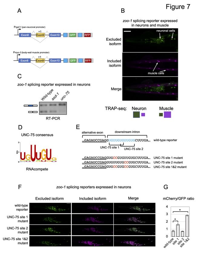
Downloaded from genome.cshlp.org on December 7, 2020 - Published by Cold Spring Harbor Laboratory Press

We have successfully adapted the Translating Ribosome Affinity Purification (TRAP) technique

(Heiman et al. 2008) for use in C. elegans and coupled it with deep-sequencing (TRAP-seq;

Gracida and Calarco 2017). In this technique, a Green Fluorescent Protein (GFP) tagged

ribosomal subunit protein, RPL-1, is expressed under the control of tissue- or cell-specific

promoters (Fig 1A). Whole animal lysates are then prepared in the presence of cycloheximide,

arresting ribosomes on mRNAs that are being actively translated. Ribosomes and their associated

mRNAs are then immuno-precipitated from the lysate via the GFP tag, thus enriching for

transcripts present in the original cell types expressing the GFP-tagged ribosomal protein. We

used TRAP-seq to obtain high coverage mRNA repertoire measurements from Caenorhabditis

elegans fourth larval stage (L4) animals for three broad tissue classes: neurons, muscle, and

intestine; and two neuronal subtypes: dopaminergic and serotonergic neurons (Fig 1B). Two

biological replicates of TRAP-enriched mRNA and matched whole animal mRNA (represented

by mRNA purified from our lysates prior to immuno-precipitation) were collected from each

transgenic strain. In total, over 2 billion uniquely mapped reads were aligned across all datasets,

with summed coverage for each tissue- or cell type-enriched transcriptome ranging from over

100 million to over 300 million reads (Fig S1).

We first mapped our TRAP-seq reads to the C. elegans genome and to existing gene models (see

Methods for details), and identified all aligned reads supporting annotated splice junctions, as

well as high-confidence unannotated splice junctions. In total, we detected 145,811 splice

junctions supported by at least 5 read counts across all samples (Supplemental Table S1). We

next investigated whether our tissue-enriched mRNA populations enabled the detection of splice

junctions that are absent due to dilution in our whole animal transcriptome data. In each tissue or

                                                                                                                 6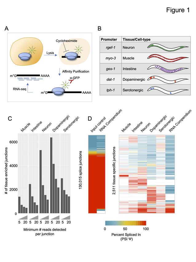
Downloaded from genome.cshlp.org on December 7, 2020 - Published by Cold Spring Harbor Laboratory Press

cell type, we detected between ~1,400 to over 6,000 splice junctions supported by at least five

reads that were absent from our whole animal transcriptome datasets (Fig 1C). Importantly, even

when requiring that tissue-enriched splice junctions be supported by more than 20 reads, we still

identified a total of 5,577 non-redundant splice junctions not detected in our whole animal data,

spanning 1,396 genes (Fig 1C).

We next asked whether our tissue-enriched splice junctions were detected in whole animal

samples upon pooling a larger number of transcriptome datasets. To perform this analysis, we

used a previously published RNA-seq compendium of reproducibly expressed splice junctions in

C. elegans, assembled from 1,682 publicly available RNA-seq experiments (Tourasse et al.

2017). Each splice junction has an associated Percent Spliced In (PSI) value, which is a measure

of the frequency of each splice junction relative to other junctions sharing the same splice donor

or acceptor sites (see Methods for details). This schema for calculating the PSI value was applied

to our data in order to compare our measurements with the RNA-seq compendium.

Both our whole animal splice junction PSI value measurements and those of the RNA-seq

compendium were strongly correlated, despite being derived from different datasets (Fig 1D; left

panel, r = 0.91, Pearson correlation coefficient). However, when comparing our TRAP-seq

tissue- and cell-type-enriched splice junctions supported by at least 20 counts with the RNA-seq

compendium, we observed that roughly 45% of these splice junctions are not detected in the

RNA-seq compendium (Fig 1D; right panel). We also asked whether the tissue-enriched splice

junctions show distinct splicing patterns across tissues. We detected 2,511 splice junctions with

evidence of alternative splicing (where a minor splice variant must have a PSI value 5%; Fig               ≥

                                                                                                                 7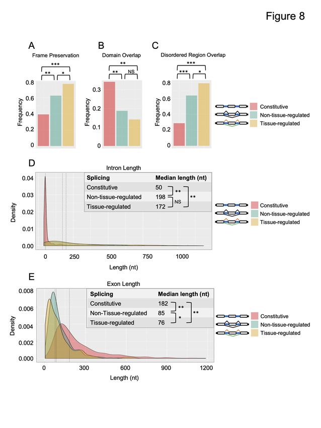
Downloaded from genome.cshlp.org on December 7, 2020 - Published by Cold Spring Harbor Laboratory Press

1D; right panel). Many of these splice junctions are not rare variants due to their high PSI values,

and also display differentially-regulated splicing patterns across tissues (Fig 1D; right panel).

Taken together, these results demonstrate that our TRAP-seq tissue-enriched mRNA profiles

reveal splice junctions that are difficult to detect in whole-animal transcriptomes.

Independent validation of TRAP-seq measurements using in vivo two-color splicing

reporters

We next sought to validate our TRAP-seq measurements through the use of an independent

approach. We utilized a new two-color fluorescent splicing reporter based on previous design

strategies (Fig 2A; Kuroyanagi et al. 2010; Norris et al. 2014). Specifically, these two-color

reporters are designed to include an alternative exon of interest and its flanking introns and exons

(referred to as a minigene). Upstream of the minigene is the promoter that drives the tissue-

specific expression of the fluorescent markers. Downstream of the minigene, we inserted

sequences encoding enhanced green fluorescent protein (EGFP) or the red fluorescent protein

mCherry, each translated in separate reading frames. The alternative exon in the minigene is then

engineered to switch between both reading frames, such that when the alternative exon is

skipped, EGFP is translated, and when the exon is included, mCherry protein is produced (Fig

2A). A similar frame-shifting strategy is applied to monitor alternative 5 and 3 splice site  ′         ′

selection events (Fig S2A).

Additionally, our reporter included 2A peptide sequences upstream of each fluorescent protein to

cleave away upstream peptides translated from the minigene exons (Ahier and Jarriault 2014).

Finally, two nuclear localization signals (SV40 and EGL-13; Lyssenko et al. 2007) were

                                                                                                                 8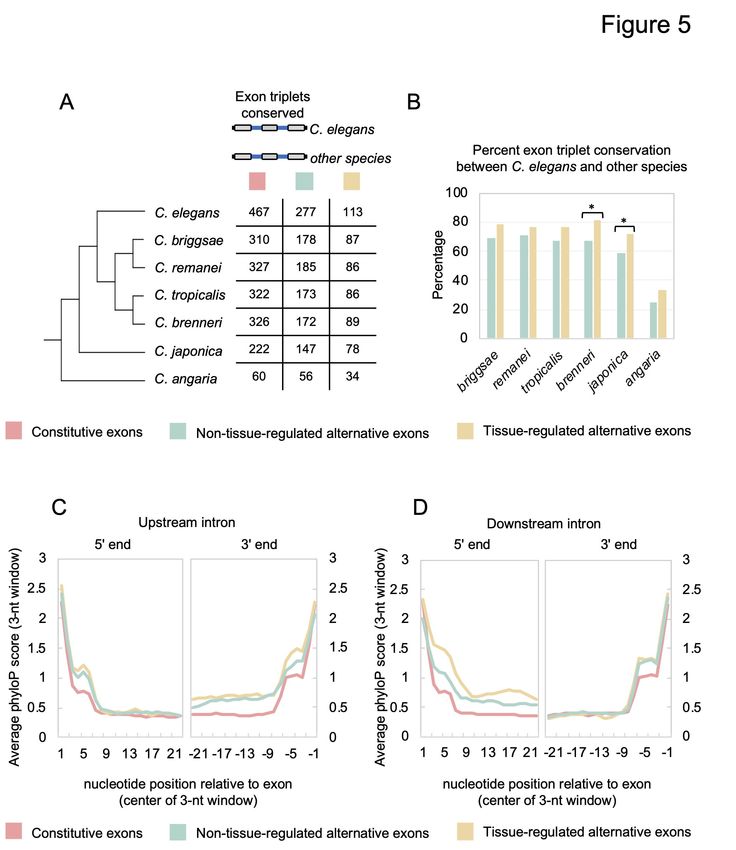
Downloaded from genome.cshlp.org on December 7, 2020 - Published by Cold Spring Harbor Laboratory Press

included at the N- and C-termini of each fluorescent protein to concentrate signal in the nucleus

of each cell expressing the reporter. With this combined set of design features, these reporters

enable the visualization of alternative splicing patterns by fluorescence microscopy in vivo and at

single-cell resolution.

Our TRAP-seq dataset was analyzed for splicing events that demonstrated switch-like splicing

patterns (a change in PSI value, or         Δ PSI, of 80%) between the broad tissue types. Four such
                                                        ≥

events were cloned into our two-color reporters (minigene sequences surrounding hlb-1 exon 12,

fhod-1 exon 8, zoo-1 exon 9, and ampd-1 exon 15), and expression of these reporters was driven

under the control of neuronal, body-wall muscle, or intestinal promoters (see Methods for

details; Fig 2B and C; Fig S2; and Fig 7A below). In all cases tested, we observed agreement

between fluorescent reporter patterns in vivo and our TRAP-seq measurements across tissues

(Fig 2B and C; Fig S2). We additionally monitored the splicing patterns of exon 7 in the gene

adr-1 gene and exon 11 in mlk-1. These splicing events were identified by TRAP-seq as

differentially regulated between serotonergic and dopaminergic neurons or between the neuronal

subtypes and the rest of the nervous system, respectively. Again, our fluorescent reporter signals

agreed with the TRAP-seq measurements (Fig 2D; Fig S2). Including these reporters, a total of

14 two-color reporters were tested, and 11 reporters displayed patterns consistent with TRAP-seq

measurements (Supplemental Table S2).

Taken together, although our TRAP-seq analysis and two-color reporters assess splicing patterns

through different means, their general correlation strongly suggests that our TRAP-seq

measurements can detect regulated splicing patterns.

                                                                                                                  9
Downloaded from genome.cshlp.org on December 7, 2020 - Published by Cold Spring Harbor Laboratory Press
TRAP-seq identifies tissue- and neuronal subtype-biased splicing patterns
We next used the MAJIQ software package (Vaquero-Garcia et al. 2016) to identify all the
alternative splicing events (defined by MAJIQ as local splicing variation) in each TRAP-seq
tissue transcriptome and all the tissue-differential splicing events within every pair of broad
tissue types (muscle, intestine, neurons) and within the nervous system (neurons, dopaminergic
neurons, serotonergic neurons; see Methods for details; Fig 3). In total, we identified 2,953
alternative splicing events in 2,196 genes across all tissues. These alternative splicing events are
distributed across five major classes: cassette-type (exon skipping), alternative 3 or 5 splice site
′ ′
usage, alternative start or terminal exons, intron retention and mutually exclusive splicing (Fig
3A; Fig S3; Supplemental Table S3). We also identified splicing events that we classified as
‘complex’ (Fig 3A; Fig S3). This latter class represents splice junctions making use of more than
one mode of splicing, and accounts for roughly 10-15% of all forms of alternative splicing in a
particular tissue or cell type. As described previously for whole animal transcriptome data
(Ramani et al. 2011), the predominant class of alternative splicing in any given tissue in C.
elegans is alternative 3 or 5 splice site usage (Fig 3A).
′ ′
Moreover, our analysis of tissue-biased alternative splicing identified 871 differentially regulated
splicing events in 689 genes across tissues and the two neuronal subtypes (Fig 3B; Supplemental
Table S4). Again, all major classes of splicing were represented among the set of differentially
regulated alternative splicing events (Fig 3B; Supplemental Table S3). However, complex
splicing events represent a much larger proportion of tissue-differential splicing when compared
with the distribution of possible splicing modes in a given tissue or cell type (compare Fig 3A
and 3B). For example, in the three broad tissue pairwise comparisons, complex splicing events
10Downloaded from genome.cshlp.org on December 7, 2020 - Published by Cold Spring Harbor Laboratory Press
accounted for between 40-50% of all differentially regulated splicing events, a significantly
increased proportion compared with individual cell or tissue types (p values all < 1 x 10⁻⁴, Chi-
squared test; compare Fig 3A and 3B).
Gene Ontology (GO) over-representation analysis was performed on the sets of genes with exons
that were differentially spliced between any two tissues. These genes with differentially spliced
exons were compared against a background set of genes expressed in the same tissues (see
Methods). Splicing events differentially regulated between muscle, intestine and neurons were
enriched in genes involved in actin/cytoskeleton organization, muscle cell differentiation,
neuronal differentiation and the regulation of neuronal projections (Supplemental Table S5),
similar to previous findings in vertebrates (Giudice et al. 2014; Zheng and Black 2013; Raj and
Blencowe 2015).
Finally, we performed clustering analysis and quantification of non-complex, differentially-
regulated splicing events expressed across at least two tissues and/or cell types (Fig 3C-E). Our
analysis revealed that fewer differences in splicing were found between muscle and intestinal
cells compared to differences between these two tissues and the nervous system (Fig 3D).
Similarly, fewer differences were detected between the dopaminergic and serotonergic neurons,
but we identified many more differences between these neuromodulatory cells and the broad
nervous system samples (Fig 3D). Accordingly, splicing patterns were correlated between
muscle and intestinal cells (r = 0.63) and between dopaminergic and serotonergic neurons (r =
0.65), but very little correlation was observed between these tissues and neuron subtypes with the
broad nervous system (r from -0.04 to 0.07) (Fig 3E).
11Downloaded from genome.cshlp.org on December 7, 2020 - Published by Cold Spring Harbor Laboratory Press
Taken together, these results indicate that tissue-regulated splicing represents a substantial and
complex layer of gene expression in C. elegans. Moreover, regulated splicing plays a particularly
important role in shaping the transcriptome of the nervous system, including the fine-tuning of
transcript variants expressed in dopaminergic and serotonergic neurons.
Expression level differences and splicing pattern differences between tissues involve largely
distinct sets of genes
Splicing differences and steady-state mRNA differences between cell and tissue types are
thought to have evolved on largely distinct sets of genes (Pan et al. 2004). To test this possibility
in our tissue-specific transcriptome data, we performed a reciprocal clustering analysis of genes
with differential expression or splicing patterns (Fig 4A and B).
First, we identified all genes that were both differentially expressed between pairs of tissues and
contain one or more alternative splicing events (13% of all differentially expressed genes, see
Methods). After clustering genes by their differential expression patterns, we observed no
obvious trends with splicing differences in the same genes (Fig 4A, Spearman’s rho values
between -0.01 to 0.04, p > 0.3). Similarly, genes with tissue-differential splicing events were
clustered according to their PSI value (Fig 4B). Again, there was no obvious correlation between
splicing patterns and steady-state RNA level differences (Fig 4B, Spearman’s rho values
between -0.04 to -0.01, p > 0.3). We observed similar patterns in comparisons between the
nervous system and neuronal subtypes (Fig S4).
12Downloaded from genome.cshlp.org on December 7, 2020 - Published by Cold Spring Harbor Laboratory Press
Consistent with these analyses above, the top over-represented GO categories for genes with
tissue-differential splicing patterns and genes with tissue-differential expression levels were
somewhat distinct (Fig 4C and D). A wide range of biological processes are enriched in
differentially expressed genes within the broad tissues, while differentially spliced genes are
enriched in vesicle transport, actin kinetics, ion channel activity and muscle/neuron
differentiation (Fig 4C, 4D; Supplemental Table S6). Taken together, our results indicate that
regulated alternative splicing has contributed to tissue and cell type diversification by influencing
a distinct set of genes compared to those regulated at the level of steady state abundance.
Tissue-regulated exons and their surrounding sequences are highly conserved across
Caenorhabditis species
Previous studies of tissue-regulated exons in vertebrates have demonstrated that sequences
flanking these exons tend to be more highly conserved (Fagnani et al. 2007; Sugnet et al. 2006).
Thus, we next assessed whether tissue-regulated exons and their flanking introns and exons
(exon triplets) were more likely to be conserved across the phylogeny of several Caenorhabditis
species (Fig 5A). For this analysis, we focused on comparing all cassette-type tissue-regulated
exons with two other groups of exon triplets (composed of three exons and two introns). One
group consisted of constitutively spliced events, where three sequential exons are always spliced
into mRNA transcripts. The other group consisted of non-tissue-regulated alternative exons and
their flanking sequences, where our TRAP-seq measurements suggest the alternative exons show
little or no tissue-specific splicing differences. Collectively, our analysis included 113 tissue-
regulated exons, 277 non-tissue-regulated exons, and 467 constitutive exons for comparison (see
Methods for details; Fig 5A).
13Downloaded from genome.cshlp.org on December 7, 2020 - Published by Cold Spring Harbor Laboratory Press
Multiple sequence alignments of C. elegans exon triplets were performed with syntenic
sequences from six species in the Caenorhabditis genus: C. briggsae, C. remanei, C. tropicalis,
C. brenneri, C. japonica, and C. angaria (Fig 5A). First, we investigated the degree of
conservation of exon triplets from each group of splicing events, defined by the presence of all
three exons and alignment of the splice sites surrounding these exons. As expected, we see a
decrease in the number of conserved exon triplets as evolutionary divergence from C. elegans
increases (Fig 5A). We next calculated the percent conservation of exon triplets between C.
elegans and the other species, excluding genes that had no clear homologous sequence
alignments over any part of their coding region (see Methods). Extending earlier comparisons
with C. briggsae and C. remanei using EST/cDNA data (Irimia et al. 2008), nearly two-thirds of
all alternative exons are conserved up to the common ancestor of C. elegans and C. japonica (Fig
5B). However, conservation of both classes of alternative exon triplets dropped substantially to
~30% in alignments between the C. elegans and C. angaria genomes (Fig 5B). Tissue-regulated
alternative exons and their neighbouring exons were found to be conserved at a modest, but
consistently higher rate throughout the phylogeny when compared with non-tissue-regulated
alternative exons (Fig 5B). In particular, C. japonica has a significantly greater proportion of
conserved tissue-regulated exon triplets with C. elegans when compared to non-tissue-regulated
alternative splicing events (72% vs. 59%, respectively; p-value = 0.02, Fisher’s exact test; Fig
5B).
Alternative splicing is known to be regulated by cis elements in exonic and intronic regions. As
such, selection pressure to preserve these elements would increase conservation in the intronic
14Downloaded from genome.cshlp.org on December 7, 2020 - Published by Cold Spring Harbor Laboratory Press
regions surrounding alternative exons compared to analogous regions flanking constitutive
exons. We investigated this characteristic by measuring average conservation in introns on an
individual nucleotide level across the seven species described above using the program phyloP
(Pollard et al. 2010). We focused on the 23 nucleotides adjacent to each splice site, because this
length would still partition our smallest introns (~40 nucleotides in length) into 5 and 3 halves. ′ ′
When comparing phyloP scores of 23 nucleotides adjacent to each splice site, we observed
increased sequence conservation surrounding alternative exons compared with constitutive exons
(Fig 5C and 5D). This increased conservation was particularly apparent in the splice sites and
neighbouring regions immediately adjacent to the alternative exons (Fig 5C and 5D, comparing
constitutive exons with both classes of alternative exons, at 3 end of upstream intron or 5 end of
′ ′
downstream intron, p-values < 2.2 x 10⁻¹⁶, Wilcoxon signed-rank test). Moreover, the intronic
regions immediately flanking tissue-regulated alternative exons were significantly more
conserved than the corresponding sequences flanking non-tissue-regulated exons (Fig 5C and
5D, 3 end of upstream intron: p-value < 2 x 10⁻⁶; 5 end of downstream intron: p-value < 2.2 x
′ ′
10-16, Wilcoxon signed-rank test).
Taken together, these data suggest that the intronic regions surrounding alternative exons are
considerably more conserved than analogous sequences in constitutive exons. Moreover, the
extended conservation required for regulation of tissue-specific exons likely creates selection
pressure to preserve neighbouring exon-intron architecture across evolutionary timescales.
Alternative and tissue-regulated exons are enriched in motifs recognized by RNA-binding
proteins
15Downloaded from genome.cshlp.org on December 7, 2020 - Published by Cold Spring Harbor Laboratory Press
Previous studies in C. elegans have demonstrated that sequence enrichment search strategies can
be effective at identifying candidate splicing regulatory signals (Kabat et al. 2006; Ramani et al.
2011). We used HOMER (Hypergeometric Optimization of Motif EnRichment; Heinz et al.
2010), which can detect enriched motifs in a given list of sequences relative to a background list
of sequences. We first compared sequences spanning alternative cassette exons and flanking
introns to equivalent sequences surrounding constitutively included exons (Fig 6A). The
resulting enriched sequences yield several expected motifs previously identified (Kabat et al.
2006; Ramani et al. 2011) and recognized by characterized RNA-binding proteins, including
FOX-1/Rbfox and the muscle-specific regulator SUP-12 (an ortholog of Rbm24 and Rbm38),
which have both been implicated in alternative splicing regulation in C. elegans (Kuroyanagi et
al. 2007). We also identified enriched cis elements recognized by UNC-75/CELF and EXC-7 (an
ortholog of Elavl4/HuD), both known to regulate tissue-specific alternative splicing within the
nervous system (Kuroyanagi et al. 2013b; Chen et al. 2016; Kuroyanagi et al. 2013a; Norris et al.
2014).
We next searched for motifs enriched in tissue-regulated alternative splicing events relative to
analogous sequences spanning non-tissue-regulated splicing events (Fig 6B). Again, we
identified motifs for known RNA-binding proteins and splicing regulators. Some of these motifs
were not identified in our alternative versus constitutive exon comparison above, suggesting that
they are selectively associated with tissue-regulated splicing patterns.
In both comparisons, we also found enrichment of many motifs that could not be connected with
a cognate RNA-binding protein, as well as motifs that matched consensus sequences from other
16Downloaded from genome.cshlp.org on December 7, 2020 - Published by Cold Spring Harbor Laboratory Press
species (Fig 6A and 6B). Taken together, our results indicate that alternative exons and, by
extension, tissue-regulated exons, are coordinately regulated by RNA-binding proteins, several
of which remain to be discovered and characterized.
UNC-75/CELF recognizes conserved motifs overlapping a 5 splice site and represses exon
′
inclusion
Both our conservation analysis (Fig 5) and motif enrichment analysis (Fig 6) indicated that
tissue-regulated exons are flanked by conserved and over-represented sequence features, likely
reflecting the presence of critical cis elements. As such, we next assessed whether these analyses
could prove useful in uncovering novel splicing regulatory mechanisms.
We identified conserved UNC-75 consensus sequences overlapping the 5 splice site in the intron
′
downstream of alternative exon 9 of the zoo-1 gene (Fig 7; Fig S5). This gene encodes an
ortholog of the Zonula Occludens tight junctional protein ZO-2, a key protein involved in cell
adhesion and cell proliferation (González-Mariscal et al. 2019). Our TRAP-seq data identified
that zoo-1 exon 9 is primarily excluded in neurons and mostly included in muscle cells (Fig 7A).
We further validated these patterns by co-expressing neuron and muscle promoter driven zoo-1
two-color splicing reporters (Fig 7A). Consistent with our TRAP-seq measurements, when signal
from both cell types are imaged together, we detected significantly preferential inclusion of exon
9 (mCherry) in muscle cells and more biased skipping of exon 9 (GFP) in neuronal cells (Fig
7B).
17Downloaded from genome.cshlp.org on December 7, 2020 - Published by Cold Spring Harbor Laboratory Press
Given that UNC-75 is broadly expressed in the nervous system (Loria et al. 2003), we speculated
that it has a direct role in blocking exon 9 inclusion through interaction with its cognate cis
elements. To test this hypothesis, we expressed a zoo-1 exon 9 splicing reporter in the nervous
system of wild-type animals, animals lacking unc-75, or animals lacking asd-1, an unrelated
RNA-binding protein that is also expressed in the nervous system. RT-PCR assays amplifying
both zoo-1 isoforms were then performed on total RNA collected from these animals (Fig 7C).
Consistent with our prediction, exon 9 is more highly included in the neurons of unc-75 loss of
function mutants when compared to wild-type animals (Fig 7C). Mutants lacking asd-1 had
splicing patterns similar to wild-type animals, suggesting that loss of unc-75 has a specific effect
on zoo-1 exon 9 alternative splicing (Fig 7C). We next generated two-color splicing reporters
with mutations targeting one or both UNC-75 consensus sequences (Fig 7D and 7E) and drove
expression of these reporters specifically in neurons (Fig 7F). When imaging the neuron-
expressed reporters in the absence of any muscle signal, the included isoform can now be
detected with imaging settings that would otherwise saturate signal in muscle cells. We next
quantified the total fluorescence signal through the head regions of animals expressing wild-type
and mutant reporters (Fig 7F and 7G). These experiments demonstrated that mutations in the first
or second cis elements increased the relative proportion of the included isoform in neurons (Fig
7F and 7G). However, mutations targeting both cis elements led to the maximum effect on
splicing, where exon 9 is significantly more included in most neurons (p-value < 0.01, Kruskal-
Wallis rank sum test, Fig 7F and 7G).
18Downloaded from genome.cshlp.org on December 7, 2020 - Published by Cold Spring Harbor Laboratory Press
Taken together, these results indicate that CELF proteins can act as repressors of alternative exon
inclusion in the nervous system when bound to cis elements directly overlapping with 5 splice ′
sites.
Alternative exons and tissue-biased exons have distinct regulatory features when compared
with constitutive exons
Previous studies of vertebrate alternatively spliced exons and their flanking introns and exons
have indicated that regulated exons have distinct sequence characteristics compared to analogous
regions surrounding constitutively spliced exons (Keren et al. 2010; Xing and Lee 2005; Yura et
al. 2006; Yeo et al. 2005; Barbosa-Morais et al. 2012). In a similar manner, we compared various
sequence features among alternative exons, constitutive exons, and their surrounding sequences.
First, we tested the frequency of internal constitutive or alternative exons to preserve the open
reading frame—specifically, if the exon is a multiple of three nucleotides (Fig 8A). Tissue-
regulated alternative exons have been shown to have increased selection pressure to be frame-
preserving in order to be included or skipped in transcripts without causing a shift in reading
frame (Modrek et al. 2001). Consistent with these observations, we observe an increase in the
frequency of alternative exons to preserve reading frame relative to constitutive exons (Fig 8A).
Frame preservation frequency is further increased among the tissue-regulated splicing events,
where nearly 80% of exons are multiples of three.
We next tested the frequency with which internal constitutive or alternative exons overlap with
conserved protein domains or intrinsically disordered regions (IDRs) (Fig 8B and C). We found
19Downloaded from genome.cshlp.org on December 7, 2020 - Published by Cold Spring Harbor Laboratory Press
that alternative exons overlap less frequently with annotated protein domains (Fig 8B), but more
frequently overlap with IDRs, with tissue-regulated alternative exons having the highest degree
of overlap with IDRs (approaching 80% of all exons) (Fig 8C). Again, this trend is consistent
with reports in vertebrates, where alternatively spliced exons are known to modulate protein-
protein interactions and the formation of multivalent protein assemblies through altering IDR
sequence features (Ellis et al. 2012; Yang et al. 2016; Buljan et al. 2012; Romero et al. 2006).
Finally, we measured the lengths of internal constitutive exons, alternative exons, and their
flanking intronic regions (Fig 8D and E). Consistent with previous observations (Kim et al. 2007;
Fox-Walsh et al. 2005; Kabat et al. 2006), introns flanking both tissue-differential and non-
tissue-differential alternative exons are significantly longer than introns flanking constitutively
spliced exons (Fig 8D). This tendency toward longer intron length is preserved in both upstream
and downstream introns flanking alternative exons, with the downstream intron having a
somewhat longer length (Fig S6). In contrast, we found that alternative exons are significantly
smaller in length compared with constitutive exons (Fig 8E). This tendency towards a shorter
length was also more pronounced in the tissue-differential alternative exons.
Collectively, these results demonstrate that alternative exons and their flanking intronic
sequences have features that distinguish them from constitutive exons. Moreover, these
identified features show hallmarks shared by other invertebrate and vertebrate alternative exons,
suggesting common evolutionary constraints and functional consequences of these exons across
metazoans.
20Downloaded from genome.cshlp.org on December 7, 2020 - Published by Cold Spring Harbor Laboratory Press
Microexons are differentially regulated across cell types and exceptionally conserved across
Caenorhabditis species
Our observations above indicated that tissue-regulated exons tend to be shorter than other classes
of exons. Upon closer examination, we identified 24 microexons ( 27 nucleotides in length)
≤
among these tissue-regulated exons (Fig 9; Supplemental Table S7). Microexons have recently
been identified as an interesting class of exons, with distinct mechanisms controlling their
splicing patterns, and demonstrated roles in neuronal development and physiology in vertebrates
(Scheckel and Darnell 2015). Moreover, the aberrant regulation of these exons has been
associated with autism spectrum disorders (Irimia et al. 2014; Gonatopoulos-Pournatzis et al.
2018; Quesnel-Vallières et al. 2016).
Our identification of tissue-regulated microexons suggested that there may be additional
microexons in our transcriptome data. We therefore slightly relaxed our filtering criteria and
detected an additional 43 alternatively spliced microexons, for a total of 67 (Supplemental Table
S7), indicating that this class of exons may be difficult to comprehensively detect in typical
transcriptome analysis with short read data from whole animal samples. Roughly one-third
(23/67) microexons were detected exclusively in our tissue-enriched TRAP-seq data, suggesting
that microexons often undergo tissue-differential splicing.
We next assessed whether microexons possessed features that distinguished them from larger
alternative exons ( 99 nucleotides). Microexons exhibit a high frequency of reading frame
≥
preservation (>80%) compared with larger exons (Fig 9A). Additionally, an examination of
intronic regions revealed that sequences surrounding microexons, particularly the 3 end of the ′
21Downloaded from genome.cshlp.org on December 7, 2020 - Published by Cold Spring Harbor Laboratory Press
upstream intron and 5 end of the downstream intron, are considerably more conserved than
′
corresponding regions surrounding larger alternative exons (p-values all < 1 x 10⁻⁷, Wilcoxon
Signed-rank test; Fig 9B). Representative multiple sequence alignments provide another view of
this conservation. For example, the six nucleotide microexon in the madd-4/punctin gene and the
15 nucleotide microexon in the spc-1 gene (a spectrin ortholog) were both found to demonstrate
high levels of conservation within the exon and flanking intronic regions among all
Caenorhabditis species examined (Fig 9C and 9D).
Taken together, our analysis has shed light on an important class of exons that are likely
influencing aspects of protein function and diversity.
Discussion
In this study, we have highlighted the utility of the TRAP-seq approach for transcriptome-wide
studies of features associated with alternative splicing across tissues and specific neuronal cell
types. Our samples are also sequenced to fairly high depths of coverage (Fig S1), which has
facilitated our ability to detect splice junctions with increased confidence. Consistent with this
point, we down-sampled our short read data and demonstrated that, although we are approaching
saturation in our ability to detect alternatively spliced junctions (Fig S7), our current results are
likely under-estimating the full complexity of splicing in the isolated tissues of the organism.
Our method provides a robust and complementary avenue for obtaining mRNA profiles along
with other approaches such as mRNA tagging (Spencer et al. 2011; Blazie et al. 2015) and
fluorescence-activated cell sorting of labeled cells (Kaletsky et al. 2016; Spencer et al. 2014).
22Downloaded from genome.cshlp.org on December 7, 2020 - Published by Cold Spring Harbor Laboratory Press
Our results demonstrate that cells of the nervous system had the highest numbers of detected
splice junctions (Fig 1C), mirroring findings in vertebrates indicating that the nervous system
exhibits relatively high levels of alternative splicing compared with other tissues (Tapial et al.
2017; Barbosa-Morais et al. 2012). It is only recently that neuronal-subtype differences in
splicing have been interrogated and have revealed differences in isoform expression (Furlanis et
al. 2019; Wang et al. 2018; Wamsley et al. 2018; Saito et al. 2019). Taken together, these
observations and our own results suggest that an expanded use of alternative splicing is a
hallmark of metazoan nervous systems, including the relatively compact nervous system of C.
elegans.
Our observation that tissue-regulated splicing is enriched in complex events (Fig 3A and 3B) in
C. elegans is consistent with a recent study indicating that complex events account for a
significant proportion of conserved differential splicing variation across mammalian tissues
(Vaquero-Garcia et al. 2016). It is interesting to speculate how complex splicing events may
arise during evolution. One likely model would involve complex events emerging from simpler
ancestral splicing events. Comparisons of genomic and transcript sequences across multiple
species may be able to provide some support of this model.
We identified highly conserved UNC-75/CELF binding sites overlapping directly with the 5 ′
splice site flanking alternative exon 9 of the zoo-1 gene. We speculate a mechanism where UNC-
75 binds to these cis elements to repress inclusion of the alternative exon in neurons (Fig 7). This
mechanism of repression contrasts with the role of UNC-75 as an activator of exon inclusion
when bound to a cis element 30 bases downstream of the 5 splice site in the unc-16 gene (Norris
′
23Downloaded from genome.cshlp.org on December 7, 2020 - Published by Cold Spring Harbor Laboratory Press
et al. 2014). Recent studies have revealed position-dependent regulation of splicing by UNC-75
(Kuroyanagi et al. 2013b). Thus, small differences in the location and position of the UNC-75 cis
elements can have distinct consequences on splicing regulation even within the same intronic
region. It will be interesting to explore the biochemical mechanisms of UNC-75 binding to target
mRNAs and investigate how prevalent this mode of CELF-mediated splicing repression is in
multicellular animals.
Our results suggest that there are shared constraints across protein coding genes in multi-cellular
animals that lead to the evolution of regulated splicing patterns. In particular, the possibility that
tissue-regulated exons are more likely to be selected for influencing disordered regions in
proteins is intriguing. Several emerging roles for disordered protein sequences include shaping
protein-protein interactions and biophysical properties, as well as serving as sites of post-
translational modifications (Wright and Dyson 2015). Given that tissue-regulated exons are
highly conserved, their influence on protein function is likely important. An important goal will
be to determine the molecular consequences of these splicing events on protein function in vivo.
Microexons have emerged as important modulators of protein function in vertebrates,
particularly in the nervous system (Li et al. 2015; Irimia et al. 2014). Our current analysis has
expanded the number of known microexons in C. elegans. Moreover, given that these
microexons are highly conserved and often frame-preserving (Fig 9), it is highly likely that these
exons will influence protein function in different tissues, as recent studies in vertebrates have
confirmed (Parras et al. 2018; Gonatopoulos-Pournatzis et al. 2020). It was proposed in a recent
study that the explosion of microexons included in neuronal transcripts may have coincided with
24Downloaded from genome.cshlp.org on December 7, 2020 - Published by Cold Spring Harbor Laboratory Press
the evolution of the enhancer of microexons (eMIC) domain in the Serine/Arginine Repetitive
Matrix (SRRM) protein family (Torres-Méndez et al. 2019). Other studies have also identified
additional factors controlling the splicing of these exons (Gonatopoulos-Pournatzis et al. 2018;
Li et al. 2015). However, SRRM proteins in Caenorhabditis species lack the eMIC domain
(Torres-Méndez et al. 2019). Thus, C. elegans represents an interesting model system to explore
ancient mechanisms governing microexon splicing.
Collectively, our work has revealed that alternative splicing represents a rich layer of gene
expression, contributing to the specialized functions of cell and tissue types in C. elegans,
particularly in the nervous system. As isoform-sensitive transcriptome profiling approaches at
single cell resolution are starting to emerge, we will likely gain an even deeper appreciation for
the extent to which transcript diversity will ultimately impact the proteome and cellular
specialization.
Methods
C. elegans maintenance and strains used in this study
Animals were maintained at 21°C and grown on nematode growth media plates seeded with
OP50-1 bacteria under standard conditions (Brenner 1974). In addition to the N2 wild-type
strain, the list of strains used in the current study are listed in Supplemental Table S8.
TRAP-seq and read alignment
cDNA libraries from whole animal input lysates and tissue-enriched data sets were created as
described previously (Gracida et al. 2017). FASTQ files from paired-end Illumina sequencing
25Downloaded from genome.cshlp.org on December 7, 2020 - Published by Cold Spring Harbor Laboratory Press
data were used as input files and reads were mapped using STAR aligner (Dobin et al. 2013), an
algorithm capable of high-speed read alignment and splice junction detection. Genome index
files were generated using C. elegans genome annotation WS251 and the default parameters.
Two-pass read alignment was performed to increase detection of novel splice junctions. SAM
and sorted BAM files were generated for subsequent analysis. Please see provided Supplemental
Material for additional analysis to identify tissue- and neuron-subtype unique junction reads.
Predicting tissue-differential splicing, categorizing splicing events into splicing classes, and
characterization of splicing events
Using sorted BAM and BAM index files from our aligned reads, the Majiq/Voila software
pipeline (Vaquero-Garcia et al. 2016) was applied to our TRAP-seq data to identify and visualize
alternative splicing events (reported as Local Splicing Variations or LSVs, within a single tissue)
and differential splicing events (between two or more tissues). Using the output files of the Majiq
analysis, more stringent filters were applied in order to obtain a set of high-confidence
alternative splicing and differential splicing events for further computational analysis. All
coding, statistical tests and graphing were performed using R 3.5.0 (3.5.1. 2018). Please see the
Supplemental Material for additional details.
Motif enrichment analysis
Motif analysis was performed using HOMER (Heinz et al. 2010) to discover annotated or de
novo motifs that were enriched in tissue-regulated splicing events, using sets of constitutive
splicing events or non-tissue-regulated splicing events as background sets. For specific details,
see Supplemental Material.
26Downloaded from genome.cshlp.org on December 7, 2020 - Published by Cold Spring Harbor Laboratory Press
GO over-representation analysis
We used the online tool, WebGestalt (Liao et al. 2019) to perform the over-representation
analyses. Sample datasets were: 1) genes that are differentially-spliced between any two tissues
(ΔPSI 0.15), and 2) Genes that are differentially-expressed between any two tissues (with a 5x
≥
up- or down-regulated). In all comparisons, we search for enrichment against background gene
sets where genes were filtered for expression (above 50 read counts) within the tissue(s)
involved. This filter controlled for simply enriching for genes that were differentially expressed
in particular tissues. Over-representation analysis was performed to identify significantly
enriched biological processes (Bonferroni multiple testing correction, corrected p-valuesDownloaded from genome.cshlp.org on December 7, 2020 - Published by Cold Spring Harbor Laboratory Press
were generated and used to obtain multiple alignment format (MAF) files from the 26-way
nematode alignment track on genome assembly ce11 (WBCel235) from the UCSC (University of
California Santa Cruz) Genome Browser. We then extracted homologous sequences from seven
species (C. elegans, C. brenneri, C. briggsae, C. remanei, C. japonica, C. tropicalis, and C.
angaria) and re-aligned relevant regions surrounding alternative or constitutive exons using
MUSCLE (Edgar 2004). In order to measure conservation patterns in a base-by-base manner, we
used the program phyloP (Pollard et al. 2010), a part of the “rphast” R package (Hubisz et al.
2011). For additional details, see Supplemental Material.
Data access
All raw and processed sequencing data generated in this study have been submitted to the NCBI
Gene Expression Omnibus (GEO; https://www.ncbi.nlm.nih.gov/geo/) under accession number
GSE106374.
Competing interest statement
The authors declare no competing interests.
Acknowledgments
We thank John Laver and Arneet Saltzman for critical reading of the manuscript. B.K. is
supported by an NSERC PGS Doctoral fellowship. Research in the Calarco Lab is also
generously supported by the Canadian Foundation for Innovation, the Ontario Research Fund,
the Canada First Research Excellence Fund (Medicine by Design), and the Canada Research
Chairs Program. Some of the analysis described in this study was performed on high
28Downloaded from genome.cshlp.org on December 7, 2020 - Published by Cold Spring Harbor Laboratory Press
performance computing hardware in the Moses Research Laboratory (University of Toronto)
supported by the Canada Foundation for Innovation. This work was supported by funding by an
NSERC Discovery Grant (2017-06573) and a CIHR Project Grant (PJT-156300), awarded to
J.A.C. This research was enabled in part by support provided by Compute Ontario
(https://computeontario.ca/) and Compute Canada (www.computecanada.ca).
Author contributions: B.K., P.P.P., X.G. and J.A.C. conceived of the study and conducted
experiments. B.K., P.P.P. and J.A.C. performed data analysis, assembled figures, and wrote the
manuscript with input from X.G. and Y.Z. L-M.S. helped with initial construction of two-color
splicing reporters. I.P. helped with analysis of overlap of alternative exons with regions encoding
intrinsically disordered proteins.
References
3.5.1. RDCT. 2018. A Language and Environment for Statistical Computing. R Found Stat
Comput 2: https://www.R-project.org. http://www.r-project.org.
Ahier A, Jarriault S. 2014. Simultaneous expression of multiple proteins under a single promoter
in Caenorhabditis elegans via a versatile 2A-based toolkit. Genetics 196: 605–613.
Baeza-Centurion P, Miñana B, Schmiedel JM, Valcárcel J, Lehner B. 2019. Combinatorial
Genetics Reveals a Scaling Law for the Effects of Mutations on Splicing. Cell 176: 549-
563.e23.
Barash Y, Calarco JA, Gao W, Pan Q, Wang X, Shai O, Blencowe BJ, Frey BJ. 2010.
Deciphering the splicing code. Nature 465: 53–59.
Barberan-Soler S, Lambert NJ, Zahler AM. 2009. Global analysis of alternative splicing
uncovers developmental regulation of nonsense-mediated decay in C. elegans. Rna 15:
1652–1660.
29Downloaded from genome.cshlp.org on December 7, 2020 - Published by Cold Spring Harbor Laboratory Press
Barberan-Soler S, Zahler AM. 2008. Alternative splicing regulation during C. elegans
development: Splicing factors as regulated targets. PLoS Genet 4.
Barbosa-Morais NL, Irimia M, Pan Q, Xiong HY, Gueroussov S, Lee LJ, Slobodeniuc V, Kutter
C, Watt S, Çolak R, et al. 2012. The evolutionary landscape of alternative splicing in
vertebrate species. Science (80- ) 338: 1587–1593.
Blazie SM, Babb C, Wilky H, Rawls A, Park JG, Mangone M. 2015. Comparative RNA-Seq
analysis reveals pervasive tissue-specific alternative polyadenylation in Caenorhabditis
elegans intestine and muscles. BMC Biol 13.
Blazie SM, Geissel HC, Wilky H, Joshi R, Newbern J, Mangone M. 2017. Alternative
polyadenylation directs tissue-specific miRNA targeting in Caenorhabditis elegans somatic
tissues. Genetics 206: 757–774.
Brenner S. 1974. The genetics of Caenorhabditis elegans. Genetics 77: 71–94.
Brown JB, Boley N, Eisman R, May GE, Stoiber MH, Duff MO, Booth BW, Wen J, Park S,
Suzuki AM, et al. 2014. Diversity and dynamics of the Drosophila transcriptome. Nature
512: 393–399.
Buljan M, Chalancon G, Eustermann S, Wagner GP, Fuxreiter M, Bateman A, Babu MM. 2012.
Tissue-Specific Splicing of Disordered Segments that Embed Binding Motifs Rewires
Protein Interaction Networks. Mol Cell 46: 871–883.
Chen L, Liu Z, Zhou B, Wei C, Zhou Y, Rosenfeld MG, Fu XD, Chisholm AD, Jin Y. 2016.
CELF RNA binding proteins promote axon regeneration in C. elegans and mammals
through alternative splicing of syntaxins. Elife 5.
Dobin A, Davis CA, Schlesinger F, Drenkow J, Zaleski C, Jha S, Batut P, Chaisson M, Gingeras
TR. 2013. STAR: Ultrafast universal RNA-seq aligner. Bioinformatics 29: 15–21.
30Downloaded from genome.cshlp.org on December 7, 2020 - Published by Cold Spring Harbor Laboratory Press
Edgar RC. 2004. MUSCLE: Multiple sequence alignment with high accuracy and high
throughput. Nucleic Acids Res 32: 1792–1797.
Ellis JD, Barrios-Rodiles M, Çolak R, Irimia M, Kim TH, Calarco JA, Wang X, Pan Q,
O’Hanlon D, Kim PM, et al. 2012. Tissue-Specific Alternative Splicing Remodels Protein-
Protein Interaction Networks. Mol Cell 46: 884–892.
Fagnani M, Barash Y, Ip JY, Misquitta C, Pan Q, Saltzman AL, Shai O, Lee L, Rozenhek A,
Mohammad N, et al. 2007. Functional coordination of alternative splicing in the
mammalian central nervous system. Genome Biol 8: R108.
Fox-Walsh KL, Dou Y, Lam BJ, Hung SP, Baldi PF, Hertel KJ. 2005. The architecture of pre-
mRNAs affects mechanisms of splice-site pairing. Proc Natl Acad Sci U S A 102: 16176–
16181.
Fu XD, Ares M. 2014. Context-dependent control of alternative splicing by RNA-binding
proteins. Nat Rev Genet 15: 689–701.
Furlanis E, Traunmüller L, Fucile G, Scheiffele P. 2019. Landscape of ribosome-engaged
transcript isoforms reveals extensive neuronal-cell-class-specific alternative splicing
programs. Nat Neurosci 22: 1709–1717.
Gerstberger S, Hafner M, Tuschl T. 2014. A census of human RNA-binding proteins. Nat Rev
Genet 15: 829–845.
Gerstein MB, Lu ZJ, Van Nostrand EL, Cheng C, Arshinoff BI, Liu T, Yip KY, Robilotto R,
Rechtsteiner A, Ikegami K, et al. 2010. Integrative analysis of the Caenorhabditis elegans
genome by the modENCODE project. Science (80- ) 330: 1775–1787.
Giudice J, Xia Z, Wang ET, Scavuzzo MA, Ward AJ, Kalsotra A, Wang W, Wehrens XHT,
Burge CB, Li W, et al. 2014. Alternative splicing regulates vesicular trafficking genes in
31You can also read