Circulating nucleic acids damage DNA of healthy cells by integrating into their genomes
←
→
Page content transcription
If your browser does not render page correctly, please read the page content below
Circulating nucleic acids damage DNA of healthy cells by integrating
                              into their genomes

    INDRANEEL MITTRA1,* , NAVEEN KUMAR KHARE1 , GORANTLA VENKATA RAGHURAM1 , ROHAN CHAUBAL2 ,
     FATEMA KHAMBATTI1 , DEEPIKA GUPTA1 , ASHWINI GAIKWAD1 , PREETI PRASANNAN1 , AKSHITA SINGH1
             AISHWARYA IYER1 , ANKITA SINGH2 , PAWAN UPADHYAY2 , NAVEEN KUMAR NAIR1 ,
                             PRADYUMNA KUMAR MISHRA1 and AMIT DUTT2
     1
         Translational Research Laboratory , 2Integrated Cancer Genomics Laboratory; Advanced Centre for Treatment,
                                               Research and Education in Cancer,
                                 Tata Memorial Centre, Kharghar, Navi Mumbai 410210, India

                                   *Corresponding author (Email, indraneel.mittra@gmail.com)

   Whether nucleic acids that circulate in blood have any patho-physiological functions in the host have not been explored. We
   report here that far from being inert molecules, circulating nucleic acids have significant biological activities of their own that
   are deleterious to healthy cells of the body. Fragmented DNA and chromatin (DNAfs and Cfs) isolated from blood of cancer
   patients and healthy volunteers are readily taken up by a variety of cells in culture to be localized in their nuclei within a few
   minutes. The intra-nuclear DNAfs and Cfs associate themselves with host cell chromosomes to evoke a cellular DNA-
   damage-repair-response (DDR) followed by their incorporation into the host cell genomes. Whole genome sequencing
   detected the presence of tens of thousands of human sequence reads in the recipient mouse cells. Genomic incorporation of
   DNAfs and Cfs leads to dsDNA breaks and activation of apoptotic pathways in the treated cells. When injected intravenously
   into Balb/C mice, DNAfs and Cfs undergo genomic integration into cells of their vital organs resulting in activation of DDR
   and apoptotic proteins in the recipient cells. Cfs have significantly greater activity than DNAfs with respect to all parameters
   examined, while both DNAfs and Cfs isolated from cancer patients are more active than those from normal volunteers. All
   the above pathological actions of DNAfs and Cfs described above can be abrogated by concurrent treatment with DNase I
   and/or anti-histone antibody complexed nanoparticles both in vitro and in vivo. Taken together, our results suggest that
   circulating DNAfs and Cfs are physiological, continuously arising, endogenous DNA damaging agents with implications for
    ageing and a multitude of human pathologies including initiation of cancer.

   [Mittra I, Khare NK, Raghuram GV, Chaubal R, Khambatti F, Gupta D, Gaikwad A, Prasannan P, Singh A, Iyer A, Singh A, Upadhyay P, Nair NK,
   Mishra PK and Dutt A 2015 Circulating nucleic acids damage DNA of healthy cells by integrating into their genomes. J. Biosci. 40 91–111] DOI
   10.1007/s12038-015-9508-6

                       1.      Introduction                                2003). It has been estimated that several hundred billion to
                                                                           a trillion cells die in the adult human body daily due to
Apoptosis is a natural biological process that leads to cellular           normal physiology to be replaced by a similar number gen-
fragmentation and release of nuclear material in the form of               erated through mitosis (Fliedner et al. 2002). The existence
mono- and oligo-nucleosomes (van Nieuwenhuijze et al.                      of an efficient scavenging system notwithstanding,

Keywords. Ageing; apoptosis; cancer; circulating chromatin; circulating DNA; circulating nucleic acids; circulating nucleosomes;
DNA damage; DNA damage response; DNA double-strand breaks; DNA repair

Supplementary materials pertaining to this article are available on the Journal of Biosciences Website at http://www.ias.ac.in/
jbiosci/mar2015/supp/Mittra.pdf

http://www.ias.ac.in/jbiosci                                 J. Biosci. 40(1), March 2015, 91–111, * Indian Academy of Sciences                   91

Published online: 4 February 2015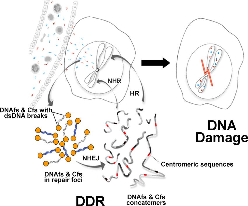
92                                                       Indraneel Mittra et al.

considerable amount of apoptotic genetic material enters the           (Leary et al. 2012), pooled plasma/serum (typically from ~5
circulation in normal individuals (Zhong et al. 2007), and in          patients) was used to isolate DNAfs and Cfs in order to
elevated levels in a multitude of acute and chronic human              maintain inter-experimental consistency.
pathologies including cancer (Holdenrieder et al. 2001;
Chang et al. 2003; Lam et al. 2003; Lui et al. 2003; Trejo-
                                                                           2.2     Isolation, characterization and quantification
Becerril et al. 2003; Zeerleder et al. 2003; Gal et al. 2004;
                                                                                               of DNAfs and Cfs
Holdenrieder and Stieber 2004; Kremer et al. 2005; Butt
et al. 2006; Chiu et al. 2006; Rhodes et al. 2006; Umetani
                                                                       DNAfs: Circulating DNAfs were isolated from plasma using
et al. 2006; Pisetsky and Ullal 2010; Tsai et al. 2011; Mittra
                                                                       NucleoSpin® Plasma XS kit (Macherey-Nagel, Germany),
et al. 2012). The levels of circulating nucleic acids increase
                                                                       which is specifically designed for this purpose. The DNA
with advancing age (Jylhävä et al. 2011; Mittra et al. 2012),
                                                                       isolated was quantified using Quanti-iTTM PicoGreen®
and foetal DNA that circulates in maternal plasma has been
                                                                       dsDNA Assay Kit (Invitrogen, USA. Catalogue No.
used for pre-natal diagnosis of genetic abnormalities (Kitz-
                                                                       P7589) and the amount of DNA was expressed as ng/mL.
man et al. 2012). However, whether nucleic acids that cir-
                                                                       The nature and integrity of DNA was determined by agarose
culate in blood have any patho-physiological role to play in
                                                                       gel electrophoresis. Because of the presence of DNase in
the host is only beginning to be explored (Mittra et al. 2006;
                                                                       circulating blood, although at low concentrations, it is pos-
Mittra et al. 2010; Mittra et al. 2012; Rekha et al. 2013). We
                                                                       sible that at least a portion of ‘free’ DNA remains within
report here results of the first systematic investigation into
                                                                       apoptotic bodies that originate during apoptosis of cancer
the biological properties of fragmented DNA (DNAfs) and
                                                                       cells (Halicka et al. 2000; Takeshita et al. 2004). However,
chromatin (Cfs) isolated from the blood of cancer patients
                                                                       since the DNA purification protocol includes use of Protein-
and healthy individuals. The objective of the present study was
                                                                       ase K and binding of DNA to silica filter, it is highly unlikely
to determine whether circulating nucleic acids have any biolog-
                                                                       that the purified DNAfs contained apoptotic bodies.
ical functions of their own. We show by a series of experiments
conducted in vitro in cultured cells as well as in vivo in mice that
DNAfs and Cfs are not inert molecules but have significant             Cfs: Circulating Cfs were isolated from serum by a method
patho-physiological activities that are deleterious to healthy cells   developed in our laboratory as described herein. Serum
of the body. They freely enter healthy cells and damage their          samples were centrifuged at 700,000g for 16 h at 4°C and
DNA by integrating into their genomes, thereby acting as a             the pellet obtained was lysed with lysis buffer provided with
physiological, continuously arising, endogenous DNA damag-             the Cell Death Detection ELISAplus kit described below
ing agents.                                                            (Roche Diagnostics GmbH, Germany). The resulting solu-
                                                                       tion was centrifuged for 16 h at 700,000g, and the pellet
                                                                       obtained was suspended in PBS and passed through an
                 2.    Materials and methods                           affinity column containing a mixture of biotinylated anti-
                                                                       histone antibodies (20 μg each of H1, H2A, H2B, H3, H4 in
                      2.1   Blood collection                           a volume of 1 mL) bound to Pierce® Streptavidin Plus
                                                                       Ultralink® Resin (Thermo Scientific, USA) (1 mL). The
Informed written consent was obtained from human subjects              washing step was omitted, and the column was directly
recruited for the study as approved by the Institutional Re-           eluted with low salt (0.25 M NaCl), and Cfs were recovered
view Board of Advanced Centre for Treatment, Research                  by ultra-centrifugation as described above. The final pellet
and Education in Cancer, Tata Memorial Centre, Navi Mum-               was re-suspended in PBS in a volume corresponding to the
bai, India. Six mL of blood was collected from patients                initial serum volume from which the pellet had been
suffering from advanced cancers of various organs (stages              obtained (1mL). The presence of chromatin in the final
III–IV). The collected blood was processed either for sepa-            suspension was confirmed by a sandwich ELISA assay,
ration of plasma or for serum for DNAfs or Cfs isolation               which is meant specifically for the detection of nucleo-
respectively. Alternatively, of the 6 mL of collected blood, 3         somes/chromatin (Cell Death Detection ELISA plus kit,
mL was used for separation of plasma for DNAfs isolation,              Roche Diagnostics GmbH, Germany). This ELISA assay
and the remaining 3 mL was used for separation of serum for            was also used for quantification of the amount of Cfs present
isolation of Cfs. Blood was also collected from age- and sex-          in the final suspension by measuring the absorbance kinetics
matched healthy volunteers and processed as above. The                 at 405 nm. The amount of chromatin present was expressed
details of cancer patients and healthy volunteers from whom            as Arbitrary Units/mL. Cfs were further characterized by
blood was collected for this study are given in supplemen-             electron microscopy as described by Vollenweider, Sogo
tary table 1. Blood was allowed to stand at room temperature           and Koller (Vollenweider et al. 1975). Grids were examined
for 2 h prior to collecting plasma or serum. Since the quan-           in a FEI Tecnai 12 BioTwin transmission electron micro-
tity of tumour-derived DNA in circulation is highly variable           scope fitted with a SIS Megaview III CCD camera. To detect

J. Biosci. 40(1), March 2015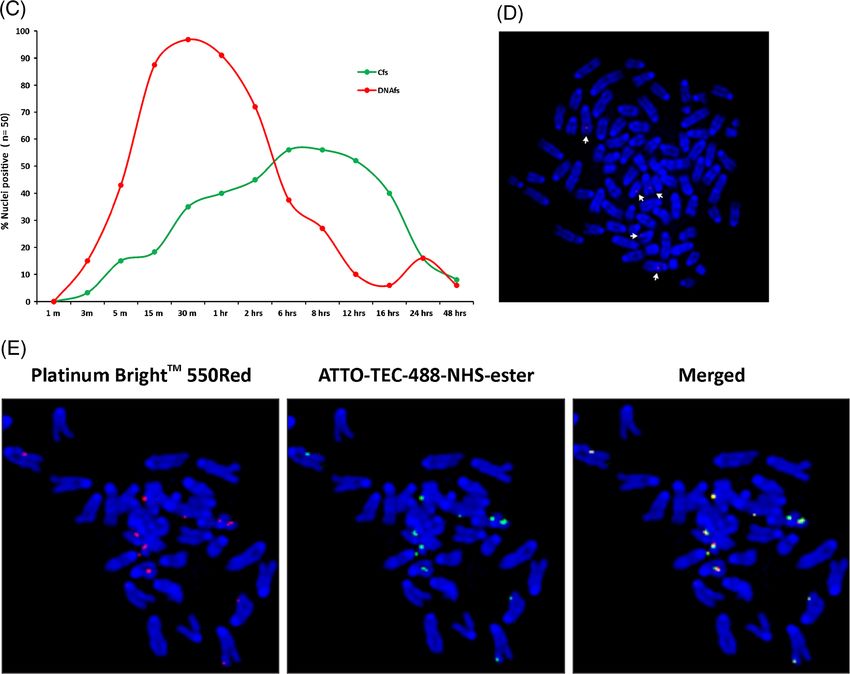
Circulating nucleic acids damage DNA of healthy cells                                     93

the presence of DNA in the isolated Cfs, the latter were         of H4 IgG: H4 IgG (200 μg/mL) was mixed with 3-
suspended in buffer containing 0.5% SDS and 0.5 mg/mL            maleimido benzoyl NHS (Sigma Aldrich™, USA) and kept
proteinase K, incubated at 50°C for 1 h and analysed by 1%       for half an hour at 25°C for the reaction to be completed; (3)
agarose gel electrophoresis (Schnitzler 2001). Western blot      conjugation of activated pullulan with activated H4 IgG:
analysis of Cfs was performed to confirm the presence of         activated H4 IgG was conjugated by vigorous stirring with
histones using polyclonal antibodies against the respective      activated pullulan to form monodispersed nanoconjugates.
histone proteins (Santa Cruz Biotechnology INC., USA).
The DNA content of Cfs was estimated using Quanti-iT™                         2.6   Treatment of cells in culture
PicoGreen® dsDNA Assay Kit (Invitrogen™, USA) and the
amount of DNA was expressed as ng/mL.
                                                                 Cell lines used in our study were: NIH3T3 (mouse fibro-
                                                                 blast), B/CMBA.OV (mouse ovary), MM55.K (mouse kid-
                                                                 ney), 3T3-L1 (mouse adipocyte), HeLa (human cervical
  2.3     Isolation, quantification and fluorescent labelling    cancer). All cell lines were obtained from the American Type
                             of RNA                              Culture Collection, USA. Cells were seeded at a density of
                                                                 6×10⁴ cells/35 mm culture dishes in 1.5 mL of Dulbecco’s
RNA from pooled plasma from cancer patients was isolated         Modified Eagle Medium (DMEM) (Gibco® Life Technolo-
using Plasma/Serum Circulating RNA Purification Kit (Nor-        gies, USA. Catalogue No. 12800-017) and maintained at
gen Biotek Corp, Canada. Catalogue No. 30000) according          37°C in the humidified atmosphere of 5% CO2 in air. After
to the manufacturer’s instructions. The kit uses a proprietary   16 h (cell number ~100,000 / dish), Cfs suspended in PBS
resin which ensures the isolation of all sizes of circulating    and DNAfs suspended in elution buffer diluted in PBS (total
and exosomal RNA, including microRNA. RNA was quan-              volume=100 μL for both) were added to the cells and treat-
tified using Nanodrop 1000 spectrophotometer (Thermo Sci-        ment was continued for periods as mentioned in the text. No
entific, USA) and labelled using ULS TM microRNA                 transfection agent(s) was used in any of our experiments.
Labeling Kit (With Cy3) (Kreatech Diagnostics, The Nether-       Control cells were treated with 100 μL PBS alone. The
lands. Catalogue No. EA-037).                                    medium was changed every 72 h when required. Equal
                                                                 concentrations of DNAfs (5 ng) and Cfs (5 ng equivalent
               2.4   Reconstitution of chromatin                 of DNA) from the same patient pool were usually used for
                                                                 treatment of cells, unless mentioned otherwise in the text.
Chromatin was reconstituted with DNA purified from pooled
plasma from cancer patients using the In Vitro Chromatin            2.7   Development of single-cell clones from DNAfs-
Assembly Kit (Diagenode, Belgium. Catalogue No. ca-vitro-                         and Cfs-treated cells
001) as per the manufacturer’s instructions. The standard
assembly reaction contained 2 μg of DNAfs and purified core      NIH3T3 cells (10×104) were treated with DNAfs and Cfs (5ng
histones at 1:1 histone:DNA ratio, 15 μL ATP, 3.5 μL remod-      DNA each) and cells were allowed to grow. After 5 passages,
eling spacing factor and nucleosome assembly protein-1 that      several single-cell clones were developed from DNAfs- and
catalyses the deposition of histones into extended periodic      Cfs-treated cells by serial dilution. Two randomly selected
nucleosomes. Efficiency of assembly formation was evaluated      clones from DNAfs-treated cells (E10 and E12) and Cfs-
by gel electrophoresis. The DNA content of reconstituted Cfs     treated cells (B2 and D5) were used for further experimentation.
(RCfs) was estimated using Quanti-iT PicoGreen kit and
amount of DNA was expressed as ng/100 μL.
                                                                    2.8 Fluorescent labelling of DNAfs, Cfs and RCfs
                                                                  and detection of their intracellular fate by laser confocal
2.5     Synthesis of pullulan-histone antibody nanoconjugates                             microscopy

Synthesis of pullulan-histone antibody nanoconjugates was        DNAfs were non-enzymatically labelled in a 50 μL reaction
performed as described in our recent publication using H4        with Platinum Bright™ 550 Red/Orange Nucleic Acid La-
IgG (Rekha et al. 2013). The process involved three steps,       beling Kit (Kreatech Diagnostics, The Netherlands. Cata-
namely, (1) activation of pullulan: typically Pullulan (Sigma    logue No. GLK-004) as per the manufacturer’s protocol.
Aldrich™, USA) was dissolved in 20 mM borax buffer,              Cfs were dually labelled; the DNA component was labelled
mixed with Traut’s reagent (2-Iminothiolane, Sigma               using Platinum Bright™ 550 Red/Orange Nucleic Acid La-
Aldrich™, USA) under continuous stirring (final pH 7.0)          beling Kit, while the protein component was labelled with
and dialysed against 0.1 M sodium phosphate (pH 7.4)             ATTO 488 NHS-ester (ATTO-TEC GmbH, Germany. Cata-
containing 0.15 M NaCl and 1 mM EDTA; (2) activation             logue No. AD 488-35) in accordance with the manufacturer’s

                                                                                                     J. Biosci. 40(1), March 2015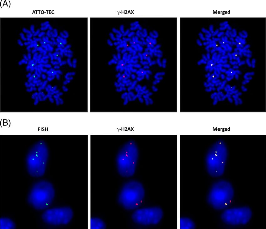
94                                                      Indraneel Mittra et al.

protocol (www.atto-tec.com). RCfs were similarly dually labelled      100 μl of saline. The human FISH probes used here were
with Platinum Bright™ and ATTO 488 NHS-ester. NIH3T3                  unreactive to mouse DNA. The protocol for in vivo experi-
cells grown overnight on coverslips were treated with 100 μL          ments in mice was approved by the Institutional Animal
of fluorescently labelled DNAfs, Cfs or RCfs (10 ng DNA each)         Ethics Committee (IAEC) of Advanced Centre for Treat-
for varying periods. After treatment, cells were fixed in 4% para-    ment, Research and Education in Cancer, Tata Memorial
formaldehyde for 20 min, washed in PBS, mounted onto clean            Centre, Navi Mumbai, India.
glass slides with Vecta-shield and intracellular uptake of labelled
DNAfs, Cfs and RCfs and their localization in DAPI-stained
                                                                       2.11   Detection of human DNA in mouse cells by whole
nuclei was visualized using Zeiss differential laser scanning
                                                                                         genome sequencing
confocal microscopy (LSCM) platform. Optical sections were
captured at depths that passed through the nuclei. Fifty nuclei
                                                                      Whole genome sequencing of two clones each derived from
were analysed in each case, and the number of nuclei showing
                                                                      DNAfs-treated (E10 and E12) and Cfs-treated (B2 and D5)
positive fluorescent signals as well as the average number of
                                                                      cells was undertaken to detect the presence of human-spe-
signals per nucleus were recorded.
                                                                      cific sequences in mouse cells. Intact high quality genomic
                                                                      DNA was isolated to generate whole genome libraries for
  2.9   Detection of chromosomal association of labelled              sequencing on the Illumina GA IIX (Genotypic Technology
                   DNAfs, Cfs and RCfs                                (P) Ltd, India). For whole genome libraries, 3 μg of genomic
                                                                      DNA was made up to 100 μL with nuclease-free water
NIH3T3 cells were treated with labelled DNAfs, Cfs and RCfs           (Ambion® Life Technologies, USA) and sonicated using a
(10 ng DNA each) and metaphase spreads were prepared after            Bioruptor (Diagenode, Belgium) (30 pulses on high at 30 s
6 h and observed under fluorescent microscope. Fifty meta-            ON and 30 s OFF) to obtain desired fragment lengths rang-
phases were analysed in each case and the numbers of meta-            ing between 150 and 600 bp. Libraries for whole genome
phases showing positive fluorescent signals as well as the            sequencing were constructed according to a modification of
average number of signals per metaphase were recorded.                the TruSeq DNA library protocol outlined in ‘TruSeq DNA
                                                                      Sample preparation guide’ (Part # 15005180; Rev. A; Nov
                                                                      2010). The libraries were quantified using Nanodrop and
2.10    Detection of human DNA sequences in mouse cells               validated for quality. The latter was performed by running
                in vitro and in vivo by FISH                          the library on a High Sensitivity Bioanalyzer Chip (Agilent)
                                                                      to check for quality and size distribution. DNA library frag-
Several single-cell clones were established from NIH3T3               ments were diluted, denatured and hybridized to a lawn of
cells treated with DNAfs and Cfs, and four randomly select-           oligonucleotides immobilized on the flow-cell surface. Hy-
ed clones were analysed by FISH for presence of human                 bridized DNA template was amplified using immobilized
DNA on metaphase spreads (DNAfs-derived clones: E10,                  oligonucleotides as primers. Each hybridized template, using
E12; Cfs-derived clones: B2, D5). A mixture of Texas Red-             the process of isothermal bridge amplification, resulted in the
labelled human whole-genomic (1 μL) and biotinylated hu-              formation of clusters comprised of roughly 1000 clonal
man pan-centrometric probes (1 μL) (Chrom-Bios GmbH,                  copies. Paired end-sequencing was performed on the Illu-
Germany. Catalogue No. HGDNAOR10 and HPANCBI10)                       mina GA IIX to generate 54-bases-long reads for Cfs-
was used. For the detection of pan-centrometric signals,              derived clones and 100-bases-long reads for DNAfs-
hybridized slides were treated with 100 μL of Avidin-FITC             derived clones respectively with an average of 1.2× coverage
conjugated secondary antibody (1:200 dilution in 4× SSC/              genome-wide across samples (supplementary table 2).
0.1 % Tween 20). Image acquisition and analysis were
performed using the Spectral Bio-Imaging System (Applied              Bioinformatics analysis: hg19 and mm9 reference sequen-
Spectral Imaging, Israel). Fifty metaphases were examined             ces were used as human and mouse references respectively.
for each clone for presence of human DNA signals. The                 BWA v 0.6.1.software was used for aligning the reads to the
human specific FISH probes were unreactive to mouse cells.            reference genomes. The sequence reads from study acces-
   For in vivo experiments, Balb/C mice (6–8 weeks old,               sion ERP000354, submitted by the Sanger center to the
weighing ~20 g) obtained from the Institute Animal House              NCBI Sequence read archive, were used as a mouse control
were used for the study. They were housed in the Animal               in our experiments. Default parameters (best hit) were used
House Facility of the Institute. Mice were injected intrave-          for alignment of sequence reads to a reference genome
nously with DNAfs (100 ng) and Cfs (100 ng DNA equiv-                 sequence. The reads were aligned to a reference genome
alent) and animals were sacrificed on day 7 by cervical               using a short read aligner, and matched to a reference ge-
dislocation and their vital organs removed, fixed in formalin         nome, after subtracting the aligned reads (supplementary
and processed for FISH. Control animals were injected with            figure 1).

J. Biosci. 40(1), March 2015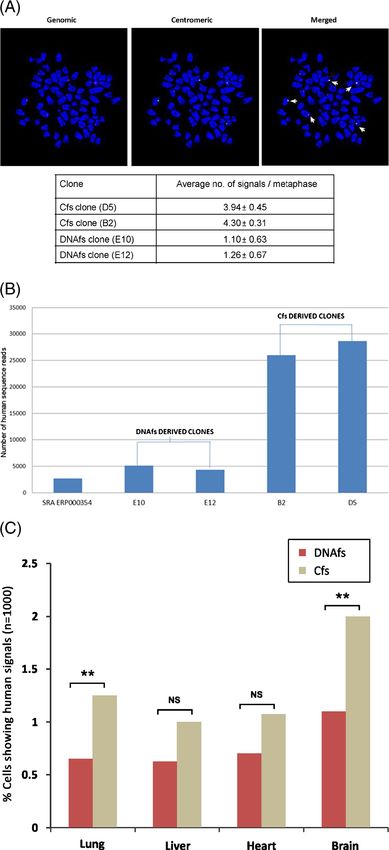
Circulating nucleic acids damage DNA of healthy cells                                       95

   To identify presence of human Alu elements in the mouse         Catalogue No. 05-479) and Caspase-3 were quantified by
cell clones, the DNA reads were compared to a database of          immuno-fluorescence using the respective antibodies and the
Alu elements specific to the hg19 human genome down-               number of cells showing positive fluorescence was recorded.
loaded from the UCSC genome browser database                       All experiments were done in duplicate; 50 cells were analysed
(Karolchik et al. 2014) employing the UCSC table browser           in each case and nuclei showing at least two foci were consid-
(Karolchik et al. 2004) using offline BLAST (Altschul et al.       ered as positive. The average number of positive cells was
1990). An e-value of 0.05 was used as a cut-off for all            recorded.
alignments generated using threshold of 80% and above
sequence identity without gaps.                                    In vivo studies: Balb/C mice (6–8 weeks old, weighing ~20
                                                                   g) were injected intravenously with DNAfs (100 ng) and Cfs
PCR amplification of human Alu elements: Genomic DNA               (containing equivalent of 100 ng DNA) in 100 μL of buffer.
was PCR amplified using primers for the Alu elements               Control mice were injected with 100 μL of saline. Two mice
(HSU14570 and HSAL002744): 5′GAATGGCGTGAACC                        were used for treatment in each case. After 24 h, animals
CGGG3′ and 5′TTTTGAGACGGAGTCTCGCTC3′ for                           were anesthetized with CO2 and blood was collected from
HSU14570; 5′CACCTTGTCCTCCCAAAGTG3′ and 5′                          orbital plexus. Animals were then sacrificed by cervical
TGCTCAGAAATCATTTCATG3′ for HSAL002744. The                         dislocation and the following organs were removed and snap
PCR products were purified using column (NucleoSpin Gel            frozen in liquid nitrogen: lung, liver, brain, heart, kidney,
and PCR Clean-up kit), sanger sequencing performed and se-         spleen, pulmonary artery, skin and muscle and processed for
quencing traces were aligned using NCBI blast as well as           cryo-sectioning. The collected blood was processed for iso-
Mutation Surveyor V 4.0.9.                                         lation of peripheral blood mononuclear cells (PBMCs) by
                                                                   Ficoll gradient centrifugation. Cryo-sections of tissues and
                                                                   PBMCs smeared on slides were processed for immuno-
 2.12 Detection of activated DDR and apoptosis-related             fluorescence staining against γ-H2AX and active Caspase-3
   proteins by immuno-fluorescence in vitro and in vivo            markers. At least 1000 DAPI-stained nuclei per animal were
                                                                   examined from 10 randomly chosen areas of various tissues,
                                                                   and in case of PBMCs, 100 nuclei per animal were exam-
In vitro studies: NIH3T3 cells were seeded on cover slips at a
                                                                   ined. In both cases, the number of nuclei showing positive
density of 6 ×10⁴ cells and allowed to grow overnight (16 h)
                                                                   foci (γ-H2AX) and number of nuclei showing positive fluo-
and subjected to treatment with DNAfs, Cfs and RCfs (5 ng
                                                                   rescence (Caspase-3) were recorded.
DNA each) for 6 h in duplicate experiments. Control cells were
treated with 100 μL PBS alone. Cells were fixed with 4% para-
formaldehyde for 20 min at room temperature, permeabilized in
0.2% Triton for 30 min, blocked in 3% BSA (Bovine Serum                    2.13    Experiments using specific inhibitors
Albumin) for 1 h and immuno-stained overnight with various                             (DNase I and CNPs)
specific antibodies against DDR proteins. Cells were immedi-
ately mounted on slides with Vecta-shield and images were          Several experiments were performed both in vitro and in vivo
acquired through Spectral Bio-Imaging System (Applied Spec-        to examine whether cellular/nuclear entry of DNAfs and Cfs
tral Imaging, Israel). In general, 50 DAPI-stained nuclei were     and the multiple biological activities that they affect could be
examined in duplicate in each experiment; the number of nuclei     inhibited by specific inhibitors, namely, DNase I and CNPs.
showing at least two foci were considered as positive. The         In case of treatment with DNAfs, inhibition experiments
antibodies used for detection of the various activated DDR         were done using DNase I while in case of Cfs, they were
proteins and their sources are given in supplementary table 3.     done using both DNase I and CNPs. For in vitro experi-
Anti-rabbit/anti-mouse secondary antibodies used were la-          ments, cells grown at a density of 10×104 were treated with
belled with FITC (Abcam®, UK. Catalogue No. ab6717/                DNAfs and Cfs (5 ng DNA each) for 6 h in the presence or
ab6785), Texas Red (Abcam® UK. Catalogue No. Ab6883)               absence of DNase I (0.05 U/mL) and/or CNPs (5 μg H4 IgG/
or Rhodamine (Merck Millipore, USA. Catalogue No.                  mL) and the treated cells were analysed for the various
AP160R) as appropriate. Onset of apoptosis was determined          specified parameters. For in vivo experiments, mice were
by assessing the status of mitochondrial membrane potential by     given a single intravenous injection of DNAfs and Cfs
labelling with BD™ MitoScreen (JC-1) kit using 5,5′,6,6′-          (100 ng DNA each) through tail vein with and without
tetrachloro-1,1′,3,3′-tetraethylbenzimidazol-carbocyanine io-      additional administration of DNase I (15 mg/kg i.p.) and/or
dide (BD Biosciences, USA. Catalogue No. 551302) and cell-         CNPs (50 μg H4 IgG/mouse i.p.). DNase I and CNPs treat-
associated fluorescence was detected after addition of JC-1 dye.   ment was started 4 h prior to DNAfs and Cfs administration
The number of cells showing green fluorescence was counted.        and continued for 24 h or 7 days as specified. DNase I was
Activation of Cytochrome-C (Merck-Millipore, Germany.              injected at 12 hourly intervals while CNPs were

                                                                                                       J. Biosci. 40(1), March 2015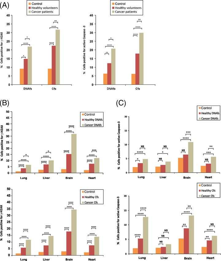
96                                                  Indraneel Mittra et al.

administered every 24 h. Animals were sacrificed and their       revealed that the uptake of DNAfs was rapid and that fluores-
vital organs, namely, heart, lung, liver and brain were re-      cent signals were detectable in nuclei of treated cells as early as
moved for analysis of the specified parameters.                  at 3 min (figure 1C). DNAfs uptake reached a maximum by
                                                                 30 min by which time almost 100% of the nuclei (49/50)
                                                                 contained fluorescent signals. Flow cytometric analysis
                  2.14    Statistical analysis                   revealed that DNAfs internalized at 30 min corresponded to
                                                                 3.88% of the genomic DNA. The proportion of cells showing
Statistical analysis was performed using GraphPad Prism 5        intracellular signals declined steadily thereafter to reach a near
(GraphPad Software, Inc., USA. Version 5.0). Data were           baseline level by 16 h, indicating that nuclear DNAfs were
compared using Chi-square analysis and Student’s t-test as       being actively degraded. The uptake of Cfs was more gradual
appropriate. The tests used have been indicated in appropri-     reaching a peak accumulation at 6–8 h when 29/50 nuclei
ate places in legends to figures.                                (58%) showed fluorescent signals. A statistical comparison
                                                                 of peak nuclear uptake of DNAfs (30 min) and Cfs (6 h)
                                                                 showed a significant greater uptake of DNAfs than Cfs
                         3.    Results                           (p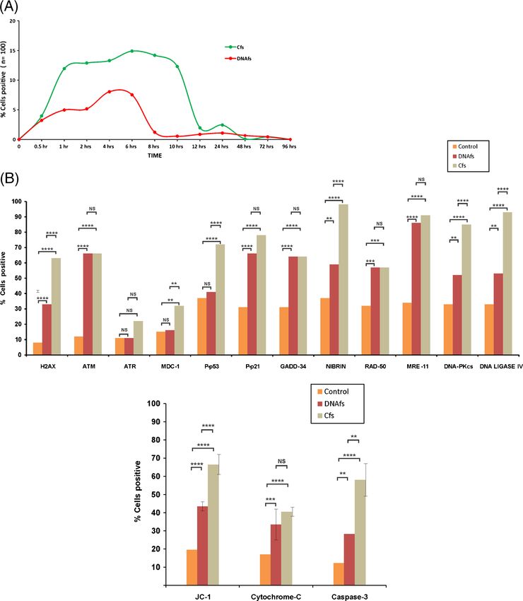
Circulating nucleic acids damage DNA of healthy cells                                       97

These conditions have been widely reported to inhibit met-        human genomic and pan-centromeric signals to co-localize
abolic activity of cultured cells (Gawel-Thompson and             on chromosomal arms indicating common sites of integra-
Greene 1989; Fulda et al. 2000; Sakurai et al. 2005; March-       tion (discussed later). These experiments were repeated at
ant et al. 2008; Pliss et al. 2013). There was a significant      least on two occasions.
reduction in nuclear uptake of DNAfs and Cfs under both
these conditions (supplementary figure 3E, F, G and H).
                                                                  In vitro experiments (using next-generation sequencing): To
Experiments using specific inhibitors (DNase I and CNPs):         further confirm our FISH findings, we undertook whole ge-
We next investigated if nuclear uptake of DNAfs and Cfs could     nome sequencing of the above DNAfs- and Cfs-derived
be prevented by treatment with DNase I (0.05 U) and/or CNPs       clones. In the DNAfs-derived clones E10 and E12, we
(5 μg of H4 IgG). Bovine pancreatic DNase I was obtained from     obtained 100-bases-long paired-end reads on Illumina GA II
Sigma Aldrich (Catalogue No. DN25-1G). DNase I significant-       X with 42 million and 40 million total paired-end reads in the
ly inhibited nuclear uptake of DNAfs (supplementary figure 3I);   two clones, respectively (supplementary table 2). In Cfs-
and with respect to Cfs, this was equally inhibited by both       derived clones B2 and D5 we obtained 54-bases-long paired-
DNase I and CNPs (supplementary figure 3J).                       end reads with 39 million and 58 million total paired-end reads
                                                                  in the two clones, respectively (supplementary table 2). Of
                                                                  these, 145364 and 144886 reads from DNAfs-derived clones
          3.3     Experiments with reconstituted Cfs              aligned to the human genome using BWA default parameters
                                                                  (best hit), while significantly higher numbers, viz. 598644 and
                                                                  889932 from Cfs-derived clones aligned to the human genome
When DNAfs purified from plasma from cancer patients
                                                                  with perfect match (supplementary figure 5). Next, to elimi-
were reconstituted in vitro into RCfs, fluorescently dual-
                                                                  nate the fraction of reads conserved between mouse and hu-
labelled, and applied to cultured cells, RCfs exhibited cellu-
                                                                  man among these aligned reads, we performed computational
lar and nuclear uptake properties that were similar to those
                                                                  subtraction by back-aligning the presumptive human reads to
seen with serum-derived Cfs with 26/50 nuclei (52%) show-
                                                                  mouse reference sequences using identical criterion of BWA
ing presence of fluorescent signals compared to 29/50 (58%)
                                                                  default parameters. The set of reads that remained unmapped
seen in case of native Cfs (supplementary figure 4A). Nota-
                                                                  after the subtractive phases were considered to be purely
bly, the kinetics of cellular uptake of RCfs, unlike that of
                                                                  human in origin. The number of strictly human reads contained
DNAfs from which they had been prepared which peaks at
                                                                  in the DNAfs-derived E10 and E12 clones were several fold
30 min, was retarded to reach a maximum at 6 h similar to
                                                                  lower than those contained in Cfs-derived B2 and D5 clones.
that seen with native Cfs (supplementary figure 4B). Further,
                                                                  In clones E10 and E12, 5106 and 4354 reads respectively were
the association of RCfs with mitotic condensed chromo-
                                                                  strictly human in nature, compared to 25979 and 28694 reads
somes could be clearly seen in 10/50 metaphases after 6 h
                                                                  in clones B2 and D5 respectively (figure 2B; supplementary
as compared to 20/50 seen in case with native Cfs (supple-
                                                                  figure 5). This indicated a significantly higher (5- to 6-fold)
mentary figure 4C). These results confirmed that reconstitu-
                                                                  efficiency of genomic integration of human DNA sequences in
tion of DNAfs into RCfs in vitro imparted properties to the
                                                                  Cfs-derived compared to DNAfs-derived clones. These find-
latter that were similar to those of serum-derived Cfs. This
                                                                  ings confirmed our results with FISH performed on the same
experiment was repeated on at least 3 occasions.
                                                                  clones which had demonstrated a ~4-fold higher human sig-
                                                                  nals in the Cfs-derived compared to the DNAfs-derived clones
       3.4      Genomic integration of DNAfs and Cfs:             (figure 2A lower panel). It needs to be mentioned that in the
                                                                  mouse reference genome used as controls downloaded from
In vitro experiments (using FISH): We analysed single-cell        NCBI, perfect match to the human reference genome was
clones derived from cells treated with DNAfs (E10 and E12)        found to be of the order of 7088 reads, of which 2720 reads
and Cfs (B2 and D5) by FISH using human whole genomic             were found to be of human in nature (supplementary figure 5).
and human pan-centromeric probes. Control experiments             These 2720 sequence reads in mouse reference genome are
confirmed that these probes did not cross-hybridize with          either an artifact of inefficiency of our alignment algorithm, or
mouse DNA. In all four clones examined we could clearly           an artifact of sequencing in the mouse reference genome.
detect positive signals indicating the presence of human              We were curious to find out whether human repetitive Alu
DNA in these mouse cell clones (figure 2A upper panel).           elements could be detected in the mouse cell clones. After
However, many more (~4-fold) human signals were detect-           adjusting for Alu reads found in the control mouse reference
able in Cfs-derived clones B2 and D5 than were detectable in      genome, we identified 47 unique Alu elements that belonged to
DNA-derived clones E10 and E12; these differences were            8 different Alu families in the Cfs-derived B2 clone and 35
highly statistically significant (p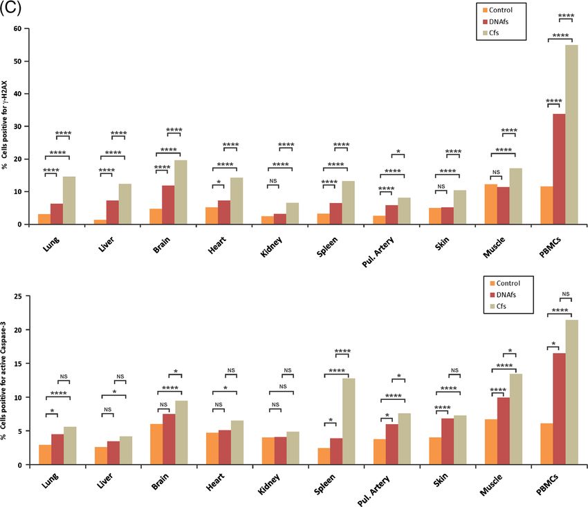
98                             Indraneel Mittra et al.

J. Biosci. 40(1), March 2015
Circulating nucleic acids damage DNA of healthy cells 99
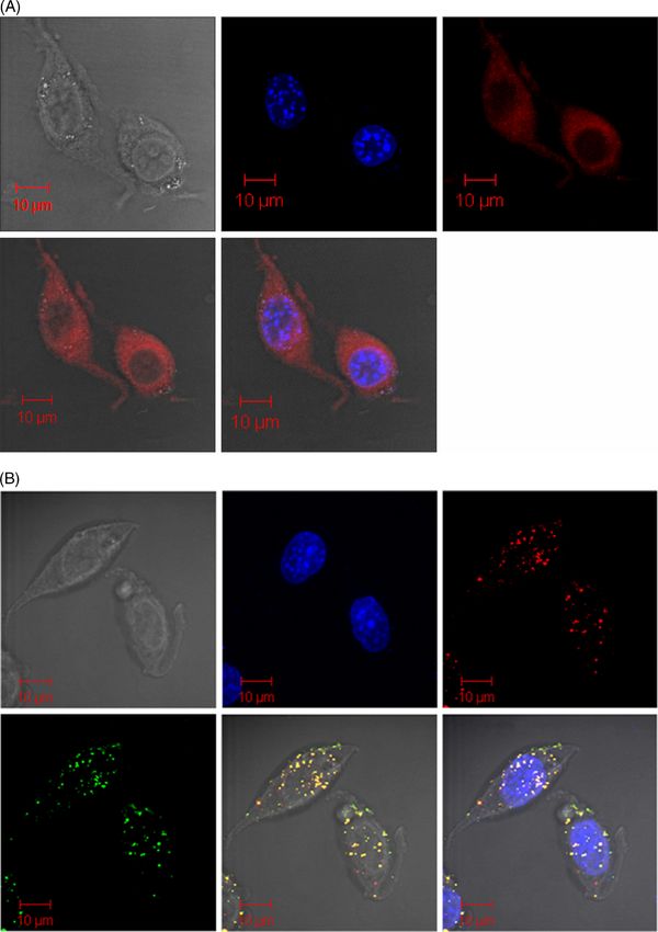
Figure 1. Cellular entry, nuclear uptake and chromosomal association of fluorescently labelled DNAfs and Cfs. NIH3T3 cells (10×104)
were treated with DNAfs labelled with ULS (red) and Cfs dual-labelled with ULS (red) and ATTO-TEC (green) (10 ng DNA in all
experiments). (A) Intracellular fate of DNAfs at 30 min as analysed by LSCM. Numerous fine fluorescent particles are seen in the
cytoplasm and in the nucleus. DIC, DAPI and ULS pictures are represented in different panels. (B) Intracellular fate of Cfs at 6 h as
analysed by LSCM. Presence of dual-labelled Cfs in the cytoplasm and nuclei are clearly seen. The red and green signals appear yellow in
colour when the images are overlapped. (C) Kinetics of nuclear uptake of fluorescently labelled DNAfs and Cfs as analysed by LSCM. Fifty
nuclei were analysed at each time-point and the percentage of positive nuclei was recorded. Nuclei containing at least two fluorescent spots
were considered as positive. (D and E) Association of fluorescently labelled DNAfs (D) and Cfs (E) with chromosomes of treated cells.
NIH3T3 cells were treated with labelled DNAfs and Cfs and metaphase spreads were prepared 6 h after treatment and analysed by
fluorescence microscopy. Note that the labelled DNA particles are considerably smaller in size than Cfs particles.
found 19 unique Alu elements representing 5 Alu families in the PCR reactions amplified human Alu from mouse genomic DNA
E10 clone, and 23 unique Alu elements representing 5 Alu extracted from NIH3T3 cells (negative control). Four randomly
families in the E12 clone (supplementary table 4; supplementary selected PCR products amplified from Cfs- and DNAfs-derived
figure 6). We validated the predicted Alu elements by PCR in 9 clones were confirmed by Sanger sequencing (supplementary
randomly chosen reads found in the Cfs- and DNAfs-derived figure 7A, B and C). This data further confirm the presence of
clones with primers designed ~100 bases apart. Seven out of 9 human DNA in the mouse cell clones.
PCR reactions amplified a ~100 bp fragment, while in human
genomic DNA extracted from DOK cells as template (positive In vivo experiments (using FISH): Since DNAfs and Cfs
control), all reactions amplified a ~100 bp fragment. None of the could access genomic DNA of cells in culture for
J. Biosci. 40(1), March 2015100 Indraneel Mittra et al. J. Biosci. 40(1), March 2015
Circulating nucleic acids damage DNA of healthy cells 101 Figure 2. Integration of DNAfs and Cfs into host cell genomes. (A) Representative FISH images showing presence of human genomic (red) and pan-centromeric (green) signals in mouse cell clone (B2) derived from NIH3T3 cells treated with Cfs. Upon merging the images, red and green signals frequently co-localize giving yellow fluorescence (marked by arrows). Summary results of FISH analysis depicted in a tabular form shows significantly higher frequency of human-specific signals (genomic + centromeric) in Cfs compared to DNAfs-derived clones. Fifty metaphases were counted in each case and the mean number of signals per metaphase was recorded and analysed by Student’s t-test. D5 vs E10 (p=0.0004); D5 vs E12 (p=0.0013); B2 vs E10 (p=0.0001); B2 vs E12 (p=0.0001). (B) Whole genome sequencing to detect presence of human sequence reads in DNAfs-derived and Cfs-derived clones. The histogram shows number of reads that are strictly human in nature in single-cell clones generated from DNAfs (E10, E12) and Cfs (B2, D5) treated NIH3T3 cells as well as in mouse reference genome (SRA ERP000354). The 2720 human sequence reads detected in the mouse reference genome are either an artifact of inefficiency of our alignment algorithm or an artifact of sequencing in the mouse reference genome. Control vs E10 (Chi-sq. 727.30; p
102                                              Indraneel Mittra et al.

general, Cfs were more active in inducing DDR than were      B/CMBA.Ov (mouse ovarian epithelial) and 3T3-L1
DNAfs.                                                       (mouse adipocytes) cells (p
Circulating nucleic acids damage DNA of healthy cells                                            103

Figure 3. Activation of DNA damage and apoptotic pathways in response to DNAfs and Cfs derived from cancer patients. (A) Kinetics of
H2AX phosphorylation in nuclei of NIH3T3 cells treated with DNAfs and Cfs. DNAfs and Cfs were isolated from pooled samples from
cancer patients. NIH3T3 cells (10×104) were treated with DNAfs and Cfs (5 ng DNA each) and cells were processed for immuno-
fluorescence to detect H2AX activation at various time points. Fifty nuclei in duplicate were analysed at each time-point and the percentage
of nuclei showing positive signals was calculated. Nuclei with two or more fluorescent foci were considered as positive. (B) Analysis of
various proteins involved in the DDR (upper panel) and apoptotic (lower panel) pathways induced by DNAfs and Cfs. NIH3T3 cells
(10×104) were treated as described above and cells were processed for immuno-fluorescence at 6 h following treatment for detection of
DDR proteins. Nuclei with two or more fluorescent foci were considered as positive. Experiments were done in duplicate; 50 cells were
counted in each case and the percentage of positive cells was calculated and analysed by Chi-square test. For analysis of activation of
apoptotic pathways, cells were treated as described above for 24 h and processed for detection of JC-1, Cytochrome-C and Caspase-3 by
immuno-fluorescence. For JC-1, cell-associated fluorescence was detected after addition of JC-1 dye and the percentage of cells showing
green fluorescence was calculated. For Cytochrome-C and Caspase-3, fluorescence was detected following antibody treatment and the
percentage of positive cells was calculated. Experiments were done in duplicate, and 50 cells were examined in each case and analysed by
Chi-square test. **p
104                                                Indraneel Mittra et al.

mediated apoptosis. DNAfs and Cfs significantly increased       brain as well as PBMCs, were examined by immuno-
the induction of all the three markers of apoptosis when        fluorescence for activation of H2AX and Caspase-3. We
compared to untreated control cells (range p
Circulating nucleic acids damage DNA of healthy cells                                             105

Figure 4. Induction of γH2AX and active Caspase-3 by DNAfs and Cfs derived from healthy volunteers. Samples from cancer patients
were also analysed for comparison. DNAfs and Cfs were isolated from pooled plasma/serum of healthy volunteers and age- and sex-
matched cancer patients. (A) In vitro analysis of γ-H2AX and active Caspase-3. NIH3T3 cells (10×104) were treated with DNAfs and Cfs (5
ng DNA each) for 6 h for detection of γ-H2AX (left) and for 24 h for detection of active Caspase -3 (right) by immuno-fluorescence. For γ-
H2AX (left-hand panel), 300 nuclei were counted and the percentage of nuclei showing positive foci were calculated and analysed by Chi-
square test. For active Caspase-3 (right-hand panel), 200 cells were counted and the percentage of cells showing positive fluorescent signals
were calculated and analysed by Chi-square test. *p
106                                                      Indraneel Mittra et al.

of Caspase-3 was evident in all organs (p
Circulating nucleic acids damage DNA of healthy cells                                       107

levels in a host of acute and chronic disease conditions           apoptotic bodies with mouse fibroblast cells leading to ma-
including ageing and cancer (Holdenrieder et al. 2001;             lignant transformation of the latter (Bergsmedh et al. 2001).
Chang et al. 2003; Lam et al. 2003; Lui et al. 2003; Trejo-        DNA released from leukemic cells in the form of nucleo-
Becerril et al. 2003; Zeerleder et al. 2003; Gal et al. 2004;      somes can integrate themselves into the chromosomes of
Holdenrieder and Stieber 2004; Kremer et al. 2005; Butt            surrounding stromal cells and induce DNA damage and
et al. 2006; Chiu et al. 2006; Rhodes et al. 2006; Umetani         apoptosis (Dvořáková et al. 2013). Mutated K-ras oncogene
et al. 2006; Swarup and Rajeswari 2007; Zhong et al. 2007;         carried in plasma from colon cancer patients can be taken up
Holdenrieder and Stieber 2009; Pisetsky and Ullal 2010;            by mouse fibroblast cells, leading to their oncogenic trans-
Rykova et al. 2010; Jylhävä et al. 2011; Tsai et al. 2011;         formation, and K-ras sequences could be detected in these
Mittra et al. 2012). Our study shows that far from being inert     cells by PCR and FISH (García-Olmo et al. 2010, Trejo-
molecules, DNAfs and Cfs have significant biological activ-        Becerril et al. 2012). Genes reconstituted into chromatin
ities of their own that are deleterious to healthy cells of the    in vitro are readily taken up by cells in culture to localize
body. Our systematic investigation of the biological proper-       in the nuclei of recipient cells, leading to the suggestion that
ties of circulating DNAfs and Cfs isolated from blood of           chromatinization of DNA maybe an efficient means for gene
cancer patients and healthy volunteers has led to the follow-      delivery (Wagstaff et al. 2008). Circulating DNA can cross
ing novel observations: (1) Fluorescently labelled DNAfs           the blood–brain barrier, and male foetal DNA have been
and Cfs are readily taken up by cells in culture to be local-      found in maternal brain cells (Chan et al. 2012). Fragmented
ized in their nuclei within minutes; (2) the intra-nuclear         DNA released by bacteria in the peritoneal cavity of frogs can
DNAfs and Cfs rapidly associate themselves with host cell          enter brain cells and synthesize RNA (Anker and Stroun
chromosomes, evoking a DDR; (3) the activated DNA repair           1972). Taken together, the above studies clearly show that
pathways facilitate the incorporation of DNAfs and Cfs into        fragmented DNA or chromatin from sources other than human
the host cell genomes; (4) FISH detected the presence of           blood or plasma can also freely enter into cells, especially their
human DNA sequences in single-cell clones derived from             nuclei, and activate biological effects in the target cells.
mouse cells treated with DNAfs and Cfs; (5) whole                     We did not attempt, in this study, to investigate the
genome sequencing of these clones confirmed the pres-              mechanism(s) by which DNAfs and Cfs are taken up by
ence of tens of thousands of human sequence reads in               recipient cells. Nonetheless, our experiments with a meta-
recipient mouse cell genomes as well as in the presence            bolic poison (Actinomycin D) and at low temperature (31°C)
of several human Alu elements; (6) genomic integration of          clearly demonstrated that cellular/nuclear uptake of DNAfs
DNAfs and Cfs leads to dsDNA breaks and activation of              and Cfs are energy dependent and require an active meta-
apoptotic pathways in the treated cells; (7) when injected         bolic machinery of the recipient cells (supplementary
intravenously, DNAfs and Cfs undergo genomic integra-              figure 3E, F, G and H). DNAfs were internalized into the
tion resulting in activation of DDR and apoptosis of cells         nuclei with greater efficiency than were Cfs; while the peak
of vital organs of mice; (8) Cfs were significantly more           DNAfs uptake occurred at 30 min with 98% of the nuclei
active than DNAfs with respect to all parameters exam-             showing presence of fluorescent signals, nuclear uptake of
ined; (9) DNAfs and Cfs from cancer patients were more             Cfs peaked at 6–8 h when 58% of the nuclei showed pres-
active than those from normal volunteers; and (10) finally,        ence of fluorescent Cfs signals (p
108                                                 Indraneel Mittra et al.

structures by complexing with newly synthesized intracellu-      figure 1) was conservative and maximally stringent allow-
lar histones. The DNA-histone assemblies are, however, not       ing zero mismatch of human reads that might be present in
as efficient as incoming native Cfs in their ability either to   the mouse cell clones when aligned to human reference
induce DDR or to integrate themselves into host cell             sequence, we anticipate a much higher underlying abun-
genomes. This interpretation may be relevant in another          dance of human sequences in these transformed mouse cell
context wherein we observed a discordance between the            clones than what we uncovered. It is likely that human
kinetics of nuclear uptake and that of H2AX activation by        reads harbouring SNPs, or those that are tumour-derived
DNAfs and Cfs. Although DNAfs were rapidly taken up by           and harbour somatic alterations, have remained unaccount-
recipient cell nuclei peaking at 30 min, as opposed to 6–8 h     ed. Taken together, these observations give additional
for Cfs (figure 1C), the kinetics of H2AX activation was         insights into the panoply of apparently random integration
similar, both reaching a maximum at ~6 h (figure 3A). This       of human DNA that occurs in mouse cells and provide the
suggests that while Cfs can activate H2AX soon after entry,      groundwork for using new sequencing technologies in
for DNAfs, there is a lag period before H2AX can be              future studies to investigate preferential integration sites,
activated. The lag period may represent the time required        if any.
for DNAfs to reconstitute themselves into Cfs-like structures        Based on these observations, we propose a new working
by complexing with intracellular histones.                       model for DNA damage and mutagenesis that are induced by
   We clearly detected the presence of human DNA signals         circulating DNAfs and Cfs (figure 6). It is well established
in single-cell clones derived from DNAfs- and Cfs-treated        from classical experiments that when genomic DNA is dam-
mouse cells using human-specific whole-genomic and pan-          aged by ionizing and UV-radiation and chemicals, the dam-
centromeric FISH probes (figure 2A). Since the threshold         age to DNA is followed by activation of DDR pathways
value for detection of FISH signals has been estimated to be     which attempt to repair the damage. The new model, on the
of the order of 30–50 kb (Frengen et al. 1997), our detection    other hand, proposes that when DNAfs and Cfs enter the
of human signals in mouse cells by FISH suggested that           recipient cell, the latter perceives the dsDNA breaks present
multiple small fragments of DNAfs and Cfs might have             in their two ends as damaged ‘self’ DNA and, in an attempt
linked up by non-homologous end-joining (NHEJ) to pro-           to repair the ‘perceived damage’, activate proteins of the
duce long concatamers of discontinuous DNA segments              DDR pathway. The activated DNA repair proteins facilitate
before integrating into host cell genomes (discussed later).     the integration of DNAfs and Cfs into host cell genomes by
NHEJ of discontinuous DNAfs and Cfs could have been              homologous and/or non-homologous recombination; and it
facilitated by DNA-ligase IV and DNA-PKcs that were              is this event of DNA integration into host cell genomes that
found to be activated during DDR (figure 3B). We clearly         brings about the damage to host cell DNA (figure 5A and B).
demonstrated that genomic integration of these concatamers       Thus, paradoxically, it is the activation of DDR that brings
involved dsDNA break repair by detecting co-localization of      about damage to DNA rather than maintenance of DNA
ATTO-TEC or FISH signals with those of γ-H2AX on                 integrity. The model further proposes that the activation of
chromosomal arms or in nuclei of treated cells (figure 5A        DNA-PKcs and DNA ligase-IV, proteins that are involved in
and B). The heterogeneously ligated concatamers apparently       NHEJ, leads to linking up of many of the internalized
elicit DNA repair pathways involving homologous and non-         DNAfs and Cfs to form long concatamers of discontinuous
homologous recombinations for their integration and repair       DNA segments which form new substrates for genomic
in the host cell genomes (discussed later).                      integration (Burma et al. 2006). The proposal of DNA con-
   Massively parallel next-generation sequencing of single-      catamerization is supported by the finding that genomic and
cell clones derived from DNAfs- and Cfs-treated mouse cells      centromeric signals frequently co-localize on chromosomal
provided additional line of evidence for the presence of         arms during FISH analysis (figure 2A), suggesting that the
human DNA in mouse-cell genomes at single base resolu-           concatamers often harbour centromeric sequences. The
tion. We found 5- to 6-fold higher abundance of human            above model of DNA damage and repair as proposed by us
DNA in Cfs-derived clones than DNAfs-derived ones                is represented schematically in figure 6. It must be pointed
(figure 2B; supplementary figure 5). This finding is consis-     out, however, that more experimental work is needed to
tent with our FISH experiments, wherein the numbers of           substantiate the model.
human signals were approximately ×4 higher in the same               It becomes clear from the present work that circulating
Cfs-derived clones compared to the DNAfs-derived clones          nucleic acids, far from being biologically inert particles,
(figure 2A). Computational analysis also detected a higher       have significant deleterious functions in the host. They freely
abundance of human Alu repeats in Cfs-derived clones com-        enter healthy cells and damage their DNA by integrating into
pared to DNAfs-derived clones (supplementary table 4; sup-       their genomes. While the genome is exposed to a number of
plementary figure 6). Given that the computational               xenobiotics and DNA damaging events, they are usually
subtraction pipeline adopted in this study (supplementary        transient and do not cause permanent damage. However,

J. Biosci. 40(1), March 2015
Circulating nucleic acids damage DNA of healthy cells                                       109

Figure 6. Schematic representation of proposed model of DNA damage. NHEJ = non-homologous end-joining; HR = homologous
recombination; NHR = non-homologous recombination.

circulating nucleic acids are ubiquitous and continuously           that are associated with elevated levels of circulating nucleic
arising, inflicting repeated damage to the somatic DNA. This        acids thereby making them one of the key players in mainte-
automatically suggests that the somatic genome may not be           nance of human health and disease. (Holdenrieder et al. 2001;
stable, but rather remains in a state of turmoil characterized by   Chang et al. 2003; Lam et al. 2003; Lui et al. 2003; Trejo-
dsDNA breaks, genomic instability and apoptosis affected by         Becerril et al. 2003; Zeerleder et al. 2003; Gal et al. 2004;
integration of circulating DNAfs and Cfs. These events may lead     Holdenrieder and Stieber 2004; Kremer et al. 2005; Butt et al.
to deletions, duplications and rearrangements causing DNA           2006; Chiu et al. 2006; Rhodes et al. 2006; Umetani et al. 2006;
mosaicism, which is being increasingly uncovered in somatic         Pisetsky and Ullal 2010; Tsai et al. 2011; Mittra et al. 2012).
cells (Jacobs et al. 2012; Laurie et al. 2012; McConnell et al.
2013). The uptake and genomic integration of circulating nucleic                       Acknowledgements
acids and the concomitant DNA damage that they induce may
contribute to ageing as well as to initiation and progression of    We sincerely thank Dr LC Padhy for most generously offer-
cancer by triggering genetic events and consequent genomic          ing his time during numerous scientific discussions, for his
rearrangements that underlie both these interrelated processes      invaluable suggestions and for his critical inputs into the
(Vijg and Dollé 2002; Hoeijmakers 2009; Stephens et al. 2009;       manuscript. We thank Dr AN Ghosh for taking the EM
Trejo-Becerril et al. 2012). DNAfs and Cfs may also be impli-       images. We acknowledge the contribution made by the many
cated in a multitude of other acute and chronic human disorders     lab members who have contributed to this project over the

                                                                                                        J. Biosci. 40(1), March 2015
110 Indraneel Mittra et al.
years. Special thanks are due to Nagnath Swamy, Shazia Gal S, Fidler C, Lo YM, Taylor M, Han C, Moore J, Harris AL and
Mansoor, Nabila Akhtar, Mansoor Ali, Rashmi Malvee, Wainscoat JS 2004 Quantitation of circulating DNA in the
Arpit Bhargava, Preetam Bala, Mihir Shetty, Ajay Choudh- serum of breast cancer patients by real-time PCR. Br. J. Cancer.
ary and Mihir Parmar. This study was supported by the 90 1211–1215
García-Olmo DC, Domínguez C, García-Arranz M, Anker P,
Department of Atomic Energy, Govt. of India, through its
Stroun M, García-Verdugo JM and García-Olmo D 2010 Cell
grant CTCTMC to Tata Memorial Centre awarded to IM. free nucleic acids circulating in the plasma of colorectal cancer
AD is supported by an Intermediate fellowship from the patients induce the oncogenic transformation of susceptible cul-
Wellcome Trust/DBT India Alliance (IA/I/11/2500278), by tured cells. Cancer Res. 70 560–567
a grant from Department of Biotechnology, Govt. of India Gawel-Thompson KJ and Greene RM 1989 Epidermal growth
(BT/PR2372/AGR/36/696/2011), and intramural grants factor: modulator of murine embryonic palate mesenchymal cell
(Seed-In-Air 2897, TMH Plan Project 2712 and IRB 92). proliferation, polyamine biosynthesis, and polyamine transport.
PU is supported by Senior Research Fellowship from Coun- J. Cell Physiol. 140 359–370
cil of Scientific & Industrial Research, Govt. of India. Halicka HD, Bedner E and Darzynkiewicz Z 2000 Segregation of
RNA and separate packaging of DNA and RNA in apoptotic
bodies during apoptosis. Exp. Cell Res. 260 248–256
Hoeijmakers JH 2009 DNA damage, aging, and cancer. N. Engl. J.
References
Med. 361 1475–1485
Holdenrieder S, Stieber P, Bodenmüller H, Busch M, Fertig G,
Altschul SF, Gish W, Miller W, Myers EW and Lipman DJ 1990 Fürst H, Schalhorn A, Schmeller N, et al. 2001 Nucleosomes
Basic local alignment search tool. J. Mol. Biol. 215 403–410 in serum of patients with benign and malignant diseases. Int. J.
Anker P and Stroun M 1972 Bacterial ribonucleic acid in the frog Cancer. 95 114–120
brain after a bacterial peritoneal infection. Science. 178 621–623 Holdenrieder S and Stieber P 2004 Apoptotic markers in cancer.
Bergsmedh A, Szeles A, Henriksson M, Bratt A, Folkman MJ, Clin Biochem. 37 605–617
Spetz AL and Holmgren L 2001 Horizontal transfer of onco- Holdenrieder S and Stieber P 2009 Clinical use of circulating
genes by uptake of apoptotic bodies. Proc. Natl. Acad. Sci. USA nucleosomes. Crit. Rev. Clin. Lab. Sci. 46 1–24
98 6407–6411 Jacobs KB, Yeager M, Zhou W, Wacholder S, Wang Z, Rodriguez-
Burma S, Chen BP and Chen DJ 2006 Role of non-homologous end Santiago B, Hutchinson A, Deng X, et al. 2012 Detectable
joining (NHEJ) in maintaining genomic integrity. DNA Repair clonal mosaicism and its relationship to aging and cancer. Nat.
(Amst). 5 1042–1048 Genet. 44 651–658
Butt AN, Shalchi Z, Hamaoui K, Samadhan A, Powrie J, Smith S, Jylhävä J, Kotipelto T, Raitala A, Jylhä M, Hervonen A and Hurme
Janikoun S and Swaminathan R 2006 Circulating nucleic acids M 2011 Aging is associated with quantitative and qualitative
and diabetic complications. Ann. NY Acad. Sci 1075 258–270 changes in circulating cell-free DNA: the vitality 90+ study.
Chan WF, Gurnot C, Montine TJ, Sonnen JA, Guthrie KA and Mech. Ageing Dev. 132 20–26
Nelson JL 2012 Male microchimerism in the human female Karolchik D, Barber GP, Casper J, Clawson H, Cline MS, Diekhans
brain. PLoS One 7 e45592 M, Dreszer TR, Fujita PA, et al. 2014 The UCSC Genome Brows-
Chang CP, Chia RH, Wu TL, Tsao KC, Sun CF and Wu JT 2003 er database: 2014 update. Nucleic Acids Res 42 D764–D770
Elevated cell-free serum DNA detected in patients with myocar- Karolchik D, Hinrichs AS, Furey TS, Roskin KM, Sugnet CW,
dial infarction. Clin. Chim. Acta. 327 95–101 Haussler D and Kent WJ 2004 The UCSC Table Browser data
Chiu TW, Young R, Chan LY, Burd A and Lo DY 2006 Plasma retrieval tool. Nucleic Acids Res. 32 D493–D496
cell-free DNA as an indicator of severity of injury in burn Kitzman JO, Snyder MW, Ventura M, Lewis AP, Qiu R, Simmons
patients. Clin. Chem. Lab. Med. 44 13–17 LE, Gammill HS, Rubens CE, et al. 2012 Noninvasive whole-
Dvořáková M, Karafiát V, Pajer P, Kluzáková E, Jarkovská K, genome sequencing of a human fetus. Sci. Transl. Med. 4 137ra76
Peková S, Krutílková L and Dvořák M 2013 DNA released by Kremer A, Wilkowski R, Holdenrieder S, Nagel D, Stieber P and
leukemic cells contributes to the disruption of the bone marrow Seidel D 2005 Nucleosomes in pancreatic cancer patients during
microenvironment. Oncogene. 32 5201–5209 radiochemotherapy. Tumour Biol. 26 44–49
Fliedner TM, Graessle D, Paulsen C and Reimers K 2002 Structure Lam NY, Rainer TH, Chan LY, Joynt GM and Lo YM 2003 Time
and function of bone marrow hemopoiesis: mechanisms of re- course of early and late changes in plasma DNA in trauma
sponse to ionizing radiation exposure. Cancer Biother. Radio- patients. Clin. Chem. 49 1286–1291
pharm. 17 405–426 Laurie CC, Laurie CA, Rice K, Doheny KF, Zelnick LR, McHugh
Frengen E, Thomsen PD, Brede G, Solheim J, de Jong PJ and CP, Ling H, Hetrick KN, et al. 2012 Detectable clonal mosai-
Prydz H 1997 The gene cluster containing the LCAT gene cism from birth to old age and its relationship to cancer. Nat.
is conserved between human and pig. Cytogenet. Cell Gen- Genet. 44 642–650
et. 76 53–57 Leary RJ, Sausen M, Kinde I, Papadopoulos N, Carpten JD, Craig
Fulda S, Meyer E and Debatin KM 2000 Metabolic inhibitors sensi- D, O′Shaughnessy J, Kinzler KW, et al. 2012 Detection of
tize for CD95 (APO-1/Fas)-induced apoptosis by down- regulat- chromosomal alterations in the circulation of cancer patients
ing Fas-associated death domain-like interleukin 1- converting with whole-genome sequencing. Sci. Transl. Med. 4
enzyme inhibitory protein expression. Cancer Res. 60 3947–3956 162ra154
J. Biosci. 40(1), March 2015You can also read