2021 American College of Rheumatology/Vasculitis Foundation Guideline for the Management of Giant Cell Arteritis and Takayasu Arteritis
←
→
Page content transcription
If your browser does not render page correctly, please read the page content below

Arthritis & Rheumatology
Vol. 0, No. 0, Month 2021, pp 1–17
DOI 10.1002/art.41774
© 2021 American College of Rheumatology. This article has been contributed to by US Government employees and their work is
in the public domain in the USA.
2021 American College of Rheumatology/Vasculitis
Foundation Guideline for the Management of Giant Cell
Arteritis and Takayasu Arteritis
Mehrdad Maz,1 Sharon A. Chung,2 Andy Abril,3 Carol A. Langford,4 Mark Gorelik,5 Gordon Guyatt,6
Amy M. Archer, Doyt L. Conn,8
7
Kathy A. Full,9 Peter C. Grayson,10 Maria F. Ibarra,11 Lisa F. Imundo,5
2 12 12
Susan Kim, Peter A. Merkel, Rennie L. Rhee, Philip Seo, John H. Stone,14
13
Sangeeta Sule,15
Robert P. Sundel,16 Omar I. Vitobaldi,17 Ann Warner,18 Kevin Byram,19 Anisha B. Dua,7 Nedaa Husainat,20
Karen E. James,21 Mohamad A. Kalot,22 Yih Chang Lin,23 Jason M. Springer,1 Marat Turgunbaev,24
4 24 25
Alexandra Villa-Forte, Amy S. Turner, and Reem A. Mustafa
Guidelines and recommendations developed and/or endorsed by the American College of Rheumatology (ACR)
are intended to provide guidance for particular patterns of practice and not to dictate the care of a particu-
lar patient. The ACR considers adherence to the recommendations within this guideline to be voluntary, with
the ultimate determination regarding their application to be made by the physician in light of each patient’s
individual circumstances. Guidelines and recommendations are intended to promote beneficial or desirable
outcomes but cannot guarantee any specific outcome. Guidelines and recommendations developed and en-
dorsed by the ACR are subject to periodic revision as warranted by the evolution of medical knowledge, tech-
nology, and practice. ACR recommendations are not intended to dictate payment or insurance decisions, and
drug formularies or other third-party analyses that cite ACR guidelines should state this. These recommenda-
tions cannot adequately convey all uncertainties and nuances of patient care.
The American College of Rheumatology is an independent, professional, medical and scientific society that
does not guarantee, warrant, or endorse any commercial product or service.
Objective. To provide evidence-based recommendations and expert guidance for the management of giant cell
arteritis (GCA) and Takayasu arteritis (TAK) as exemplars of large vessel vasculitis.
Methods. Clinical questions regarding diagnostic testing, treatment, and management were developed in the population,
intervention, comparator, and outcome (PICO) format for GCA and TAK (27 for GCA, 27 for TAK). Systematic literature reviews
were conducted for each PICO question. The Grading of Recommendations Assessment, Development and Evaluation
methodology was used to rate the quality of the evidence. Recommendations were developed by the Voting Panel, comprising
adult and pediatric rheumatologists and patients. Each recommendation required ≥70% consensus among the Voting Panel.
Results. We present 22 recommendations and 2 ungraded position statements for GCA, and 20 recommendations
and 1 ungraded position statement for TAK. These recommendations and statements address clinical questions
relating to the use of diagnostic testing, including imaging, treatments, and surgical interventions in GCA and TAK.
Recommendations for GCA include support for the use of glucocorticoid-sparing immunosuppressive agents and the
use of imaging to identify large vessel involvement. Recommendations for TAK include the use of nonglucocorticoid
immunosuppressive agents with glucocorticoids as initial therapy. There were only 2 strong recommendations; the
remaining recommendations were conditional due to the low quality of evidence available for most PICO questions.
Conclusion. These recommendations provide guidance regarding the evaluation and management of patients
with GCA and TAK, including diagnostic strategies, use of pharmacologic agents, and surgical interventions.
1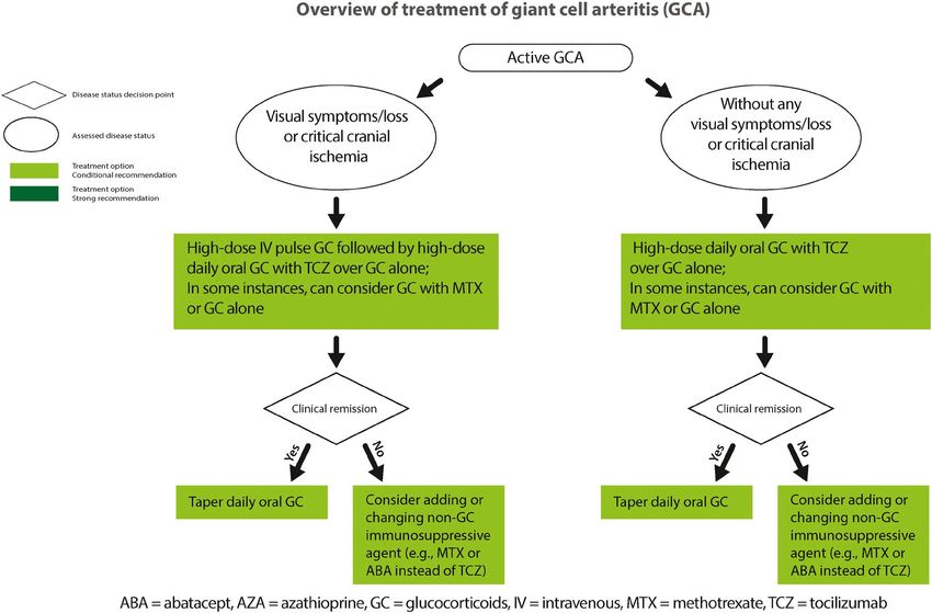
2 | MAZ ET AL
INTRODUCTION METHODS
Giant cell arteritis (GCA) and Takayasu arteritis (TAK) are sys- This guideline followed the American College of Rheu-
temic vasculitides that primarily affect large-and medium-sized matology (ACR) guideline development process (https://www.
vessels (1). GCA can present with both cranial and extracra- rheumatology.org/Practice-Quality/Clinical-Support/Clinical-
nial manifestations. Cranial manifestations include headaches, Practice-Guidelines) using the Grading of Recommendations
scalp tenderness, vision loss, and jaw claudication. Large vessel Assessment, Development and Evaluation (GRADE) methodol-
(“extracranial”) involvement results in arterial stenosis and aneu- ogy to rate the quality of evidence and develop recommenda-
rysms, causing absent pulses and limb claudication (2). GCA tions (13–15). ACR policy guided the management of conflicts
is more common in individuals of Northern European descent of interest and disclosures (https://www.rheum atology.org/
who are older than 50 years of age. Diagnosis is based on clinical Practice-Quality/Clinical-Support/Clinical-Practice-Guidelines/
presentation, pathologic abnormalities on temporal artery biopsy, Vasculitis). Supplementary Appendix 1 (available on the Arthri-
and/or evidence of large vessel involvement on vascular imaging tis & Rheumatology website at http://onlinelibrary.wiley.com/
(1–6). Glucocorticoids are the mainstay treatment for GCA, but doi/10.1002/art.41774/abstract) presents a detailed descrip-
tocilizumab has been approved by the US Food and Drug Admin- tion of the methods. Briefly, the Literature Review team under-
istration for the treatment of GCA (7,8). took systematic literature reviews for predetermined questions
TAK causes granulomatous inflammation of the aorta and specifying the clinical population, intervention, comparator, and
its branches. It is more common in younger women (9,10). Clini- outcomes (PICO). An in-person Patient Panel of 11 individuals
cal manifestations include constitutional symptoms, elevated levels with different types of vasculitis (3 patients with GCA or TAK)
of inflammation markers, and arterial stenosis and/or aneurysms was moderated by a member of the Literature Review team
resulting in limb claudication and absent pulses (11). Treatment (ABD). This Patient Panel reviewed the evidence report (along
options include glucocorticoids, nonglucocorticoid immunosup- with a summary and interpretation by the moderator) and pro-
pressive agents, and surgical management of vascular abnormal- vided patient perspectives and preferences about their personal
ities (12). experiences regarding clinical and treatment aspects of their
As GCA and TAK share clinical manifestations, similar ques- disease. The Voting Panel comprised 9 adult rheumatologists,
tions arise regarding their treatment and management. Recent 5 pediatric rheumatologists, and 2 patients; they reviewed the
studies have broadened treatment options for GCA, and vascular Literature Review team’s evidence summaries and, bearing
imaging is increasingly used for diagnosis and management. This in mind the Patient Panel’s deliberations, formulated and voted
guideline was developed to provide evidence-based recommen- on recommendations. A recommendation required ≥70% con-
dations for the evaluation and management of GCA and TAK. sensus among the Voting Panel.
The article is published simultaneously in Arthritis Care & Research. Rheumatology, Atlanta, Georgia; 25Reem A. Mustafa, MD, FACP, MPH,
Supported by the American College of Rheumatology and the Vasculitis PhD: University of Kansas Medical Center, Kansas City, and McMaster
Foundation. University, Hamilton, Ontario, Canada.
1
Mehrdad Maz, MD, Jason M. Springer, MD, MS: University of Kansas Drs. Maz, Chung, and Abril contributed equally to this work.
Medical Center, Kansas City; 2Sharon A. Chung, MD, MAS, Susan Kim, Dr. Langford has received consulting fees, speaking fees, and/or
MD: University of California, San Francisco; 3Andy Abril, MD: Mayo Clinic, honoraria from Bristol Myers Squibb (less than $10,000) and research
Jacksonville, Florida; 4Carol A. Langford, MD, Alexandra Villa-Forte, MD, grants from Bristol Myers Squibb, GlaxoSmithKline, and Genentech.
MPH: Cleveland Clinic Foundation, Cleveland, Ohio; 5Mark Gorelik, MD, Dr. Grayson has submitted a patent application for the diagnosis and
Lisa F. Imundo, MD: Columbia University, New York, New York; 6Gordon treatment of vacuoles, E1 enzyme, x-linked, autoinflammatory, somatic
Guyatt, MD, MSc, FRCP, OC: McMaster University, Hamilton, Ontario, (VEXAS) syndrome. Dr. Merkel has received consulting fees, speaking fees,
Canada; 7Amy M. Archer, MD, PhD, Anisha B. Dua, MD, MPH: Northwestern and/or honoraria from AbbVie, Biogen, Bristol Myers Squibb, CSL Behring,
University, Chicago, Illinois; 8Doyt L. Conn, MD: Emory University, Atlanta, Genentech/Roche, Genzyme/GlaxoSmithKline, Insmed, Janssen, Kiniksa,
Georgia; 9Kathy A. Full, BA, MA: Springfield, Illinois; 10Peter C. Grayson, Kyverna, Pfizer, Sparrow, and Takeda (less than $10,000 each) and from
MD, MSc: National Institute of Arthritis and Musculoskeletal and Skin AstraZeneca, InflaRx, and ChemoCentryx (more than $10,000 each), research
Diseases, NIH, Bethesda, Maryland; 11Maria F. Ibarra, MD: Children’s support from AstraZeneca, Boehringer Ingelheim, Bristol Myers Squibb,
Mercy Hospital, Kansas City, Missouri; 12Peter A. Merkel, MD, MPH, Celgene, ChemoCentryx, Forbius, Genentech/Roche, Sanofi-Genzyme,
Rennie L. Rhee, MD: University of Pennsylvania, Philadelphia; 13Philip GlaxoSmithKline, and InflaRx, and royalties from UpToDate. Dr. Stone has
Seo, MD, MHS: Johns Hopkins University, Baltimore, Maryland; 14John H. received consulting fees, speaking fees, and/or honoraria from Roche and
Stone, MD, MPH: Massachusetts General Hospital, Boston; 15Sangeeta Genentech (less than $10,000 each). Dr. Sule holds a patent related to the
Sule, MD, PhD: Children’s National Hospital, Washington, DC; 16Robert P. development of the Glucocorticoid Toxicity Index. Dr. Sundel has received
Sundel, MD: Boston Children’s Hospital, Boston, Massachusetts; 17Omar royalties from UpToDate. Dr. Dua has received consulting fees, speaking fees,
I. Vitobaldi, MSEE: Chicago, Illinois; 18Ann Warner, MD: Saint Luke’s Health and/or honoraria from ChemoCentryx and AbbVie (less than $10,000 each).
System, Kansas City, Missouri; 19Kevin Byram, MD: Vanderbilt University, No other disclosures relevant to this article were reported.
Nashville, Tennessee; 20Nedaa Husainat, MD: SSM Health–St. Mary’s Address correspondence to Sharon A. Chung, MD, MAS, 513 Parnassus
Hospital, St. Louis, Missouri; 21Karen E. James, MD, MSCE: University of Avenue, Medical Sciences S865, UCSF Box 0500, San Francisco, CA 94143.
Utah, Salt Lake City; 22Mohamad A. Kalot, MD: State University of New Email: Sharon.Chung@ucsf.edu.
York at Buffalo; 23Yih Chang Lin, MD: University of South Florida, Tampa; Submitted for publication September 25, 2020; accepted in revised form
24
Marat Turgunbaev, MD, MPH, Amy S. Turner: American College of April 13, 2021.2021 AMERICAN COLLEGE OF RHEUMATOLOGY/VASCULITIS FOUNDATION GUIDELINE FOR GCA AND TAK | 3
How to interpret the recommendations recommendation or did not favor one intervention over another.
However, the Voting Panel believed that the PICO question
A strong recommendation is typically supported by moderate-
addressed a commonly encountered clinical question which has
to high- quality evidence (e.g., multiple randomized controlled
not been fully clarified and requires further investigation, and thus
trials). For a strong recommendation, the recommended course
felt that providing guidance for this question was warranted. For
of action would apply to all or almost all patients. Only a small
these situations, we present “ungraded position statements,”
proportion of clinicians/patients would not want to follow the rec-
which reflect general views of the Voting Panel.
ommendation. In rare instances, a strong recommendation may
In this evidence- based guideline, we explicitly used the
be based on very low– to low-certainty evidence. For example,
best evidence available and present that in a transparent man-
an intervention may be strongly recommended if it is considered
ner for the clinician reader/user (10). In some instances, this
low-cost, without harms, and the consequence of not perform-
includes randomized trials in which the interventions under
ing the intervention may be catastrophic. An intervention may be
consideration are directly compared. The GRADE system rates
strongly recommended against if there is high certainty that the
evidence that comes exclusively from the collective experience
intervention will lead to more harm than the comparison with very
of the Voting Panel and Patient Panel members as “very low–
low or low certainty about its benefit (16).
quality” evidence (15).
A conditional recommendation is generally supported by
For each recommendation, details regarding the PICO ques-
lower- quality evidence or a close balance between desirable
tions and the GRADE evidence tables can be found in Supple-
and undesirable outcomes. For a conditional recommendation,
mentary Appendix 2 (http://onlin elibr
ary.wiley.com/doi/10.1002/
the recommended course of action would apply to the majority
art.41774/abstract).
of the patients, but the alternative is a reasonable consideration.
Conditional recommendations always warrant a shared decision-
RESULTS
making approach. We specify conditions under which the alterna-
tive may be considered. For the GCA evidence report, 399 articles were reviewed to
In some instances, the committee found that the evi- address 27 PICO questions. For the TAK evidence report, 347
dence for a particular PICO question did not support a graded articles were reviewed to address 27 PICO questions.
Table 1. Definitions of selected terms used in the recommendations and ungraded position statements for GCA and TAK*
Term Definition
Disease states
Suspected disease Clinical signs and/or symptoms suggestive of GCA/TAK and not explained by other conditions
Active disease New, persistent, or worsening clinical signs and/or symptoms attributed to GCA/TAK and not related to
prior damage
Severe disease Vasculitis with life-or organ-threatening manifestations (e.g., vision loss, cerebrovascular ischemia, cardiac
ischemia, limb ischemia)
Nonsevere disease Vasculitis without life-or organ-threatening manifestations (e.g., constitutional symptoms, headache, jaw
claudication, symptoms of polymyalgia rheumatica)
Remission Absence of clinical signs or symptoms attributed to active GCA/TAK, on or off immunosuppressive therapy
Refractory disease Persistent active disease despite an appropriate course of immunosuppressive therapy
Relapse Recurrence of active disease following a period of remission
Cranial ischemia Visual and neurologic involvement including amaurosis fugax, vision loss, and stroke
Treatments
IV pulse GCs IV methylprednisolone 500–1,000 mg/day (adults) or 30 mg/kg/day (children; maximum 1,000 mg/day) or
equivalent for 3–5 days
High-dose oral GCs Prednisone 1 mg/kg/day up to 80 mg or equivalent
Moderate-dose oral GCs Prednisone 0.5 mg/kg/day (generally 10–40 mg/day in adults) or equivalent
Low-dose oral GCs Prednisone ≤10 mg/day or equivalent
Non-GC nonbiologic Azathioprine, leflunomide, methotrexate, mycophenolate mofetil, cyclophosphamide
immunosuppressive therapy
Biologics Abatacept, tumor necrosis factor inhibitor, tocilizumab
Surgical intervention Angioplasty, stent placement, vascular bypass, vascular graft
Disease assessments
Clinical monitoring Assessing for clinical signs and symptoms of active disease, obtaining 4 extremity blood pressures, and
obtaining clinical laboratory results, including inflammation marker levels
Inflammation markers Erythrocyte sedimentation rate, C-reactive protein level
Noninvasive imaging Computed tomography angiogram, magnetic resonance angiogram, positron emission tomography scan,
vascular ultrasound, magnetic resonance imaging of temporal and scalp arteries
Invasive imaging Conventional catheter-based angiogram
* GCA = giant cell arteritis; TAK = Takayasu arteritis; IV = intravenous; GCs = glucocorticoids.4 | MAZ ET AL
Recommendations and ungraded position a missed diagnosis. This recommendation is conditional due to
statements for the management of GCA a lack of high-quality evidence, but the Voting Panel emphasized
obtaining longer biopsy specimens when possible (18,19).
Table 1 presents definitions of selected terms used in the rec-
ommendations, including disease states such as severe disease,
Recommendation: For patients with suspected GCA,
dosing ranges for glucocorticoids, categorization of medications,
we conditionally recommend obtaining a temporal artery
and disease assessments. Tables 2 and 3 present the recom-
biopsy specimen within 2 weeks of starting oral glucocorti-
mendations with their supporting PICO questions and levels of
coids over waiting longer than 2 weeks for a biopsy.
evidence. We present 22 recommendations and 2 ungraded
Overall, biopsy specimens should be obtained as soon as
position statements for GCA. All but 1 of the recommendations
possible to maximize the likelihood of detecting histopathologic
are conditional due to very low– to low-quality evidence. Figure 1
changes. Studies suggest that histopathologic changes indicating
presents key recommendations for the treatment of GCA.
GCA are more likely to be detected in a temporal artery biopsy if
obtained within 2 weeks of starting glucocorticoids; however, his-
topathologic changes have been detected in biopsy specimens
Diagnostic testing
obtained much later than 2 weeks after the start of glucocorticoid
Recommendation: For patients with suspected GCA, treatment (20–28). A biopsy specimen obtained 2 weeks after
we conditionally recommend an initial unilateral temporal starting glucocorticoids could be informative and may be consid-
artery biopsy over bilateral biopsies. ered at the discretion of the physician and patient.
Initially, a unilateral biopsy is recommended. However, bilateral
temporal artery biopsies may be appropriate if the symptoms are Recommendation: For patients with suspected GCA, we
not clearly localized to 1 temporal artery. Proceeding with the con- conditionally recommend temporal artery biopsy over tem-
tralateral biopsy is also appropriate if the unilateral biopsy result is poral artery ultrasound for establishing a diagnosis of GCA.
negative and additional evidence for cranial GCA is sought (17). In general, rheumatologists and radiologists in the US are
less experienced in using ultrasound to diagnose temporal artery
Recommendation: For patients with suspected GCA, involvement in GCA compared to their counterparts in Europe.
we conditionally recommend a long- segment temporal Therefore, temporal artery biopsy remains the optimal approach
artery biopsy specimen (>1 cm) over a short-segment tem- to diagnosing GCA in the US, because ultrasound is operator-
poral artery biopsy specimen (1 cm) over a short-segment temporal artery
biopsy specimen (2021 AMERICAN COLLEGE OF RHEUMATOLOGY/VASCULITIS FOUNDATION GUIDELINE FOR GCA AND TAK | 5
Table 3. Recommendations/statements for treatment (medical management and surgical intervention) and clinical/laboratory monitoring in
GCA*
GCA PICO
question informing
recommendation Level of
Recommendation/statement and discussion evidence
Medical management
Recommendation: For patients with newly diagnosed GCA without manifestations of cranial ischemia, 11 Very low to low
we conditionally recommend initiating treatment with high-dose oral GCs over IV pulse GCs.
Recommendation: For patients with newly diagnosed GCA with threatened vision loss, we 12 Very low
conditionally recommend initiating treatment with IV pulse GCs over high-dose oral GCs.
Recommendation: For patients with newly diagnosed GCA, we conditionally recommend dosing oral 18 Low
GCs daily over an alternate-day schedule.
Recommendation: For patients with newly diagnosed GCA, we conditionally recommend initiating 14 Very low to low
treatment with high-dose oral GCs over moderate-dose oral GCs.
Recommendation: For patients with newly diagnosed GCA, we conditionally recommend the use of 15, 16, 17 Low to high
oral GCs with tocilizumab over oral GCs alone.
Recommendation: For patients with GCA with active extracranial large vessel involvement, we 21 Very low to low
conditionally recommend treatment with oral GCs combined with a non-GC immunosuppressive
agent over oral GCs alone.
Ungraded position statement: The optimal duration of therapy with GCs for GCA is not well 20 Low to moderate
established and should be guided by the patient’s values and preferences.
Recommendation: In patients with newly diagnosed GCA, we conditionally recommend against the 19 Very low
use of an HMG-CoA reductase inhibitor (“statin”) specifically for the treatment of GCA.
Recommendation: For patients with GCA who have critical or flow-limiting involvement of the 13 Very low to
vertebral or carotid arteries, we conditionally recommend adding aspirin. moderate
Recommendation: For patients with GCA who experience disease relapse while receiving moderate- Relapse 2 †
to-high–dose GCs, we conditionally recommend adding a non-GC immunosuppressive drug.
Recommendation: For patients with GCA who experience disease relapse with symptoms of cranial Relapse 1, 3 †
ischemia, we conditionally recommend adding a non-GC immunosuppressive agent and increasing
the dose of GCs over increasing the dose of GCs alone.
Recommendation: For patients with GCA who experience disease relapse with symptoms of cranial Relapse 4 †
ischemia while receiving GCs, we conditionally recommend adding tocilizumab and increasing the
dose of GCs over adding methotrexate and increasing the dose of GCs.
Surgical intervention
Ungraded position statement: For any patient requiring surgical vascular intervention for GCA, the ‡ ‡
type and timing of intervention should be a collaborative decision between the vascular surgeon
and rheumatologist.
Recommendation: For patients with severe GCA and worsening signs of limb/organ ischemia 24 Very low to low
who are receiving immunosuppressive therapy, we conditionally recommend escalating
immunosuppressive therapy over surgical intervention with escalation of immunosuppressive
therapy.
Recommendation: For patients with GCA undergoing vascular surgical intervention, we conditionally 27 Very low
recommend the use of high-dose GCs during the periprocedural period, if the patient has active
disease.
Clinical/laboratory monitoring
Recommendation: For patients with GCA in apparent clinical remission, we strongly recommend 10 Very low to low
long-term clinical monitoring over no clinical monitoring.
Recommendation: For patients with GCA who have an increase in levels of inflammation markers 23 Very low
alone, we conditionally recommend clinical observation and monitoring without escalation of
immunosuppressive therapy.
* For the population, intervention, comparator, and outcome (PICO) questions used in the Grading of Recommendations Assessment,
Development and Evaluation methodology, as developed for giant cell arteritis (GCA), please refer to Supplementary Appendix 2 (available on
the Arthritis & Rheumatology website at http://onlinelibrary.wiley.com/doi/10.1002/art.41774/abstract). GCs = glucocorticoids; IV = intravenous;
HMG-CoA = hydroxymethylglutaryl-coenzyme A.
† PICO question was developed after completion of literature review and evidence reports. Data from studies already included in evidence
reports were reviewed, but no dedicated literature review was performed for these questions. Recommendation was formed from available
evidence and expert opinion.
‡ Ungraded position statement was not based on a specific PICO question.
Recommendation: For patients with suspected GCA, we can be helpful to establish a diagnosis of GCA (30,31,34–
conditionally recommend temporal artery biopsy over mag- 37). However, lack of technical expertise with this modality in
netic resonance imaging (MRI) of the cranial arteries for the US, as well as the lack of widespread validation of this
establishing a diagnosis of GCA. approach, limits the applicability of MRI with contrast of the
Protocols to image the cranial vessels using differ- cranial vessels as a replacement for temporal artery biopsy at
ent modalities, including MRI, have been developed, which the current time.6 | MAZ ET AL
Figure 1. Overview of treatment of giant cell arteritis.
Recommendation: For patients with suspected GCA Medical management
and a negative temporal artery biopsy result (or results), we
Recommendation: For patients with newly diagnosed
conditionally recommend noninvasive vascular imaging of
GCA without manifestations of cranial ischemia, we condi-
the large vessels with clinical assessment to aid in diagno-
tionally recommend initiating treatment with high-dose oral
sis over clinical assessment alone.
glucocorticoids over intravenous (IV) pulse glucocorticoids.
Imaging the large vessels may provide additional evi-
Cranial ischemic manifestations include visual and neurologic
dence of disease (e.g., extracranial GCA) when the diag-
involvement such as amaurosis fugax, vision loss, and stroke.
nosis is uncertain following negative temporal artery biopsy
Some studies have suggested that the use of IV pulse glucocor-
results (28,34,38–44). Potential diagnostic imaging modali-
ticoids in this patient group could decrease disease relapse and
ties include MR or computed tomography (CT) angiography
increase remission rates. However, routine use of IV pulse gluco-
of the neck/chest/abdomen/pelvis, ultrasonography, and
18 corticoids can also be associated with increased risks, including
F-fluorodeoxyglucose positron emission tomography (FDG-
infections, that may outweigh the benefits, especially in the elderly
PET) (43,45).
(47,48).
Recommendation: For patients with newly diagnosed
GCA, we conditionally recommend obtaining noninvasive Recommendation: For patients with newly diagnosed
vascular imaging to evaluate large vessel involvement. GCA with threatened vision loss, we conditionally recom-
Baseline noninvasive imaging with MR or CT angiography of mend initiating treatment with IV pulse glucocorticoids
the neck/chest/abdomen/pelvis in patients with newly diagnosed over high-dose oral glucocorticoids.
GCA can detect large vessel involvement and may be compared Studies investigating the effect of IV pulse glucocorticoids in
with subsequent routine monitoring if indicated (46). In a patient patients with GCA and cranial ischemia have demonstrated con-
with large vessel involvement, routine noninvasive vascular imaging flicting results. However, this population is at high risk for vision
can identify early and long-term complications, such as aneurysms loss as well as toxicity from glucocorticoid use. IV pulse gluco-
and stenoses, and assess stability of existing lesions. In patients corticoids can be used in patients with the highest risk of vision
without large vessel involvement, routine and repeated monitoring loss, but this decision should be guided by the patient’s clinical
with vascular imaging may or may not be necessary. condition, values, and preferences (49,50).2021 AMERICAN COLLEGE OF RHEUMATOLOGY/VASCULITIS FOUNDATION GUIDELINE FOR GCA AND TAK | 7
Recommendation: For patients with newly diagnosed (e.g., imaging findings) attributed to GCA can include the addition
GCA, we conditionally recommend dosing oral glucocorti- of nonglucocorticoid immunosuppressive agents. These agents
coids daily over an alternate-day schedule. include biologic agents (e.g., tocilizumab) as well as oral therapies
This recommendation is conditional solely due to the low level (e.g., methotrexate) (56,57). However, the Voting Panel recognizes
of evidence, which indicates higher remission rates in patients that there are few high-quality studies evaluating the efficacy of
receiving daily dosing. The panel did not identify any situations in these agents for this patient group. While there is stronger clinical
which alternate-day dosing of prednisone would be preferred (51). evidence supporting the use of tocilizumab compared to meth-
otrexate for the treatment of GCA, methotrexate can be consid-
Recommendation: For patients with newly diagnosed ered for patients unable to use tocilizumab due to factors such
GCA, we conditionally recommend initiating treatment with as recurrent infections, history of gastrointestinal perforations or
high- dose oral glucocorticoids over moderate- dose oral diverticulitis, and cost.
glucocorticoids.
We recommend starting high- dose oral glucocorticoids Ungraded position statement: The optimal duration of
to achieve rapid disease control followed by tapering the glu- therapy with glucocorticoids for GCA is not well established
cocorticoid dose (weeks to months) to avoid prolonged high- and should be guided by the patient’s values and preferences.
dose treatment and reduce toxicity. The dosing and duration Factors that may influence the duration of therapy include the
of oral glucocorticoid therapy can be variable depending on a patient’s clinical manifestations, toxicity related to glucocorticoid
patient’s manifestations and comorbidities and whether the use use, number of flares, the physician’s experience, and the patient’s
of a glucocorticoid-sparing agent was also initiated. Studies sup- preferences (8). Overall, the Patient Panel emphasized minimizing
porting the efficacy and lower toxicity of moderate-dose gluco- the use of glucocorticoids as much as possible but recognized that
corticoids are of low quality, which prevents the Voting Panel from longer-term use may be needed in some patients to avoid flares.
recommending moderate-dose glucocorticoids as initial therapy.
Moderate-dose glucocorticoids may be used in patients with sig- Recommendation: In patients with newly diagnosed
nificant risk of severe glucocorticoid toxicity and in patients with GCA, we conditionally recommend against the use of a
low risk of vision loss or other life-or organ-threatening compli- hydroxymethylglutaryl- coenzyme A reductase inhibitor
cations (48–53). (“statin”) specifically for the treatment of GCA.
The use of statins is not known to provide a clinically sig-
Recommendation: For patients with newly diagnosed nificant immunosuppressive effect for GCA. Whether statins are
GCA, we conditionally recommend the use of oral gluco- warranted to decrease the patient’s risk of cardiovascular events
corticoids with tocilizumab over oral glucocorticoids alone. is a separate clinical question and depends on the patient’s risk
A trial published in 2017 (8) demonstrated that tocilizumab factors for cardiovascular disease (58–60).
has a significant glucocorticoid-sparing effect in GCA, and thus,
tocilizumab should be considered for initial treatment. How- Recommendation: For patients with GCA who have crit-
ever, methotrexate with glucocorticoids, as well as glucocorti- ical or flow-limiting involvement of the vertebral or carotid
coids alone, can also be considered as initial treatment for newly arteries, we conditionally recommend adding aspirin.
diagnosed GCA. The decision to treat with tocilizumab and glu- There are few data regarding this clinical question, but the
cocorticoids, methotrexate and glucocorticoids, or glucocorti- antiplatelet activity of aspirin may be beneficial in preventing
coid monotherapy for initial therapy should be made based on the ischemic events in patients with vascular narrowing causing
physician’s experience and the patient’s clinical condition, values, decreased cerebral blood flow (61–64). The efficacy of aspirin to
and preferences. Lack of long-term follow-up data on tocilizumab prevent ischemic events in patients without vertebral or carotid
and cost may limit its use (8,54). Abatacept with glucocorticoids narrowing remains unclear at this time.
can also be considered if these other agents are not effective (55).
Recommendation: For patients with GCA who expe-
Recommendation: For patients with GCA with active rience disease relapse while receiving moderate-to-high–
extracranial large vessel involvement, we conditionally dose glucocorticoids, we conditionally recommend adding
recommend treatment with oral glucocorticoids combined a nonglucocorticoid immunosuppressive drug.
with a nonglucocorticoid immunosuppressive agent over Relapses of any type while receiving moderate-to-high–dose
oral glucocorticoids alone. glucocorticoids indicate that it is unlikely that it will be possi-
Management of GCA in patients with new, persistent, or ble for glucocorticoids to be tapered to a low dose. Therefore,
worsening extracranial symptoms (e.g., limb claudication) or signs glucocorticoid-sparing therapy should be considered.8 | MAZ ET AL
Recommendation: For patients with GCA who experi- periprocedural period in GCA, and thus, support for this recom-
ence disease relapse with symptoms of cranial ischemia, we mendation is based in part on their use in TAK. As in TAK, high
conditionally recommend adding a nonglucocorticoid immu- doses of oral glucocorticoids in the perioperative setting are rec-
nosuppressive agent and increasing the dose of glucocorti- ommended if the disease is active or if the clinician is concerned
coids over increasing the dose of glucocorticoids alone. that the patient may have active disease.
Nonglucocorticoid immunosuppressive agents considered
in this situation include tocilizumab and methotrexate (8,65,66).
Clinical/laboratory monitoring
Relapses with symptoms of polymyalgia rheumatica may be con-
trolled by increasing the dose of glucocorticoids alone. Recommendation: For patients with GCA in apparent
clinical remission, we strongly recommend long-term clini-
Recommendation: For patients with GCA who experience cal monitoring over no clinical monitoring.
disease relapse with cranial symptoms while receiving gluco- The optimal frequency and length of monitoring are not well
corticoids, we conditionally recommend adding tocilizumab established and depend on factors including the duration of remission,
and increasing the dose of glucocorticoids over add- site of involvement, risk of disease progression, whether the patient
ing methotrexate and increasing the dose of glucocorticoids. is receiving immunosuppressive therapy, and reliability of the patient
Tocilizumab is an effective glucocorticoid-sparing agent for GCA to report new signs or symptoms (48,69). Clinical monitoring may
(8,54). While there are no comparative studies, the glucocorticoid- include history taking, examinations, and laboratory and imaging
sparing effect seen with methotrexate is smaller than the effect seen studies. This is a strong recommendation given the minimal risks and
with tocilizumab (8,55,65–67). While the glucocorticoid-sparing effect potential catastrophic outcomes if monitoring is not performed.
of tocilizumab is best quantified using the subcutaneous formulation
(8), IV tocilizumab has also been shown to be glucocorticoid-sparing Recommendation: For patients with GCA who have
(54). Again, methotrexate can be considered for patients who are an increase in levels of inflammation markers alone, we
unable to tolerate or have limited access to tocilizumab. conditionally recommend clinical observation and moni-
toring without escalation of immunosuppressive therapy.
Increases in levels of inflammation markers such as eryth-
Surgical intervention
rocyte sedimentation rate and C-reactive protein can be non-
Ungraded position statement: For any patient requiring specific (69). Therefore, increasing immunosuppressive therapy
surgical vascular intervention for GCA, the type and timing is not warranted in the setting of increased levels of inflamma-
of intervention should be a collaborative decision between tion markers in the absence of other signs of disease activity.
the vascular surgeon and rheumatologist. However, these increased levels may warrant more frequent clin-
ical and/or radiographic assessments for active disease.
Recommendation: For patients with severe GCA and
worsening signs of limb/organ ischemia who are receiving
Recommendations and ungraded position
immunosuppressive therapy, we conditionally recommend
statement for the management of TAK
escalating immunosuppressive therapy over surgical inter-
vention with escalation of immunosuppressive therapy. Table 1 presents definitions of selected terms used in the rec-
Because patients can develop collateral blood vessels to ommendations, and Tables 4 and 5 present the recommendations
improve distal blood flow, immunosuppressive therapy is recom- with their supporting PICO questions and levels of evidence. We
mended as initial therapy in patients with GCA and worsening present 20 recommendations and 1 ungraded position statement
limb/organ ischemia. However, clinical situations that would war- for TAK. All recommendations except for 1 are conditional due to
rant consideration of immediate surgical intervention include aor- the availability of only very low– to low-quality evidence. Figure 2
tic aneurysms at high risk for rupture and impending/progressive presents key recommendations for the treatment of TAK.
tissue or organ infarction or damage (68–70).
Medical management
Recommendation: For patients with GCA undergo-
ing vascular surgical intervention, we conditionally rec- Recommendation: For patients with active, severe
ommend the use of high-dose glucocorticoids during the TAK who are not receiving immunosuppressive therapy,
periprocedural period, if the patient has active disease. we conditionally recommend initiating treatment with high-
This recommendation pertains to patients with GCA who dose oral glucocorticoids over IV pulse glucocorticoids fol-
are undergoing a vascular surgical intervention due to a com- lowed by high-dose oral glucocorticoids.
plication of GCA (e.g., aneurysm or stenosis). There are limited There is no evidence that IV pulse glucocorticoids are more
data regarding the use of high-dose glucocorticoids during the effective than high-dose oral glucocorticoids in this setting.2021 AMERICAN COLLEGE OF RHEUMATOLOGY/VASCULITIS FOUNDATION GUIDELINE FOR GCA AND TAK | 9
IV pulse glucocorticoids may be considered for patients with with newly active, nonsevere disease (e.g., patients with constitu-
life-or organ-threatening disease. In children, alternate ste tional symptoms and without limb ischemia) (72).
roid dosing regimens (e.g., IV pulse glucocorticoids with low
daily oral dosing) may be preferred to improve compliance and Recommendation: For patients with TAK who achieved
potentially reduce adverse consequences such as impacting remission while receiving glucocorticoids for ≥6–12 months,
growth (71). we conditionally recommend tapering off glucocorticoids over
long-term treatment with low-dose glucocorticoids for remis-
Recommendation: For patients with newly active, severe sion maintenance.
TAK, we conditionally recommend initiating treatment with The optimal duration of glucocorticoid use in TAK is unknown.
high-dose glucocorticoids over low-dose glucocorticoids. Glucocorticoid exposure should be limited if possible in order
A higher dose of glucocorticoids is recommended due to the to minimize toxicity. Glucocorticoids may be continued for a longer
potential for organ damage or life-threatening events. However, duration if disease is not adequately controlled or if the patient
lower doses of glucocorticoids may be considered for patients experiences frequent disease relapse.
Table 4. Recommendations/statement for treatment (medical management and surgical intervention) in TAK*
TAK PICO question
informing
recommendation Level of
Recommendation/statement and discussion evidence
Medical management
Recommendation: For patients with active, severe TAK who are not receiving immunosuppressive 6 Very low
therapy, we conditionally recommend initiating treatment with high-dose oral GCs over IV pulse GCs
followed by high-dose oral GCs.
Recommendation: For patients with newly active, severe TAK, we conditionally recommend initiating 5 Very low
treatment with high-dose GCs over low-dose GCs. to low
Recommendation: For patients with TAK who achieved remission while receiving GCs for ≥6–12 months, 15 Very low
we conditionally recommend tapering off GCs over long-term treatment with low-dose GCs for
remission maintenance.
Recommendation: For patients with active TAK, we conditionally recommend the use of a non-GC 7, 8, 9 Low
immunosuppressive agent plus GCs over GCs alone.
Recommendation: For patients with active TAK, we conditionally recommend the use of other non-GC 8, 10, 11, 12 Very low
immunosuppressive therapy over tocilizumab as initial therapy. to low
Recommendation: For patients with TAK that is refractory to treatment with GCs alone, we conditionally 14 Very low
recommend adding a tumor necrosis factor inhibitor over adding tocilizumab.
Recommendation: For patients with TAK and asymptomatic progression of a previously identified vascular 16 Very low
lesion seen on imaging, without evidence of inflammation, we conditionally recommend continuing
current therapy over escalating/changing immunosuppressive therapy.
Recommendation: For patients with active TAK and critical cranial or vertebrobasilar involvement, we 13 Low
conditionally recommend adding aspirin or another antiplatelet therapy.
Surgical intervention
Ungraded position statement: For any patient requiring surgical vascular intervention, the type and timing † †
of intervention should be a collaborative decision between the vascular surgeon and rheumatologist.
Recommendation: In patients with known TAK and persistent limb claudication without evidence of 20 Very low
ongoing active disease, we conditionally recommend against surgical intervention. to low
Recommendation: For patients with known TAK with worsening signs of limb/organ ischemia while 21, 24 Very low
receiving immunosuppressive therapy, we conditionally recommend escalating immunosuppressive
therapy over surgical intervention with escalation of immunosuppressive therapy.
Recommendation: For patients with TAK with renovascular hypertension and renal artery stenosis, we 26 Very low
conditionally recommend medical management over surgical intervention. to low
Recommendation: For patients with TAK and stenosis of a cranial/cervical vessel without clinical 22 Very low
symptoms, we conditionally recommend medical management over surgical intervention. to low
Recommendation: For patients with TAK with worsening signs of limb/organ ischemia, we conditionally 23 Very low
recommend delaying surgical intervention until the disease is quiescent over performing surgical to low
intervention while the patient has active disease.
Recommendation: For patients with TAK who are undergoing surgical intervention, we conditionally 25 Very low
recommend the use of high-dose GCs in the periprocedure period if the patient has active disease. to low
* For the population, intervention, comparator, and outcome (PICO) questions used in the Grading of Recommendations Assessment,
Development and Evaluation methodology, as developed for Takayasu arteritis (TAK), please refer to Supplementary Appendix 2 (available on
the Arthritis & Rheumatology website at http://onlinelibrary.wiley.com/doi/10.1002/art.41774/abstract). GCs = glucocorticoids; IV = intravenous.
† Ungraded position statement was not based on a specific PICO question.10 | MAZ ET AL
Table 5. Recommendations for clinical/laboratory monitoring and vascular imaging in TAK*
TAK PICO question
informing
recommendation Level of
Recommendation and discussion evidence
Clinical/laboratory monitoring
Recommendation: For patients with TAK, we conditionally recommend adding inflammation markers to 2 Very low
clinical monitoring as a disease activity assessment tool. to low
Recommendation: For patients with TAK in apparent clinical remission, we strongly recommend long- 4 Very low
term clinical monitoring over no clinical monitoring.
Recommendation: For patients with TAK in apparent clinical remission but with an increase in levels 19 Very low
of inflammation markers, we conditionally recommend clinical observation without escalation of
immunosuppressive therapy.
Vascular imaging
Recommendation: For patients with TAK, we conditionally recommend the use of noninvasive imaging 1 Low
over catheter-based dye angiography as a disease activity assessment tool.
Recommendation: For patients with known TAK, we conditionally recommend regularly scheduled 3 Very low
noninvasive imaging in addition to routine clinical assessment. to low
Recommendation: For patients with TAK in apparent clinical remission but with signs of inflammation 17, 18 Very low
in new vascular territories (e.g., new stenosis or vessel wall thickening) on vascular imaging, we to low
conditionally recommend treatment with immunosuppressive therapy.
* For the population, intervention, comparator, and outcome (PICO) questions used in the Grading of Recommendations Assessment,
Development and Evaluation methodology, as developed for Takayasu arteritis (TAK), please refer to Supplementary Appendix 2 (available on
the Arthritis & Rheumatology website at http://onlinelibrary.wiley.com/doi/10.1002/art.41774/abstract).
Recommendation: For patients with active TAK, we glucocorticoid-related toxicity. Methotrexate is often used as the
conditionally recommend the use of a nonglucocorticoid initial nonglucocorticoid immunosuppressive agent, but other ther-
immunosuppressive agent plus glucocorticoids over gluco- apies such as tumor necrosis factor inhibitors and azathioprine
corticoids alone. can be considered as well (70–73). Methotrexate is often pre-
Nonglucocorticoid immunosuppressive agents are recom- ferred for use in children since it is usually well tolerated. Gluco-
mended over monotherapy with glucocorticoids to minimize corticoid m onotherapy can be considered for mild disease or if the
Figure 2. Overview of treatment of Takayasu arteritis based on clinical and radiographic assessments.2021 AMERICAN COLLEGE OF RHEUMATOLOGY/VASCULITIS FOUNDATION GUIDELINE FOR GCA AND TAK | 11
diagnosis is uncertain. Patient-specific factors such as alcohol use, fibrosis” in response to effective treatment. Intervention is not
plans for childbearing, medication compliance, and medical comor- always needed, since collateral circulation frequently develops
bidities may influence the choice of immunosuppressant (73,74). over time. However, the location and the extent of the lesion of
the affected vessel should be considered. Escalating immunosup-
Recommendation: For patients with active TAK, we pressive therapy may be warranted if significant progression has
conditionally recommend the use of other nonglucocorti- developed rapidly (e.g., weeks to months) after a period of stable
coid immunosuppressive therapy over tocilizumab as initial disease (80,81).
therapy.
As discussed above, nonglucocorticoid immunosuppressive Recommendation: For patients with active TAK and
agents such as methorexate, tumor necrosis factor inhibitors, and critical cranial or vertebrobasilar involvement, we condi-
azathioprine can be used as initial therapy in TAK. We recommend tionally recommend adding aspirin or another antiplatelet
these agents over tocilizumab for initial therapy, because the effi- therapy.
cacy of tocilizumab in TAK is not established at this time. While Small observational studies suggest a decreased risk of
tocilizumab has been shown to be efficacious for GCA, the primary ischemic events with antiplatelet therapy but an increased risk
efficacy end point was not achieved in the only randomized trial of bleeding (82). Therefore, antiplatelet therapy is usually used
of tocilizumab in TAK conducted thus far (74,75). Tocilizumab may for patients at higher risk of ischemic events (e.g., patients with
be considered for patients with inadequate response to other flow-limiting vertebrobasilar disease or stents). Antiplatelet therapy
immunosuppressive therapies. Abatacept is not recommended, should be used with caution after surgical procedures or if there is
since it has been shown in a small randomized controlled trial to an increased risk of bleeding (81).
not be efficacious in TAK (74,76).
Clinical/laboratory monitoring
Recommendation: For patients with TAK that is refrac-
tory to treatment with glucocorticoids alone, we condition- Recommendation: For patients with TAK, we
ally recommend adding a tumor necrosis factor inhibitor onditionally recommend adding inflammation markers to
c
over adding tocilizumab. clinical monitoring as a disease activity assessment tool.
We recognize that among biologic therapies, some practi- While inflammation markers are an imperfect indicator
tioners favor TNF inhibition, while others favor interleukin-6 inhi- of disease activity, they may be helpful for clinical monitoring
bition (tocilizumab) in this situation. Overall, the Voting Panel (80,83).
favored tumor necrosis factor inhibitors over tocilizumab,
since there is more clinical experience with and data on Recommendation: For patients with TAK in apparent
tumor necrosis factor inhibitors in TAK compared to tocilizumab. clinical remission, we strongly recommend long-term clini-
In observational studies, tumor necrosis factor inhibitors have been cal monitoring over no clinical monitoring.
shown to induce remission and decrease relapses (77–79). Clini- The frequency of monitoring depends on factors including
cal experience with tocilizumab in TAK has been demonstrated in the duration of remission, sites of involvement, risk of disease
a randomized controlled trial and small case series. In the rand- progression, the patient’s immunosuppressive regimen, and the
omized trial, a trend toward a longer time to relapse was seen in the ability and likelihood of the patient reliably reporting new signs or
tocilizumab arm, but the difference was not statistically significant. symptoms of TAK. This is a strong recommendation given the min-
However, that study was felt to be underpowered (36 participants). imal risks and potential catastrophic outcomes without monitoring
Of note, tocilizumab use also affects acute- phase reactants, (80,83).
which may impact ability to gauge disease activity. Therefore,
while the panel favors tumor necrosis factor inhibitor use, we rec- Recommendation: For patients with TAK in apparent
ognize that tocilizumab may also be considered, especially when clinical remission but with an increase in levels of inflam-
tumor necrosis factor inhibitors are contraindicated (75). mation markers, we conditionally recommend clinical
observation without escalation of immunosuppressive
Recommendation: For patients with TAK and asymptom therapy.
atic progression of a previously identified vascular lesion As discussed above in the GCA recommendations, increases
seen on imaging, without evidence of inflammation, we in levels of inflammation markers can be nonspecific, and inten-
conditionally recommend continuing current therapy over sifying immunosuppressive therapy in the setting of increased
escalating/changing immunosuppressive therapy. inflammation markers alone may not be warranted. More frequent
Vascular lesions can progress due to a number of fac- clinical and/or radiographic assessments for active disease can
tors that may not be related to active disease, such as “healing be considered (77,80,83).12 | MAZ ET AL
Vascular imaging help determine whether the observed imaging changes repre-
sent active disease.
Recommendation: For patients with TAK, we condi-
tionally recommend the use of noninvasive imaging over
Surgical intervention
catheter- based dye angiography as a disease activity
assessment tool. Ungraded position statement: For any patient requir-
Noninvasive imaging such as CT angiography, MR angi- ing surgical vascular intervention, the type and timing of
ography, or FDG-PET are recommended because these imag- intervention should be a collaborative decision between
ing modalities provide information regarding vascular wall the vascular surgeon and rheumatologist.
inflammation, while catheter- based angiography primarily pro-
vides information regarding the vascular lumen. Catheter-based Recommendation: In patients with known TAK and
angiography can be used to accurately determine central blood persistent limb claudication without evidence of ongoing
pressures, as part of surgical planning, or if noninvasive modalities active disease, we conditionally recommend against surgi-
do not provide adequate information. Identifying active disease cal intervention.
based on noninvasive imaging at this time can be challenging, Patients with TAK can develop collateral circulation that
since the hallmarks of active disease have not been definitively bypasses the stenosis causing limb claudication, and thus, sur-
established (43,45,84). gical intervention may not be needed (87). However, surgical
intervention can be considered for patients whose activities are
Recommendation: For patients with known TAK, we significantly impacted by limb claudication.
conditionally recommend regularly scheduled noninvasive
imaging in addition to routine clinical assessment. Recommendation: For patients with known TAK with
Routine imaging is recommended since vascular changes in worsening signs of limb/organ ischemia while receiving
TAK can occur when the disease is considered clinically quiescent. immunosuppressive therapy, we conditionally recommend
The optimal interval between imaging studies is not well established, escalating immunosuppressive therapy over surgical inter-
and ranges vary (e.g., every 3–6 months or longer). The interval may vention with escalation of immunosuppressive therapy.
be shorter early in the disease course and longer with established, Immunosuppressive therapy is recommended to control
quiescent disease. Since sedation may be required for imaging vascular inflammation in order to improve or prevent worsening
studies in children and can be associated with potential risks and blood flow. However, clinical situations that could warrant imme-
complications, routine imaging of inactive disease in children is at diate surgical intervention include coronary artery involvement
the discretion of the treating clinician, while considering risks and and impending/progressive tissue or organ infarction (88–90).
benefits (85,86).
Recommendation: For patients with TAK with reno-
Recommendation: For patients with TAK in apparent vascular hypertension and renal artery stenosis, we con-
clinical remission but with signs of inflammation in new ditionally recommend medical management over surgical
vascular territories (e.g., new stenosis or vessel wall thick- intervention.
ening) on vascular imaging, we conditionally recommend Medical management includes antihypertensive drugs and
treatment with immunosuppressive therapy. immunosuppressive therapy if TAK is active. Surgical intervention
A new arterial stenosis is concerning as it can indicate (including catheter-based interventions) may be warranted for
recent active disease, and thus usually warrants immunosup- hypertension that is refractory to medical management in spite of
pressive therapy. Other findings suggestive of active disease on optimized immunosuppressive therapy or in the setting of worsen-
MR angiography or CT angiography include vascular edema, ing renal function (12,91–94).
contrast enhancement, and increased wall thickness, and may
result in luminal damage over time. Findings of active disease Recommendation: For patients with TAK and stenosis
by FDG-PET are defined by supraphysiologic FDG uptake in of a cranial/cervical vessel without clinical symptoms, we
the arterial wall. However, abnormal findings in the vascular conditionally recommend medical management over sur-
wall identified by imaging are not necessarily specific to vascu- gical intervention.
lar inflammation. The implication of finding vessel wall edema Medical therapy is recommended if only a single vessel is
or enhancement on imaging remains an area of investigation, involved, due to the substantial risks of surgery. Surgical inter-
and the clinical importance of such findings on CT angiography, ventions can be considered if multiple vessels are involved. This
MR angiography, or FDG-PET is not certain (43,45,80,83–86). recommendation is based on indirect evidence obtained from
Therefore, all therapeutic decision-making in this context should neurologic experience and studies, because there is no direct evi-
occur after reviewing the imaging findings with a radiologist to dence for TAK (90,95–98).You can also read