Hydroxychloroquine and Chloroquine Retinopathy: Recommendations on Monitoring - Clinical Guidelines - RCOphth
←
→
Page content transcription
If your browser does not render page correctly, please read the page content below
Clinical Guidelines

Hydroxychloroquine and
Chloroquine Retinopathy:
Recommendations
on Monitoring
January 2020
Review date: December 2020
The Royal College of Ophthalmologists (RCOphth) is the professional body for
eye doctors, who are medically qualified and have undergone or are undergoing
specialist training in the treatment and management of eye disease, including surgery.
As an independent charity, we pride ourselves on providing impartial and clinically
based evidence, putting patient care and safety at the heart of everything we do.
Ophthalmologists are at the forefront of eye health services because of their extensive
training and experience. The Royal College of Ophthalmologists received its Royal
Charter in 1988 and has a membership of over 4,000 consultants of all grades. We
are not a regulatory body, but we work collaboratively with government, health and
charity organisations to recommend and support improvements in the coordination
and management of eye care both nationally and regionally.

© The Royal College of Ophthalmologists 2018. All rights reserved.
For permission to reproduce any of the content contained herein please contact contact@rcophth.ac.uk

Contents
1.    Executive Summary                                                                     5

2.    Key Recommendations and good Practice Points (GPP) for Implementation                 6
      2.1   Monitoring Criteria                                                              6
      2.2   Monitoring Protocol: Baseline Examination                                        6
      2.3   Monitoring Protocol: Monitoring Tests                                            7
      2.4   Interpretation of Monitoring Results                                             7
      2.5   Management of Patients with Possible Retinopathy                                 8
      2.6   Management of Patients with Definite Toxicity                                    8
      2.7   Termination of Monitoring                                                        8
      2.8   Organisation of Services                                                         8
      2.9   Work Commitment                                                                  9

3.    Monitoring for hydroxychloroquine retinopathy: Lay Summary                            10

4.    Introduction                                                                          12
      4.1   Population to whom the Guideline applies e.g. the age range, gender, clinical
            description (ICD10) and co-morbidity (ICD10) and any exclusions                 13
      4.2   Paediatric Indications for Monitoring                                           13
      4.3   Current practice, and why there is scope for change                             13

5.    Objectives                                                                            15
      5.1   Description of the key stakeholders and end users                               15

6.    Methods                                                                               16
      6.1   Methodology                                                                     16
      6.2   Search strategy                                                                 16
      6.3   Levels of evidence and Grades of Recommendations                                16

7.    Results                                                                               18
      7.1   Summary of the results                                                          18
      7.2   Monitoring                                                                      21
      7.3   Patient information                                                             31

8.    Summary of Review                                                                     34
      8.1   Benefits and risks                                                              34
      8.2   Limitations of the evidence                                                     34
      8.3   Limitations of the guidelines                                                   34
      8.4   Identify any organisational barriers that may exist                             34
      8.5   Recommendations for implementation                                              34
      8.6   Consideration of clinical audit                                                 34

9.    References                                                                            35

10.   Acknowledgements                                                                      38

11.   Details of the source of any funding                                                  38

12.   Details of the external peer-reviewers                                                38

13.   Membership of the Guideline Development Group                                         39
      13.1 Contribution of authors                                                          39

                                                      3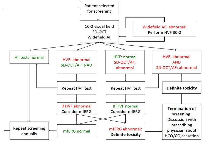
14. Revisions since last version of guidelines 39
15. Details of the electronic searches performed 40
Appendix A: assessment algorithm 50
15.1 Baseline assessment algorithm 50
15.2 Monitoring algorithm 50
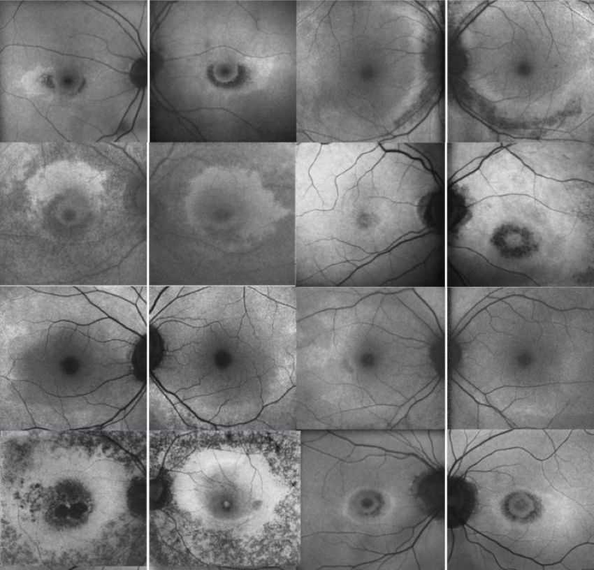
Appendix B: Examples of hydroxychloroquine retinopathy 51
15.3 Visual field 51
15.4 Spectral domain optical coherence tomography imaging 51
15.5 Fundus autofluorescence 52
Appendix C: Defining disease severity 54
16. Referral form… 55
16.1 Definitions of severity 57
17. Additional comments following consultation on guidelines 58
17.1 Termination of monitoring 58
17.2 Capacity to deliver guidelines 58
17.3 Future research 58
17.4 Audit 58
List of Tables
Table 1: Scottish Intercollegiate Guidelines Network framework (SIGN 50) 16
Table 2: Grade of recommendation 17
Table 3: Hydroxychloroquine use by disease 18
Table 4: NHS Digital Data Strengths and Limitations 19
4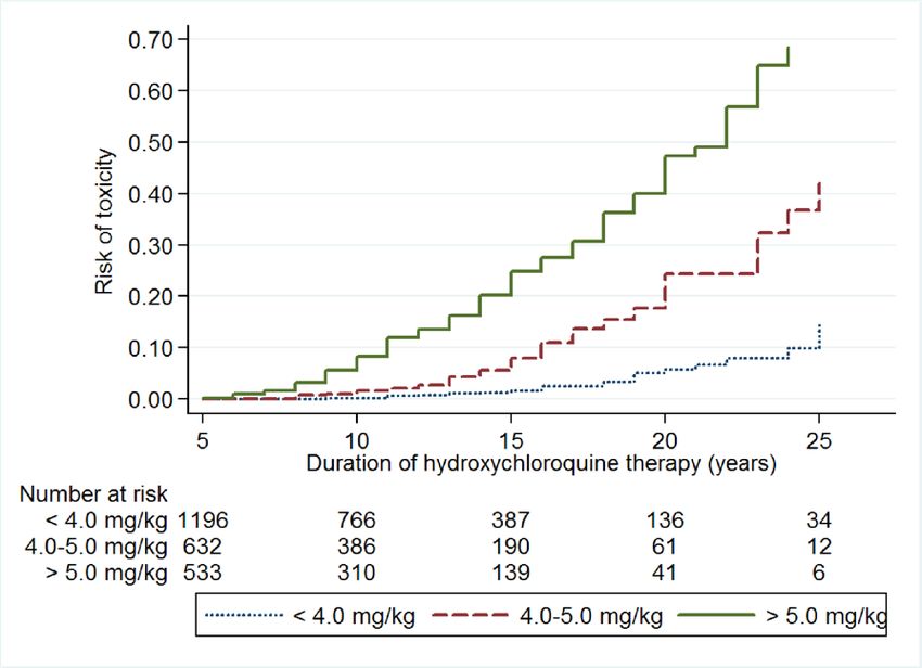
1. Executive Summary
Recent data have highlighted that hydroxychloroquine retinopathy is more common than previously reported.
The prevalence in long term use patients appears to be around 7.5% and depending on dose and duration of
therapy can increase to 20-50% after 20 years of therapy. Risk increases for patients taking more than 5mg/
kg/day. The retinopathy is manifest as damage to the photoreceptors and subsequent degeneration of the
retinal pigment epithelium (RPE). This may produce a “Bull’s eye maculopathy” and central visual loss. This is
important as the only intervention to prevent further damage is stopping the drug. The risk is increased for
patients taking more than 5mg/kg/day, those also taking Tamoxifen, and those with renal impairment. In
some patients, toxicity may first present as pericentral retinopathy and thus requires monitoring outside the
macula. We assume chloroquine retinopathy follows a similar course as hydroxychloroquine retinopathy and so
these guidelines also apply to patients taking chloroquine therapy.
After careful review of the existing peer reviewed literature, we recommend that all patients planning to take
hydroxychloroquine long term i.e. over five years have a baseline examination in a hospital eye department
ideally within six months, but definitely within 12 months, of starting therapy with a colour retinal photograph
and spectral domain optical coherence tomography (SD-OCT) scans of the macula.
Patients should be referred for annual monitoring after five years of therapy and be reviewed annually
thereafter whilst on therapy. At each monitoring visit patients should undergo 10-2 Humphrey visual field
testing, followed by pupillary dilation and imaging with both SD-OCT and widefield fundus autofluorescence
imaging (FAF). If widefield FAF is not available, FAF can be acquired in several photographic fields to
encompass the macula and extra-macular areas. Patients with abnormalities on widefield FAF with normal
10-2 visual field test results should undergo 30-2 visual field testing on another date. Patients with persistent
and significant visual field defects consistent with hydroxychloroquine retinopathy, but without evidence of
structural defects on SD-OCT or FAF may be considered for multifocal electroretinography. Monitoring may
be commenced before five years of therapy if additional risk factors exist e.g. very high dose of drug therapy,
concomitant Tamoxifen therapy or renal insufficiency. Adequate monitoring may not be possible with retinal
co-pathology.
Chloroquine appears to be more retinotoxic than hydroxychloroquine and so we recommend identical baseline
and monitoring tests, but that monitoring begins after one year of therapy for all patients on chloroquine.
Monitoring may be best incorporated into the hospital eye service via virtual clinics. The results of monitoring
should be communicated back to the prescribing doctor, patient and GP as normal, possible or definite
hydroxychloroquine retinopathy. It is the prescribing doctor’s responsibility to ensure their patients are
adequately monitored and to act on the results of monitoring. A useful aide memoir for these guidelines for
hydroxychloroquine is the 5 x 5 rule (ideally keep dosage < 5mg/kg/day and monitor after five years of drug
use.

                                                         5
2. Key Recommendations and good Practice
Points (GPP) for Implementation
The criteria used for the summary of grades of recommendations are found in Table 1 below.
Grade Explanation
A At least one meta-analysis, systematic review, or RCT rated as 1++, and directly applicable to the
target population; or
A body of evidence consisting principally of studies rated as 1+, directly applicable to the target
population, and demonstrating overall consistency of results
A body of evidence including studies rated as 2++, directly applicable to the target population,
and demonstrating overall consistency of results; or Extrapolated evidence from studies rated as
1++ or 1+
B A body of evidence including studies rated as 2+, directly applicable to the target population and
demonstrating overall consistency of results; or Extrapolated evidence from studies rated as 2++
C Evidence level 3 or 4; or
Extrapolated evidence from studies rated as 2+
GPP Good Practice Points based upon consensual expert opinion where the evidence base did not
support an A-C grading.
2.1 Monitoring Criteria
Criteria Level of
evidence
All individuals who have taken hydroxychloroquine for greater than five years should receive B
annual monitoring for retinopathy.
All individuals who have taken chloroquine for greater than one year should receive annual B
monitoring for retinopathy.
All individuals taking hydroxychloroquine who have additional risk factors for retinal toxicity GPP
may be monitored annually from the baseline visit or annual monitoring commenced before five
years of treatment completed. This is to be decided by a consultant ophthalmologist following
the baseline visit. Additional risk factors: Concomitant Tamoxifen use, impaired renal function
(estimated glomerular filtration rate of less than 60ml/min/1.73m2), does of hydroxychloroquine
greater than 5mg/kg/day.
It is the responsibility of the prescribing physician (as per GMC guidelines) to refer patients GPP
eligible for monitoring to the local hospital eye service.
The referring clinician should be encouraged to complete a standardised referral proforma GPP
specifying the key clinical details relevant to monitoring for retinal toxicity. This will allow a
determination of risk toxicity and interpretation of test results.
62.2 Monitoring Protocol: Baseline Examination
Criteria Level of
evidence
All patients planning to be on therapy long term (≥ five years for hydroxychloroquine and >one C
year for chloroquine) should receive baseline examination ideally within six months of starting
hydroxychloroquine or chloroquine and definitely within 12 months.
Baseline examination should include a fundus photography and spectral domain optical GPP
coherence tomography.
If the baseline examination demonstrates macular pathology, a baseline Humphrey 10-2 visual GPP
field test may be undertaken.
2.3 Monitoring Protocol: Monitoring Tests
The following is a standardised protocol for all patients.
Criteria Level of
evidence
In addition to oral communication, written information about hydroxychloroquine retinopathy GPP
and monitoring for hydroxychloroquine retinopathy should be given to all patients.
All patients should undergo 10-2 Humphrey visual fields testing (using a white stimulus), followed B
by pupillary dilation and imaging with both spectral domain optical coherence tomography (SD-
OCT) and widefield fundus autofluorescence (FAF).
Patients with abnormalities on widefield fundus autofluorescence with normal 10-2 visual field C
test results should undergo 30-2 visual field testing on another date.
Patients with persistent and significant visual field defects consistent with hydroxychloroquine C
retinopathy, but without evidence of structural defects on SD-OCT or FAF may be considered for
multifocal electronicretinography.
Some patients at risk of hydroxychloroquine retinopathy may not be able to undertake the required monitoring
tests, and in some there may be ocular co-pathology that prevents interpretable imaging.
Criteria Level of
evidence
Where a patient taking hydroxychloroquine or chloroquine cannot undergo monitoring (i.e. GPP
cannot perform visual field testing), or in whom retinal imaging cannot be performed or images
interpreted, a discussion between the patient and the prescribing physician is recommended to
determine whether hydroxychloroquine treatment should be continued without retinal monitoring.
2.4 Interpretation of Monitoring Results
Criteria Level of
evidence
No toxicity: No abnormalities suggestive of toxicity detected on any test. B
Possible toxicity: One test result (which in the case of visual fields should be reproducible) typical GPP
of hydroxychloroquine retinopathy, but typical abnormalities not present in other tests.
Definite toxicity: Two test results (one subjective test and one objective test) with abnormalities B
typical of hydroxychloroquine retinopathy.
72.5 Management of Patients with Possible Retinopathy
Criteria Level of
evidence
Patients with possible hydroxychloroquine retinopathy should continue drug treatment. This will GPP
reduce the risk of inappropriate treatment cessation.
Patients with one abnormal test result on retinal imaging (SD-OCT & widefield FAF) but normal GPP
visual fields (including 30-2 protocol (if appropriate) should return for annual review as per the
monitoring schedule. This will reduce the risk of inappropriate treatment cessation.
Patients with persistent visual field abnormalities in the context of normal structural imaging GPP
(SD-OCT and widefield FAF) may be referred for multifocal electroretinography. Treatment should
continue until the outcome of electrophysiology is known.
2.6 Management of Patients with Definite Toxicity
Criteria Level of
evidence
A recommendation to stop hydroxychloroquine should be made to the prescribing physician to B
facilitate further discussion between specialist (for the treatment indication) and patient about
the risk of stopping hydroxychloroquine and the options for alternative drug therapy.
Some description by the ophthalmology of disease severity (mild, moderate, or severe) may be GPP
helpful to facilitate this discussion between patient and prescribing physician.
It would be inappropriate for ophthalmologists to stop hydroxychloroquine treatment. GPP
Patients should be referred for appropriate support at the point of detection of GPP
hydroxychloroquine retinopathy. This may involve low vision or eye clinic liaison officer (ECLO)
services, certification of vision impairment, and referral to local and/or national charities.
Patients who are drivers should be advised not to drive until an Estermann visual field test confirms GPP
it is legal to do so. The patient should inform the Driver and Vehicle Licensing Agency (DVLA).
2.7 Termination of Monitoring
Criteria Level of
evidence
Monitoring for hydroxychloroquine retinopathy should be discontinued if patients stop taking C
hydroxychloroquine (due to retinal toxicity or for other reasons).
2.8 Organisation of Services
Criteria Level of
evidence
Monitoring for hydroxychloroquine retinopathy should take place in the hospital eye service. GPP
Monitoring for hydroxychloroquine retinopathy may most effectively take place in virtual clinics where GPP
visual field testing and dilated retinal imaging is undertaken before later being interpreted by either an
ophthalmologist or an allied health professional under the supervision of a consultant ophthalmologist.
Written communication from the ophthalmologist indicating the outcome of a monitoring GPP
episode should be sent to the patient, prescribing physician and general practitioner.
In the event of failure to attend monitoring, patients should not be automatically discharged. Patients GPP
should be reminded of the purpose of monitoring and the approximate interval to the next monitoring
appointment stated.
82.9 Work Commitment
Criteria Level of
evidence
Ophthalmologists who regularly complete the interpretation of hydroxychloroquine retinopathy GPP
monitoring test results should have sessional commitments allocated within their work plan.
93. Monitoring for hydroxychloroquine
retinopathy: Lay Summary
Hydroxychloroquine is a medicine that is effective in treating various long-term inflammatory disorders of
the joints and skin. In general, hydroxychloroquine is a safe and cost-effective medication, particularly when
compared to newer anti-inflammatory medicines which can more significant adverse effects on the body.
However, some patients taking hydroxychloroquine, or a similar medication called chloroquine, can suffer
permanent loss of vision due to the harmful long-term effect of hydroxychloroquine on the retina. The retina is
the light sensitive layer at the back of the eye which allows light to be sensed and relayed to the brain so that
an image is perceived or “seen” by an individual. This condition where hydroxychloroquine can affect the retina
and vision when taken for a long period of time is called “hydroxychloroquine retinopathy”.
Hydroxychloroquine retinopathy becomes more likely the longer any individual is taking the medication.
The disorder is rarely seen within the first five years of treatment, but becomes more common with a longer
duration of use. Between 20 and 50% of people taking hydroxychloroquine for more than 20 years may have
some signs of hydroxychloroquine retinopathy. Overall, 7.5% of individuals taking hydroxychloroquine for more
than five years may have some signs of retinal damage detected on specialised tests.
If advanced, hydroxychloroquine retinopathy can cause symptoms of loss of peripheral vision, and then in
later stages, central vision can become affected too. If hydroxychloroquine retinopathy is advanced, it can
result in permanent loss of sight in both eyes that can impact quality of life and activities such as driving and
reading. It is unlikely that all the visual field will be lost, even in advanced hydroxychloroquine retinopathy.
However, once hydroxychloroquine retinopathy results in noticeable loss of vision, the damage to the retina is
permanent and often continues to get worse even if the medication is stopped.
It is possible to detect early signs of hydroxychloroquine retinopathy using specialised techniques that can
look at layers of the retina with photographs of the eye, and by visual field testing. Visual field testing is tested
by an individual pressing a button when they see a light on a specialised type of eye test. These tests, when
taken together, can detect early signs of hydroxychloroquine retinopathy before the condition is noticed by an
individual (before it causes symptoms) and therefore are able to detect the condition at a much earlier stage.
Looking for a particular condition in a person thought to be at risk, in order to detect it early and minimize
the risk of harm is a process called “monitoring”. In the United Kingdom, monitoring for hydroxychloroquine
retinopathy is now recommended on the National Health Service for all individuals taking either
hydroxychloroquine or chloroquine, who are expected to remain on the medication for more than five years.
The aim of monitoring for hydroxychloroquine retinopathy is to detect the earliest definite signs of the
condition to allow those individuals to seek alternative medications in consultation with their doctor. This will
reduce the amount of sight that is lost at the time of detection (diagnosis), and reduce the risk of the sight
getting any worse (by stopping the medication).
Monitoring for hydroxychloroquine retinopathy involves having “baseline tests” at the eye service where the
monitoring will take place. This will mean having a photograph taken of the retina (after the instillation of
dilating eye drops) within a year of starting hydroxychloroquine medication. The reason for these baseline
tests is to determine whether an individual can undergo monitoring, and whether any conditions of the
retina or the eye already exist which may make monitoring difficult or impossible. Thereafter, most patients
will be monitored after five years of taking the medication, and will be monitored annually thereafter,
with a combination of retinal photographs and visual field tests. Those patients considered at higher risk
of developing hydroxychloroquine retinopathy (such as those who also take Tamoxifen, those who take
chloroquine, those who have impaired kidney function, and those who are taking a high daily dose of
hydroxychloroquine) will be seen annually after they start taking the medication. Should the standard
monitoring tests prove inconclusive, it may be necessary to repeat some tests, or rarely, have a patient undergo
more specialist tests at a different eye centre where such tests are available.
The result of monitoring (whether any given individual has any signs of retinopathy on the tests or not) will
be communicated to that individual, the General Practitioner and the prescribing physician (such as the
rheumatologist or dermatologist) if relevant. If an individual is diagnosed with having hydroxychloroquine
retinopathy, they will have an appointment in the hospital eye service so that the results of the tests can
be discussed further, any questions can be answered and any additional support can be provided to that
individual. It is also expected that before stopping hydroxychloroquine, a consultation will be arranged
10with the prescribing doctor so that an alternative medication can be identified if the decision to stop
hydroxychloroquine has been recommended by the monitoring process.
More information on hydroxychloroquine and monitoring for hydroxychloroquine retinopathy is available from:
The Macular Society: www.macularsociety.org/
British Association of Dermatologists: www.bad.org.uk/for-the-public/patient-information-leaflets
114. Introduction
Hydroxychloroquine is used increasingly in the treatment of autoimmune disease with established roles in
rheumatology and dermatology (through inhibition and modulation of immune responses) and emerging roles
in oncology (through inhibition of autophagy).1 Chloroquine has a similar mechanism of action although is
used far less frequently as it is more toxic to the retina.2 The increasing use of hydroxychloroquine is for two
main reasons:
1. Systemic safety: Hydroxychloroquine has a favourable systemic safety profile when compared to
other immunosuppressive agents. Disturbances of hepatic and renal function, skeletal muscle and
cardiotoxicity are considered rare.
2. Efficacy: Hydroxychloroquine has efficacy data in systemic lupus erythematosus (SLE) and rheumatoid
arthritis (RA). Data from the LUMINA studies (a multi-ethnic US cohort study) have identified a survival
benefit of hydroxychloroquine in patients with SLE.3 This finding is probably due to the inhibitory effect
that hydroxychloroquine has on mechanisms of cardiovascular disease - thrombosis, diabetes and
dyslipidaemia.4-6 It is also protective against further renal damage in patients with lupus nephritis.7
Whilst previously avoided in pregnancy, hydroxychloroquine is now recommended in pregnancy
with improved outcomes for mother and neonate.8 Some rheumatologists now advocate the use of
hydroxychloroquine in all patients with SLE.5, 9 In RA, hydroxychloroquine is indicated only as part of
a combination therapy regimen with other anti-rheumatic agents.10 The evidence for efficacy in RA
demonstrates that it is an inferior agent to other available options.11 However, due to its tolerability
profile it remains in frequent use.
Anti-malarial drugs were shown to be useful in the management of SLE and discoid lupus as early as 1951,12
but were identified as having toxic effects on the retina as early as 1963.13 In the absence of modern retinal
imaging techniques, retinopathy was typically detected once it became symptomatic and retinal pigment
epithelial damage had become established. At this stage, hydroxychloroquine retinopathy is visible on
fundoscopy as a complete or incomplete annular, parafoveal pattern of retinal pigment epithelial loss (Bull’s
eye maculopathy) associated with severe, bilateral concentric paracentral visual field loss. Further deterioration
of visual function is very likely at this stage despite discontinuation of hydroxychloroquine.14 Using advanced
retinal pigment epithelial damage as a definition of toxicity (i.e. detectable on fundoscopy), the prevalence of
hydroxychloroquine retinopathy was estimated 0.5% after six years of treatment in a Greek study,15 and 0.48%
in patients taking a dose of greater than 6.5mg/kg/day in a US study.16
The advent of modern retinal imaging techniques (spectral domain optical coherence tomography and fundus
autofluorescence) has permitted detection of disease at an early, pre-symptomatic stage before retinal
pigment epithelial damage is detectable clinically. The recommendation of screening for hydroxychloroquine
retinopathy by the American Academy of Ophthalmology in 2002,17 (revised in 201118 and 201619) has
allowed large case-control studies to define the prevalence of toxicity using modern techniques. When
hydroxychloroquine retinopathy is diagnosed on the basis of one subjective test (central, static visual
field testing, such as Humphrey 10-2), and one objective test (such as SD-OCT), the overall prevalence of
toxicity in long-term hydroxychloroquine users has been estimated at 7.5%.20 It is therefore likely that
hydroxychloroquine retinopathy represents a greater public health problem than previously estimated.
Hydroxychloroquine retinopathy results in largely irreversible structural and functional retinal deficits. The
earlier disease is diagnosed, and hydroxychloroquine discontinued (if appropriate), the less severe the visual
deficits are at the point of detection, and the less likely they are to progress.21 The aim of monitoring is not to
prevent hydroxychloroquine retinopathy, but to detect the earliest definitive signs of pre-symptomatic toxicity.
This will facilitate an informed discussion between the patient and prescribing physician on treatment options
(continuing hydroxychloroquine versus seeking an alternative treatment).
Chloroquine is associated with a higher prevalence of toxic retinopathy when compared to
hydroxychloroquine;22 consequently, the use of hydroxychloroquine has grown although chloroquine is still
used rarely in clinical practice. For this reason, clinical guidelines on hydroxychloroquine monitoring include
chloroquine users within their remit.19, 23 Chloroquine has a very similar molecular structure, is assumed to
have a very similar mechanism of action, and produces a similar retinal phenotype.24 Guidance on safe dosing
suggests a dose of less than 2.3 milligrams per kilogram per day for chloroquine: a dose suggested from
extrapolation of data from the older literature using comparisons with toxic doses of hydroxychloroquine.19
12The higher risk of retinal toxicity in patients taking chloroquine suggest it should be considered as an
additional risk factor, and therefore qualify for annual monitoring from baseline.
A full and current review of hydroxychloroquine retinopathy is available free to members of The Royal College
of Ophthalmologists: www.ncbi.nlm.nih.gov/pubmed/28282061
4.1 Population to whom the Guideline applies e.g. the age range, gender,
clinical description (ICD10) and co-morbidity (ICD10) and any exclusions
This guideline applies to male and female adult patients (over the age of 18) who are users of
hydroxychloroquine and chloroquine (but not mepacrine/quinacrine as there is a lack of substantive evidence
that this drug is associated with retinal toxicity).
There are no relevant exclusions to those hydroxychloroquine users recommended to undergo monitoring for
retinopathy. However, it is likely that some patients may not be assessable using the monitoring protocol (who
cannot undergo retinal imaging, or undertake a visual field test), and some may have co-pathology which
makes image interpretation impossible. In such cases, communication between ophthalmologist, prescribing
physician and patient may determine whether hydroxychloroquine treatment without retinal monitoring is
appropriate in their case, and whether any ophthalmic intervention may be necessary to facilitate monitoring
(i.e. cataract surgery).
The ICD-10 codes that describe hydroxychloroquine most approximately are:
• T37.8X5A (Adverse effect of other specified systemic anti-infectives and antiparasitics, initial encounter)
• H35.00 (unspecified background retinopathy)
• H35.89 (other specified retinal disorder)
4.2 Paediatric indications for monitoring
There are an increasing number of paediatric indications for hydroxychloroquine drug therapy.1 There are
no reports of hydroxychloroquine retinopathy in patients under the age of 18, or evidence for monitoring
paediatric patients for drug toxicity. However, long-term users of hydroxychloroquine under the age of 18 who
otherwise satisfy the monitoring criteria should be referred for monitoring.
4.3 Current practice, and why there is scope for change
Current practice in the United Kingdom is guided by recommendations made by the RCOphth guideline
development group in 2009 which determined that monitoring for hydroxychloroquine retinopathy was not
recommended.25 This recommendation was made on the best available evidence at the time which suggested
that hydroxychloroquine retinopathy was very rare,15, 16 and it was felt that insufficient evidence existed for the
benefits of detecting hydroxychloroquine at an early stage.
However, recent epidemiological data from a high quality case-control study indicate that the prevalence of
toxicity amongst long-term hydroxychloroquine users may be around 7.5%.20 This risk may be as high as 20-
50% in those taking the drug more than 20 years, depending on the summation of risk factors in particular
individuals.20 Additionally, the tests used to diagnose hydroxychloroquine toxicity (automated central static
visual field testing, spectral domain optical coherence tomography imaging and fundus autofluorescence
imaging) have been proven to be effective at detecting presymptomatic disease, and reducing the risk of
progression of visual loss by detecting disease at an early stage.19-21, 23, 26 These tests are widely available in
ophthalmic clinics, practical to undertake and are likely to be acceptable to patients. It is now likely that the
justification given in 2009 for not supporting a systematic screening programme for hydroxychloroquine are
no longer valid considering new data.
Additionally, increasing awareness of emerging epidemiological data on the prevalence of hydroxychloroquine
retinopathy in long-term users20 - amongst patients and ophthalmologists - has led to some ad hoc monitoring
in ophthalmic clinics. Such clinical episodes are often initiated by patients themselves who are concerned
about undetected retinal toxicity, who may be aware of screening programmes in other countries and wish
to have the same diagnostic tests. Ophthalmologists may have encountered cases of severe and irreversible
loss of vision in some patients with hydroxychloroquine retinopathy who become symptomatic and present
to ophthalmic clinics.27 This opportunistic monitoring disadvantages those patients less motivated to seek
13monitoring or less aware of the need for it. Where monitoring does occur, there is likely to be some variation in
the monitoring schedule (the frequency of monitoring) and monitoring methods (what tests are undertaken)
between clinicians which may adversely affect the efficacy and cost effectiveness of monitoring.
A national consensus recommendation on systematic monitoring for hydroxychloroquine retinopathy within
the National Health Service is needed now. The purpose of this guidance is to systematically review the
literature to provide a current best practice recommendation for hydroxychloroquine retinopathy monitoring.
The aim is to reduce the risk of sight loss associated with undetected hydroxychloroquine retinopathy by
detecting it at the earliest opportunity. Furthermore, the guidance intends to reduce inequality in access
to monitoring, encourage consistency of monitoring schedule and methods, and clarify best practice
for all stakeholders including patients, ophthalmologists, prescribing physicians (general practitioners,
rheumatologists, dermatologists), optometrists and commissioners.
In view of changing practice, we stress we no longer recommend monitoring with a reading chart (as per 2009
guidelines), or annual optometrist assessment.
145. Objectives
The overall objectives are:
1. To systematically re-evaluate the literature to determine whether sufficient evidence exists to
recommend monitoring for hydroxychloroquine retinopathy in the National Health Service.
2. To make recommendations on the criteria for inclusion for monitoring, the schedule of monitoring and
the method of monitoring for hydroxychloroquine retinopathy which is practical and achievable for most
hospital eye departments.
3. To provide guidance on the interpretation of monitoring test results and present recommendations on
the subsequent management of patients at the point of detection of hydroxychloroquine retinopathy,
and in those with diagnostic uncertainty.
4. To consult with key stakeholders, including ophthalmologists, rheumatologists, dermatologists and
patient groups on issues that relate to monitoring for hydroxychloroquine retinopathy.
The clinical questions covered by the guideline are:
Epidemiology
1. How many patients are currently receiving treatment and how many new patients are prescribed
treatment each year?
2. What is the risk of toxicity retinopathy for patient sub-groups (by disease and ethnic groupings)?
3. What is the risk of progression of toxicity retinopathy for patient sub-groups?
Monitoring
1. Who should initiate the toxicity monitoring process?
2. Which patients require monitoring for hydroxychloroquine retinopathy?
3. When should patients be monitored for hydroxychloroquine retinopathy?
4. What tests should be performed on patients as part of the monitoring schedule?
5. In what setting should monitoring for hydroxychloroquine retinopathy be performed?
6. What are the signs from monitoring tests that indicate hydroxychloroquine retinopathy?
7. What action should be taken for patients with hydroxychloroquine retinopathy?
8. What are the costs or economic implications of monitoring for hydroxychloroquine retinopathy?
9. What is the efficacy of monitoring protocols for detecting early hydroxychloroquine retinopathy?
10. Who is responsible for monitoring patient participation in monitoring?
11. What information should be obtained from referring clinicians to ophthalmologists at the initiation of monitoring?
12. What information should be communicated by ophthalmologists after a monitoring episode has been
undertaken?
Patient information
1. What information should be given to patients at initial monitoring?
2. What information should be given to patients regarding treatment options at the point of detection of
hydroxychloroquine retinopathy?
5.1 Description of the key stakeholders and end users
Key stakeholders:
1. Patients at risk of hydroxychloroquine retinopathy
2. Ophthalmologists (in particular, medical retina specialists)
3. Rheumatologists
4. Dermatologists
5. General Practitioners
6. Optometrists
7. Patient groups
8. Health care commissioners
156. Methods
6.1 Methodology
This guideline has been developed in accordance with the Guideline Development Manual of The Royal
College of Ophthalmologists (found at www.rcophth.ac.uk) following the prespecified stages to ensure that the
recommendations are aligned with the strength of evidence available from the review of the literature.
6.2 Search strategy
Key questions for the guideline were developed using the PICO framework to provide a structured basis
for identifying the evidence. A systematic review of the literature was undertaken using the explicit search
strategies devised in collaboration with the Cochrane Eyes and Vision Group. Databases searched include
Medline, Embase, and the Cochrane Library for literature published between 2000 & 2017. Further searches
were undertaken on various websites including the US National Guidelines Clearinghouse.
The evidence base for this guideline was identified and synthesized in accordance with the accepted
methodology. Each of the selected papers was evaluated by the guideline development group using standard
checklists before conclusions were considered as acceptable evidence. The literature search focused on the best
available evidence to address the key review questions by including the following types of evidence
• Published guidelines
• Systematic reviews
• Randomised controlled trials
• Cohort and case control studies
• Case series
Papers not published in the English language, conference abstracts and letters were excluded.
6.3 Levels of evidence and Grades of Recommendations
Evidence was graded by the Guideline Development Group according to its strength using the Scottish
Intercollegiate Guidelines Network framework (SIGN 50 – Table below). The strength of each recommendation
considered the quality of the evidence. Table from Scottish Intercollegiate Guidelines Network framework
(SIGN 50).
Table 1: Scottish Intercollegiate Guidelines Network framework (SIGN 50)
Type of Description
Evidence
1++ High-quality meta-analyses, systematic reviews of RCTs, or RCTs with a very low risk of bias
1+ Well-conducted meta-analyses, systematic reviews of RCTs, or RCTs with a low risk of bias
1- Meta-analyses, systematic reviews of RCTs, or RCTs with a high risk of bias*
2++ High-quality systematic reviews of case–control or cohort studies
High-quality case–control or cohort studies with a very low risk of confounding, bias or chance
and a high probability that the relationship is causal
2+ Well-conducted case–control or cohort studies with a low risk of confounding, bias or chance
and a moderate probability that the relationship is causal
2- Case–control or cohort studies with a high risk of confounding, bias, or chance and a
significant risk that the relationship is not causal
3 Non-analytic studies (for example, case reports, case series)
4 Expert opinion, formal consensus
16Using the evidence identified the Guideline Development Group determined the guideline recommendations.
The strength of each recommendation has been based upon the quality of the evidence and the potential for
patient benefit.
This guideline makes a strong recommendation where:
• The evidence is of high quality
• Estimates of the effect of an intervention are precise (i.e. there is a high degree of certainty that effects will
be achieved in practice)
• There are few downsides of therapy
• There is a high degree of acceptance among patients
And a conditional recommendation is made where:
• There are weaknesses in the evidence base
• There is a degree of doubt about the size of the effect that can be expected in practice
• There is a need to balance the upsides and downsides of therapy
• There are likely to be varying degrees of acceptance among patients
The strength of the recommendation has been graded by the Guideline Development Group using the
methodology from the Scottish Intercollegiate Guidelines Network (SIGN 50)9. The grade of recommendation
relates to the strength of the evidence on which the recommendation is based (Table 2). It does not reflect the
clinical importance of the recommendation.
Table 2: Grade of recommendation
Grade Explanation
A At least one meta-analysis, systematic review, or RCT rated as 1++, and directly applicable to
the target population; or
A body of evidence consisting principally of studies rated as 1+, directly applicable to the
target population, and demonstrating overall consistency of results
A body of evidence including studies rated as 2++, directly applicable to the target population,
and demonstrating overall consistency of results; or Extrapolated evidence from studies rated
as 1++ or 1+
B A body of evidence including studies rated as 2+, directly applicable to the target population
and demonstrating overall consistency of results; or Extrapolated evidence from studies rated
as 2++
C Evidence level 3 or 4; or Extrapolated evidence from studies rated as 2+
GPP Good practice points based upon consensual expert opinion where the evidence base does not
support A-C grading
177. Results
7.1 Summary of the results
Epidemiology
1. How many patients are currently receiving treatment and how many new patients are prescribed
treatment each year?
• Over 4,000, and up to 8,000 people a year commence hydroxychloroquine in England
• Up to 320,000 people are currently taking hydroxychloroquine in England
• Between 50-70% of use is for rheumatoid arthritis
• Estimates are not currently available for how many people have been on hydroxychloroquine > five years
• Hydroxychloroquine use has more than doubled in the last decade
This section provides information of the number of patients who may need retinal monitoring due to
hydroxychloroquine use. A review of the literature has been unhelpful in determining estimates specific to
England and Wales. We set out to explore the questions using data drawn from three sources:
1. A single-centre electronic prescribing record
2. National Early Inflammatory Arthritis Audit
3. NHS Digital Summary Prescribing Data
Single Centre Experience
Rheumatoid arthritis is estimated to affect 400,000 people in England. There are 146 NHS trusts in England and Wales
with rheumatology services. The average adult catchment population for rheumatology departments is 330,000.
King’s College Hospital (KCH) rheumatology department has an electronic database that allows extraction
of all patients on hydroxychloroquine, broken down by indication (Table 3). KCH is a 1000-bedded teaching
hospital. The Rheumatology department is slightly larger than average, with an adult catchment population
approximately 400,000 adults. In total, the department cares for approximately 3,000 patients with rheumatic
disease, including 1,300 people with Rheumatoid arthritis. 489 patients are currently on hydroxychloroquine,
suggesting that approximately 35% of rheumatoid patients are on this medication.
The breakdown across diseases suggests that prescribing accounts for just over half of hydroxychloroquine use
in rheumatology.
KCH may be an outlier, as they are a specialist commissioning unit for connective tissue disease, so it is likely
that it has a larger burden of diseases like Lupus compared to the average unit.
Table 3: Hydroxychloroquine use by disease
Indication N %
Rheumatoid Arthritis 489 55.5
Connective Tissue Disease 286 32.5
Other 52 5.9
Undifferentiated Inflammatory Arthritis 24 2.7
Inflammatory Myopathy 14 1.6
Seronegative Spondyloarthropathy 9 1
Possible Early Inflammatory Arthritis 6 0.7
Vasculitis 1 0.1
Total 881 100
The most common dose of hydroxychloroquine was 200mg daily, with 97% of patients receiving doses below
6.5mg/kg.
18National Early Inflammatory Arthritis Audit
The Healthcare Quality Improvement Project commissioned a national audit of early inflammatory arthritis
in England and Wales, conducted between 2014-2016, over 21 months. In total 136/146 units participated.
During the recruitment window, 5,675 incident cases of rheumatoid arthritis were captured. This equated
to an annualised incidence of rheumatoid of 9/100,000 adults. It is recognised that the National Audit had
incomplete capture of incidence cases, as data from the Clinical Practice Research Datalink suggest the true
incidence is 15/100,000. Therefore, it has been estimated that the Audit collected data on approximately 60%
of incident cases.
In the National Audit, hydroxychloroquine was commenced in 2,467 patients (51.7% of newly diagnosed
rheumatoid arthritis cases).
NHS Digital Summary Prescribing Data
NHS Digital provides summary data from England on the use and expenditure associated with medications
prescribed in the community. In 2016, there were 117,247,136 hydroxychloroquine 200mg tablets dispensed
across all indications. Allowing for 365/days therapy per subject at 200mg once daily, and assuming no
duplicate prescriptions, this equates to 321,225 prevalent users. This will be an overestimate of true user
prevalence, as some patients will be on 400mg daily.
Discussion
Rheumatoid arthritis accounts for around half of hydroxychloroquine use at KCH. Whilst this may be an
underestimate at National level (KCH is a specialist centre for lupus), any guideline needs to consider that there
will be implications for other diseases.
Between 30-50% (KCH: 30%; National Audit: 50%) of rheumatoid arthritis patients are on hydroxychloroquine.
The higher figure from the National Audit is likely to reflect an incident user population. A proportion of
patients will not respond to hydroxychloroquine, or experience side effects (e.g. nausea / rash) that lead to
discontinuation.
Comparing National Audit with NHS Digital
NHS Digital covers only England, and suggests approximately 320,000 users of hydroxychloroquine. This will be
an overestimate due to the methodology (no accounting for duplicate prescriptions or patients taking >200mg
daily). However, even if 50% of rheumatoid arthritis patients were taking HCQ, this would still only be 200,000
individuals.
Comparing KCH with NHS Digital
KCH rheumatology has 800 people on hydroxychloroquine. From 146 rheumatology units in England and
Wales, if each unit had 800 people on hydroxychloroquine, this would only be 116,800 people. The discrepancy
between the KCH and NHS Digital estimates likely reflects use of hydroxychloroquine in other specialities.
Table 4: NHS Digital Data Strengths and Limitations
Strengths Limitations
High level of data completeness Only covers England (in contrast to National Audit, which
covers Wales as well)
All prescriptions which are dispensed in England need to be
submitted to NHS Business Services Authority if the dispenser Does not provide information on incident use (i.e. new
is to be reimbursed and so coverage should be complete starters)
Data quality audit reports >99% accuracy No information on dose per person
Covers use for all conditions No data on how many users have been on therapy >five years
Estimate of incident use:
Incident and prevalent hydroxychloroquine need to be considered separately. It has not been possible to estimate
the number of new starters by indication; however, the incidence of the diseases is important to bear in mind:
• Rheumatoid incidence: 15/100,000
• Lupus incidence: 3/100,000
19It can be inferred that rheumatoid arthritis is a more common indication for starting hydroxychloroquine,
although compared to lupus a higher proportion are likely to discontinue (due to the lower efficacy and
availability of alternative options in rheumatoid).
Based upon the National Audit Data, we could expect approximately 4,100 new hydroxychloroquine users in
England and Wales for the indication of rheumatoid arthritis.
The estimate needs inflating to allow for starters with other conditions. Comparing across the datasets, it
seems likely that rheumatoid arthritis accounts for between 50 – 70% of use. Based upon available evidence,
the incidence of new hydroxychloroquine users may be as high as 8,000 people per year in England and Wales.
Estimate of prevalent use:
NHS Digital provides an estimate of the number of patients established on hydroxychloroquine: 320,000
people. For reasons already explained, this number is an upper boundary, and true numbers will be lower. The
NHS Digital data do not provide information on what proportion of these patients have remained on drug for
>five years.
Horizon scanning and time trends:
A further consideration is whether hydroxychloroquine is likely to remain stable in future years. It is relevant
to consider that within rheumatology, treatment guidelines have substantially evolved in the last decade, with
recommendations for more intensive therapy of rheumatoid arthritis, advocating targets of disease remission.
Several strategy trials have also demonstrated substantial cost benefits to utilising combination DMARD
therapy (e.g. including hydroxychloroquine) prior to initiating biologics. Current NICE guidance (TA375)
specifically require a failure of combination therapy prior to biologic therapy. NHS Digital provides a useful
data source for examining the time trends of hydroxychloroquine use (Figure 1).
Figure 1: Source: http://content.digital.nhs.uk/catalogue/PUB23631/pres-cost-anal-eng2016-trend.zip
(accessed 14th May 2017)
2. What is the risk of hydroxychloroquine retinopathy for patient sub-groups (by disease and ethnic
groupings)?
The risk of hydroxychloroquine retinopathy in patients of different ethnic groups has not been determined
in any study. However, there are variations in the pattern of hydroxychloroquine retinopathy between ethnic
groups;28 pericentral retinopathy occurs at a higher frequency, but not exclusively, in “Asian patients”.28 Korean
patients have demonstrated a high incidence of pericentral disease in a separate case series (eight out of nine
cases had a pericentral distribution of disease).29
20It is important to note that whilst Melles and co-authors did not state the ethnic provenance of Asian patients
in their influential study undertaken in California,30 census data from California in 201031 identifies Philippino,
Chinese and Vietnamese populations as more numerous than Indian and Pakistani populations. In the UK
Census (2011), 4.9% of the population were Indian, Pakistani or Bangladeshi, compared to 0.7% who identified
themselves as Chinese.32 Such ethnic differences are important when interpreting the study data28, 29 in the
context of the UK population. It may be likely that more Caucasian patients with pericentral disease are
identified than “Asian” patients with pericentral disease given the population demographics in the United
Kingdom. This may have implications for the selection of monitoring tests recommended in the monitoring
protocol.
There is no study that seeks specifically to identify the risk of hydroxychloroquine retinopathy by disease group
(i.e. RA, SLE etc.). However, one study did reveal a small increased risk of hydroxychloroquine retinopathy in SLE
compared to RA, although no statistical analysis was performed.30 This difference is unlikely to be significant.
What is the risk of progression of hydroxychloroquine retinopathy for patient subgroups?
Hydroxychloroquine retinopathy can continue to progress after cessation of hydroxychloroquine therapy.21
There is limited evidence of progression of retinopathy (defined as worsening of SD-OCT appearance) if retinal
pigment epithelial loss is detected on autofluorescence or spectral domain OCT imaging at the point of drug
cessation.
An additional study identified longitudinal changes in a cohort of 11 patients with confirmed
hydroxychloroquine retinopathy. In two patients with early disease there was no progression at three years,
whereas in the other nine patients, progression of retinal degeneration occurred with loss of visual acuity.33
This may be explained in part by the development of cystoid macular oedema and epiretinal membrane
formation secondary to advanced hydroxychloroquine retinopathy in some patients. A further study identified
that preservation of the external limiting membrane on SD-OCT was a favourable prognostic sign in cases of
hydroxychloroquine retinopathy.14
This evidence suggests that progression of retinopathy is linked closely to the disease severity at the point of
detection of toxicity (level C). This emphasizes the need for early detection of hydroxychloroquine retinopathy.
7.2 Monitoring
1. Who should initiate the referral for monitoring for hydroxychloroquine retinopathy?
Several guidelines provide guidance on who should initiate the referral for monitoring for hydroxychloroquine
retinopathy. The American College of Rheumatology position statement recommends that “patients beginning
therapy with hydroxychloroquine should be informed of potential adverse drug reactions including retinal
toxicity and that periodic monitoring and early recognition can limit the impact of macular toxicity.34 All
individuals starting these drugs should have a complete baseline ophthalmologic examination within the first
year of treatment.”34
The General Medical Council (GMC) states that the duty of the doctor prescribing any drug is “to reach
an agreement with the patient on the treatment proposed, explaining the likely benefits, risks and
burdens including serious and common side effects”, including an explanation of the “arrangements for
monitoring…”.35
We therefore recommend that the physician prescribing hydroxychloroquine (rheumatologist,
dermatologist, general practitioner etc.) should initiate the referral of patients at risk of retinal toxicity from
hydroxychloroquine or chloroquine to a hospital eye department for monitoring. If the patient is no longer
under the care of the original prescriber (i.e. rheumatologist), this responsibility lies with the individual issuing
repeat prescriptions, which in most cases will be their general practitioner.
2. Which patients require monitoring for hydroxychloroquine retinopathy?
In order to determine which patients require monitoring; the major risk factors for hydroxychloroquine
retinopathy should be considered. A large case-control study involving 2,361 participants was undertaken
in the US, each of whom had taken hydroxychloroquine for greater than five years and had had an
automated visual field test and SD-OCT imaging as part of retinal monitoring.20 The long-term prevalence of
hydroxychloroquine retinopathy amongst this patient group was 7.5% but varied with daily consumption (odds
ratio, 5.67; 95%CI, 4.14-7.79 for doses of hydroxychloroquine >5.0mg/kg) and with duration of use (odds ratio,
3.22; 95%CI, 2.20-4.70 for >10 years of drug treatment).
21For daily consumption of 4.0 to 5.0 mg/kg, the prevalence of retinal toxicity remained less than 2% within
the first 10 years of use but rose to almost 20% after 20 years of use. Other major risk factors include kidney
disease (odds ratio, 2.08; 95%CI, 1.443.01 – this correlated with falling estimated glomerular filtration rate)
and concurrent Tamoxifen citrate therapy (odds ratio, 4.59; 95%CI, 2.05-10.27).20
This new epidemiological data, acquired following the recommendation for a screening programme for
hydroxychloroquine retinopathy in the US in 2012, suggest that retinal toxicity is more common than
previously thought. Estimates of prevalence of hydroxychloroquine retinopathy amongst those taking the drug
for more than five years range between 4 and 7.5%.20, 28 In consideration of these data, it is very likely that
modern diagnostic techniques (automated visual field testing, FAF, SD-OCT, and multifocal ERG if necessary)
can detect disease at an early stage. Patients at risk of retinal toxicity should be monitored to detect toxicity at
the earliest opportunity at which point alternative drug treatment may be considered.
3. When should patients be monitored for hydroxychloroquine retinopathy?
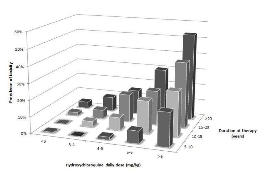
Melles and co-authors determined that the risk of toxicity is proportional to duration of therapy and dosage,
concomitant Tamoxifen use and renal disease (Figure 1, reproduced with permission20). Whilst patients taking
>5mg/kg of absolute body weight were shown by this data to be in the highest risk group, many patients
are taking greater than 6mg/kg, as hydroxychloroquine tablets are available in 200mg or 400mg doses. It
is likely therefore, that many patients are taking higher than the recommended doses (previously, a dose of
hydroxychloroquine of less than 6.5mg/kg/day was considered safe dosing).
The risk of toxicity is low for the first five years of therapy, but increases to 20% after five years for those
patients in the highest dose group (>5mg/kg). Therefore, monitoring should begin after five years from the
start of hydroxychloroquine therapy. For those patients who have had long periods off hydroxychloroquine
treatment (during the winter months for photosensitive dermatoses, for example), a cumulative duration of
drug exposure may be determined.
It is unclear whether those patients with one major risk factor, or more than one major risk factor should
be monitored earlier than five years after starting therapy. Very high doses of hydroxychloroquine used in
oncology trials (where doses can be as high as two grams per day) have identified patients with retinal toxicity
within two years of exposure to the drug.36-41 This highlights the observation that it is possible to develop
toxicity before five years of hydroxychloroquine therapy if risk factors are present, if risk factors are severe in
magnitude or multiple (in which case, the risks may be considered additive). Therefore, patients with severe
risk factors (renal impairment defined as an eGFR of less than 60ml/min/1.73m2, Tamoxifen use, high doses
of hydroxychloroquine >5.0 mg/kg, or any patient taking chloroquine > 2.3 mg/kg may be monitored yearly
from baseline (but not more frequently). The risk factors for hydroxychloroquine retinopathy are specified
in the referral proforma. Patients who may be considered for monitoring before five years of treatment
may be identified at the baseline visit. It is the Consultant Ophthalmologist (or other clinician / allied health
professional working under their supervision) interpreting the baseline tests on a patient who may make a
recommendation to begin monitoring earlier than five years on the basis of specified risk factors.
22Figure 2: The prevalence of hydroxychloroquine retinopathy by duration of therapy and daily dose of
hydroxychloroquine retinopathy (courtesy of Dr. Ronald Melles M.D.)
Figure 3: Risk of hydroxychloroquine retinopathy by daily dose and duration of drug therapy (courtesy of Dr.
Ronald Melles M.D.)
23You can also read