HEPATOLOGY FORUM Volume 2 Supplement 1 January 2021 - 2021 Hepatology ...
←
→
Page content transcription
If your browser does not render page correctly, please read the page content below
Official Journal of the Turkish Association for the Study of the Liver     ISSN 1307-5888
                                                                         E-ISSN 2757-7392

HEPATOLOGY
FORUM
                                                                             Volume 2
                                                                         Supplement 1
                                                                         January 2021
Hepatology Forum                                                                     ISSN: 1307-5888 / E-ISSN 2757-7392

                                                    Editorial Board

                      Editor-in-Chief                                           Hüseyin Savaş Göktürk
                    Bulent Degertekin                           Department of Gastroenterology Başkent University Konya
  Department of Gastroenterology Acıbadem Mehmet Ali                Uygulama ve Araştırma Hospital, Konya, Turkey
  Aydınlar University School of Medicine İstanbul, Turkey                      savasgokturk@yahoo.com
                degertekinb@hotmail.com
                                                                                 Murat Harputluoğlu
           Associate Editors (Alphabetically)                   Department of Gastroenterology İnönü University School of
                     Mevlüt Başkol                                             Medicine, Malatya, Turkey
Department of Gastroenterology Erciyes University School of                   mharputluoglu@hotmail.com
                Medicine Kayseri, Turkey
                 mbaskol@erciyes.edu.tr                                           Gökhan Kabaçam
                                                                 Department of Gastroenterology Güven Hospital, Ankara,
                    Yasemin Balaban                                                      Turkey
Department of Gastroenterology Hacettepe University School                    gokhankabacam@yahoo.com
               of Medicine Ankara, Turkey
               ybalaban@hacettepe.edu.tr                                           Aydın Şeref Köksal
                                                               Department of Gastroenterology Sakarya University School of
                     Murat Dayangaç
                                                                               Medicine, Sakarya, Turkey
Department of Gastroenterology Medipol University School of
                                                                                  koksalas@yahoo.com
                Medicine Istanbul, Turkey
                mdayangac@hotmail.com
                                                                                    Zarife Kuloğlu
                                                                  Department of Pediatrics Ankara University School of
                     Levent Doğanay
                                                                              Medicine, Ankara, Turkey
 Department of Gastroenterology Ümraniye Eğitim Araştırma
                                                                              zarifekuloglu@yahoo.com
                Hospital, Istanbul, Turkey
                 ldoganay@hotmail.com
                                                                                Aslı Çiftçibaşı Örmeci
                                                                 Department of Gastroenterology Haseki Eğitim Araştırma
                     Hakan Dursun
Department of Gastroenterology Atatürk University School of                    Hospital, Istanbul, Turkey
               Medicine, Erzurum, Turkey                                         aslic79@yahoo.com.tr
                 hadursun@hotmail.com
                                                                                     Berna Savaş
                       Cumali Efe                                 Department of Pathology Ankara University School of
Department of Gastroenterology Diyarbakır Eğitim Araştırma                     Medicine, Ankara, Turkey
               Hospital, Diyarbakır, Turkey                                     bernasavas@gmail.com
                 drcumi21@hotmail.com
                                                                                      İlker Turan
                    Cemal Nuri Erçin                            Department of Gastroenterology Ege University School of
 Department of Gastroenterology Sağlık Bilimleri University                    Medicine, İzmir, Turkey
       Gülhane School of Medicine, Ankara, Turkey                               ilkerturan@gmail.com
                  cnercin@hotmail.com
                                                                                    Enver Üçbilek
                       Hale Gökcan                             Department of Gastroenterology Mersin University School of
Department of Gastroenterology City Hospital, Ankara, Turkey                   Medicine, Mersin, Turkey
                  halesumer@yahoo.com                                          enucbilek@hotmail.com
Suna Yapalı                                               Ahmet Gürakar, USA
   Department of Gastroenterology Acıbadem Mehmet Ali              Department of Gastroenterology and Hepatology, Johns
   Aydınlar University School of Medicine İstanbul, Turkey         Hopkins University School of Medicine, Baltimore, USA
                  sunayapali@yahoo.com                                              aguraka1@jhmi.edu

                      Nuray Yazıhan                                                Azra Husic-Selimovic
Department of Pathophysiology and Internal Medicine Ankara        General Hospital Abdulah Nakas, Bosnia and Herzegovina
      University School of Medicine, Ankara, Turkey                                husic_azra@yahoo.com
      Ankara University, Institute of Health Sciences,
 Interdisciplinary Food, Metabolism and Clinical Nutrition                        Patrick Kamath, USA
                Department, Ankara, Turkey                      Department of Gastroenterology and Hepatology, Mayo Clinic
                 nurayyazihan@yahoo.com                                 College of Medicine, Rochester, MN, USA
                                                                                kamath.patrick@mayo.edu
                       Yusuf Yılmaz
Department of Gastroenterology Marmara University School of                          Paul Kwo, USA
                 Medicine Istanbul, Turkey                        Department of Gastroenterology and Hepatology, Stanford
                dryusufyilmaz@gmail.com                                    University Medical Center, CA, USA
                                                                                   pkwo@stanford.edu
                       Alper Yurci
                                                                                 Alexandre Louvet, France
 Department of Gastroenterology Kayseri Erciyes University
                                                                              University Hospital of Lille, France
           School of Medicine, Kayseri, Turkey
                                                                                alexandre.louvet@chru-lille.fr
                 alperyurci@yahoo.com

                                                                             Georgios Papatheodoridis, Greece
                      Müjdat Zeybel
                                                                 Department of Gastroenterology and Hepatology, National and
  Department of Gastroenterology Koç University School of
                                                                Kapodistrian University of Athens Medical School, Athens, Greece
                 Medicine İstanbul, Turkey
                                                                                    gepapath@med.uoa.gr
                    mzeybel@ku.edu.tr
                                                                                   Timucin Taner, USA
     International Associate Editors (Alphabetically)
                                                                  Department of Surgery, Mayo Clinic College of Medicine,
                    Ayşe Aytaman, USA                                              Rochester, MN, USA
Department of Gastroenterology and Hepatology VA New York                        taner.timucin@mayo.edu
            Harbor Healthcare System Brooklyn
                   ayse.aytaman@va.gov                                           Ming-Hua Zheng, China
                                                                NAFLD Research Center, Department of Hepatology, the First
                  Ali Canbay, Germany                            Affiliated Hospital of Wenzhou Medical University, China
              General Hospital Abdulah Nakas,                                     zhengmh@wmu.edu.cn
                 ali.canbay@med.ovgu.de
                                                                                     Managing Editors
                   Şükrü Emre, USA                                                   Fatih Oğuz Önder
Transplant and Immunology Center Yale School of Medicine,          Department of Gastroenterology Acıbadem Mehmet Ali
                  New Haven, CT, USA                               Aydınlar University School of Medicine İstanbul, Turkey
                  sukru.emre@yale.edu                                           oguz.onder@acibadem.com

                   Jordan Feld, Canada                                              Arif Mansur Coşar
Department of Gastroenterology and Center for Liver Diseases,        Department of Gastroenterology Karadeniz Technical
          University of Toronto, Toronto, Canada                       University School of Medicine, Trabzon, Turkey
                    jordan.feld@uhn.ca                                                arif@doctor.com

                                                                                                                 Hepatology Forum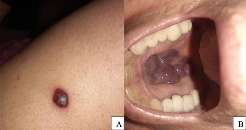
Salih Boğa                                                         Fulya Günşar
      Department of Gastroenterology Memorial Hospital,                  Department of Gastroenterology Ege University School of
                        İstanbul, Turkey                                                 Medicine, İzmir, Turkey
                    salihboga@yahoo.com                                                    fgunsar@yahoo.com
                  Consulting Biostatistician                                            Sabahattin Kaymakoğlu
                                                                        Department of Gastroenterology Istanbul University School of
                      Beyza Doğanay                                                     Medicine, Istanbul, Turkey
   Department of Biostatistics Ankara University School of                             kaymakoglus@hotmail.com
                 Medicine, Ankara, Turkey
            bdoganay@medicine.ankara.edu.tr                                                  Murat Akyıldız
                                                                          Department of Gastroenterology Koç University School of
                       Editors Emeriti                                                   Medicine Istanbul, Turkey
             Nurdan Tozun, Founding Editor                                                akyildizmr@yahoo.com
    Department of Gastroenterology Acıbadem Mehmet Ali
   Aydınlar University School of Medicine, İstanbul, Turkey                                   Mesut Akarsu
                 nurdantozun@hotmail.com                                   Department of Gastroenterology Dokuz Eylül University
                                                                                    School of Medicine, İzmir, Turkey
            TASL Governing Board 2019–2020                                               mesut.akarsu@deu.edu.tr
                     Ramazan İdilman
 Department of Gastroenterology Ankara University School of                                   Mehmet Demir
                 Medicine, Ankara, Turkey                                 Department of Gastroenterology Mustafa Kemal University
         ramazan.idilman@medicine.ankara.edu.tr                                      School of Medicine Hatay, Turkey
                                                                                        drmehmetdemir@yahoo.com
                       Zeki Karasu
  Department of Gastroenterology Ege University School of
                 Medicine, İzmir, Turkey
                 zekikarasu@gmail.com

Contact
Editor in Chief:   Prof. Bulent Degertekin                              Fax:            +90 212 234 19 60
Address:           Ankara Acıbadem Hastanesi Turan Güneş Bulvarı,       Web:            www.tasl.org.tr
                   630. Sk. No:6, 06450 Çankaya/Ankara                  E-mail:         tasl@tasl.org.tr
Phone:             +90 532 2740569
Fax:               +90 312 4903465                                      Publisher:      Kare Publishing
E-mail:            degertekinb@hotmail.com                              Address:        Concord İstanbul, Dumlupınar Mah. Cihan Sk. No: 15
                                                                                        B Blok Da: 162, 34720 Kadıköy, İstanbul-Turkey
Turkish Association for the Study of the Liver                          Phone:          +90 216 550 61 11
Address:           İnönü mah. Cumhuriyet cad. No:131 Mutlu apt. Kat:4   Fax:            +90 216 550 61 12
                   D: 5 Harbiye İstanbul/Turkey                         Web:            www.karepb.com
Phone:             +90 212 244 30 71                                    E-mail:         kare@karepb.com

Hepatology Forum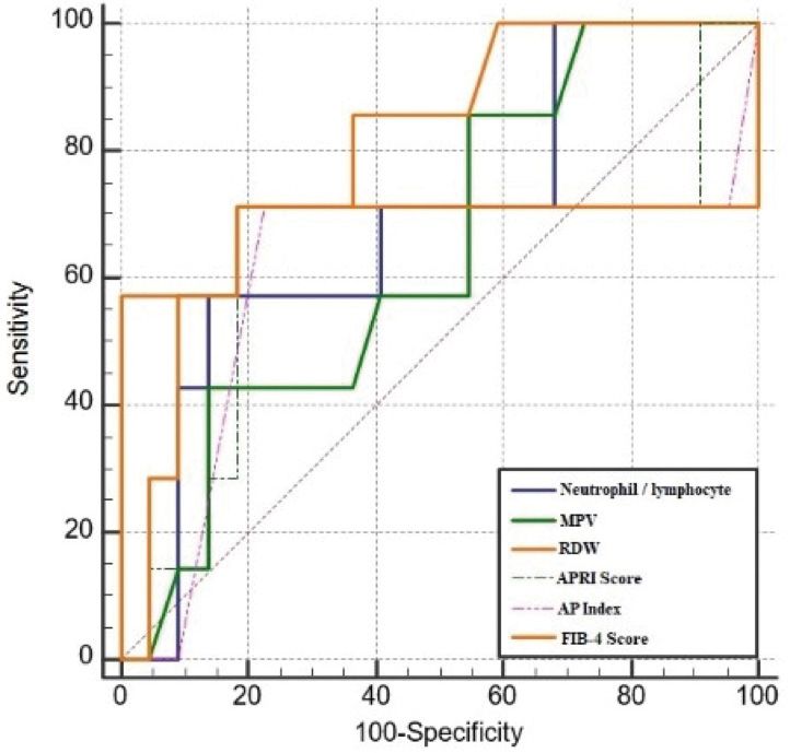
Committees
  TASL Organizer
Ramazan İdilman, M.D.

 AASLD Organizer
  W. Ray Kim, M.D.

Scientific Secreteriat
 Murat Akyıldız, M.D.
 Mehmet Demir, M.D.
Invitation
Dear Colleagues,

It is with great pleasure that the American Association for the Study of Liver Diseases (AASLD) and the Turkish Association for the
Study of the Liver (TASL) announce our second joint conference to CONNECT physicians and health care providers in Turkey with
experts from the USA and Turkey in hepatology.

AASLD is the global leader focused on advancing the science and practice of hepatology. Content delivered from our programs and
journals, combined with local perspectives from experts in Turkey, have facilitated the incorporation of cutting edge research data
into practice and offer the best possible atmosphere to encourage collaborative research between Turkish and American physicians.

This year, we have decided to take on the current challenges presented by the ongoing pandemic head-on and created a unique and
exciting collaborative opportunity to link two meetings into a seamless, productive interactive experience. AASLD and TASL are
working together to offer TASL attendees in our joint AASLD-TASL Digital Hepatology Connect to also register (at no additional
cost) for The Liver Meeting Digital Experience™ 2020.

We truly believe that this arrangement for TASLD audience to participate in the two meetings will maximize learning about the
latest liver disease research being presented at The Liver Meeting Digital Experience,™ followed, with the Hepatology Connect,
by bringing the knowledge to life with more in-depth, case study-based discussions for state-of-the-art diagnosis and treatment
approaches. The opportunity to interact with the unprecedented number of AASLD faculty and with local and regional experts in the
science and practice of hepatology will make this a must-attend combination of events for the TASL audience.

We would be grateful to partner with organizations and companies that share our vision of collaboration and exchange. Support
of this meeting is vital so that we can provide attendees with a quality meeting experience. Specifically, your support will help us
deliver an exceptional scientific program via a digital platform that allows for large didactic lectures and smaller breakout sessions,
as well as other opportunities for faculty and attendees to interact. Your participation will be mutually beneficial. There will be
opportunities for satellite symposia and virtual exhibition space, and your involvement will provide exposure and brand recognition
to extend your reach to hundreds of attendees.

On behalf of AASLD and TASL, we would like to thank you in advance for your financial contribution. Your support will play
a crucial role in promoting liver health, providing quality patient care, and advancing the study and practice of hepatology in the
region, especially in the area of viral hepatitis, chronic liver disease and hepatocellular carcinoma. It is because of supporters like
you that we are able to plan and implement programs of this magnitude and collaboration on a global level. Your generosity directly
allows us to carry out our mission to prevent and ultimately cure liver disease in Turkey, the US, and the world over.

Sincerely,

W. Ray Kim, MD                                                            Ramazan Idilman, MD
Board Liaison, AASLD Global Outreach Committee                            President, TASL
Co-organizer, AASLD-TASL Connect                                          Co-organizer, AASLD-TASL Connect
Content
                              E-Presentations...................................................... 1–22
                              Oral Presentations................................................ 22–25

The abstracts are being reprinted without Journal editorial review.The opinions expressed in this supplement
are those of the panelists and are not attributable to the sponsor or the publisher, editor, or editorial board of the
Hepatology Forum. Clinical judgment must guide each physican in weighing the benefits of treatment against the
risk of toxicity. References made in the articles may indicate uses of drugs at dosages, for periods of time, and in
combinations not included in the current prescribing information.
Abstract Book AASLD TASL Digital Hepatology Connect
EP-01 higher in patients with HCC; It was 11.74±4.193 kPa versus 15.86±2.806 kPa
(p=0.027). The cut off value of LSM to predict HCC was calculated as 14.1 kPa.
Association of genetic polymorphisms of OATP with CONCLUSION: Patients and clinicians will possess the right information about
susceptibility to hepatocellular carcinoma in hepatitis the expected risk of HCC on an individual basis and more importantly, it will
serve to stress the importance of continuing HCC screening (especially in the
C patients who achieved SVR by direct acting
high-risk groups) and maintaining adherence to these programs by the patients.
antivirals Keywords: Hepatitis C, hepatocellular cancer, liver stiffness measurement
Zuhal Mert Altintas1, Serkan Yaras2, Engin Altintas2
Mersin University, Faculty of Medicine, Medical Genetics
1
Mersin University, Faculty of Medicine, Gastroenterology Department
2
EP-03
OBJECTIVES: Simeprevir, daclatasvir, ledipasvir, paritaprevir and ritonavir Re-evaluating diagnoses of autoimmune liver diseases
are all substrates and inhibitors of the organic anion transporting polypeptide in patients followed up at Hacettepe University
(OATP1B1 transporter, whereas sofosbuvir, ombitasvir and dasabuvir are not
substrates. The purpose of this study is to evaluate the association of organic
Sefika Nur Ayar1, Cem Şimşek2, Elif Soyak3, Deniz Çağdaş
anion transporting polypeptide (OATP) gene polymorphism and hepatocellular
carcinoma in Hepatitis C patients who achieved SVR by direct acting antivirals.
Ayvaz3, Yasemin Balaban2
MATERIALS & METHODS: Four single-nucleotide polymorphisms (SNPs) (388 1
Hacettepe University, Medical Faculty, Department of Internal Medicine, An-
A>G, 521 T>C, 334 T>G, and 699 G>A) within the OATP gene were genotyped kara, Turkey
by PCR-RFLP in 200 patients with chronic HCV infection (CHC) treated with 2
Hacettepe University, Medical Faculty, Department of Internal Medicine, Divi-
DAAs, Laboratory work up and abdominal ultrasound was performed at baseline, sion of Gastroentology, Ankara, Turkey
at 12 weeks after end of treatment and then at every 6 months of follow up (FU). 3
Hacettepe University, Ihsan Dogramaci Children’s Hospital, Department of Pe-
RESULTS: The overall SVR12 rate was 99.5%. The SVR12 rate was similar be- diatric Immunology, Ankara, Turkey
tween the patients with HCC and those without HCC (100% vs 99.4%, p=0.49).
OBJECTIVES: Autoimmune liver diseases (ALDs), which consist of autoim-
HCC developed in 10 (5%) of the patients, approximately 11 (6-36 months) after the
mune hepatitis (AIH), primary biliary cholangitis (PBC), primary sclerosing
end of the treatment. No significant differences were found regarding OATP gene
cholangitis (PSC) and overlap syndrome (OS), are a group of diseases charac-
polymorphisms among the case groups (including CHC and HCC) and no matter in
terized by immune attack to hepatocytes and/or cholangiocytes. Although each
comparisons of alleles, genotypes, or haplotypes. Similar insignificant results were
also observed when subgroup analyses were performed in different gender. ALDs has own diagnostic criteria, they can share some clinical, laboratory and
also histological features at initial diagnosis or follow up. This heterogeneous
CONCLUSION: Our observation suggests that SNPs 388 A>G, 521 T>C, 334 nature of ALDs leads to diagnostic difficulties. We aimed to re-evaluate diagno-
T>G, and 699 G>A of OATP gene might not contribute to the development of ses of our patients according to updated diagnostic criteria of ALDs.
HCC in Hepatitis C patients who achieved SVR by direct acting antivirals.
MATERIALS & METHODS: We included 90 patients with ALDs who are on
Keywords: Hepatitis C, hepatocellular carcinoma, organic anion transporting active follow up at Hacettepe University Gastroenterology Unit. Re-evaluation
polypeptide of diagnoses were done based on last accepted universal diagnostic criteria and
guidelines for each disease; AIH revised diagnostic criteria (1) for AIH, current
AASLD guidelines published in 2018 and 2010 for PBC and PSC (2,3), respec-
tively and Paris criteria (4) for OS (AIH and PBC).
EP-02
RESULTS: Out of 90 patients with ALDs, the previous diagnoses were 38
The evaluation of the role of liver stiffness (42%) AIH, 29 (32%) PBC, 5 (6%) PSC and 18 (20%) OS (Table1). The revised
diagnoses of ALDs in 89 patients distributed as 43 (49%) AIH, 32 (36%) PBC,
measurement in predicting hepatocellular carcinoma 5 (6%) PSC and 8 (9%) OS. The kappa value was 0,73 (substantial agreement).
in chronic hepatitis C patients However revised diagnosis differed from previous one in 16 (18%) out of 90
patients. Previous diagnoses did not change in patients with AIH and PSC. How-
Serkan Yaras, Osman Ozdogan, Hatice Rizaoglu Balci, Ferzan ever, the diagnosis of PBC changed in 4 (14%) patients (2 to OS, 1 to PSC and
Aydin, Mustafa Zanyar Akkuzu, Enver Ucbilek, Fehmi Ates, 1 to non-ALD). Strikingly, the diagnosis was changed in most of patients with
Orhan Sezgin, Engin Altintas AIH-PBC variant OS (12 patients (67%); 7(39%) to PBC and 5 (28%) to AIH.
Reasons for misdiagnosis of OS with no other features compatible with Paris
Mersin University, Faculty of Medicine, Gastroenterology Department criteria were the presence of histological interface hepatitis and ANA positivity
(3 cases), interface hepatitis (2 cases), high IgG level (1 case), high ALT level
OBJECTIVES: Due to their safety profile and low side effects any patient in
together with ANA positivity (1 case) in 7 patients with PBC; and were elevated
any stage of chronic liver disease related with Hepatitis C can be treated with
ALP levels (2 cases), AMA positivity (2 cases), histological bile duct injury (1
direct-acting antivirals (DAA). Therefore, it is important to know which patients
case) in 5 patients with AIH.
will be prone to develop liver-related complications, such hepatocellular carci-
noma. However, in the DAA era the association between liver stiffness measure- Table 1. Previous and revised diagnoses of ALD patients
ment (LSM) improvement post-therapy and de novo development of HCC has
not been well studied.
MATERIALS & METHODS: This prospective study included 200 Hepatitis C
patients who had been treated with DAA and had achieved SVR. Laboratory work
up and LSM was performed at baseline and at every 6 months of follow up (FU).
RESULTS: The mean age was 60.5. HCC developed in 10 (5%) of the pa-
tients, approximately 11 (6-36 months) after the end of the treatment. LSM was
January 15–16, 2021 1AASLD TASL Digital Hepatology Connect Abstract Book
CONCLUSION: Type of ALDs was misdiagnosed in 18% of patients. Although
AIH and PSC were accurately diagnosed, cholestatic features and biliary injury
cause false diagnosis of PBC, and leaded to the major mistake as 67% overdi-
agnosis of OS.
Keywords: Autoimmune liver diseases, autoimmune hepatitis, primary biliary
cholangitis, primary sclerosing cholangitis, overlap syndrome
EP-04 Figure 1. (A) Brown pigments can be seen in liver biopsy with hemotoxylin&eosin
stain (x200). (B) Excessive iron deposition is demonstrated by Prussian blue
Iron Man: Distinguishing hereditary and secondary staining (x400).
iron overload in the setting of autoantibody positivity
Table 1. HFE gene mutation analysis studies in Turkey
Nur Yazdalı Köylü1, Bahadır Köylü1, Cenk Sökmensüer2,
Yasemin Balaban3
1
Hacettepe University, Faculty of Medicine, Department of Internal Medicine,
Ankara, Turkey
2
Hacettepe University, Faculty of Medicine, Department of Pathology, Ankara,
Turkey
3
Hacettepe University Faculty of Medicine, Department of Internal Medicine,
Division of Gastroenterology, Ankara, Turkey
OBJECTIVES: Iron overload can cause diagnostic dilemma in case of chron-
ic liver disease. Abnormal serum iron studies and iron overload can be either
primary as in the hereditary hemochromatosis (HH) or secondary to chronic
inflamation as in other chronic liver diseases such as alcoholic liver disease,
non-alcoholic fatty liver disease, chronic viral hepatitis and, even -very rarely-
autoimmune hepatitis (AIH). HH is characterized by abnormal accumulation of
iron not only in parenchymal cells of liver, but also in endocrine glands, heart
and skin. Whereas, secondary iron accumulation mostly affects reticuloendo-
thelial cells. Here, we report a cirrhotic patient with iron overload who has also
new-onset diabetes, hypergammaglobulinemia and high titers of auto-antibodies.
MATERIALS & METHODS: A 38-year old male, with new-onset diabetes, has
been referred to Gastroenterology clinic because of bicytopenia. He had complaints
of malaise and weight lose. At physical examination, there were dark-yellow discol-
oration of skin and splenomegaly. He denied any alcohol, intravenous drug or herb-
al product use. He has no significant family history for liver diseases. Laboratory
tests revealed thrombocytopenia, leucopenia, elevated unconjugated bilirubin lev-
el, and normal transaminase levels. Viral hepatitis was ruled out with negative viral
panel. Anti-nuclear antibody (ANA) and anti-smooth muscle antibody (ASMA)
EP-05
were positive with titers of 1/160 and 1/320 along with hypergammaglobulinemia.
Transferrin saturation was 92% and ferritin was 498 mcg/L. MRI revealed cirrhotic Long-term follow-up liver stiffness results of chronic
liver, multiple siderophilic nodules in the liver, portal vein occlusion, atrophic pan-
creas, splenomegaly, splenorenal shunt and esophageal varices. Pancreatic atrophy hepatitis C patients treated with direct-acting
resulting in diabetes was attributed to iron accumulation rather than immune caus- antivirals
es. No other endocrinopathy was detected. The liver biopsy showed mild lympho-
cytic inflammatory infiltrate in portal areas, local piecemeal necrosis and hepato- Gözde Derviş Hakim1, Ayşe Gökçen Tufan2
cyte-predominant iron deposition. Modified HAI score was 3/18 and fibrosis score 1
Tepecik Training and Research Hospital, Department of Gastroenterology,
was 5/6. HFE gene test revealed C282Y heterozygote mutation.
İzmir, Turkey
RESULTS: In our case, high titer auto-antibody positivity and hypergamaglob- 2
Çiğli Research Hospital, Department of Internal Medicine, İzmir, Turkey
ulinemia created a diagnostic confusion, although HH was suspected because
of new-onset diabetes and skin pigmentation in a young male patient. The di- BACKGROUND & PURPOSE: It was aimed to evaluate the liver stiffness
agnosis of HH was supported by elevated ferritin level together with markedly measurements detected by transient elastography (Fibroscan®), FIB-4 (Fibro-
increased transferrin saturation and demonstration of iron accumulation in liver sis-4) and APRI (AST (aspartate aminotransferase) to platelet ratio index) scores
by MRI. The liver biopsy not only confirmed primary iron accumulation, also in patients diagnosed with chronic hepatitis C (CHC) treated with direct-acting
excluded AIH. C282Y mutation is of high frequency in Europe, and the diagnos- antivirals (DAAs) in long term follow up.
tic algorithm of HH is based on mutation analysis. However, we could not detect MATERIALS & METHODS: Liver stiffness measurements carried out with
any C282Y mutation in our previous study among 2677 Turkish healthy blood transient elastography, FIB-4, APRI scores and biochemical data before and af-
donors. Futhermore, C282Y mutation was absent among our HH patients in a ter long term follow up of the treatment of 26 patients with CHC treated with
small study. The presence of heterozygote C282Y mutation was considered as a DAA were reviewed. Patients receiving Paritaprevir + Ritonavir/Ombitasvir
supportive finding for HH in our case. +Dasabuvir were included in group 1 (n=13), and patients receiving Sofosbuvir
Keywords: Hereditary hemochromatosis, iron overload, autoimmune hepatitis + Ledipasvir ± Ribavirin in group 2 (n=13).
2 January 15–16, 2021Abstract Book AASLD TASL Digital Hepatology Connect RESULTS: There was no significant difference in gender (group 1: 5 men/8 wom- FINDINGS: Of the patients included in the study 14 (16.6%) of them were en, group 2: 4 men/9 women), age (group 1: 60.38±10,87, group 2: 58.54±15.03) child A. Of the patients, 56 (66.6%) had genotype 1, and 28 (33.3%) had gen- or the follow up time (group1:27 months (min-max: 12-34), group 2: 28 months otype 3. Basal LS was found to decrease significantly after AVT (8.00±2.56 (min-max: 22-38) between the groups. Mean liver stiffness measurement of kpa vs 6.95±2.86 kpa and p 0.05). It was observed that Δ-LS value after 12.15±1.84 kPa (min-max: 4.30-42.00 kPa) end of treatment and 9.73±1.57kPa AVT was lower in patients with Child A than patients without cirrhosis (p (min-max 3.0-42.2 kPa) 28 months after treatment. Significant regressions were
AASLD TASL Digital Hepatology Connect                                                                                                                  Abstract Book

EP-07                                                                                    Table 1. Demographic, non-invasive and laboratory findings in patients with
                                                                                         HbeAg (+) fibrozis 2
RDW predicts fibrosis in patients with chronic hepatitis
B infection having persistently normal ALT levels
Basak Yılmaz Guller1, Erdinc Gulumsek2,
Hilmi Erdem Sumbul1, Adnan Tas2
1
 Department of Internal Medicine, University of Health Sciences, Adana Health
Practice and Research Center, Adana, Turkey
2
 Department of Gastroenterology, University of Health Sciences, Adana Health
Practice and Research Center, Adana, Turkey

INTRODUCTION: Although there are studies on determination of hepatic fi-
brosis with noninvasive markers, the data on liver biopsy results and noninva-
sive markers in patients with Chronic Hepatitis B (CHB) is limited.
OBJECTIVE: To examine the relationship between liver histology findings and
noninvasive markers in CHB diagnosed patients, with persistently normal alanin
aminotransferaz (ALT) levels, who have undergone liver biopsy; and to deter-
mine a marker that predicts the fibrosis.
MATERIAL & METHOD: A total of 122 CHB patients, comprising 29 HbeAg
(+) and 93 HbeAg (-), were included in the study, who were 30 years of age and
older, HBV DNA> 2000 IU/ml and had persistently normal serum ALT levels
which were analyzed four times in the last year in three months periods. Patients’
demographic features, laboratory parameters, histological activity index (HAI)
and fibrosis values obtained from liver biopsy, and non-invasive markers (AP
(age-platelet) index, APRI (AST/platelet ratio) and FIB-4 score, neutrophil/lym-
phocyte ratio (NLR), mean platelet volume (MPV) and erythrocyte distribution
width (RDW) were recorded.
RESULTS: It was found that the relation between RDW value and fibrosis was
statistically significant in HbeAg (+) group and in all group independently of
HbeAg (p2
MPV with fibrosis were not statistically significant (p>0.05 for each).
CONCLUSION: It has been shown that the RDW value can be used to predict
fibrosis in HbeAg (+) CHB patients with persistently normal ALT levels and the
cut off value is 12.
Keywords: Chronic hepatitis B, Liver fibrosis, APRI, FIB-4, RDW, MPV


    Figure 1. ROC curve of HbeAg (+) patients with fibrozis >2.

4                                                                                                                                                    January 15–16, 2021
Abstract Book AASLD TASL Digital Hepatology Connect
EP-08 Table 2. Logistic regression analysis for PVT risk factors in liver transplant
candidates
Risk factors for portal vein thrombosis in 121
consecutive liver transplant candidates
Gupse Adalı1, Pınar Gökçen1, Aylin Acar2,
Abdulbaki Ağaçkıran3, Levent Doğanay1, Kamil Özdil1
1
Department of Gastroenterology, Ümraniye Training and Research Hospital,
Health Sciences University, Istanbul, Turkey
2
Department of General Surgery, Ümraniye Training and Research Hospital,
Health Sciences University, Istanbul, Turkey
3
Department of Radiology, Ümraniye Training and Research Hospital, Health
Sciences University, Istanbul, Turkey
BACKGROUND: Portal vein thrombosis (PVT) is particularly detected in ad-
vanced liver cirrhosis patients. Prevalence rates in patients with cirrhosis have
recorded between 1.3% and 9.8%.1 However, the prevalence of PVT in cirrhotic
patients evaluating for liver transplantation varies between 2-26%.2 We aimed to
analyze the risk factors for PVT in liver transplant candidates.
METHODS: Dataset for consecutive 121 cirrhotic patients who were evaluat-
ed for liver transplantation between September 2018 and September 2020 were
retrospectively analyzed. We sorted patients into two groups: Patients with
PVT and patients without PVT. Included variables were: age, sex, etiology of
liver disease, BMI, MELD-Na Score, Child-Pugh Score, platelet count, pres-
ence of ascites, diabetes and hepatocellular carcinoma. Univariate and multi-
variable logistic regression analysis were used to identify risk factors of PVT.
RESULTS: Of 121 liver transplant candidates the prevalence of PVT was 26.4%
(32 with PVT and 89 without PVT) (Table 1). Male sex, Child Pugh Class B, presence of ascites and lower platelet levels were risk factors for PVT in univar-
iate analysis. In logistic regression analysis, transplant candidates with higher
Table 1. Clinical Characteristics of Patients with and without PVT
BMI values (relative risk [RR], 1.13; 95% confidence interval [CI], 1.02-1.25;
P=0.021) and male sex (relative risk [RR], 6.9; 95% confidence interval [CI],
1.42-33.61; P=0.017) had an increased risk for PVT.
CONCLUSION: In liver transplant candidates, the prevalence of PVT is not
low. The development of PVT is associated with male gender and BMI values.
Prospective, large cohort studies needed for risk stratification and early recogni-
tion of PVT prior to liver transplantation.
Keywords: Portal vein thrombosis, liver transplantation, cirrhosis
EP-09
SARS-CoV-2 seroprevelance and clinical features of
COVID-19 in liver transplant recipients:
A single center study
Derya Arı1, Dilara Turan1, Adalet Aypak2, Meral Akdoğan1
1
Department of Gastroenterology, Ankara City Hospital, Ankara, Turkey
2
Department of Infectious Disease, Ankara City Hospital, Ankara, Turkey
BACKGROUND: Liver transplant (LT) recipients with COVID-19 have been
reported as a high-risk population for severe disease through the COVID-19
pandemic. Recent studies have shown that liver transplantation did not signifi-
cantly increase the risk of death and severe disease in patients with SARS-CoV-2
infection (1). Also, patients with COVID-19 can be asymptomatic. Here we re-
port the first results of our ongoing SARS-CoV-2 seroprevalence study.
RESULTS: During November 2020, we collected data and serum samples for
30 liver transplant recipients at Ankara City Hospital (median age 54.2 years
[IQR 24-68]; 23 (76.7%) men, 7 (23.3%) women). Hypertension was the most
common comorbid disease (8/30, 26.6%), followed by type 2 diabetes (3/30,
10%). All study participants were older than 18 years, and they provided in-
formed consent enrolment. Study enrolment was performed when patients pre-
January 15–16, 2021 5AASLD TASL Digital Hepatology Connect Abstract Book
sented for scheduled routine follow-up. All participants completed a question- of post-transplant hepatic steatosis and liver fibrosis were slightly higher in
naire querying information including clinical symptoms in the last three months. recipients with non-alcoholic fatty liver disease-related cirrhosis than those
In addition to all routine blood sample tests, Anti-SARS-CoV-2 IgM and IgG with viral hepatitis-related etiologies (44% vs 27%, p=0.43 and 44% vs 30%,
was determined with enzyme-linked immunosorbent assays (ELISA). All par- p=0.45, respectively).
ticipants with positive results were checked for anti-SARS-CoV-2 PCR by the
CONCLUSIONS: MRE and TE are accurate in assessing liver fibrosis in the
nasopharyngeal swab. We further collected 30 serum samples, and of these 30
liver transplant setting. Obesity and underlying etiology of primary liver disease
samples, 7 (23.3%) tested positive for anti-SARS-Cov-2 IgM and IgG. Two pa-
tients had a known history of symptomatic COVID-19 in the last three months. do not influence the measurements.
We evaluated 5 patients for the COVID-19 PCR test, and all of them were neg- Keywords: Liver transplant, transient elastography, magnetic resonance elastog-
ative. Overall, only one patient declared symptoms of flu-like upper respiratory raphy, fibrosis, steatosis
tract infection, while 4 did not (1/5, 20%). The diseases of two participants with
apparent COVID-19 (2/7) were mild-moderate.
EP-11
CONCLUSION: We documented past SARS-CoV-2 infection in 23.3% of
our LT recipients during the study, and the majority were asymptomatic. Se- Changing of non-invasive fibrosis index values in hepatitis
roprevalence in the general population are lacking for our country and city.
We are planning to include all LT recipients with follow-up in our hospital in C patients treated with direct-acting antiviral agents
our study.
Derya Arı1, Dilara Turan Gökçe1, Hale Gökcan2, Ferhat
Keywords: Anti-SARS-CoV-2 IgG, liver transplant recipients, asymptomatic
COVID-19
Bacaksız3, Ömer Öztürk1, Sabite Kacar1, Meral Akdoğan1
1
Ankara City Hospital, Department of Gastroenterology
2
Ankara University Medical School, Department of Gastroenterology
3
Diyarbakır SBÜ Gazi Yaşargil Education Training and Research Hospital,
EP-10 Gastroenterology
Evaluation of magnetic resonance elastography and INTRODUCTION: Hepatitis C virus (HCV) infection, which may lead to
transient elastography for liver fibrosis and steatosis cirrhosis and hepatocellular carcinoma, is a major cause of chronic liver dis-
ease worldwide. Histological staging of liver fibrosis is essential for treatment
assessments in the liver transplant setting decision-making and prognostication in patients with chronic HCV infection.
DAAs (direct-acting antiviral agents) was known to reduce fibrosis. Owing to
Zeynep Melekoğlu Ellik1, Ramazan Idilman1, the invasive nature of liver biopsy, many noninvasive fibrosis indices have been
Ilkay Sedakat Idilman2, Aysun Kartal1, Yasemin Balaban3, developed to assess the stage of liver fibrosis. Among these indices, the aspartate
Atilla Halil Elhan4, Muşturay Karçaaltıncaba2, aminotransferase (AST)/platelet ratio index (APRI), FIB-4 index, AST/alanin
Hasan Özkan1 aminotransferase (ALT) ratio are commonly used. The present study investigat-
ed the temporal effect of DAAs on the noninvasive index values of patients with
1
Department of Gastroenterology, Ankara University School of Medicine, An- chronic HCV infection at baseline, week 4, month 3, month 6 and month 12.
kara, Turkey
2
Department of Radiology, Hacettepe University School of Medicine, An- METHODS: The data of 88 chronic HCV infection who received a complete
kara,Turkey course of DAA therapy, between 2015 to 2018 were enrolled in this retrospective
3
Department of Gastroenterology, Hacettepe University School of Medicine, analysis. Inclusion criteria were as follows: age ≥18 years, presence of the serum
Ankara,Turkey anti-hepatitis C virus (HCV) antibody for >6 months and detectable HCV RNA, and
4
Department of Biostatistics, Ankara University School of Medicine, Ankara, completion of DAA therapy. Demographic, laboratory characteristics were com-
Turkey pared. Changes in APRI,FIB-4 and AST/ALT index were compared with Willcoxon
signed rank test. SPSS 22 computer program was used for statistical evaluation.
BACKGROUND: Extensive clinical data on the utility and performance of RESULTS: A total of 88 patients were enrolled retrospectively; their median age
elastography in native livers are available in the literature. However, few stud- was 59 (23–80) years, and 46 (52,2%) of them were men. The median follow-up
ies have evaluated the accuracy of magnetic resonance elastography (MRE) time was 28.2 month.The baseline median AST, ALT and total bilirubin levels were
and transient elastography (TE) in assessing liver fibrosis in the liver transplant 55 (19–247) U/L, 56 (15–178) U/L, and 0.9 (0.3–3.3) mg/dL, respectively. The me-
setting. The aims of the present study were to evaluate the accuracy of MRE dian platelet count was 175 (66–360) × 109/L. Furthermore, 21 (23.8%) patients had
and TE in the assessment of liver fibrosis in liver transplant recipients, and to liver cirrhosis. Eighty-five (96.5%) patients received diagnoses of HCV genotype
determine the posttransplant hepatic steatosis and liver fibrosis recurrence rates. (GT) 1 infections. The baseline median APRI value was 1.01 (0.15–6.18), and the
median FIB-4 value was 3.26 (0.34–12.01). In patients who received DAA therapy,
METHODS: Between September 2019 and March 2020, a total of 126 consec- the median APRI and FIB-4 values decreased from week 4 until month 12 (Figure
utive liver transplant recipients were included. MRE and TE were performed for 1). The median APRI value decreased from 1.01 at baseline to 0.44, 0.39, 0.39, and
liver stiffness measurements. 0.38 at week 4, month 3, month 6 and month 12 respectively (all PAbstract Book AASLD TASL Digital Hepatology Connect
EP-12
Melkerson Rosenthal syndrome
Vedat Goral
Department of Gastroenterology, Istanbul Medipol University, Istanbul, Turkey
INTRODUCTION: Melkerson Rosenthal syndrome (MRS) is a rare disease. Its
incidence was determined as 0.08%. It is more common in women between the
ages of 20–40. Recurrence consists of peripheral paralysis, orofacial edema and
fissured tongue, triple symptom. It is more common if they are monosymptom-
atic or oligosymptomatic. Coexistence of these three criteria is present in 25%
of the patients. The disease also affects the nervous system, musculoskeletal sys-
tem, liver, bone, spleen, bone marrow, salivary gland, heart and some other or-
gans. In this case report, a case of MRS with elevated liver enzymes is presented.
RESULTS: Our patient, a 25-year-old male patient who was followed up in
neurology, after various examinations have been performed due to episodic fe-
ver, swelling of the face and eyes since 2014 and he had various tests and was
diagnosed with Melkerson R syndrome. FMF gene: heterozygous mutation +, V
72 6A mut and there were contractions in the legs that had been in attacks for
the last 1 year. ALT 153 IU/ml, AST 172 IU/ml, ALP 574 IU/ml, GGT 155 IU/ Figure 1.
ml, total bilirubin 3.295 mg/dl, direct bilirubin 2.6 mg/dl, iNR: 2.59, albumin
and total protein and albumin were slightly low, protrombin time (PT) was long Table 1. Results of the study parameters
(20.8 sec). Liver biopsy was performed in hospital in 2018 and no primary liver
disease was detected. The patient did not use NSAIDs and alcohol, HBV, HDV,
HCV, autoantibodies to the liver, celiac test were negative, ceruloplasmin, iron
Fe++ BK were normal. Liver biopsy was requested to the patient again in this
time, but the patient refused. Liver enzyme profile was evaluated as liver in-
volvement of the disease.
CONCLUSIONS: The etiology of MRS disease is not fully known. Bacterial
and viral causes, especially HSV, granulomatous diseases, additives, hypersen-
sitivity to proteins and heavy metals, genetic predisposition and many autoim-
mune diseases are held responsible. The treatment of our patient is carried out
by the neurology clinic and follow-up was recommended. Also, he was receiving
ursodeoxycholic acid medication 3 times per day. This case is presented because
it is very rare and interesting.
RESULTS: There was no statistically significant difference in comorbidities
Keywords: Melkerson Rosenthal syndrome, liver, neurological signs
and medications between the groups. In the measurements taken; patient group
had statistically significantly higher FLI’s (p=0.042), there was no statistically
significant difference between the groups regarding BMI, waist circumference,
EP-13
T2DM diagnosis duration. Patient group had statistically significantly higher
The relationship between serum fgf23 levels and liver ALT levels than the control group (p=0.008), there was no statistically signif-
icant difference between the groups regarding BUN, creatinine, eGFR, AST,
steatosis in individuals with type 2 diabetes mellitus GGT, 25-(OH)-vitamin D, phosphorous, ALP and PTH levels. Finally, there was
no statistically significant difference between the groups in terms of their serum
Kadir Bilgi1, Meltem Gürsu2, Şahabettin Selek3 FGF-23 concentrations (p=0.786). There was no correlation of FGF-23 with FLI
(p=0.130) (r=-0.164). There was a correlation between eGFR and FGF-23 even
1
Faculty of Medicine, Bezmialem Vakif University, Istanbul, Turkey
though eGFR levels were above 80 ml/min/1.73 m2.
2
Department of Nephrology, Faculty of Medicine, Bezmialem Vakif University,
Istanbul, Turkey CONCLUSION: Although we could find no relationship between serum FGF-
3
Department of Biochemistry, Faculty of Medicine, Bezmialem Vakif University, 23 levels and the presence of NAFLD in T2DM patients, this study had time
Istanbul, Turkey and resource limitations therefore further experimental and long-term clinical
studies with larger sample sizes are needed in this area.
INTRODUCTION: Non-alcoholic fatty liver disease (NAFLD) is one of the Keywords: FGF23, non-alcoholic liver disease, type 2 diabetes mellitus
most common causes of liver failure today and usually accompanies Type 2 dia-
betes mellitus (T2DM). Fibroblast growth factor-23 (FGF-23) is a hormone that
is known for its role in phosphate regulation, but recent studies found connec-
tions between FGF-23 and liver disease. The aim of this study was to compare EP-14
the serum FGF-23 levels of T2DM patients with and without NAFLD to provide
a better understanding of the relationship between FGF-23 and NAFLD. Celiac disease and NASH association
METHODS: We included 54 volunteers with hepatosteatosis as the patient
group and 33 volunteers without hepatosteatosis as the control group. Waist cir-
Vedat Goral
cumference, height and weight were measured, BMI and FLI were calculated. Department of Gastroenterology, Istanbul Medipol University, Istanbul. Turkey
The routine control test results were recorded. Calcium, phosphorus, GGT, ALP,
25-(OH)-vitamin D, PTH and FGF-23 were measured in serum samples. INTRODUCTION: Nonalcoholic fatty liver disease (NAFLD) is the most com-
January 15–16, 2021 7AASLD TASL Digital Hepatology Connect Abstract Book mon cause of chronic liver disease in children and adolescents. Celiac disease (CD) is associated with both acute and chronic liver diseases, especially au- toimmune liver disease. In this Case report, the association of celiac disease and NASH is presented. CASE: A 19-year-old female patient was admitted to our clinic with high liver enzymes, diarrhea and weakness. In liver function tests, ALT was 36.1 IU/ml (n:
Abstract Book                                                                                                       AASLD TASL Digital Hepatology Connect

EP-16                                                                                     EP-17
Diagnostic validity of non-invasive tests for predicting                                  Evaluation of prothrombin index in chronic hepatitis
liver fibrosis stage in chronic hepatitis B patients with                                 B patients
HBV DNA>2000 IU/mL, ALT>ULN
                                                                                          Şükran Köse1, Didem Çelik2
Didem Çelik , Bengü Tatar , Şükran Köse
                   1                  2                  2
                                                                                          1
                                                                                            Department of Infectious Diseases and Clinical Microbiology, University of
1
 Department of Infectious Diseases and Clinical Microbiology, Bakırçay Uni-               Health Sciences Tepecik Education and Research Hospital, İzmir, Turkey
versity Çiğli Education and Research Hospital, İzmir, Turkey
                                                                                          2
                                                                                            Department of Infectious Diseases and Clinical Microbiology, Bakırçay Uni-
2
 Department of Infectious Diseases and Clinical Microbiology, University of               versity Çiğli Education and Research Hospital, İzmir, Turkey
Health Sciences Tepecik Education and Research Hospital, İzmir, Turkey
                                                                                          INTRODUCTION: Prothrombin index (PI) is one of the early indicators of liver
INTRODUCTION: Determining the stage of liver fibrosis in chronic hepatitis B              damage and reflects lung synthesis capacity. There are studies indicating that it
                                                                                          can be used to predict advanced liver failure and esophageal varices. The aim of
(CHB) patients is the main step of treatment decision. However, this method is
                                                                                          this study was to investigate the association of PI with other liver function tests
invasive, difficult, expensive and have many complication risks. The aim of our
                                                                                          and hepatitis B virus serum levels and to determine whether it can be used to
study was to evaluate noninvasive markers of liver fibrosis; aspartate transami-
                                                                                          predict significant liver fibrosis or not.
nase (AST)-platelet ratio index (APRI), 4 factors based fibrosis index (FIB-4),
AST-Alanine transaminase ratio (AAR), age-platelet index (API), gamma-glu-                METODS: A total of 547 treatment naiv patients aged 18 years or older with
tamyl transpeptidase-platelet ratio(GPR), RDW-platelet ratio (RPR), King’s                chronic hepatitis B were included in the study. Patients with coinfections and
score, Fibro quotient (Fibro Q), mean platelet volume (MPV).                              comorbidities were excluded from the study. Demographic data and laboratory
                                                                                          values were retrospectively reviewed from patient files. Liver biopsy results was
MATERIAL & METHOD: In this study, 143 treatment naiv CHB patients with
                                                                                          calculated according to Ishak scoring system. The patients’ SPSS 22.0 program
HBVDNA>2000 IU/mL and ALT>ULN were included. Data were obtained retro-                    was used for statistical analysis.
spectively from patients’ follow-up files. Liver histopathology was calculated ac-
cording to Ishak scoring system. The data evaluated using SPSS IBM 22.0 program.          RESULTS: Of the patients included in the study, 284 (51.9%) were female and
                                                                                          the median age was 41/year. The number of patients according to the stages of
RESULTS: Of all patients 48.25 (n:69) were female and the mean age was 44.3/              fibrosis were F0, 94 (17.2%); F1, 144 (26.3%); F2, 218 (39.9%); F3, 55 (10.1%);
years. Distribution of each stage of fibrosis were F0; 18(12.6%), F1; 26(18.2%),          F4, 21 (3.8%); F5, 13 (2.4%); F6 was 2 (0.4%). HbeAg positivity was 7.1%
F2; 54 (37.7%), F3; 23(16.1%), F4; 12(8.4%), F5; 8(5.6%), F6; 2(1.4%). Of the             (n:39). Spearman correlation analysis showed significant correlation with PI and
11 noninvasive tests, 7 had the power to predict ≥F2 and 8 had ≥F3. The best              age, AST, ALT, INR, platelet, HBV DNA, HBsAg quantitative levels (p ULN and HBV DNA>2000 IU/mL                        Table 1. Correlation analysis of Prothrombin Index with age, AST, ALT, GGT,
                                                                                              HBV DNA, HbsAg quantitative, Fibrosis and Histological Activity Index


January 15–16, 2021                                                                                                                                                               9
AASLD TASL Digital Hepatology Connect Abstract Book
EP-18 Table 1. Demographics of the patients and donors
The effect of sarcopenia in liver transplantation
Genco Gencdal1, Şencan Acar2, Utku Alkara3, Ayhan Dinçkan4,
Çiğdem Arıkan5, Murat Akyıldız1
1
Koc University, School of Medicine,Liver Transplantation Center, Department
of Gastroenterology & Hepatology, Istanbul, Turkey
2
Sakarya University, School of Medicine, Department of Gastroenterology &
Hepatology, Adapazarı, Turkey
3
Yeni Yüzyıl University, School of Medicine, Liver Transplantation Center, De-
partment of Interventional Radiology, Istanbul, Turkey
4
Istinye University, School of Medicine, Liver Transplantation Center, Depart-
ment of General Surgery, Istanbul, Turkey
5
Koc University, School of Medicine, Liver Transplantation Center, Department
of Pediatric Gastroenterology & Hepatology, Istanbul, Turkey
BACKGROUND & AIM: Nutritional impairment and loss of muscle mass in the
liver cirrhosis negatively affect morbidity and mortality, and usually does not return
after liver transplantation (LT). In this study, we aimed to investigate the incidence
of sarcopenia in cirrhotic patients before LT and the effects of sarcopenia on LT.
MATERIALS & METHODS: 108 LT recipients (live donor LT and cadaveric
LT performed between May 2015 and March 2017) were analyzed retrospective-
ly. CT images of patients were measured blindly by a single radiologist using
the psoas muscle and other skeletal muscles from the intervertebral disc line
between the 3rd and 4th lumbar vertebrae using Myrian Software®.
RESULTS: Sarcopenia was observed in 20 of 108 patients (15 females, 5 males).
The demographic datas of the patients are shown in table 1. While there was no sig- EP-19
nificant relationship between sarcopenia and BMI, body weight was significantly
lower in sarcopenic cases (p0.05). The survival curve is shown in
figure 1. Overall survival was 97% at 6 months and 92.7% at 1 year, and there was
experience
no significant difference between the two groups in terms of 1-year survival.
Ferit Çelik1, Ali Şenkaya1, Nalan Gülşen Ünal1, Seymur
CONCLUSION: Recent Studies showed that sarcopenia significantly affects
morbidity and mortality in cirrhotic patients while adversely affecting liver
Aslanov1, Alper Uysal1, Ayşın Zeytinoğlu2, Ilker Turan1, Murat
transplant results. On the other hand, in the present study sarcopenia did not Zeytunlu3, Ömer Özütemiz1, Ulus Salih Akarca1, Zeki Karasu1,
significantly affect the transplant results different from the literature. We inter- Fulya Günşar1
pret that discrepancy, firstly, because of the majority of cases were live donor 1
Ege University Department of Internal Medicine, Division of Gastroenterology,
LT who had lower MELD scores than western countries where the most of liver
transplantations were from cadaveric with higher MELD score especially over Izmir, Turkey
than 20 and secondly, we usually give supportive treatment and have arranged
2
Ege University Department of Medical Microbiology, Izmir, Turkey
the timig of transplantation according to patient status.
3
Ege University Faculty of Medicine Department of General Surgery, Izmir, Turkey
Keywords: Sarcopenia, liver, transplantation BACKGROUND: Hepatitis E virus (HEV) may cause chronic liver disease in
solid organ transplant recipients. We determined the HEV seroprevalence and
associated factors in liver transplant recipients.
MATERIALS & METHODS: Patients followed at the outpatient clinic of liver
transplantation between January 2019 and January 2020 were screened retro-
spectively for HEV serology (HEV immunoglobulin M [IgM] and HEV IgG).
RESULTS: Among the 150 patients (male/female, 104/46; age, 55.4±13.2
years) studied, anti-HEV IgG was positive in 31 (20.7%) and anti-HEV IgM
was negative in all. Mean time after liver transplantation (72 [48%] deceased
and78 [52%] living donors) was 81±78.5 months. Drinking water consisted
of carboy water in 88 (58.7%) patients and tap water in 62 (41.3%). Of the
patients, 120 (80%) lived in urban and 30 (20%) in rural areas. On com-
parison, a statistically significant difference was detected between the an-
ti-HEV IgG-positive and –negative groups in terms of age, place of birth,
water supply, and donor type, (p=0.007, p=0.000, p=0.034, and p=0.049,
respectively).
CONCLUSION: HEV seroprevalence was more frequent in liver transplant re-
cipients compared to the normal population. Older age, watersupply, and place
of birth were risk factors for HEV seroprevalence.
Figure 1. Survival comparison of sarcopenics with non-sarcopenics Keywords: Anti-HEV IgG, liver transplantation, seroprevalence
10 January 15–16, 2021Abstract Book AASLD TASL Digital Hepatology Connect
Table 1. Seroprevalence of hepatitis E virus in liver transplant recipients was performed. A lesion consistent with HCC, measuring approximately 15 mm
according to sociodemographic characteristics, liver function tests, and risk factors in size in liver segment 8, was enhanced in the arterial phase and washout in
late images (Figure-1). Serum alpha-fetoprotein level was totally normal. Based
on these findings, the final diagnosis of HCC associated with sarcoidosis was
confirmed by biopsy. Infliximab treatment was stopped. The patient’s MELD
score was evaluated as 10 points, and the Child-Pugh score as A. Transarterial
chemoembolization treatment was applied to the HCC lesion present in the liver
of the patient. Our patient remained healthy without recurrence or metastasis at
the last follow-up visit.
DISCUSSION: Cirrhosis or portal hypertension has been reported in ≤1% of all
sarcoidosis cases. In addition, the association of hepatic sarcoidosis with HCC
is thought to be considerably rare. When we evaluated the previous cases, we
found that there is an increased risk for HCC in sarcoidosis patients with or
without cirrhosis.
CONCLUSION: In patients with systemic sarcoidosis, especially in the pres-
ence of liver cirrhosis, regular liver checkups should be performed.
Keywords: Hepatocelular carcinoma, sarcoidosis, infliximab, TACE
Figure 1. HCC lesion on MRI, hypointense mass in the hepatobiliary phase (A),
and contrast-enhancing mass in the arterial phase (B).
EP-20
Systemic sarcoidosis-associated hepatocellular
carcinoma: A case report
EP-21
Derya Arı1, Dilara Turan1, Ömer Öztürk1, Mustafa Özdemir2,
Nesrin Turhan3, Bahar Özdemir4, Meral Akdoğan1 Serum levels of ADAMTS-7 and 12 as hepatic fibrosis
markers in patients with chronic hepatitis B
1
Ankara City Hospital, Department of Gastroenterology
2
Ankara City Hospital, Department of Interventional Radiology
Fatih Kıvrakoğlu1, Zahide Şimşek2, Ilhami Yüksel1
3
Ankara City Hospital, Department of Pathology
4
Ankara City Hospital, Department of Rheumatology 1
Department of Gastroenterology, Ankara Yıldırım Beyazıt University, Ankara
City Hospital, Ankara, Turkey
INTRODUCTION: Sarcoidosis is a multisystem granulomatous disorder 2
Department of Gastroenterology, Dışkapı Yıldırım Beyazıt Education and Re-
of unknown ethology that affects individuals worldwide and is characterized search Hospital, Ankara, Turkey
pathologically by the presence of noncaseating granulomas in involved organs.
The association of hepatic sarcoidosis with hepatocellular carcinoma (HCC) is Hepatitis B is a life-threatening liver infection that is common in our country
considerably rare. Here we report a rare case of HCC associated with systemic and in the world. The appropriate treatment of the patient based on serum ALT
sarcoidosis. level, serum HBV DNA level and liver histology. Liver biopsy is recommend-
CASE: A 65-year-old male with a history of systemic sarcoidosis, diabetes mel- ed for the determination of liver fibrosis and for the planning of treatment in
litus, and hypothyroid consulted our clinic. The patient had been followed up for patients with established criteria. Liver biopsy is accepted as the gold standard
an 11-year active pulmonary and musculoskeletal symptoms and bone marrow in determining fibrosis. However, non-invasive fibrosis markers have become
involvement confirmed by biopsy. Imaging studies on his first admission re- important because of the invasiveness of biopsies, complications, sampling
vealed bilateral pulmonary infiltrates with a ground-glass had an appearance. errors, differences in evaluation among pathologists, and difficulty in rebiop-
He had pulmonary and musculoskeletal symptoms, and his symptoms and the sy. In recent years, the role of “A Disintegrin-like and Metalloproteinase with
radiologic infiltrates were resolved by treatment with corticosteroids. Cirrhosis Thrombospondin type-1 motif (ADAMTS)” genes in the etiopathogenesis of
findings had encountered during imaging in another centre. Liver sarcoidosis various diseases is being investigated. Studies that investigating the relationship
was diagnosed with the sample obtained in a needle biopsy examination in 2019. between ADAMTS and fibrosis in the literature were made at the tissue level and
During the follow-up of the patient, hyperintense lesions were observed in T2 there is no study in made with serum. This study was planned to investigate the
measured as 8 mm in liver segment 3, 7.4 mm in segment 6 and 12 mm in utility of serum ADAMTS-7 and 12 levels as noninvasive markers in patients
segment 8, and hypointense lesions that did not show contrast enhancement in with Chronic Hepatitis B (CHB). This study includes 77 CHB and 30 healthy as
contrast-enhanced series. Infliximab (anti-TNF) treatment had initiated in the a control who was treated and followed-up in Ankara Dışkapı Yıldırım Beyazıt
patient due to the symptoms of the musculoskeletal system unresponsive to Education and Research Hospital Gastroenterology Clinic-Hepatology depart-
steroids. In the first year of infliximab treatment, the patient’s complaints im- ment. In our study, ADAMTS-7 and 12 levels were statistically significantly
proved almost completely, and abdominal magnetic resonance (MR) imaging lower in patients with CHB than in the healthy control group (pAASLD TASL Digital Hepatology Connect Abstract Book
these two markers were compared between the mild fibrosis/advanced fibro- Table 1. The seroprevalence rates of HEV infection in healthy population in Turkey
sis groups, no significant difference was found (for ADAMTS-7 p=0.701, for
ADAMTS-12 p=0.342). According to these results, low serum ADAMTS-7 and
12 levels in patients with CHB indicate the presence of liver fibrosis. But they
were found to be inadequate to determine fibrosis level.
Keywords: Chronic hepatitis B, non-invasive markers, Adam-ts 7 and 12
EP-22
The prevalance of hepatitis-E virus infection in
adult Turkish population: Systematic review of the
literature and prevalence study in blood donors in
mersin province
Orhan Sezgin1, Serkan Yaras1, Seda Tezcan Ulger2, Gonul
Aslan2, Eyup Naci Tiftik3
1
Mersin University Faculty of Medicine, Department of Gastroenterology
2
Mersin University Faculty of Medicine, Department of Microbiology
3
Mersin University Faculty of Medicine,Department of Hematology
OBJECTIVE: Hepatitis-E virus (HEV) causes acute hepatitis but can rarely be-
come chronic in immunocompromised patients. The frequency of HEV infection
varies depending on factors such as geographical region, socioeconomic level
and age; the prevalence of HEV according to the results of a limited number
of studies based upon the adult population of Turkey is 10%. There is no suffi-
cient up-to-date information on the frequency of HEV infection in our city. For
that reason, we aimed to investigate the prevalance of HEV infection in healthy
blood donors in Mersin and to review all Turkey-based scientific results of HEV
infection in the adult population.
EP-23
MATERIAL & METHOD: A total of 900 healthy volunteers who applied to
donate blood to the University Hospital Blood Center and accepted the use of Hepatic steatosis effects hepatitis B virus surface
their data were enrolled in the study. Five ml of blood samples were taken and
antigen seroclearance in individuals with chronic
centrifuged, and the serum portions were stored at -80°C until analyzed. Anti
HEV IgG Ab was examined by ELISA method. Volunteers’ age, gender, profes- hepatitis B virus infection
sion, information about the region they live in, and contact with pets or livestock
were recorded. Also Turkey-based HEV seroprevalence studies from 1990-2020 Zeynep Melekoğlu Ellik, Serkan Duman, Abdullah Mübin
investigating the adult population in Turkish or English full text and conference Özercan, Mesut Gümüşsoy, Emin Bodakçi, Muhammed Fatih
papers were evaluated. General frequency results were evaluated chronologically Karakaya, Ramazan Erdem Er, Hale Gökcan, Ramazan Idilman
by city and region. Risk factors and results in risky populations were evaluated.
Department of Gastroenterology, Ankara University School of Medicine, Anka-
RESULTS: The average age of the volunteers was 35.22±9.60 years and 98.7% ra, Turkey
were men. Anti HEV IgG was positive in 12.8% of study population. The average
age of the seropositive individuals was 40.40±9.72 years and 98.2% were men. BACKGROUND & AIMS: The aim of the present study was to evaluate the effect
There was no statistical difference between the mean age of the volunteers and of baseline hepatic steatosis on the development of hepatitis B virus (HBV) sur-
the positive cases (p=0.282). 40% of the participants were rural residents, and face antigen (HBsAg) seroclearance in individuals with chronic HBV infections.
60% were urban residents. The seropositivity rates of these groups were 43.9% MATERIALS & METHODS: A total of 49 individuals with chronic HBV in-
and 56% respectively. Among all participants, the rate of those with a history of fection (M/F: 18/31) was included into analysis. The diagnosis of chronic HBV
contact with livestock was 5.6%. No association was found between anti-HEV infection was made based on clinical, biochemical and histological. The median
IgG positivity and occupation, place of living, and encounter with animals. Also follow-up period was 159 months (interquartile range: 141.5-174.5 months).
total of 57 studies were analyzed to assess the prevelance of HEV in Turkey: 34 of
them constituted healthy population screenings, while 23 constituted screenings RESULTS: The mean age was 53.7±8.7 years. The mean serum ALT level was
of at risk patients. The average seroprevalence of HEV infection of the healthy 22.8±8.7 U/L; the mean AST level was 23.6±6.0 U/L. Of the 49 individuals,
population in Turkey was found to be 9.52% (Table). The highest prevalance 12% had diabetes mellitus, 31% had hypertension. Twenty-seven patients had
rate was in Southeast region of Turkey (Figure). According to studies conducted hepatic steatosis (>5%) in their baseline liver biopsies, whereas the remain-
in risky groups, the highest seroprevalence rate was 73.3% in those with acute ing 22 patients had no steatosis. Of the patients’ characteristics, patients’ age,
hepatitis, followed by 42% in hemodialysis patients. The frequency is higher than body mass index, the controlled attenuation parameter (CAP) score, serum ALT
the general population in those working in farms and animal husbandry. and fasting glucose levels were significantly higher in HBV-infected individ-
uals with hepatosteatosis than those of in individuals without hepatosteatosis
CONCLUSION: The anti-HEV IgG seropositivity rate in healthy blood donors (pYou can also read