Glutathione As Biomarker for Neurodegenerative Diseases
←
→
Page content transcription
If your browser does not render page correctly, please read the page content below
1
  ISSN 2693-2504
              Glutathione As Biomarker for Neurodegenerative Diseases
Journal of Bioscience & Biomedical Engineering                                                                       Mini Review

Shimon Shatzmiller*, Inbal Lapidot

                                                                     *
                                                                      Correspondence author
                                                                     Shimon Shatzmiller
                                                                     Department of Biological Chemistry
 Department of Biological Chemistry, Ariel University of Samara,
 40700 Ariel, Israel                                                 Ariel University of Samara
                                                                     40700 Ariel
                                                                     Israel

                                                                     Submitted : 24 Aug 2020 ; Published : 18 Sept 2020

      Abstract
      Brain imaging is one of the main obstacles of modern disease diagnostics since it must allow accurate and quantitative
      measurements on a living organ that is placed in adults (in neonatal humans’ skull penetration is somewhat easier)
      inside a bone cage. Inside is the brain, the main function controller of our body that must be done in a non- invasive
      way on the living operation brain. Many spectroscopic methods are trying to overcome the difficult access to the brain:
      Photoacoustic microscopy, Confocal microscopy, Two-photon microscopy, Optical coherence tomography, Scanning
      Laser Acoustic Microscopy, Acoustic microscopy, Ultrasonography, positron tomography, fluorescence methods, photo
      caustic microscopy and multi (two) photon imaging spectroscopy. The situation is complex since the brain is wrapped
      in the blood brain barrier allowing only selected molecules to pass from the blood stream to and out of the brain. In
      this chapter, we will survey the current situation of brain diagnostics with the aid of the spectroscopic methods. Brain
      research is integrated in aging research as a major area of interest. Aging is in most cased coupled with the loss of
      brain function and dementia. The neurodegenerative diseases, although identified by Dr. Alzheimer and his collaborators
      more than a century ago, continue as the leading causes of mortality among the elderly. Brain research in trying to
      give hope to those people but unfortunately our understanding in this area is limited. Oxidative stress contributes to
      neurodegenerative diseases pathophysiology and progression. The target was to describe central and peripheral
      metabolites of redox metabolism and to describe correlations between glutathione status, age, and disease severity.

Opening Words

The brain is the organ known to have its own guarding system,       thereby disrupt the function of cells, tissues and organs of
a huge blood vessels net that allows the entry of essential         the body.). Retinopathies are the abnormal accumulation and
nutrients while blocking other substances. Unfortunately,           toxicity of proteins in certain disease states. [3] Also, selective
this barrier is so effective in protecting against the passage of   hyper proteolytic diseases have been referred to this category,
foreign substances that it may prevent life-saving drugs from       e.g. critical illness myopathies or tumor cachexia [2]. The
being able to repair the damaged brain [1,2]. A partial list of     retinopathies comprise at least 30 diseases that affect a variety
more than 20 amyloid-related diseases includes Alzheimer’s          of organs and tissues, including Alzheimer’s Disease (AD),
Disease, Parkinson’s Disease, Huntington’s Disease, prion           Parkinson’s Disease, type 2 diabetes, amyloidosis, and a wide
diseases, familial amyloidosis, type II diabetes, Creutzfeldt–      range of other disorders [4,5].
Jakob disease, Lewy Body Dementia and more than 20
more uncurbable and therefore fatal diseases. New studies           In some retinopaths, an abnormal assembly can be designed
are guiding researchers toward a breakthrough in the cure           on an exogenous protein, usually in the folded form of the
of non-infectious neurodegenerative diseases are associated         same protein. In this way the disease state in a susceptible host
with the accumulation of fibrillar proteins. These diseases         can be induced by inserting a diseased tissue extract from an
exhibit features that are reminiscent of those of prionopathies,    infected donor. The best known form of such an infectious
including phenotypic diversity and the propagation of               (or transmitted) protopathy is a fertility disease, which can
pathology of Proteopathy ( refers to a class of diseases in         be transmitted by exposure of a host organism to a purified
which certain proteins become structurally abnormal, and            fertility protein in a disease-causing structure [6,7]. There
J B & Bio Engine; 2020                               www.unisciencepub.com                                         Volume 1 | Issue 3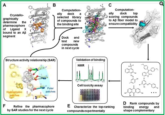
2
is now evidence that other protopaths can be injected by a
similar mechanism, including amyloidosis AA, apolipoprotein
AII amyloidosis, and amp amyloidosis [8,9]. In all of these
cases, an aberrant form of the protein itself appears to be
the pathogenic agent. Already one hundred years ago, Jacob
Heinrich Lewy described intracellular eosinophilic inclusions
in the brains of patients with “paralysis agitos “, commonly
known as Parkinson’s Disease [10].

                                                                     Amyloid Beta Hypothesis Oxidative stress Hypothesis,
                                                                                    Glutathione [12-14]

A group of psychiatrists at the Psychiatric Clinic at the
University of Munich: A. Alzheimer and Solomon

Fuller sit in the front row; Standing in the back row, from
left to right: Bronciini, Bronciini, von Norbert, Ranky, and
unidentified.

Mighty progress has been made over the past decades in
analyzing the content and formation of Levy bodies and            Schematic illustration of the Aβ-amyloid cascade from APP
their relationship to degenerative diseases. Multitudes of        cleavage by secretases to generate Aβ monomers, to plaque
researchers have been engaged in the last hundred years since     formation, via oligomers, protofibrils, and fibrils. Causative
the identification of the phenomenon. However, only little        factors for neuronal injury are indicated in italic letters under
has been achieved in the diagnosis of the initial stages of the   the Aβ pathway. Anti-amyloid agents are also shown in solid
diseases. Here we try to survey the chemistry aspect of the       white letters above the therapeutic targets in the Aβ pathway
diagnostic efforts.                                               [1].
                                                                  Glutathione (GSH) is the major cellular thiol present in
The health authorities in the USA gathered some information       mammalian cells and is critical for maintenance of redox
on the effort in the chemistry of neurodegenerative diseases. A   homeostasis [15]. However, current assay systems for
sample could be found on the Internet [11].                       glutathione lack application to intact animal tissues. Although
                                                                  reports exist on the quantitative imaging of glutathione
                                                                  in hippocampal neurons and glia in culture using bimane
                                                                  fluorescence, there is an urgent need to map the levels of
                                                                  glutathione in intact brain with cellular resolution (acute tissue
                                                                  slices and live animals) [16]. Glutathione is a major antioxidant
                                                                  system in the mammalian central nervous system (CNS).
                                                                  Abnormalities of GSH metabolism have been associated
                                                                  with many disorders of the CNS, including Parkinson’s,
                                                                  Alzheimer’s, and Huntingdon’s diseases and ischemic/
                                                                  reperfusion injury. Investigation of GSH levels in the CNS
                                                                  generally relies on biochemical assays from cultures enriched
                                                                  for different cell types.

                                                               There is Oxidative Damage Is the Earliest Event in Alzheimer
                                                               Disease [17]. Glutathione S- transfer, commonly abbreviated
                                                               (GST), refers to a group of enzymes that employ glutathione
    J B & Bio Engine; 2020                         www.unisciencepub.com                                  Volume 1 | Issue 3
3
in many reactions that contribute to the conversion of many
compounds such as therapeutic drugs, carcinogens, and
products involved in oxidative stress. The evaluation of the
enzyme Glutathione Transferases (GST), a method that may
become instrumental in the choosing glutathione Transferases
level as a biomarker for Alzheimer’s Disease [18,19]. Scientist
developed a spectrophotometric assay for the glutathione
conjugation and determined specific activities with a range of
human GSTs as well as some rat GSTs for comparison. The
ubiquitous GST P1-1 showed the highest activities with the
6-halogenopurines, which bodes well for the application of
pro-probes for human investigations.

The acceptance of the “oxidative stress” as a major process
in the early evolution of neurodegenerative pathology has
become a major trend in this field of research.

                 Oxidative stress early stages

                                                                  Alternative splitting b synuclein enzymes, digestion of beta-
                                                                  amyloid precursor protein [20]. Is it a secondary effect or
                                                                  byproduct that arises from but does not causally influence a
                                                                  process, in particular.

               Amyloid hypothesis early stages

Digestion of beta-amyloid precursor protein is supposed to Such defects are considered to the postulated initiation of the
produce fractures that do not harm or harm fractures. There cascade leading to Metabolic abnormities In Alzheimer’s Disease
is an evolving consensus seeing amyloid cerebrospinal fluid to confirm the characteristics of metabolic abnormalities in
(Aß) as a key biological marker for the Alzheimer’s disease Alzheimer’s Disease, regional metabolic activity summarized
stage of mild cognitive impairment. Aβ is directly involved in tennis of quantitative cerebral glucose metabolic rate.
in the pathogenesis of AD or in close correlation with other
primary pathogenic factors. It is produced from amyloid
                                                                 Introduction
precursor protein (APP) by proteolytic processing dependent
on the enzyme 1-closed AP-site and the γ-secretase complex,
                                                                 Glutathione (GSH) is required for many critical cell processes,
and is degraded by a wide variety of proteases. This review
                                                                 buy plays a particularly key role in the care and regulation of
summarizes targeted proteolytic studies of Aβ in biological
                                                                 the thiol-redox status of the cell. GSH is the most important
fluids and identifies clinical useful markers of Aβ homeostasis
                                                                 endogenous antioxidant and plays an important role in the
in AD disruption.
J B & Bio Engine; 2020                               www.unisciencepub.com                                    Volume 1 | Issue 3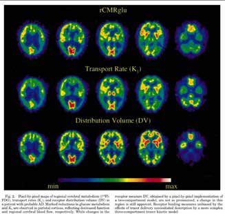
4
detoxification of xenobiotics and their metabolites, as well as
in the maintenance of the intracellular redox balance.

With current estimates of 36 million people affected worldwide.
Alzheimer’s Disease (AD) - a disorder characterized by
impossible progressive impairment of memory and other
cognitive functions - is the most prevalent form of dementia.
Therefore, identifying biological markers that can serve as a
reliable surrogate for the onset of the disease disorder and its
progression is of paramount importance for information on
intervention. Oxidative stress, which is a common denominator
of a number of pathophysiological events associated with
AD, appears to be a major factor in the pathogenesis and
progression of AD. Alzheimer’s Disease (AD) represents
one of the great and unresolved medical needs facing society
during this thousand years. Despite considerable work over the
last quarter. There are no drugs that attack the pathophysiology
underlying the Disease. One of the cardinal characteristics
of counting Disease is the placement of platelets composed
of peptides (3-amyloid, A, s) in the brain. Especially in areas
related to cognition and memory overproduction of A that
appears to be directly neurotoxic. Can be detected in the
earliest stages of AD and. in fact.

Before cognitive function can be discerned a father is extracted
from a protein from its flesh. Amyloid precursor protein (APP),
by proteolytic processing at its N- and C-terminals by enzymes
α,β- and γ-secretase enzymes [21,22].

Two main hypotheses are today directing neurodegenerative
diseases research. The Amyloid hypothesis and the oxidative
stress mechanism. Although the accumulation of data supports
the Amyloid beta aggregation hypothesis, in conclusion, the
oxidative stress hypothesis (inflammation) of AD is very much
alive and viable, but a great deal of work needs to be done
to design future studies and appropriate clinical trials that
will conclusively establish the role of oxidative stress in AD        Amyloid Beta Hypothesis Oxidative stress Hypothesis
pathogenesis.                                                                       Glutathione [24, 25]

One high hurdle is the lack of a quantitative instrumental         Schematic illustration of the Aβ amyloid cascade from APP
method to diagnose and follow the Disease from initial stages.     cleavage by secretases to generate Aβ monomers, to plaque
The development of new drugs depends a lot of such a helpful       formation, via oligomers, protofibrils, and fibrils. Causative
device.                                                            factors for neuronal injury are indicated in italic letters
                                                                   under the Aβ pathway. Anti-amyloid agents are also shown
Alzheimer’s Disease is one of the many neurodegenerative           in solidwhite letters above the therapeutic targets in the Aβ
disorders that are tormenting many, older people. It is in the     pathway [9].
unflavored situation where many theories on what initiates the
cascade of events and when exactly it started to affect the life   The oxidative (inflammatory) step Is the Earliest Event in
of the ill people [23].                                            Alzheimer Disease [26,27]. Observations indicate that increased
                                                                   oxidative damage is an early event in AD that decreases with
Today, only a post mortem analysis of the patient brain can        disease progression and lesion formation. These findings
diagnose 100% the Alzheimer’s Disease.                             suggest that AD is associated with compensatory changes
                                                                   that reduce damage from reactive oxygen. The activities and
                                                                   expression of several antioxidant enzymes such as Cu/Zn- and
                                                                   Mn-superoxide dismutase, glutathione peroxidase, glutathione
                                                                   reductase, and catalase have been studied in AD and could be
                                                                   in part responsible for the decrease in oxidative damage we
                                                                   observed.

    J B & Bio Engine; 2020                          www.unisciencepub.com                                      Volume 1 | Issue 3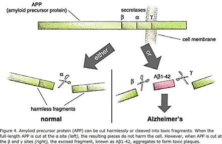
5
“Clinical criteria for the diagnosis of AD include dementia         - what is known as Aβ1 - 40 is most common - and some
established by clinical examination and neuropsychological          forms are worse than others, with the most toxic peptide being
testing, deficits in two or more areas of cognition, progressive    Aβ1-42. This fraction includes the first 42 amino acids after
worsening of memory and other cognitive functions, no               the ß-secretase site and easily forms insoluble lumps in the
disturbance in consciousness, onset between ages 40 and 90,         brain. These aggregates are toxic and aggressively lead to
and absence of systemic disorders or other brain disease to         dysfunction of nearby brain cells and their resulting death and
account for the progressive cognitive decline. A diagnostic         removal. Once these brain cells disappear, there is currently no
laboratory test for AD has not been found and AD remains a          way to replace them [31a-b].
diagnosis of exclusion. A definitive diagnosis cannot be made
without neuropathological confirmation.                             Disruption of glutathione homeostasis and changes in
                                                                    glutathione-dependent enzyme activity are increasingly
Two neuropathological criteria are available for the diagnosis      involved in the induction and progression of neurological
of AD. The major microscopic alterations in AD are SP and           diseases, including Alzheimer’s Disease, Parkinson’s and
NFT formation, selective neuron loss and shrinkage, synapse         Huntington’s, amyotrophic atherosclerosis, and Friedrich’s
loss, neuropil thread formation, and amyloid antipathy”[28].        ataxia [32].
Decrease in glutathione is also a major event that is associate
with neurodegenerative (Alzheimer’s, Parkinson’s, as                Various lines of evidence suggest that the operating system in
examples) diseases [29]. Most antioxidant defenses (SOD,            the brain (oxidative stress) is an underlying factor underlying
GSH-PX, a-tocopherol) do not seem to be substantially changed       the etiology of AD. GSH levels have been consistently proven
in the aging brain, but glutathione (GSH). Concentration and        to reflect operating system status. Furthermore, the literature
the glutathione redox index are lowered [30].                       reviewed so far reveals a strong correlation between pathology
                                                                    and counting and reduced GSH levels. These findings spurred
Looking At Glutathione in the Brain                                 the development of tests for GSH levels as a biological marker
The event in which polypeptides are cleaved in the inner brain      for AD. A number of methodologies have been developed to
is the creator of a chain of other events that brings about the     evaluate GSH levels in peripheral biological samples, such as
formation of fibrils and plaques that finally kill the neurons      blood. Recent advances in technology have also enabled non-
in the brain of the patients. This process is going on for many     invasive in vivo measurement of GSH directly in different
tears, decades. The first stages have very little expression on     brain regions using MRS. We discuss recent findings from
the behavior, memory of the sick. Under thesis circumstances.       studies using different GSH measurement methodologies and
                                                                    evaluate their relative potential to serve as a reliable measure
                                                                    of GSH levels.

                                                                    Comments on new blood test suggestions for accurate early
                                                                    detection of Alzheimer’s can be found, samples available
                                                                    online [33].

                                                                    We suggest measuring a “biphotonic laser scanning
                                                                    microscopy” (TPLSM) for direct measurement of glutathione
                                                                    (GSH) as a combination of its S-Bimane glutathione (GSB)
                                                                    in blood samples after AIB-CYS-BIMANE injection (there is
                                                                    an analogue to S- bimanylmercaptoacetic acid as), Followed
                                                                    by sleep of the animal [34]. A Decrease in GSH is indicative
                                                                    for Oxidative Stress (OS) , In the past, the presence of an
                                                                    efflux system in mouse cerebral micro vessel endothelial cells
What’s wrong with an Alzheimer’s patient? There are about           was examined in vitro by using a fluorescent glutathione-
half a dozen different genetic circumstances that can trigger       bimane (GS-B) conjugate [35,36]. Oxidative stress and the
the disease, and probably others that are not currently known.      diminishing Glutathione because of this early process suggest
They all lead to the same molecular pathology - the formation       that Glutathione can be viewed as a molecular whistleblower
of aggregates of an “unfolded” fraction of a normal protein.        for the Alzheimer’s Disease [37].
This normal protein, the amyloid precursor protein (or APP)
[a-b], is embedded in the outer membrane of cells in a variety It was found (Post Mortem) that in certain regions of the
of tissues. During its normal function, the APP is cut into brain, GSH reduction in these regions correlated with decline
segments, or peptides, at three specific sites targeted by α-, β-, in cognitive functions [38]. Receiver operator characteristics
and γ-secretase enzymes, respectively. During the development analyses evidenced that hippocampal GSH robustly
of Alzheimer’s disease, the APP protein is cleaved at the β and discriminates between mild cognitive impairment (MCI) and
γ sites, resulting in a fracture that folds itself into a sticky, self- healthy controls with 87.5% sensitivity, 100% specificity, and
accumulating form. This peptide can have between 39 and 43 positive and negative likelihood ratios of 8.76/.13, whereas
amino acids, given the different variability of the β-secretase cortical GSH differentiates MCI and AD with 91.7% sensitivity,
cleavage site. Not all variants are produced in equal amounts
J B & Bio Engine; 2020                                     www.unisciencepub.com                                    Volume 1 | Issue 3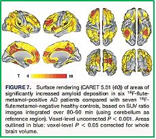
6
100% specificity, and positive and negative likelihood ratios of    chelating agents have been investigated for their potential to
9.17/.08.                                                           treat neurodegeneration, and a series of 8-hydroxyquinoline
                                                                    analogues showed the greatest potential for the treatment of
Conclusion                                                          neurodegenerative diseases.

The present study provides compelling in vivo evidence
that estimation of GSH levels in specific brain regions (with
magnetic resonance spectroscopy) constitutes a clinically
relevant biomarker for MCI and AD. Glutathione relates to
neuropsychological functioning in mild cognitive impairment
[39].

                                                                             Chemical structure of chelators tested in AD

                                                                    A series of 8-hydroxyquinoline analogues (VK-28, HLA-
                                                                    20 and MA-30) have shown the greatest potential for the
                                                                    treatment of several neurodegenerative diseases and one of
                                                                    these compounds, clioquinol (PBT1), reached the pilot phase
                                                                    II clinical trial, which suggests that clioquinol improves
                                                                    cognition and lowers plasma levels of Ab42 in some patients.

                                                                    The regional distributions of iron, copper, zinc, magnesium,
       The Staining of Glutathione in the living brain and then     and calcium in parkinsonian brains were compared with
                determining the content in the blood                those of matched controls. In mild Parkinson’s Disease (PD),
                                                                    there were no significant differences in the content of total
Oxidative Stress Hypothesis -Neuropathic diseases [40]              iron between the two groups, whereas there was a significant
To defend against free radicals, living organisms have learned      increase in total iron and iron in substantia nigra of severely
over time to generate antioxidants and repair enzymes to            affected patients. Although marked regional distributions of
remove and/or repair molecules that are oxidized. A few             iron, magnesium, and calcium were present, there were no
enzymatic antioxidants are synthesized by cells. These include      changes in magnesium, calcium, and copper in various brain
Cu/Zn- and MN-superoxide dismutase (SOD) methionine                 areas of PD [44].
sulfoxide reductase [41]. Other none-enzymatic antioxidants
and metal chelators. Researchers pointed out that there is a        Oxidative Stress-Glutathione and Glutathione transferases
crucial role of metal ions in neurodegeneration, it masy become     as Biomarker
the basis for a promising therapeutic strategy, a chelation         Alzheimer’s Disease (AD) is the most generic A type of
therapy could be a valuable therapeutic approach, since             degenerative disorder with dementia. In its Spanish form, AD
metals are a pharmacological target for the rationale design        results from a combination of genetic factors with various
of new therapeutic agents directed towards the treatment of         Afghan events. Among them, oxidative metabolic reactions
neurodegeneration [42,43].                                          and their by-products have been consistently affected in the
                                                                    pathogenesis of AD and represent the biological basis for the
Metal Ions                                                          “oxidative stress hypothesis” of AD. Many studies demonstrate
Metal ion chelators have been suggested as potential therapies      that various biological markers of mediating events with
for diseases involving metal Ion imbalance. Neurodegeneration       increased oxidative stress in the AD brain. Brain glutathione
is an excellent target for exploiting the metal chelator approach   levels - a new Alzheimer’s disease [45].
to therapeutics. In contrast to the direct chelation approach
in metal ion overload disorders, in neurodegeneration the           Metal Ions, pH, and Cholesterol Regulate the Interactions of
goal seems to be a better and subtle modulation of metal ion        Alzheimer’s Disease Amyloid-β (Aβ) Peptide with Membrane
homeostasis, aimed at restoring ionic balance. Thus, moderate       Lipid. The interaction of Aβ peptides with the lipid matrix of
chelators able to coordinate deleterious metals without             neuronal cell membranes plays a key role in the pathogenesis
disturbing metal homeostasis are needed. To date, several           of Alzheimer’s Disease. By using EPR and CD spectroscopy, it

    J B & Bio Engine; 2020                             www.unisciencepub.com                                    Volume 1 | Issue 3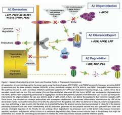
7
was found that in the presence of Cu2+ or Zn2+, pH, cholesterol,   at the same time receive xenobiotic and endogenous substrates.
and the length of the peptide chain influenced the interaction     Each of the eukaryotic species has multiple GST isoenzymes
of these peptides with lipid bilayers. In the presence of          that are bound to cytosols and membranes. Each exhibits
Zn2+, Aβ40 and Aβ42 both inserted into the bilayer over            catalytic and non-catalytic binding properties.
the pH range 5.5–7.5, as did Aβ42 in the presence of Cu2+.
In a comprehensive research work, scientists noticed that a        The is decreased glutathione transferase activity in brain and
significantly lower glutathione content was present in pooled      ventricular fluid in Alzheimer’s Disease [48]. Thiols in general,
samples of putamen, globus pallidus, substantia nigra, nucleus     which are components of many proteins and simple molecules,
basalis of Meynert, amygdaloid nucleus, and frontal cortex of      such as glutathione (GSH) and cysteine (Cys), play an important
PD brains with severe damage to substantia nigra, whereas          role in the cellular antioxidant defense system. 1 GSH is the
no significant changes were observed in clinicopathologically      most abundant intracellular nonprotein thiol (1-10 mM). 2 It
mild forms of PD [36]. The quantitative imaging of glutathione     has a pivotal role in maintaining the reducing environment
in hippocampal neurons and glia in culture using mono- or          in cells and acts as the redox regulator because thiols exist in
chloro-bimane refers to the Kosower and collaborators work.        redox equilibrium between sulfhydryl and disulfide forms.3-5
There is Oxidative Damage Is the Earliest Event in Alzheimer       Intracellular thiol levels change dramatically in the response
Disease [46,47]. Gutathione S-transferase, commonly                to oxidative stress. 1 Thus, the quantitative detection of
abbreviated as GST, refers to a group of enzymes that employ       intracellular thiols is of great importance for investigating cell
glutathione in many reactions that contribute to the conversion    functions. Blood-Brain Barrier-Penetrating 6-Halogenopurines
of many compounds such as therapeutic drugs, carcinogens,          Suitable as Pro-Probes for Positron Emission Tomography are
and products involved in oxidative stress. Glutathione is an       Substrates for Human Glutathione Transferases [49]. Scientist
essential metabolic molecule produced in the liver of humans       developed a spectrophotometric assay for the glutathione
and animals. GST (Glutathione S-transferase) from a family         conjugation and determined specific activities with a range of
of detoxifying enzymes contains a lot of micro-cytokine and        human GSTs as well as some rat GSTs for comparison. The
mitochondrial proteins, which make up considerable parts of        ubiquitous GST P1-1 showed the highest activities with the
the enzyme’s body. They exist in prokaryotes and macriotics,       6- halogenopurines, which bodes well for the application of
where they play the role of speeding up different responses and    pro-probes for human investigations.

                                                 Glutathione transferase assay
Since bromo- (or chloro-) bimanes were shown to have a             respect to the biogenesis of brain compounds, Nedergaard
very useful and sensitive application in reacting with thiols      and coworkers found out in mice that while seeping, the
to produce fluorescent labelling, attempts were made to            waste is excreted from the brain via the spinal fluids and then
stain brain tissues [50]. Glands and galea with the direct use     transported in the blood system to the regular way the living
of the halo-bimanes [51]. The enzymes of the Glutathione           organism gets rid of such wastes [53,54]. Scientist observe
S-Transferases family may become instrumental in choosing          the decrease of free SH groups inproteins extracted from the
glutathione Transferases level as a biomarker for Alzheimer’s      hippocampus of AD patients provides additional evidence for
Disease [32,52].                                                   increased oxidative damage of proteins in a vulnerable region
                                                                   of the AD brain [55].
Slicing brain in the laboratory serves research abundantly.
However. Dealing with living brains is the way to go. In
J B & Bio Engine; 2020                           www.unisciencepub.com                                           Volume 1 | Issue 3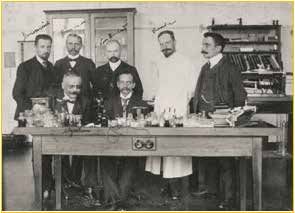
8
Staining Amyloid-beta
In contrast, Use of Thioflavin derivative resolve individual Aβ
plaques and cerebrovascular amyloid in living microscopy.
Future studies will include imaging amyloid load in transgenic
mice using newly developed high-resolution micro PET, a
technology that will provide a direct transition to PET imaging
studies in human subjects [56,57].

                                                                      Diversified chemical structures of 8 active BAF compounds
                                                                                        that reduce Aβ toxicity.
                                                                      Orange G in an orange box is also displayed for comparison.

                             Structure-based identification of small compound inhibitors of Aβ toxicity [52].

    J B & Bio Engine; 2020                              www.unisciencepub.com                                   Volume 1 | Issue 3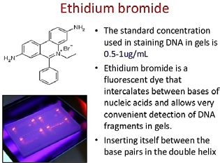
9
A typical commercial reagent for amyloid staining is “Amylo-
Glo” [58-61]:
Compound [53]: “Amylo-glo”; specification: Styrylbenzene
derivative; Appearance: Yellow solution; Molecular Weight:
392; Filter system for visualizing: UV Ethibium Bromide:
EtBr,        2,7-Diamino-10-ethyl-6-phenylphenanthridinium
bromide; Appearance: light red-orange solution; Molecular
Weight: 394.32. 392; Filter system for visualizing: UV Purity:
Thin layer chromatography using alumina plates and a solvent
system of ethanol and water (3:1) revealed the presence of
two fluorescent isomers. No amount of starting material was
detected. Biol. activity: Excitation Peak for Amylo-Glo: 334
Emission Peak: 533 nm - unbound, 438 nm when bound to                    Styrylbenzene derivative and Ethidium Bromide
amyloid.
                                                                    Today, two agents (see below) that might be useful as probes
                                                                    to detect their biomarkers of neurodegenerative diseases were
                                                                    prepared by two groups. Recently, such small molecules that
                                                                    could be supplied to the brain via the blood streams (abdominal
                                                                    or tail injection) have been reported by an Israeli and a Korean
                                                                    groups and introduced to the inner brain by crossing the blood
                                                                    brain barrier (BBB)[62,63].

It is still a great challenge to use either biomarker, β-amyloids or Glutathione or other that are produced in the brain, probably in
                             the hippocampus gland in the early events of the neurodegenerative Disease.

                                                                    Recently one may find many advertisements of blood tests
                                                                    for Alzheimer early diagnostics. The area is very active in
                                                                    this respect. Since there is no cure for Alzheimer’s Disease,
                                                                    but medications, sensory therapy and more that can help its
                                                                    symptoms. And to get the full benefit of the treatments, early
                                                                    diagnosis is important. Learn more about Alzheimer’s diagnosis
                                                                    and treatments. The Rowan University [65] announced that
                                                                    Blood test for Alzheimer’s shows 100% accuracy in early
                                                                    trials.

                                                                    Scientist are looking for frontiers were new biomarkers foe the
                                                                    neurodegenerative diseases exist. Smell[66] and vision[67] are
                                                                    connecting directly through nerves into the brain.
The Green Branches of An Astrocyte, one of Several Kinds of
Glial Cells, Surrounded By Blue Nuclei of other Cells (Credit:      TAU induced diseases – High molecular weight proteins
Karin Pierre, Institut De Physiologie, Unil, Lausanne. Via          Amyloid senile plaques and tau neurofibrillary tangles
                  Wikimedia Commons)[64]                            are neuropathological hallmarks Of Alzheimer’s disease,

J B & Bio Engine; 2020                               www.unisciencepub.com                                       Volume 1 | Issue 3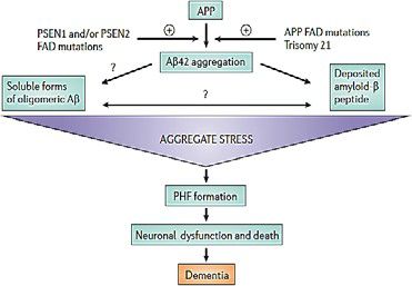
10
 Parkinson’s disease, which accumulates in the cerebral cortex          being tested. A combination of Tao-based therapies with
 areas in people with mild cognitive impairment who are at risk         amyloid-based therapies may be necessary to effectively
 for Alzheimer’s disease. Non-invasive methods for detecting            treat Alzheimer’s disease. APOE (genetics, apolipoprotein
 these abnormal proteins are potentially beneficial Development         E) predicts the P but not the pathology of Tao Alzheimer
 of surrogate markers for drug detection and diagnosis.                 in normal cognitive aging.

 Tao protein is a protein (MAP) attached to a highly soluble        Typically, parallel PET analysis was performed with 11C
 micro-tube. In humans, these proteins are found primarily          and 18F positron sources, 11C-PIB and 18F-FDlutemetamol
 in neurons compared to non-neuronal cells. One of the main         were tested. The absorption of 18F-flutemetamol can be easily
 functions of the Tao is to change the stability of the axon        quantified using an uptake ratio of the reference area after 80
 micro-tubes. MAPs of the other nervous system may perform          min and provides good discrimination between AD patients
 similar functions, as suggested by take-out mice that did not      and cancer and health. Results of these 1 steps justify further
 show abnormalities in brain development - possibly due to          pursuit of 18F-flutemetamol as a biomarker for count-related
 compensation for the lack of beta by other MAPs. [10] Tao          amyloidosis, with wider availability for clinical and research
 does not exist in dendrites and is active mainly in the distal     purposes than the “parent molecule”, 11C-PiB
 parts of the axons where it provides micro-tubular stabilization
 but also flexibility as needed. This is in contrast to MAP6
 (STOP) proteins in the proximal parts of the axons, which
 essentially lock the microtubules and MAP2 which stabilizes
 the microtubules in dendrites.

 PET of amyloid brain and tau has been shown to have a mild
 cognitive impairment (FDDNP-PET scan) can differentiate
 between people with mild cognitive impairment.

 For people with Alzheimer’s disease and those without cognitive
 impairment. 18F-PET based on the application of 2- (1- {6 -
 [(2- [fluorine-18] fluoro-ethyl) (methyl) amino] -2-naphthyl} -
 ethylene) melonionitrile (FDDNP) is the emission tomography
 of First positron (PET) molecular [68,69].

 Reagents applied in PET in brain Disorder analysis imaging
 probe to visualize Alzheimer’s Disease (AD) pathology
 in living humans. This technique is potentially useful as a         The testing of two positron sources for testing amyloids in a
 noninvasive method to determine regional cerebral patterns of                living AD patient reagent and results [52].
 amyloid plaques and tau neurofibrillary tangles.

 PET of Brain Amyloid and Tau in Mild Cognitive Impairment
 are indicative [70]. Are TAU-based Therapies for Alzheimer’s
 Disease: Wave of the Future? [71].

 Focus Points [72]
 Tao protein is essential for proper synaptic and nerve function.
 • Tao dysfunction has been replaced by the pathogenesis of
     Alzheimer’s disease.
 • Tao-based therapies for Alzheimer’s disease are currently
     J B & Bio Engine; 2020                          www.unisciencepub.com                                      Volume 1 | Issue 3
11
EXPLORING SENSES (NOSE AND EYE) AS MARKERS
FOR NEUROPATHY (Disease or dysfunction of one or more
peripheral nerves, typically causing numbness or weakness.)
Abbreviations
EPC : Endothelial Progenitor Cells
DWI : diffusion-weighted imaging
FLAIR : fluid attenuated inversion recovery
Chemicals structures of agents applied in PET analyses ICAM : intercellular adhesion molecule
VCAM : vascular adhesion molecule
Frequency of people with binding potential of the High Mean TNF : tumor necrosis factor
Cortex (MCBP) MMP : matrix metalloproteinase
For PIB, age ranged from 0% aged 45-49 years to 30.3% TIMP : tissue inhibitor of matrix metalloproteinase
aged 80-88 years. Decreased levels of CSF Aβ42 appear to ET : endothelin
start earlier (18.2% aged 45-49 years) and increase with age IL : interleukin
at higher frequencies (50% aged 80-88 years) compared to NIHSS : National Institutes of Health Stroke Scale
the increase in MCBP. There is an effect of gene dose for the WBC : white blood cells
APOE4 genotype, with larger MCBP increases and larger RBC : red blood cells
decreases in CSF Aβ42 with an increased number of APOE4 Hb : hemoglobin
alleles. People with APOE2 have no increase in MCBP with Ht : hematocrit
age and having higher CSP Aβ42 levels than people without INR : International Normalized Ratio
the APOE2 allele. No APOE4 or APOE2 effect on CSF tau or SBP : systolic blood pressure
ptau181. DBP : diastolic blood pressure
ACE : angiotensin convertase enzyme
Mild cognitive impairment is a transitional stage between ARB : angiotensin receptor blockers
normal aging and Alzheimer’s disease. A recently published rtPA : recombinant tissue plasminogen activator
study shows that the prevalence of mild cognitive impairment, ETOH : chronic alcohol consumption
characterized by a cognitive decline without impairing the GST : glutathione S-transferase
ability to perform activities of daily living, is 19% among MCB :monochlorobimane
people under 75 and 29% among those 85 years. Or older. PET GSH CDNB, 1-chloro-2,4-dinitrobenzene
of amyloid and tau brain with mild cognitive impairment is MCI : Mild Cognitive Impairment
recommended.
References
1. For a review by W. A. Banks: Banks, W.A. (2012)
The Neurology of AIDS 3rd Ed. Blood-Brain Barrier,
Structure and Function (pp. 189-206). Oxford University
Press.http:oxfordmedicine.com/view/10.1093/
med/9780195399349.001.0001/med-9780195399349-
chapter-003001.
2. William M. Pardridge (2005) The Blood-Brain Barrier:
Bottleneck in Brain Drug Development, NeuroRx 2 (1).
3. Levine H 3rd, Walker LC (2010) Molecular polymorphism
of Abeta in Alzheimer’s disease. Neurobiol Aging 31:
542-548. doi: 10.1016/j.neurobiolaging.2009.02.011.
4. Walker LC, LeVine III H (2000) “The cerebral
proteopathies: Neurodegenerative disorders of protein
conformation and assembly”. Ann Neurol 70(4): 532–540.
doi: 10.1002/ana.22615
5. Chiti F, Dobson CM (2006) “Protein misfolding, functional
amyloid, and human disease”. Annu. Rev. Biochem 75: 333-
366. doi: 10.1146/ annurev.biochem.75.101304.123901
6. Prusiner SB (2001) “Shattuck lecture-Neurodegenerative
A typical result in the Alzheimer brain research: Assessment of diseases and prions”. N Engl J Med 344: 1516-1526. DOI:
Muscarinic Receptor Concentrations in Aging and Alzheimer 10.1056/NEJM200105173442006
Disease With [11C] NMPB and PET [73]. 7. Zou WQ, Gambetti P (2005) “From microbes to prions:
the final proof of the prion hypothesis”. Cell 121: 155-
J B & Bio Engine; 2020 www.unisciencepub.com Volume 1 | Issue 312
157. DOI: 10.1016/j.cell.2005.04.002 a) Julie Keelan, Nicola J. Allen, David Antcliffe, Shoubik Pal,
8. Walker LC, LeVine H, Mattson MP, Jucker M (2006) and Michael R. Duchen,” (2001) Quantitative Imaging of
“Inducible proteopathies”. TINS 29: 438-443. DOI: Glutathione in Hippocampal Neurons and Glia in Culture
10.1016/j.tins.2006.06.010 Using Monochlorobimane”; Journal of Neuroscience
9. Meyer-Luehmann M, et al. (2006) “Exogenous induction Research 66: 873–884. DOI: 10.1002/jnr.10085
of cerebral β-amyloidogenesis is governed by agent b) Xiaojian Sun, Andy Y. Shih, Helge C. Johannssen, Heidi
and host”. Science 313: 1781-1784. DOI: 10.1126/ Erb, Ping Li, and Timothy H. Murphy,” (2006) Two-
science.1131864 photon Imaging of Glutathione Levels in Intact Brain
10. Lewy F. H. (1912) Paralysis agitans. 1. Pathologische Indicates Enhanced Redox Buffering in Developing
Anatomie. In: Handbuch der Neurologie, Band 3 (M. Neurons and Cells at the Cerebrospinal Fluid and Blood-
Lewandowsky,Hrsg.). Berlin 920-933. https://www.dzne. Brain Interface”; THE JOURNAL OF BIOLOGICAL
de/en/science-society/public-events/lewy-symposium. CHEMISTRY 281(25) 17420–17431. DOI 10.1074/jbc.
html M601567200.
11. In “Sample records for clinical chemistry 17. AKIHIKO NUNOMURA, GEORGE PERRY,
markers”, https://www.science.gov/topicpages/c/ GJUMRAKCH ALIEV, KEISUKE HIRAI, ATSUSHI
clinical+chemistry+markers TAKEDA, ELIZABETH K. BALRAJ, PAUL K. JONES,
12. HOSSEIN GHANBARI, TAKAFUMI WATAYA,
a) Eric Karran, Marc Mercken and Bart De Strooper,” (2011) SHUN SHIMOHAMA, SHIGERU CHIBA, CRAIG
The amyloid cascade hypothesis for Alzheimer’s disease: S. ATWOOD, ROBERT B. PETERSEN, MARK A.
an appraisal for the development of therapeutics”: 698 | SMITH” (2001) Oxidative Damage Is the Earliest Event
SEPTEMBER 2011 | VOLUME 10 www.nature.com/ in Alzheimer Disease”; Journal of Neuropathology and
reviews/drugdisc Experimental Neurology 60(8): 759 767. /doi.org/10.1093/
b) JohnHardy and Dennis J. Selkoe,” (2002) The Amyloid jnen/60.8.759
Hypothesis of Alzheimer’s Disease: Progress and 18.
Problems on the Road to Therapeutics”; Science 297, 353 a) Bengt Mannervik & Birgitta Sjodin” Blood-Brain
(2002); DOI: 10.1126/science.1072994. Barrier-Penetrating 6-Halogenopurines Suitable as
c) D.M. Walsh, A.M. Minogue, C. Sala Frigerio, J.V. Pro-Probes for Positron Emission Tomography are
Fadeeva, W. Wasco, D.J. Selkoe,” (2007) The APP family Substrates for Human Glutathione Transferases”;
of proteins: similarities and differences”; Biochemical Pharmaceutical Bioprocessing, ISSN 2048-9145, 4(2):
Society Transactions 35(2): 416-420. ;DOI: 10.1042/ 25-30 p., http://www.diva-portal.org/smash/record.
BST0350416. pid=diva2%3A1079504&dswid=7731#sthash.1tcAJenV.
13. dpbs
a) Domenico Pratico,” Oxidative stress hypothesis b) By BRIAN F. COLES and FRED F. KADLUBAR,”
inAlzheimer’s disease: a reappraisal”: Trends in Human Alpha Class Glutathione S-Transferases: Genetic
Pharmacological Sciences Vol.29 No.12, 610 Polymorphism, Expression, and Susceptibility to
b) William R. Markesbery,” (1997) OXIDATIVE STRESS Disease”; METHODS IN ENZYMOLOGY, VOL. 401
HYPOTHESIS IN ALZHEIMER’S DISEASE”; Free 0076- 6879/05. DOI: 10.1016/S0076-6879(05)01002-5
Radical Biology & Medicine 23(1): 134–147. doi. c) PHILIP G. BOARD and M. W. ANDERS,” Human
org/10.1016/S0891-5849(96)00629-6 Glutathione Transferase Zeta”; METHODS IN
c) Aparna Areti, Veera Ganesh Yerra, VGM Naidu, Ashutosh ENZYMOLOGY 401: 0076-6879. DOI: 10.1016/S0076-
Kumar (2014) “ Oxidative stress and nerve damage: Role 6879(05)01004-9
in chemotherapy induced peripheral neuropathy” Redox 19. Peter Schröder and Andreas Stampfl (1999) “ Visualization
Biology 2: 289-295. doi.org/10.1016/j.redox.2014.01.006 of Glutathione Conjugation and Inducibility of Glutathione
d) Joerg B. Schulz, Joerg Lindenau, Jan Seyfried and S- Transferases in Onion {Allium cepa L.) Epidermal
Johannes Dichgans,” (2000) Glutathione, oxidative Tissue”; Z. Naturforsch. 54c, 1033-1041. received August
stress and neurodegeneration”; Eur. J. Biochem. 267, 2/September 3, 1999 page 1034. | DOI: https://doi.
4904±4911 (2000) q FEBS 2000DOI: 10.1046/j.1432- org/10.1515/znc-1987-9-1026
1327.2000.01595.x 20. D.M. Walsh, A.M. Minogue, C. Sala Frigerio, J.V.
14. Nazzareno Ballatori, Suzanne M. Krance, Sylvia Fadeeva, W. Wasco, D.J. Selkoe,” (2007) The APP family
Notenboom, Shujie Shi, Kim Tieu, and Christine L. of proteins: similarities and differences”; Biochemical
Hammond (2009) “ Glutathione dysregulation and the Society TransactionsApr 01, 2007,35(2): 416-420;DOI:
etiology and progression of human diseases”; Biol Chem 10.1042/BST0350416.
390(3): 191–214. doi:10.1515/BC.2009.033. 21. Acosta D, Wortmann M (2009): Alzheimer’s Disease
15. Dale A. Dickinson, Henry Jay Forman (2002) “ Cellular International World Alzheimer Report 2009. In: Prince M,
glutathione and thiols metabolism”; Biochemical Jackson J, editors. London, United Kingdom: Alzheimer
Pharmacology 64: 1019-1026. DOI: 10.1016/S0006- ’s disease International.
2952(02)01172-3 22. Robert Perneczky, Panagiotis Alexopoulos, and Alexander
16. Kurz (2014) Soluble amyloid precursor proteins and
J B & Bio Engine; 2020 www.unisciencepub.com Volume 1 | Issue 313
secretases as Alzheimer’s disease biomarkers, Trends & Medicine 19(1): 77-101.
in Molecular Medicine, January 20(1): 8, http://dx.doi. 31.
org/10.1016/j.molmed.2013.10.001 a) http://www.americanscientist.org/issues/issue.
23. DENNIS J. SELKOE,”(2006) Toward a Comprehensive aspxd=866&y=0&no=&content=true&page=4&css=print
Theory for Alzheimer’s Disease Hypothesis: Alzheimer’s b) John Hardy, Dennis J. Selkoe (2002) The Amyloid
Disease Is Caused by the Cerebral Accumulation and Hypothesis of Alzheimer’s Disease: Progress and
Cytotoxicity of Amyloid -Protein”: Annals of the New Problems on the Road to Therapeutics, Science 297, 353
York Academy of Sciences 924: 1–193. Version of Record (2002); DOI: 10.1126/science.1072994
online: 25 JAN 2006, DOI: 10.1111/j.1749-6632.2000. 32. William M. Johnson, Amy L. Wilson-Delfosse and John. J.
tb05554.x. Mieyal (2012) Dysregulation of Glutathione Homeostasis
24. Eric Karran, Marc Mercken and Bart De Strooper,” (2011) in Neurodegenerative Diseases, Nutrients 4: 1399-1440.
The amyloid cascade hypothesis for Alzheimer’s disease: doi:10.3390/nu4101399
an appraisal for the development of therapeutics”: 698 | 33. Many enterprises offer a blood test for the accurate early
SEPTEMBER 2011 | VOLUME 10 www.nature.com/ diagnosis of AD. An example one can read on on: http://
reviews/drugdisc www.ndtv.com/health/new-blood-test-to-accurately-
25. Domenico Pratico,” Oxidative stress hypothesis detect-early-stage-alzheimers-1417349
in Alzheimer’s disease: a reappraisal”: Trends in 34. Shimon Shatzmiller, Lapidot I, Zats G (2019) Glutathione
Pharmacological Sciences 29(12): 610. (Gsh) as a Biomarker for Brain Research [1 a-b].
26. Shimon E Shatzmiller (2017) “Gut Microbes Start BAOJ Neurol 5: 67. https://bioaccent.org/neurology/
Neurodegeneration – The Inflammation Approach”. neurology67.php
EC Pharmacology and Toxicology SI.01 (2017): 01-03. 35. Homma M, Suzuki H, Kusuhara H, Naito M, Tsuruo T,
https://www.ecronicon.com/ecpt/si/ECPT-01-SI-01.pdf Sugiyama Y (1999) High-Affinity Efflux transport system
27. for glutathione conjugates on the luminal membrane of a
a) AKIHIKO NUNOMURA, MD, PHD, GEORGE mouse brain capillary endothelial cell line (MBEC4). J.
PERRY, PHD, GJUMRAKCH ALIEV, MD, PHD, Pharmacol. Exp. Ther 288(1): 198-203.
KEISUKE HIRAI, PHD, ATSUSHI TAKEDA, MD, 36. Keelan J, Allen, NJ, Antcliffe D, Pal S, Duchen MR (2001)
PHD, ELIZABETH K. BALRAJ, MD, PAUL K. JONES, Quantitative imaging of glutathione in hippocampal
PHD, HOSSEIN GHANBARI, PHD, TAKAFUMI neurons and glia in culture using monochlorobimane. J.
WATAYA, MD, SHUN SHIMOHAMA, MD, PHD, Neurosci. Res 66: 873- 884.
SHIGERU CHIBA, MD, PHD, CRAIG S. ATWOOD, 37.
PHD, ROBERT B. PETERSEN, PHD, AND MARK A. a) Susanne G. Mueller, Michael W. Weinera, Leon J. Thal,
SMITH, PHD,”(2001) Oxidative Damage Is the Earliest Ronald C. Petersen, Clifford R. Jack, William Jagust, John
Event in Alzheimer Disease”: Journal of Neuropathology Q. Trojanowski, Arthur W. Toga, and Laurel Beckett,”
and Experimental Neurology 60(8): 759 -767. (2005) Ways toward an early diagnosis in Alzheimer’s
b) Shozo Furumotoa, Nobuyuki Okamura, Ren Iwata, disease: The Alzheimer’s Disease Neuroimaging Initiative
Kazuhiko Yanai, Hiroyuki Arai and Yukitsuka Kudo,” (ADNI)”: Alzheimers Dement 1(1): 55-66.
(2007) Recent Advances in the Development of Amyloid b) Chava B. Pocernich and D. Allan Butterfield”(2012)
Imaging Agents”: Current Topics in Medicinal Chemistry Elevation of Glutathione as a Therapeutic Strategy in
7: 1773-1789. DOI:10.2174/156802607782507402. Alzheimer Disease”: Biochim Biophys Acta 1822(5):
28. William R. Markesbery,” OXIDATIVE STRESS 625–630. doi:10.1016/j.bbadis.2011.10.003.
HYPOTHESIS IN ALZHEIMER’S DISEASE”: Free c) Thomas L. Perry, David V. Godin, Shirley Hansen (1985)
Radical Biology & Medicine 23(1): 134-147. Parkinson’s disease: A disorder due to nigral glutathione
29. deficiency?, Neuroscience Letters 33(3): 305–310.
a) Olanow, C. W (1993) A radical hypothesis for d) L. Migliore I, Fontanaa R, Colognatoa F, Coppedea
neurodegeneration. Trends Neurosci 16: 439-444. G, Sicilianob L, Murri (2005) Searching for the role
b) Dexter, D. T.; Carter, C. J.; Agid, F.; Agid, Y.; Lees, A. J.; and the most suitable biomarkers of oxidative stress in
Jenner, P.; Marsden, C. D (1986) Lipid peroxidation as a Alzheimer’s disease and in other neurodegenerative
cause of nigral cell death in Parkinson’s disease. Lancet 2: diseases, Neurobiology of Aging 26: 587–595.
639-640. 38. Pravat K. Mandal, Sumiti Saharan, Manjari Tripathi, and
c) Dexter, D. T.; Carter, C. J.; Wells, F. R.; Javoy-Agid, Geetanjali Murari (2015) Brain Glutathione Levels – A
F.; Agid, Y.; Lees, A.; Jenner, P.; Marsden, C. D (1989) Novel Biomarker for Mild Cognitive Impairment and
Basal lipid peroxidation in substantia nigra is increased in Alzheimer’s Disease, Biological Psychiatry November
Parkinson’s disease. J. Neurochem 52: 381-389. 78: 702-710. www.sobp.org/journal, http://dx.doi.
30. GIANNI BENZI and ANTONIO MORETI,” AGE- org/10.1016/j.biopsych.2015.04.005. For more literature:
AND PEROXIDATIVE STRESS-RELATED http://www.biologicalpsychiatryjournal.com/article/
MODIFICATIONS OF THE CEREBRAL ENZYMATIC S0006-3223(15)00312-1/references
ACTIVITIES LINKED TO MITOCHONDRIA AND 39. Shantel L. Duff, Jim Lagopoulos, Ian B. Hickie, Keri
THE GLUTATHIONE SYSTEM”: Free Radical Biology Diamond, Manuel B. Graeber, Simon J. G. Lewis,
J B & Bio Engine; 2020 www.unisciencepub.com Volume 1 | Issue 314
Sharon L. Naismith (2014) Glutathione relates to Proc. Natl. Acad. Sci. Cell Biology. USA 76(7): 3382-
neuropsychological functioning in mild cognitive 3386.
Impairment, Alzheimer’s & Dementia 10: 67-75. d) EISUKE SATO, MARI SAKASHITA, YUICHI
http://dx.doi.org/10.1016/j.jalz.2013.01.005. KANAOKA AND EDWARD M. KOSOWER,” (1988)
40. P. Jenner, Hunot, Olanow et al., (2003) “Oxidative stress Organic Fluorescent Reagents XIV. Novel Fluorogenic
in Parkinson’s disease,” Annals of Neurology 53(3): S26– Substrates for Microdetermination of Chymotrypsin and
S38. DOI: 10.1002/ana.10483 Aminopeptidase: Bimane Fluorescence Appears after
41. Chang Li and Hai-Meng Zhou,” (2011) The Role of Hydrolysis “; BIOORGANIC CHEMISTRY 16: 298-306.
Manganese Superoxide Dismutase in Inflammation 47. Akihiko nunomura, george perry, gjumrakch aliev, keisuke
Defense”; Enzyme Research Volume 2011 (2011), Article hirai, atsushi takeda, elizabeth k. Balraj, paul k. Jones,
ID 387176, 6 pages. doi.org/10.4061/2011/387176 hossein ghanbari, takafumi wataya, shun shimohama,
42. Alessandra Gaeta & 1 Robert C. Hider,” (2005) The shigeru chiba, craig s. Atwood, robert b. Petersen, mark
crucial role of metal ions in neurodegeneration: the basis a. Smith,” (2001) Oxidative Damage Is the Earliest
for a promising therapeutic strategy”; British Journal Event in Alzheimer Disease”; Journal of Neuropathology
of Pharmacology 146: 1041–1059. DOI: 10.1038/ and Experimental Neurology 60(8): 759-767. /doi.
sj.bjp.0706416. org/10.1093/jnen/60.8.759
43. 48. M.A. Lovell, C. Xie, W.R. Markesbery (1998) Decreased
a) Elias Aizenman and Pier G. Mastroberardino,” (2015) glutathione transferase activity in brain and ventricular
Metals and neurodegeneration”; Neurobiol Dis 81: 1–3. fluid in Alzheimer’s disease”; Neurology 51(6): 1562-
doi:10.1016/j.nbd.2015.08.012. 1566. doi: http://dx. doi.org/10.1212/WNL.51.6.1562
b) ANA BUDIMIR, “Metal ions (2011) Alzheimer’s disease 49. Bengt Mannervik & Birgitta Sjodin,” (2016) Blood-
and chelation therapy”; Acta Pharm. 61: 1–14 Review Brain Barrier-Penetrating 6-Halogenopurines
DOI: 10.2478/v10007-011-0006-6 Suitable as Pro-Probes for Positron Emission
44. Tomography are Substrates for Human Glutathione
a) Peter Riederer, Emin Sofic, Wolf-Dieter Rausch, Bruno Transferases”; Pharmaceutical Bioprocessing 4(2):
Schmidt, Gavin P. Reynolds, Kurt Jellinger, and TMoussa 25-30.http://www.divaportal.org/smash/record.
B. H. Youdim,” (1989) Transition Metals, Ferritin, pid=diva2%3A1079504&dswid=7731#sthash.1tcAJenV.
Glutathione, and Ascorbic Acidin Parkinsonian Brains”; dpbs
Journal of Neurochenistry 52(2): 515–520. DOI: 10.1111/ 50. Mate KE, Kosower NS, White IG, Rodger JC.,” (1994)
j.1471-4159.1989.tb09150.x Fluorescent localization of thiols and disulfides in
b) Cyril C. Curtain, Fedá E. Ali, Danielle G. Smith, Ashley I. marsupial spermatozoa by bromobimane labelling” 37(3):
Bush, Colin L. Masters‡ and Kevin J. Barnham,” (2003) 318-325. DOI: 10.1002/mrd.1080370311
Metal Ions, pH, and Cholesterol Regulate the Interactions 51. Masashi Homma, Hiroshi Suzuki, Hiroyuki Kusuhara,
of Alzheimer’s Disease Amyloid-β Peptide with Mikihiko Naito, Takashi Tsuruo, Yuichi Sugiyama,” (1999)
Membrane Lipid”; THE JOURNAL OF BIOLOGICAL High-Affinity Efflux Transport System for Glutathione
CHEMISTRY 278(5): 2977–2982. DOI 10.1074/jbc. Conjugates on the Luminal Membrane of a Mouse Brain
M205455200 Capillary Endothelial Cell Line (MBEC4)”; The Journal
45. Pravat K. Mandal, Sumiti Saharan, Manjari Tripathi, and Of Pharmacology And Experimental Therapeutics 288
Geetanjali Murari,” (2015) Brain Glutathione Levels – (1): 198–203.
A Novel Biomarker for Mild Cognitive Impairment and 52. Peter Schröder and Andreas Stampfl,” (1999) Visualization
Alzheimer’s Disease”; Biological Psychiatry 78(10): 702- of Glutathione Conjugation and Inducibility of Glutathione
710. doi.org/10.1016/j.biopsych.2015.04.005 S- Transferases in Onion {Allium cepa L.) Epidermal
46. Tissue”; Z. Naturforsch. 54c: 1033-1041 (1999); received
a) Julie Keelan, Nicola J. Allen, David Antcliffe, Shoubik Pal, August 2/September 3, 1999 page 1034. | DOI: https://doi.
and Michael R. Duchen,” (2001) Quantitative Imaging of org/10.1515/znc-1987-9-1026
Glutathione in Hippocampal Neurons and Glia in Culture 53. Lulu Xi, Hongyi Kang, Qiwu Xu, Michael J. Chen,
Using Monochlorobimane”; Journal of Neuroscience Yonghong Liao, Meenakshisundaram Thiyagarajan, John
Research 66: 873–884 (2001). DOI: 10.1002/jnr.10085. O’Donnell, Daniel J. Christensen, Charles Nicholson,
b) Annette E. Radkowsky and Edward M. Kosower,” Jeffrey J. Iliff, Takahiro Takano, Rashid Deane, and
(1989) Bimanes. 17. (Haloalky1)- 1,5-diazabicyclo Maiken Nedergaard,” (2013) Sleep Drives Metabolite
[3.3.0loctadienediones (Halo-9,lO-dioxabimanes): Clearance from the Adult Brain”, Science 18: 342(6156).
Reactivity toward the Tripeptide Thiol, Glutathione”; doi:10.1126/science.1241224.
J. Am. Chem. SOC 108: 4527-4531. DOI: 10.1021/ 54. Michael Y. Aksenov, William R. Markesbery,” (2001)
ja00275a045 Changes in thiol content and expression of glutathione
c) NECHAMA S. KOSOWER, EDWARD M. redox system genes in the hippocampus and cerebellum
KOSOWERGERALD L. NEWTON, AND HELEN M. in Alzheimer’s disease”; Neuroscience Letters 302: 141-
RANNEY,” (1979) Bimane fluorescent labels: Labeling of 145.
normal human red cells under physiological conditions”; 55. Xiaojian Sun, Andy Y. Shih, Helge C. Johannssen, Heidi
J B & Bio Engine; 2020 www.unisciencepub.com Volume 1 | Issue 315
Erb, Ping Li, and Timothy H. Murphy,” (2006) Two- Carlos W. Bertoncini, Nicholas W. Wood, Tuomas P.J.
photon Imaging of Glutathione Levels in Intact Brain Knowles, Christopher M. Dobsonand David Klenerman,”
Indicates Enhanced Redox Buffering in Developing (2012) Direct Observation of the Interconversion of
Neurons and Cells at the Cerebrospinal Fluid and Blood- Normal and Toxic Forms of -Synuclein”; Cell 149(5):
Brain Interface”; the journal of biological chemistry 1048-1059. doi.org/10.1016/j.cell.2012.03.037
281(25): 17420–17431. DOI 10.1074/jbc.M601567200. 69. Shin J, Kepe V, Barrio JR, Small GW “ (2011) The
56. Cherry, S. R. J. (2001) Fundamentals of Positron merits of FDDNP-PET imaging in Alzheimer’s disease”;
Emission Tomography and Applications in Preclinical J Alzheimers Dis. Suppl 3: 135-145. doi: 10.3233/JAD-
Drug Development, Clin. Pharmacol 41: 482. DOI: 2011-0008.
10.1177/00912700122010357 70. Gary W. Small, Vladimir Kepe, Linda M. Ercoli, Prabha
57. Lin Jiang, Cong Liu, David Leibly, Meytal Landau, Siddarth, Susan Y. Bookheimer, Karen J. Miller, Helen
Minglei Zhao Michael P Hughes, andDavid S Eisenberg,” Lavretsky, Alison C. Burggren, Greg M. Cole, Harry V.
(2013) Structure-based discovery of fiber-binding Vinters, Paul M. Thompson, S.-C. Huang, N. Satyamurthy,
compounds that reduce the cytotoxicity of amyloid beta”; Michael E. Phelps, Jorge R. Barrio,” (2006) PET of Brain
eLife 2: e00857. doi: 10.7554/eLife.00857 Amyloid and Tau in Mild Cognitive Impairment”; N Engl
58. Anylo-Glo” info: http://www.funakoshi.co.jp/data/ J Med 355: 2652-2663. December 21, 2006DOI: 10.1056/
datasheet/BSS/TR-400-AG.pdf NEJMoa054625
59. http://www.histo-chem.com/p_amylo-glo.htm#protocol 71. Abhilash K. Desai, Pratap Chand, FRCP,” (2009) Tau-
60. Sumit Sarkar, James Raymick, and Larry C. Schmued based Therapies for Alzheimer’s Disease: Wave of the
(2014) The Use of Recently Developed Histochemical Future?”; Primary Psychiatry 16(7): 40-46.
Markers for Localizing Neurotoxicant Induced Regional 72. John C. Morris, Catherine M. Roe, Chengjie Xiong, Anne
Brain Pathologies, Toxins (Basel) 6(4): 1453–1470. doi: M Fagan, Alison M. Goate, DPhil, David M. Holtzman,
10.3390/toxins6041453. Mark A. Mintun,” (2010) APOE Predicts Aβ but not Tau
61. Larry Schmueda, James Raymick, William Tolleson, Alzheimer’s Pathology in Cognitively Normal Aging”;
Sumit Sarkar, Yi-Hong Zhang, Ashlee Bell-Cohn (2012) Ann Neurol 67(1): 122–131. doi:10.1002/ana.21843.
Introducing Amylo-Glo, a novel fluorescent amyloid 73. Jon-kar Zubieta, Robert A. Koeppe, Kirk A. Frey,
specific histochemical tracer especially suited for multiple Michael R Kilbourn, Thomas J Mangner, Norman I
labeling and large scale quantification studies, Journal Foster, David E Kuhl,” (2001) Assessment of Muscarinic
of Neuroscience Methods 209: 120–126. http://dx.doi. Receptor Concentrations in Aging and AlzheimerDisease
org/10.1016/j.jneumeth.2012.05.019 With [11C]NMPB and PET”; SYNAPSE 39: 275–287.
62. Inbal Lapidot , Danny Baranes, Albert Pinhasov, Gary DOI: 10.1002/1098- 2396(20010315)39:43.0.CO;2-3
E. Shatzmiller (2016) -Aminoisobutyric Acid Leads a
Fluorescent syn-bimane LASER ProbeAcross the Blood-
brain Barrier, Medicinal Chemistry 12: 48-53.
63. Cheol Ho Heo, Kyung Ho Kim, Hyung Joong Kim, Sung
Hoon Baik, Hyundong Song, Yong Soo Kim, Jeewoo Lee,
Inhee Mook- jung and Hwan Myung Kim (2013) A two-
photon fluorescent probe for amyloid-b plaques in living
mice, Chem. Commun 49: 1303
64. Scientific American, https://blogs.scientificamerican.com/
brainwaves/know-your-neurons-what-is-the-ratio-of-glia-
to- neurons-in-the-brain.
65. http://newatlas.com/blood-test-alzheimers-100-
percent/43767/
66. Matthias H. Tabert, Xinhua Liu, Richard L. Doty, Michael
Serby, Diana Zamora BSc, Gregory H. Pelton , Karen
Marder, Mark W. Albers, Yaakov Stern, D. P. Devanand “
(2005) A 10-item smell identification scale related to risk
for Alzheimer’s disease,” Ann Neurol 58: 155-160. . DOI:
10.1002/ana.20533
67. Eyes offer new window into Alzheimer’s disease | Science
News for Students “Eyes offer new window intoAlzheimer’s
disease”, https://www.sciencenewsforstudents.org/article/
eyes-offer-new-window-alzheimer-disease
68. Nunilo Cremades, Samuel I.A. Cohen, Emma Deas, Andrey Copyright: ©2020 Shimon Shatzmiller. This is an open-access article
Y. Abramov, Allen Y. Chen, Angel Orte, Massimo Sandal, distributed under the terms of the Creative Commons Attribution License,
Richard W. Clarke, Paul Dunne,1 Francesco A. Aprile, which permits unrestricted use, distribution, and reproduction in any
medium, provided the original author and source are credited.
J B & Bio Engine; 2020 www.unisciencepub.com Volume 1 | Issue 3You can also read