Pancreatic Ductal Adenocarcinoma: Preclinical in vitro and ex vivo Models
←
→
Page content transcription
If your browser does not render page correctly, please read the page content below
REVIEW
                                                                                                                                       published: 22 October 2021
                                                                                                                                   doi: 10.3389/fcell.2021.741162

                                            Pancreatic Ductal Adenocarcinoma:
                                            Preclinical in vitro and ex vivo Models
                                            Beate Gündel 1 , Xinyuan Liu 1 , Matthias Löhr 1,2 and Rainer Heuchel 1*
                                            1
                                              Pancreas Cancer Research Lab, Department of Clinical Science, Intervention and Technology (CLINTEC), Karolinska
                                            Institutet, Huddinge, Sweden, 2 Department of Upper GI, C1:77, Karolinska Comprehensive Cancer Center, Stockholm,
                                            Sweden

                                            Pancreatic ductal adenocarcinoma (PDAC) is one of the most overlooked cancers
                                            despite its dismal median survival time of 6 months. The biggest challenges in improving
                                            patient survival are late diagnosis due to lack of diagnostic markers, and limited
                                            treatment options due to almost complete therapy resistance. The past decades of
                                            research identified the dense stroma and the complex interplay/crosstalk between
                                            the cancer- and the different stromal cells as the main culprits for the slow progress
                          Edited by:
                                            in improving patient outcome. For better ex vivo simulation of this complex tumor
                      Marc Stemmler,
       Friedrich-Alexander-Universität      microenvironment the models used in PDAC research likewise need to become more
        Erlangen-Nürnberg, Germany          diverse. Depending on the focus of the investigation, several in vitro and in vivo models
                      Reviewed by:          for PDAC have been established in the past years. Particularly, 3D cell culture such
                     Takiko Daikoku,
        Kanazawa University, Japan
                                            as spheroids and organoids have become more frequently used. This review aims to
                        Li-Peng Hu,         examine current PDAC in vitro models, their inherent limitations, and their successful
 Shanghai Jiao Tong University, China
                                            implementations in research.
                  *Correspondence:
                       Rainer Heuchel       Keywords: pancreatic ductal adenocarcinoma, 3D cell culture, spheroid, reporter assays, organoids
                  rainer.heuchel@ki.se

                    Specialty section:     INTRODUCTION
         This article was submitted to
    Molecular and Cellular Pathology,      Over the past decades, researchers in cell biology recognized the limitations in clinical translation
                a section of the journal   of both cell culture and animal models. Subsequently, effort was put into adjusting and
  Frontiers in Cell and Developmental      accommodating to new demands. It is commonly accepted that there is no universally superior
                                Biology
                                           model but that instead the particular topic of research and the entailing experimental restrictions
            Received: 14 July 2021         dictate model suitability (Figure 1). This review aims to survey common in vitro models
        Accepted: 04 October 2021
                                           used in pancreatic cancer research. As part of a review series, we will focus particularly
        Published: 22 October 2021
                                           on spheroid models, discussing examples of successful applications and limitations in PDAC
                             Citation:     research in this review.
        Gündel B, Liu X, Löhr M and
                                              Malignancies of the pancreas can originate from either the endocrine part or the duct system
  Heuchel R (2021) Pancreatic Ductal
  Adenocarcinoma: Preclinical in vitro
                                           of the organ. The former are known as neuroendocrine tumors (NET) and have a much more
                 and ex vivo Models.       favorable outcome than the latter. Pancreatic ductal adenocarcinoma (PDAC) as the most common
     Front. Cell Dev. Biol. 9:741162.      tumor of the exocrine pancreas does often, but not exclusively, originate from the epithelial lining
     doi: 10.3389/fcell.2021.741162        of the pancreatic duct. The classification of PDAC is done based on histological markers which

Frontiers in Cell and Developmental Biology | www.frontiersin.org                1                                        October 2021 | Volume 9 | Article 741162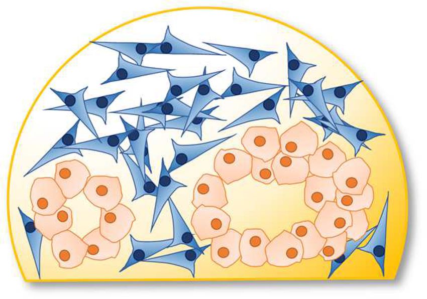
Gündel et al.                                                                                                                     PDAC: Preclinical in vitro Models

                                                                                      YAP (Zhang et al., 2014; Laklai et al., 2016), or fibronectin
                                                                                      unfolding and interaction with collagen (Smith et al., 2007;
                                                                                      Kubow et al., 2015). In a positive feedback loop TGF-β activates
                                                                                      PSCs to differentiate into myofibroblasts which increase the
                                                                                      contractile strain in the tissue (Biffi et al., 2019). Thus, solid stress
                                                                                      causes a stiffening of the already dense matrix, exacerbating the
                                                                                      restrictive nature of the TME in PDAC.
                                                                                          The physical changes of the TME have great ramifications
                                                                                      for cancer therapy in PDAC. Commonly, blood vessels in solid
  FIGURE 1 | Illustrating the codependences of model, reporter system and             tumors such as PDAC are leaky. Their formation is faulty due
  biological question. A relevant model needs to be suitable to emulate the
  biological environment dictated by the research question. Additionally, there
                                                                                      to the uncoordinated neo-angiogenesis found in tumors. This
  needs to be a reporter system which will allow the observation of explicit          causes drugs of high molecular weight to exit the blood stream
  parameters suitable to answer that question. Finally, the model and reporter        more easily in a tumor than in healthy tissue which leads to a drug
  system need to compatible.                                                          enrichment in the target tissue (Maeda, 2012). This phenomenon
                                                                                      is known as enhanced permeability and retention (EPR) effect
                                                                                      and is widely exploited to reduce off-site effects in therapy
overlap with those of healthy ductal epithelium. Of all the                           (Maeda et al., 2000; Maeda, 2012). The EPR effect, however, is
exocrine tumors, PDAC is by far the most common and accounts                          compromised in PDAC. Tumor blood vessels collapse due to their
for the majority of deaths linked to pancreatic cancer. With a                        flawed construction and the pressure induced by the stroma and
devastating median survival time of just 4.6 months (Carrato                          blood flow is more strongly directed out of the tumor rather
et al., 2015), PDAC was the 4th leading cause for cancer related                      than into it (Maeda, 2012). Unlike many other cancers, neo-
deaths in Europe in 2020 (European Union, 2021) and is                                angiogenesis is not common in PDAC either and the resulting
projected to become the 2nd leading cause of cancer related death                     lack of blood flow creates a highly hypoxic TME.
by 2030 (Rahib et al., 2014).                                                             Hypoxia in tumors has long been linked to increased
    Pancreatic ductal adenocarcinoma (PDAC) is characterized                          metastatic potential and, consequently, poor patient outcome
by dense and abundant desmoplastic stroma. The amount of                              in PDAC and other cancer types (Brizel et al., 1996; Höckel
cancer cells is often estimated at only 20% based on histological                     et al., 1996; Chang et al., 2011). Hypoxia represents evolutionary
tissue inspection (Leppanen et al., 2019; Mayer et al., 2020).                        pressure for cancer cells. The ones which manage to adapt, are
The most common cells in the tumor microenvironment (TME)                             more resilient toward poor metabolic conditions as a result.
are cancer associated fibroblasts (CAFs). They often stem from                        Additionally, hypoxia itself sharply reduces the effectivity of
pancreatic stellate cells (PSCs) but can also arise from resident                     radiotherapy as it relies on generating reactive oxygen species
fibroblasts (Haber et al., 1999; Shek et al., 2002) and can be                        from elemental oxygen.
recruited from bone marrow derived stem cells (Marrache et al.,                           Unsurprisingly, these circumstances have made treatment
2008; Iwamoto et al., 2021). Upon injury to the tissue, caused by                     difficult. Where applicable, surgery remains the best option
trauma or infection, these cells differentiate into myofibroblasts,                   for survival for now. However, improving in vitro models
which are rich in α-smooth muscle actin (α-SMA, ACTA2), and                           harbors two major advancements to improve patient outcome:
built up the major part of the extracellular matrix (ECM) (Haber                      the development of in vitro models describing the tumor
et al., 1999). However, cancer cell lines also produce considerable                   microenvironment more accurately and thus furthering drug
amounts of matrix components (Löhr et al., 1994) and induce                           discovery; and by facilitating personalized medicine in the
ECM formation in fibroblast by secretion of transforming growth                       form of patient derived organoids (PDOs) or patient derived
factor β (TGF-β) (Abetamann et al., 1996; Löhr et al., 2001).                         xenografts (PDXs). Combined, as in vitro models increasingly
Recent LC-MS/MS proteomics revealed that this property is not                         reflect the complexity of PDAC more accurately, they represent
due to cell culture effects but verified the same findings in patient                 a better chance to identify ways to overcome resistance to
samples and a human-to-mouse orthotopic xenograft model                               conventional treatments.
(Tian et al., 2019). Furthermore, the type of secreted matrix
components is not constant but changes as the cancer progresses.
The main matrix components found in ECM are collagen types                            2D CELL CULTURE
I and III, laminin, fibronectin, and hyaluronic acid (HA) (Fries
et al., 1994; Löhr et al., 1994; Abetamann et al., 1996; Bachem                       2D cell culture has been the standard of operating procedure
et al., 2005; Naba et al., 2012). Like other glycosaminoglycans, HA                   for molecular life sciences for good reasons. It is easy to
is highly hygroscopic, causing a localized trapping of water which                    control and manipulate in experiments and hence provided
leads to increased interstitial fluid pressure and thus swelling of                   good understanding for the fundamental processes in living
the tissue. This swelling exerts pressure, both onto the tumor                        cells. Compared to cell-free systems, e.g., in drug design screens,
itself and the surrounding healthy tissue, which is commonly                          the introduction of cellular systems introduced parameters
summarized as solid stress (Nia et al., 2016; Voutouri et al.,                        like membrane permeability, the impact of naturally occurring
2016). This compression triggers a number of mechano-sensitive                        agonists and antagonists on the intended target and of course
responses, such as activation of latent TGF-β (Wipff et al., 2007),                   the cytotoxicity of the tested compounds (Blay et al., 2020). The

Frontiers in Cell and Developmental Biology | www.frontiersin.org                 2                                       October 2021 | Volume 9 | Article 741162
Gündel et al.                                                                                                                        PDAC: Preclinical in vitro Models

technical benefit of using 2D cell culture was indeed so great that                    they all have in common is that cells are not cultivated as a
commercial assays are often designed based on 2D cell culture,                         monolayer. By various means, which will be discussed in detail
also given how widespread and easily accessible this model is.                         below, some aspects of the three-dimensionality of tissue are
    This simplicity, however, is not sufficient when investigating                     simulated or even preserved. Another commonality is that these
increasingly complex systems such as cancer. Understanding the                         models are not as established yet despite simulating in vivo
microenvironment of a tumor has been recognized as essential                           conditions better compared to 2D cell culture models. For
in eventually overcoming the challenges and heterogeneity of                           example, failure of potential drug candidates at early stages of
cancer. Pancreatic cancer in particular has a highly altered                           drug development in more advanced models might prevent costly
microenvironment (Kleeff et al., 2007), marked by excessive                            failures at later stages.
desmoplasia, hypoxia and poor nutrition. 2D cell culture fails to                          However, more advanced models also have drawbacks.
simulate this environment sufficiently on several accounts. Most                       More complex models are inherently less consistent as simpler
strikingly, cells grow in monoculture unlike tissue, which consists                    models. Consequently, more complex models also generate less
of a multitude of different cell types. The surroundings of cells                      consistent samples. This reduced model fidelity carries the risk
are also severely altered in 2D cell culture where there is none                       of masking significant results. It is hence an ongoing struggle to
of the extracellular matrix preserved or commonly replicated.                          minimize this sample background heterogeneity by streamlining
An inevitable drawback shared by any in vitro system is the                            established protocols.
selection for fast growing cells. In contrast, healthy tissue grows
slowly and tightly regulated, even after injury. Additionally, while                   Spheroids
incubation chambers are supplied with carbon dioxide, they rely                        Spheroids are solid cell clusters generated from established 2D
on normoxia when it comes to the oxygen content in the medium.                         cell lines and do not require many additional changes in culture
    However, there are ways which enhance the biological                               conditions compared to the 2D requirements. There are different
relevance of this cell culture system. A more hypoxic                                  types of spheroids and several ways to generate spheroids and
environment can be achieved by using hypoxia chambers                                  based on the method used, the properties of the spheroids
which are commercially available (Abdalla et al., 2019). With                          will differ (Table 1). Consequently, it is important to keep
these, the cells can be cultivated under an altered atmosphere                         the methodology in mind as a source of heterogeneity when
with partial gas pressures that resembles the ones found in                            comparing different findings, especially when the conclusions
tissue more closely (Geismann and Arlt, 2020). Another example                         drawn from the experiments contradict one another.
is the use of trans-well plates which allows the cultivation                              Common to all 3D culture is that the cells are forced not to
of different cell types in one well. The medium containing                             adhere to the plastic surfaces of the culture vessels, but instead
signaling molecules and metabolites can diffuse through a                              aggregate with other cells. To this end ultra-low attachment
membrane enabling crosstalk of solvent molecules. However,                             plastics as well as less costly methods using simple non-adherent
cell-cell-contact and its subsequent signaling is not possible.                        plates in combination with crowding agents like methylcellulose
    Conclusively, PDAC research has recognized the complexity                          or agarose coated plastics have been developed (Carlsson and
and significance of the TME with high levels of dense ECM. More                        Yuhas, 1984; Longati et al., 2013). Spheroids can be either grown
detailed investigations into PDAC consequently require models                          just in liquid (media), embedded in or on matrix or by using a
which include these properties.                                                        microfluidic platform.
                                                                                          The most common matrices used for spheroid generation
                                                                                       are Matrigel and collagen hydrogels. These two natural matrices
3D CELL CULTURE                                                                        supply common ECM proteins, such as collagens and fibronectin,
                                                                                       which allow cell-cell and cell-matrix interactions, e.g., by
When it comes to 3D cell culture, there are several levels of                          cadherins and integrins. A more specialized matrix is HA-
model complexity and consequently biological relevance. What                           based and for PDAC spheroids of particular interest as HA is

TABLE 1 | Comparison of different techniques used for spheroid formation and their attributes (Merck KGaA, 2021; The Cell Culture Dish, 2021).

Attributes                              Matrix-embedded                     Hanging         ULA-plate        ULA-plate with          Bioreactor        Microfluidic
                                                                             drop                            crowding agent                              system
                               Animal derived            Synthetic
                                 matrices                hydrogels

Consistency of samples                +                      ++              +++               ++                   ++                   −                  ++
Cost efficiency                       −                       −                +               +++                 +++                   −                   −
High throughput                       +                       +               ++               ++                   ++                  +++                  −
Long term culture                    ++                      ++                +               ++                    +                  +++                +++
Sample retrieval                      +                       +               ++               +++                 +++                  +++                  −
Image analysis                        +                       +                +               ++                   ++                   −                 +++

Suitability: +++ = High; ++ = Medium; + = Low; − = Very low. ULA = ultra low attachment.

Frontiers in Cell and Developmental Biology | www.frontiersin.org                  3                                        October 2021 | Volume 9 | Article 741162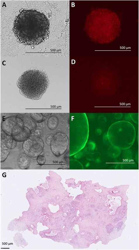
Gündel et al.                                                                                                         PDAC: Preclinical in vitro Models

a main component of the TME and the major driving force                    exterior layers still proliferate. As such spheroids might be viewed
for the tumor interstitial fluid pressure (Scaife et al., 2008;            as avascular minitumors.
Liu et al., 2018). Tissue stiffness was determined by atomic                   Dense stroma is a key hallmark of PDAC. Spheroids were
force microscope (AFM) to increase from 0.4 kPa in normal                  shown to build up some matrix components (Longati et al.,
pancreas tissue to 1.2 kPa of a PDAC tumor (Rice et al., 2017).            2013), however, fall short in mimicking the combined physical
Collagen matrices have a compression force more akin to healthy            properties of the tumor, like stiffness and compression, and thus
tissue while modified HA matrices reach compression forces up              cannot inherently model solid stress. External induction of solid
to 1.5 kPa which compares well to the pressure measured in                 stress can be simulated using a collagen-1-matrix for embedding.
PDAC tumors (Chang and Lin, 2021). A major drawback of                         Spheroids can be grown in coculture in several ways
using natural compounds is their batch-to-batch variability. The           to investigate cell-cell-communication. The before-mentioned
substitution with synthetic polymers alleviates this problem and           trans-well method can be used, e.g., a spheroid in solution and
introduces more control and consistency to the model. The most             a 2D cell culture of CAFs, or a solid spheroid with immune cells
widely used synthetic polymer is polyethylene glycol (PEG), a              in solution (Kuen et al., 2017). Even a combination of 3 types of
substance widely used in biomedical context due to its non-                cells was reported with heterospheroids of different pancreatic
toxicity and non-immunogenicity. Polyethylene glycol can be                cancer cell lines and CAFs being exposed to monocytes.
modified to show properties similar to ECM proteins and enable             Histology allowed then to assess infiltration and drug response
cell aggregation (Liu and Vunjak-Novakovic, 2016).                         (Kuen et al., 2017).
    While organoid culture, described below, relies heavily on                 Additionally, spheroids can also be generated from more than
these matrices, spheroid culture has alternative options by                one cell type forming heterospheroids (Norberg et al., 2020).
using microfluidic systems. The simplest way is preventing cell            Subsequent analysis of crosstalk between the different cell types
adhesion by using coatings, such as agarose (Carlsson and                  requires pre-labeling of these. For example, cell lines expressing
Yuhas, 1984; Ma et al., 2012). This approach is quite easy                 fluorophores are often used to distinguish cell types within the
to accomplish and inexpensive but lacks spheroid uniformity,               spheroid or during cell sorting or image analysis (Öhlund et al.,
a feature important for consistent results. The same problem               2017). Cell sorting comes with the drawback of having to generate
arises when using bioreactors, such as the spinner flask.                  single cells from spheroids which entails substantial stress on the
While this method has a very high yield of spheroids and                   cells and sample loss. Other pre-labeling techniques like the use
enables ongoing growth and culture, the produced spheroids                 of isobaric tags have been carried out to investigate the proteomic
come in any shape which limits their experimental use                      shift between 2D and 3D culture in PDAC (Samonig et al., 2020)
(Cui et al., 2017). As reproducibility and uniformity of size              and to show how drug-induced Akt-inhibition increases the
and shape is very important in research, the hanging drop                  stemness of pancreatic cancer spheroids (Arasanz et al., 2020).
method was favored over the previously mentioned methods                       Alternative to pre-labeling, different cell types can also be
(Timmins and Nielsen, 2007). Although more labor intensive                 vitrually “sorted” after cultivation by using cells from different
than other spheroid generation methods and smaller overall                 species and exploiting the small differences in species specific
size of spheroids, it provided more consistency and allowed                DNA/RNA sequences (Conway et al., 2012; Liu et al., 2021).
medium throughput analyses (Cui et al., 2017). A recent                    Our lab established one such model by generating heterospecies
advancement was combining aspects of microfluidics and matrix              heterospheroids using human Panc1 cells and immortalized
assisted growth by supplying the growth medium with the                    mouse pancreatic stellate cells (imPSCs) (Norberg et al., 2020; Liu
crowding agent methylcellulose (MC) in combination with non-               et al., 2021). This allowed for the analysis of cell-cell crosstalk and
adherent cell culture vessels (Longati et al., 2013). Like solid           could show that the coculture in heterospheroids substantially
matrices, MC increases the viscosity of the medium enough                  changed the gene expression pattern in key cancer pathways,
to prevent cell sedimentation but unlike natural polymers                  PDAC type stratification as well as PSC/CAF phenotype (Liu
it is not subject to batch-to-batch-variation. Spheroids are               et al., 2021). The RNA profile of Panc-1 in heterospheroids was
grown one per well, in scale to the well size, so it does                  more reflecting squamous like phenotype compared to Panc-
not reach the high throughput levels of bioreactors. However,              1 in monospheroids. The CAF phenotype was shifting from
with liquid handling equipment commonly available at high                  myCAF to iCAF due to the presence of Panc-1 cells in the
throughput facilities spheroids were successfully grown even in            heterospheroids.
1536 well plates (Madoux et al., 2017). It grows consistently                  It could also be repeatedly shown that the spatial distribution
sized spheroids which are considerably larger than those                   of cells and the resulting gradients have substantial effects on
produced by the hanging drop method, creating more distinct                chemosensitivity. Especially in the context of pancreatic cancer
internal gradients.                                                        research with a large focus on drug discovery, these findings add
    Even the most basic form of spheroid culture which only uses           weight to the progression toward three-dimensional cell culture
a single cell type improves on mirroring the conditions of tissue          as the base line for medical research which aims to identify novel
compared to 2D cell culture (Friedrich et al., 2009; Longati et al.,       treatment options.
2013; Wagner et al., 2013). By shielding the core of the spheroid              As spheroids are grown from 2D cell culture sources, some
from the medium, gradients of oxygen, nutrients, metabolic waste           of the limitations carry over while some characteristics are
products and signaling molecules are generated. Often the core             improved upon. Cell line identity can drift the longer a cell line
of the spheroid shows necrosis upon prolonged culture while the            is kept in culture. Especially cancer cells, which have lost many

Frontiers in Cell and Developmental Biology | www.frontiersin.org      4                                      October 2021 | Volume 9 | Article 741162
Gündel et al.                                                                                                                PDAC: Preclinical in vitro Models

DNA control/repair mechanisms, are more prone to accumulate                  More detailed analyses of the medium are being carried out
mutations. In order to limit the influence of loss of cell line           as well. Metabolomics, i.e., detection of radioactively labeled
identity, cell lines should be tested regularly by short tandem           substrates in combination with NMR have been successfully
repeat analysis and discontinued after 40-50 passages (Reid et al.,
2013). Another limitation is that also spheroids cannot model the
heterogeneity of PDAC. For one, established pancreatic cancer
cell lines are limited in number (Moore et al., 2001) and do not
reflect the genetic heterogeneity of mutations found in patients.
Nor do co-cultured cell lines such as PSCs as recently shown
(Lenggenhager et al., 2019). An additional problem lies in that
not all cancer cell lines form spheroids and even less form
heterospheroids with other cell types. Consequently, any findings
carried out using only one model are not guaranteed to apply to
all cell lines, all models and ultimately not all PDAC patients.
PDAC, like many other cancers, is inherently heterogeneous
and has several sub types. In this context, different 2D cell
lines grown as spheroids can be viewed as models for different
subpopulations of patients.
    Other methods rely on single cell sedimentation into cell
clusters (Lee et al., 2019). Uniform indentations in a silicon
matrix allow for the sedimentation of 2D cells which then pack
densely as a result of gravity. This is one of the novel approaches
to develop 3D cell culture further, but it is too early to be
assessed/discussed in a greater context for now.

Reporter Assays for Spheroid Cultures
Modern research in the field of cell biology is mostly dependent
on reporters that rely on optical permeability at one point of the
analysis with only few exceptions, e.g., radioactive tracers. We
then interpret these optical readings in a biological/physiological
context to draw conclusions. As such, models heavily rely on
allowing light transmission.
    Methods which treat spheroids as small tissue pieces were
designed to deal with samples which cannot readily pass light.
Consequently, practices like paraffin or OCT (optimal cutting
temperature) compound embedding for immuno-histochemistry
(Maftouh et al., 2014) following sectioning the sample are
readily compatible with spheroid research and are being
carried out routinely.
    Commercially available biochemical assays have simplified
sample analysis substantially by standardizing and simplifying
protocols hence making experiments more reproducible between               FIGURE 2 | Limitations of light microscopy for different in vitro models. (A)
different research groups. However, most of these assays were              Bright field (BF) image of Panc-1 and imPSC heterospheroid at 4d. (B)
designed for 2D cell culture which readily fulfills the important          Detection of mCherry fluorophore in Panc-1 and imPSC heterospheroid at 4d.
prerequisite of allowing light passage. Spheroids on the other             Evenly distributed fluorescent signaling despite unevenly distributed cells in a
hand exhibit a compact structure which allows for very little light        spheroid. (C) BF image of Panc-1 and imPSC heterospheroid at 4d after
                                                                           clearing procedure. Clearing involved dehydration with ascending
transmission. Consequently, adapting reporter systems to be used           concentrations of ethanol causing some shrinking of the spheroid. (D)
for spheroid research is not trivial.                                      Detection of mCherry fluorescent protein in Panc-1 and imPSC
    There are some classes of reporter assays which commonly               heterospheroid at 4d after clearing. Fluorescent signal diminished due to
adapt well to spheroid culture: those that involve lysis of the            clearing procedure but distribution of the signal aligns with cellular distribution
                                                                           with more concentrated signal in the center. (E) BF image of hollow organoids
spheroid and those that test only a reporter in the culture
                                                                           derived from a mouse tumor at 2d. (F) Detection of eGFP fluorophore in
medium. As mentioned previously, spheroid culture can be                   hollow organoids derived from a mouse tumor at 3d. The transparency of the
carried out without using a solid matrix but an altered                    organoid line allows unhindered observation. (G) Organotypic slice culture
medium instead. The combination of a liquid medium-based                   image after 72h in ex vivo culture. H&E staining. Images (E,F) were kindly
spheroid culture with colorimetric or fluorescent reporters which          provided by Daniel Öhlund, Umeå University, Sweden. Image (G) was kindly
                                                                           provided by Carlos Fernández Moro, Karolinska Institutet, Stockholm,
only samples the medium even allows a set up for high
                                                                           Sweden.
throughput screens.

Frontiers in Cell and Developmental Biology | www.frontiersin.org     5                                             October 2021 | Volume 9 | Article 741162
Gündel et al.                                                                                                          PDAC: Preclinical in vitro Models

applied to Panc-1 spheroids, or more precisely to the medium                 the spatial aspect to this data would offer even more insight.
they were cultivated in Fan et al. (2018).                                   High definition spatial transcriptomics (HDST) can be carried
    An example for a commercial adaptation to 3D samples is                  out with a resolution of 2 µm (Vickovic et al., 2019). Given
the cell viability kit CellTiterGlo 3DTM with enhanced lytic                 the diameter of spheroids varying between 200 and 600 µm in
capabilities. As a result this assay is widely and successfully used         diameter, based on cultivation method, spatial transcriptomics
(Norberg et al., 2020; Liu et al., 2021), and is the golden standard         would provide a differentiated view on the impact of metabolic
for cell viability in high throughput screens using spheroids. The           gradients on RNA expression.
APH assay is another cell viability assay optimized for 3D cell                  Fourier-transform infrared (FTIR) spectroscopy imaging was
culture (Longati et al., 2013). It measures the activity of acid             successfully used for detecting necrosis in melanoma spheroids, a
phosphatase in the cells. However, this assay requires a high                useful tool when determining toxicity of compounds in research
pH step which impedes its implementation in high throughput                  (Srisongkram et al., 2020). The advantage of this method over
settings due to its corrosive effects on metal parts of liquid               others is the multivariate analysis that it is commonly linked
handling equipment.                                                          with. It can quantify RNA, lipids and DNA and even subclasses
    While these options offer a wide field of research, the most             of proteins present, as well as their folding status. As such it is a
common reporter remains elusive: fluorescence. We previously                 more comprehensive analysis of fewer samples. This method has
mentioned fluorescence as a simple read out to distinguish                   not been carried out with PDAC spheroids yet but could provide
different cell types in live cell imaging by making use of                   interesting insights, for example when trying to determine which
continuously expressed fluorescent proteins, but many available              changes occur to the ECM during different treatments. This may
assays rely on activatable fluorescent probes as well. However,              reveal insights as to why certain drugs fail in 3D which seemed
the larger the spheroid, the less applicable fluorescence as a               promising in 2D as FTIR imaging was also able to find substantial
reporter becomes. The sheer density of the “tissue” prevents                 differences in biochemical composition between the two models
light passage (Figures 2A,B). Confocal microscopy can be used                (Srisongkram et al., 2020). Additionally, FTIR imaging can also
to observe tissues too dense for brightfield imaging. However,               be taught to identify certain compounds similarly to MALDI-MSI
the more elaborate image capture then limits the throughput                  by incorporating specialized spectral libraries and so could also
capacity. Tissue clearing alleviates this problem altogether but             serve to keep track of drug delivery and metabolization.
also relies on embedding the tissue and precludes further                        However, what image-based analyses of spheroids have in
culturing (Figure 2C). Spheroid protocols which provide highly               common is that they are not adaptable to high throughput yet
homogenous spheroids can overcome this difficulty with larger                and so cannot replace 2D-cell-culture oriented assays. In the
sample number, so that different spheroids serve as single data              future, with implementation of machine learning, improvements
points in timelines rather than one spheroid being continuously              in image acquisition and a more automated image analysis,
observed. A common problem with clearing protocols is                        image-based analysis has the potential to enhance and replace
the diminishing of pre-labeling fluorophores (Figure 2D).                    some of the methods for spheroid research described here. In
Fluorophores used on already cleared samples, e.g., during                   the meantime, more work needs to be invested into establishing
immunohistochemical staining, are not affected.                              robust and efficient reporting systems as spheroids represent a
    Single cell-analysis like flow cytometry relies on creating single       significant improvement compared to 2D cell culture.
cells from a spheroid which often entails substantial loss of                    Particularly methods which are high-throughput ready need
cells. Additionally, there is uncertainty about the homogeneity of           to be established on a broader base. The main parameter currently
single cell origin. Central parts of the spheroid may not be fully           is cell viability but in order to distinguish PDAC-specific toxicity
separated into single cells and gated out of the analysis as a result.       from general cell toxicity more information must be acquired
This would then bias the analysis toward cells on the outside of             in large drug screens, as for instance metabolic parameters such
the spheroid which were less subjected to existing gradients. In             as pH, lactate or glutamate accumulation and consumption of
the case different cell types are mixed in spheroids, the methods            glucose and glutamine.
used to prepare single cell suspensions might preferentially harm
one cell type introducing a different type of bias.                          Organoids
    An ongoing trend in research is to add a spatial parameter to            Since this review is part of a review series, we will not focus
any given optical output and create chemical images of samples.              as much on organoids as this topic is being covered in several
One such method is matrix assisted laser desorption ionization               entries of the series.
mass spectrometry imaging (MALDI-MSI). The spatially targeted                   In comparison, organoids are 3D structures that mimic
MS-analysis allows for example to determine drug penetration in              properties and tissue organization of the organ the cells are
spheroids (Mittal et al., 2019).                                             derived from. An important aspect of organoids is the self-
    To our knowledge spatial transcriptomics has not yet been                assembly into a micro tissue. As such their structure is more
introduced in PDAC spheroid research. This method, however,                  complex than that of spheroids and 2D cell culture while still
would be interesting to use in order to investigate the RNA profile          being grown under laboratory conditions. While spheroids are
along the gradients that build up in a spheroid. While single                grown from established 2D cell lines, organoids also known
cell RNAseq (Liu et al., 2021) and transcriptomic profiling (Yang            as PDOs are cultivated using primary cells. Consequently, they
et al., 2018) have already provided much information about gene              represent the heterogeneity of cancer much more accurately than
expression in pancreatic cancer spheroids, succeeding in adding              spheroids or 2D cell lines. Organoids often preserve the polarity

Frontiers in Cell and Developmental Biology | www.frontiersin.org        6                                     October 2021 | Volume 9 | Article 741162
Gündel et al. PDAC: Preclinical in vitro Models
and gene expression pattern of the original tumor during early as well (Frappart et al., 2020). In very recent years, protocols
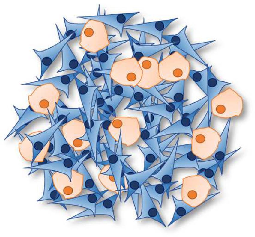
passages (Baker et al., 2016). Organoids can grow as hollow to derive organoids from fine needle aspiration biopsies are
spheres (Figure 2E) or small, more solid spheres which allow being established which could provide the necessary drug testing
light passage and the use of fluorescence for analysis (Figure 2F). platform for patients not suitable for tumor resection (Tiriac
All parameters considered, organoids seem like a better suited et al., 2018b). However, acquiring enough cancer cells from the
candidate for in vitro drug testing. sampled tissue currently necessary for organoid establishment
However, the requirements for successful culture are presents a major hurdle, especially when using fine needle
considerably higher and more expensive than that of spheroids. biopsies in PDAC.
Spheroids are easily propagated, quickly grown to the required
amount and inexpensive to maintain. On the other hand, the Organotypic Slice Culture
self-organization of organoids into a tissue relies on signaling, This model uses precision cut slices of tissue which is cultured
both via solvent molecules, cell-matrix- and cell-cell-interactions. submerged in medium adjusted to the tissue used (Moro, 2021).
The most widely used matrix to facilitate this signaling is Only recently has this model been used in PDAC research
Matrigel . It is animal-cell derived and provides a complex mix
R
(Jiang et al., 2017; Misra et al., 2019; Moro, 2021).
of matrix components that is able to mimic the natural matrix An outstanding benefit of this type of in vitro model is the
composition of the basement membrane. However, also due maintaining of the TME and the spatial information of the
to its animal origin it is subject to significant batch-to-batch tumor (Figure 2G) in combination with time resolved analysis.
variations which negatively affect the reproducibility of organoid The activation and effects of CD8+ T-cells following treatment
research. In addition, Matrigel is rather soft compared to the stiff was demonstrated using live-cell imaging in combination with
collagen-I-rich stroma characteristic for PDAC (Chang and Lin, confocal microscopy (Seo et al., 2019). Additionally to the tissue,
2021). Attempts to move into a more homogenous matrix have the supernatant can be analyzed as well, e.g., to detect soluble
not been successful yet but remain a target of intensive research. signaling molecules (Jiang et al., 2017; Seo et al., 2019).
This obligatory embedding in matrix also poses a problem Organotypic slice culture can be viewed as an alternative
for sample accessibility. Whereas spheroids grown in liquid to organoid culture in personalized medicine. Like organoids,
medium can be collected and split with ease, organoids need to it maintains the genetic tumor heterogeneity of the patient’s
be separated from the matrix. Precise organoid sample aliquots tumor. Additionally, it retains the stromal environment, which
are not possible without breaking them down to single cell level, gets inevitably lost during organoid generation. In detail, it
a condition some organoid lines have problems to recover from. could be shown that proliferation rate and grade of tumor
Given these difficulties, organoids have not yet firmly differentiation could be maintained throughout culture duration
established their implementation into high throughput drug of 4 days (Misra et al., 2019). Tissue slices also responded in a
screening. However, with further improvements of the model, dose and time dependent manner to drug treatment which was
organoids will also play an increasingly important part in the 3Rs confirmed by immunohistochemical measurements of cellularity
of humane animal research, to replace, refine and reduce animal and cleaved caspase-3 positive cells (Misra et al., 2019). A benefit
experimentation in modern research as well as become a platform compared to organoid culture is its immediate availability.
for drug screens of varying scale. We discussed earlier the time sensitivity of PDAC treatment
The most striking benefit of organoid research are the and how organoid culture takes too long under current
possibilities for clinical application. As previously mentioned, conditions to be a tool in personalized medicine. Organotypic
PDOs are generated from patient tumor tissue. Hence these tissue slices, on the other hand, are available quickly and can
organoid lines closely resemble the status quo of an individual be used to screen for specifically effective anti-cancer drugs
patient. Potential therapies can then be tested in vitro for (Ghaderi et al., 2020). Naturally, the size of the tumor limits
effectiveness before being administered to the patient, avoiding the number of samples and the number of drugs which can
unnecessary side effects from ineffective treatments (Huang be tested. Consequently, organotypic slice culture cannot serve
et al., 2015, 2020; Tiriac et al., 2018a). However, as before as a model for drug discovery but remains an interesting tool
mentioned the median survival time for PDAC is around for the advancement and clinical translation of personalized
6 months, meaning that a significant number of PDAC patients medicine in PDAC.
have a shorter life expectance than it takes to generate enough Conclusively, 3D cell culture is a diverse field of ongoing
organoid material and perform the drug testing. To make a research with significant improvements compared to 2D cell
difference for patient therapy, organoid establishment, drug culture in modeling a complex disease like PDAC (Table 2).
screen and data analysis must be carried out within a time frame However, more reporter systems which are tailored to these
short enough to warrant delaying immediate treatment. Current models are still required to make them an even more
approaches cannot supply this information quickly enough yet. advantageous part of PDAC research.
With better diagnostic markers being investigated in parallel by
many research groups, this strained schedule could become more
relaxed in the future. Until then, only the roughly 25 percent of SPHEROIDS AS A STEPPINGSTONE
patients amenable to surgery and thus with longer life expectancy
can take advantage of this approach. Proposals to incorporate Spheroids are not exclusively used as a model for research.
small scale PDO screens into clinical practice have been made Instead, they are also used as a tool to refine animal models
Frontiers in Cell and Developmental Biology | www.frontiersin.org 7 October 2021 | Volume 9 | Article 741162Gündel et al. PDAC: Preclinical in vitro Models
TABLE 2 | Comparison of relevant parameters of spheroid, organoid and organotypic slice model.
Spheroid model Organoid model Organotypic slice culture
Derived of established 2D cell lines primary tissue Primary tissue
Complexity homogenous sample generation with consistent varied organoid growth density and cultivation of precision-cut tumor slices
growth progression limited growth predictability retains TME, its spatial distribution and
cannot model tumor heterogeneity retains the genetic expression pattern of tumor differentiation/grade
the original tumor
Co-culture system • heterospheroid formation limited to few cell lines • co-culture inside Matrigel dome • contains all cells of the patient’s TME
• trans-well co-culture with monospheroids • co-culture with suspended cells • additional non-adhesive cells can be
• differentiated cell-type-specific analysis of added to medium to observe infiltration
crosstalk possible
Availability compatible with regular cell laboratory facilities requires additional storage, management compatible with regular cell laboratory
and cultivation resources facilities
Costs similar to 2D culture, depends on protocol additional costs caused by Matrigel and additional medium components necessary
medium supplements
Most applicable reporter + analyses of the medium + image-based analysis with or without + immunohistochemistry
systems + imaging after embedding embedding + live-cell imaging with confocal
+ analyses of lysates + analysis of lysates microscopy
use of radioactive tracers + use of radioactive tracers + medium analysis
Complications for reporter limited light transmission of whole spheroids limited accessibility due to matrix tissue architecture prevents
systems embedding and matrix interactions cell-type-specific biochemical analyses
High throughput analysis of medium and spheroid lysis AI-assisted image analysis Not applicable to high throughput due to
application very small sample size
and to advance other in vitro models. This last part of the collect cells for sampling so any results need to be image-based
review will focus on the implementation of spheroids in other (Tomás-Bort et al., 2020).
preclinical models. Models including spheroids grown from PDAC lines were
established on several accounts. Established PDAC cell lines were
embedded in collagen and offered ongoing nutritional perfusion
Xenografts (Beer et al., 2017). This model proved highly resistant to cisplatin
Xenografts are an in vivo model commonly used in translational
and identified the matrix-interaction as a crucial factor in model
research. Typically, suspended cells are injected orthotopically
establishing. Despite not forming spheroids, the cells responded
or subcutaneously. However, when injecting spheroids instead
more akin to those in spheroids rather than those in ECM-free
of 2D cells the resulting tumors grew more homogenously
2D culture (Beer et al., 2017).
as well as more successfully. As such the implementation of
An organ on chip model was also designed to investigate solid
spheroids reduced the number of animals subjected to cell
stress found in PDAC. By increasing the interstitial fluid pressure
injection as well as refining the in vivo model (Valta et al., 2016;
to match that of a patient tumor an upregulation of the multidrug
Durymanov et al., 2019; Huang et al., 2020).
resistance protein family could be observed (Kramer et al., 2019).
For PDAC there was one more substantial improvement.
A microfluidic system of Panc-1 and PSC cells was established
When transplanting spheroids consisting of Panc1 cancer cells
to demonstrate the promotive effect of PSCs on cell motility (Lee
and 3T3 fibroblasts, the resulting tumors contained more
et al., 2018). Additionally, an increased gemcitabine resistance
stroma than when suspended cells were injected, hence creating
facilitated by PSCs was demonstrated. The addition of medium
tumors which also more accurately resemble a patient’s tumor
circulation represents a platform to not only test for drug
(Durymanov et al., 2019).
responses but also to test for dosage and treatment schedules.
A relatively new application is investigating multi-organ
Organ on Chip crosstalk (Wagner et al., 2013; Schimek et al., 2020). There, a
Organ on chip systems use cells suspended in hydrogels in a small liver spheroid was introduced to metabolize administered drugs
glass chamber. This model specializes on mimicking the influence to observe any possible adverse effect not only of the original drug
of tissue-tissue interfaces or fluid-tissue interfaces. The latter is but also its metabolized products.
achieved by using microhydraulic systems which simulate blood While not yet attempted to our knowledge, the combination
circulation. The central drawback of this model is the inability to of liver metabolism to PDAC drug trials would represent a
Frontiers in Cell and Developmental Biology | www.frontiersin.org 8 October 2021 | Volume 9 | Article 741162Gündel et al. PDAC: Preclinical in vitro Models
considerable advancement. Not only due to the added metabolic the methods by which we build, interrogate and interpret
degradation of the drugs but also to investigate liver toxicity. these models must keep pace and develop further to meet
As metastases of PDAC are commonly observed in the liver, the changing and increasingly complex questions of frontline
this organ is of additional interest in research seeking to research. Spheroids in particular exhibit great balance in
improve PDAC treatment. recapitulating tissue conditions more authentically while also
allowing controllable conditions which can be easily manipulated
3D Bioprinting in experiments. Alongside spheroids, all 3D cell culture models
Bioprinting uses cells typically suspended in hydrogels or other will further expand our understanding how the TME can
solidifying scaffolds to precisely determine the distribution of be modified in order to improve patient treatment. High
different cell types to one another. Unlike previously mentioned throughput drug screenings and personalized medicine are
methods it thus seeks to recapitulate the morphology of organs merely the most prominent examples how 3D cell culture
or organ systems. models could translate into relevant preclinical applications;
The use of scaffold also serves the purpose to give cells the the clinical effectiveness and truth of which will only be
correct cues for migration and differentiation which are naturally revealed by time.
provided by the ECM.
Another novel approach was using scaffold embedded
spheroids instead of scaffold embedded cells (Goulart et al., AUTHOR CONTRIBUTIONS
2019). Compared to single cells, hepatic spheroids showed a more
balanced metabolism and more importantly preserved the cell All authors contributed to collecting literature, and to writing and
identity of the hepatocytes even in prolonged culture. revising the review.
The bioprinting of neural spheroids was also recognized as
more advantageous compared to single cell printing. Again a
prolonged longevity of the culture could be observed, based on FUNDING
the enhanced self-renewal (Han and Hsu, 2017).
The importance of modeling the appropriate ECM for PDAC This study was supported with financial grants by
was also recognized in the area of bioprinting. However, only Vetenskapsrådet (Grant Number: K2013-67 × 22322–01-3),
few studies have so far been carried out with PDAC cells. PDX RaHFo (Grant Numbers: 111252 and 131163), EPC-TM-Net (EU
derived cells were embedded along PSCs and human umbilical Grant Number: 256974) and PRE- CODE (EU Grant Number:
vein endothelial cells (HUVECs) (Langer et al., 2019) which 861196) to ML, CancerFonden (Grant Numbers: CAN2013/780,
created a dense and active stroma over time. CAN2017/615 and 20 1356 PjF 01 H) to RH, and China
To summarize, spheroids seem to represent a way to stabilize Scholarship Council (scholarship number: 201700260279) to XL.
the cellular identity. This conservation is especially important for
fields of bioprinting which are slowly progressing into clinical
application. Additionally, however, the benefit of in vitro models ACKNOWLEDGMENTS
aiding in improving upon established models like the murine
We thank Daniel Öhlund and his lab members Parniyan
xenograft should not be overlooked.
Maneshi and Tommy Lidström of the Department of Radiation
Sciences and Wallenberg Centre for Molecular Medicine,
CONCLUSION Umeå University, Sweden, for providing us with organoid
images (Figures 2E,F). We also thank Carlos Fernández
To model complex and heterogenous pathologies like cancer, Moro of the Department of Pathology/Cytology, Karolinska
in vitro models must move beyond 2D cell culture as a failure- University Hospital, Stockholm, Sweden for providing images of
rich history in PDAC research has demonstrated. Likewise, organotypic cultures (Figure 2G).
REFERENCES Bachem, M. G., Schünemann, M., Ramadani, M., Siech, M., Beger, H., Buck,
A., et al. (2005). Pancreatic carcinoma cells induce fibrosis by stimulating
Abdalla, M. Y., Ahmad, I. M., Rachagani, S., Banerjee, K., Thompson, proliferation and matrix synthesis of stellate cells. Gastroenterology 128, 907–
C. M., Maurer, H. C., et al. (2019). Enhancing responsiveness of 921. doi: 10.1053/j.gastro.2004.12.036
pancreatic cancer cells to gemcitabine treatment under hypoxia by heme Baker, L. A., Tiriac, H., Clevers, H., and Tuveson, D. A. (2016). Modeling pancreatic
oxygenase-1 inhibition. Transl. Res. 207, 56–69. doi: 10.1016/j.trsl.2018. cancer with organoids. Trends Cancer 2, 176–190. doi: 10.1016/j.trecan.2016.
12.008 03.004
Abetamann, V., Kern, H. F., and Elsässer, H. (1996). Differential expression of the Beer, M., Kuppalu, N., Stefanini, M., Becker, H., Schulz, I., Manoli, S., et al.
hyaluronan receptors CD44 and RHAMM in human pancreatic cancer cells. (2017). A novel microfluidic 3D platform for culturing pancreatic ductal
Clin. Cancer Res. 2, 1607–1618. adenocarcinoma cells: comparison with in vitro cultures and in vivo xenografts.
Arasanz, H., Hernández, C., Bocanegra, A., Chocarro, L., Zuazo, M., Gato, Sci. Rep. 7:1325.
M., et al. (2020). Human pancreatic cancer cells undergo profound Biffi, G., Oni, T. E., Spielman, B., Hao, Y., Elyada, E., Park, Y., et al. (2019).
metabolic reprogramming towards cellular stemness as adaptation to IL1-induced JAK/STAT signaling is antagonized by TGFbeta to shape CAF
inhibition of the Akt pathway. bioRxiv [Preprint]. doi: 10.1101/2020.04.01.0 heterogeneity in pancreatic ductal adenocarcinoma. Cancer Discov. 9, 282–301.
20446 doi: 10.1158/2159-8290.cd-18-0710
Frontiers in Cell and Developmental Biology | www.frontiersin.org 9 October 2021 | Volume 9 | Article 741162Gündel et al. PDAC: Preclinical in vitro Models Blay, V., Tolani, B., Ho, S. P., and Arkin, M. R. (2020). High-throughput screening: pluripotent stem cell–and patient-derived tumor organoids. Nat. Med. 21, today’s biochemical and cell-based approaches. Drug Discov. Today 25, 1807– 1364–1371. doi: 10.1038/nm.3973 1821. doi: 10.1016/j.drudis.2020.07.024 Huang, Y., Lu, Y., Vadlamudi, M., Zhao, S., Felmlee, M., Rahimian, R., et al. Brizel, D. M., Scully, S. P., Harrelson, J. M., Layfield, L. J., Bean, J. M., Prosnitz, (2020). Intrapulmonary inoculation of multicellular spheroids to construct an L. R., et al. (1996). Tumor oxygenation predicts for the likelihood of distant orthotopic lung cancer xenograft model that mimics four clinical stages of metastases in human soft tissue sarcoma. Cancer Res. 56, 941–943. non-small cell lung cancer. J. Pharmacol. Toxicol. Methods 104:106885. doi: Carlsson, J., and Yuhas, J. (1984). “Liquid-overlay culture of cellular spheroids,” in 10.1016/j.vascn.2020.106885 Spheroids in Cancer Research, Vol. 95, eds H. Acker, J. Carlsson, R. Durand, Iwamoto, C., Ohuchida, K., Shinkawa, T., Okuda, S., Otsubo, Y., Okumura, T., et al. and R. M. Sutherland (Berlin: Springer),1–23. doi: 10.1007/978-3-642-8 (2021). Bone marrow-derived macrophages converted into cancer-associated 2340-4_1 fibroblast-like cells promote pancreatic cancer progression. Cancer Lett. 512, Carrato, A., Falcone, A., Ducreux, M., Valle, J. W., Parnaby, A., Djazouli, K., et al. 15–27. doi: 10.1016/j.canlet.2021.04.013 (2015). A systematic review of the burden of pancreatic cancer in Europe: real- Jiang, X., Seo, Y. D., Chang, J. H., Coveler, A., Nigjeh, E. N., Pan, S., et al. (2017). world impact on survival, quality of life and costs. J. Gastrointest. Cancer 46, Long-lived pancreatic ductal adenocarcinoma slice cultures enable precise study 201–211. doi: 10.1007/s12029-015-9724-1 of the immune microenvironment. Oncoimmunology 6:e1333210. doi: 10.1080/ Chang, C.-Y., and Lin, C.-C. (2021). Hydrogel models with stiffness gradients for 2162402x.2017.1333210 interrogating pancreatic cancer cell fate. Bioengineering 8:37. doi: 10.3390/ Kleeff, J., Beckhove, P., Esposito, I., Herzig, S., Huber, P. E., Löhr, J. M., et al. (2007). bioengineering8030037 Pancreatic cancer microenvironment. Int. J. Cancer 121, 699–705. Chang, Q., Jurisica, I., Do, T., and Hedley, D. W. (2011). Hypoxia predicts Kramer, B., De Haan, L., Vermeer, M., Olivier, T., Hankemeier, T., Vulto, P., aggressive growth and spontaneous metastasis formation from orthotopically et al. (2019). Interstitial flow recapitulates gemcitabine chemoresistance in grown primary xenografts of human pancreatic cancer. Cancer Res. 71, 3110– a 3D microfluidic pancreatic ductal adenocarcinoma model by induction 3120. doi: 10.1158/0008-5472.can-10-4049 of multidrug resistance proteins. Int. J. Mol. Sci. 20:4647. doi: 10.3390/ Conway, T., Wazny, J., Bromage, A., Tymms, M., Sooraj, D., Williams, E. D., ijms20184647 et al. (2012). Xenome—a tool for classifying reads from xenograft samples. Kubow, K. E., Vukmirovic, R., Zhe, L., Klotzsch, E., Smith, M. L., Gourdon, D., et al. Bioinformatics 28, i172–i178. (2015). Mechanical forces regulate the interactions of fibronectin and collagen Cui, X., Hartanto, Y., and Zhang, H. (2017). Advances in multicellular spheroids I in extracellular matrix. Nat. Commun. 6:8026. formation. J. R. Soc. Interface 14:20160877. doi: 10.1098/rsif.2016.0877 Kuen, J., Darowski, D., Kluge, T., and Majety, M. (2017). Pancreatic cancer Durymanov, M., Kroll, C., Permyakova, A., O’Neill, E., Sulaiman, R., Person, cell/fibroblast co-culture induces M2 like macrophages that influence M., et al. (2019). Subcutaneous inoculation of 3D pancreatic cancer spheroids therapeutic response in a 3D model. PLoS One 12:e0182039. doi: 10.1371/ results in development of reproducible stroma-rich tumors. Transl. Oncol. 12, journal.pone.0182039 180–189. doi: 10.1016/j.tranon.2018.10.003 Laklai, H., Miroshnikova, Y. A., Pickup, M. W., Collisson, E. A., Kim, G. E., Barrett, European Union (2021). ECIS – European Cancer Information System. Available A. S., et al. (2016). Genotype tunes pancreatic ductal adenocarcinoma tissue online at: https://ecis.jrc.ec.europa.eu (accessed March 15, 2021). tension to induce matricellular fibrosis and tumor progression. Nat. Med. 22, Fan, T. W.-M., El-Amouri, S. S., Macedo, J. K., Wang, Q. J., Song, H., Cassel, T., 497–505. doi: 10.1038/nm.4082 et al. (2018). Stable isotope-resolved metabolomics shows metabolic resistance Langer, E. M., Allen-Petersen, B. L., King, S. M., Kendsersky, N. D., Turnidge, to anti-cancer selenite in 3D spheroids versus 2D cell cultures. Metabolites 8:40. M. A., Kuziel, G. M., et al. (2019). Modeling tumor phenotypes in vitro with doi: 10.3390/metabo8030040 three-dimensional bioprinting. Cell Rep. 26, 608–623.e6. Frappart, P.-O., Walter, K., Gout, J., Beutel, A. K., Morawe, M., Arnold, F., Lee, D., Pathak, S., and Jeong, J.-H. (2019). Design and manufacture of 3D cell et al. (2020). Pancreatic cancer-derived organoids–a disease modeling tool to culture plate for mass production of cell-spheroids. Sci. Rep. 9:13976. predict drug response. United Eur. Gastroenterol. J. 8, 594–606. doi: 10.1177/ Lee, J.-H., Kim, S.-K., Khawar, I. A., Jeong, S.-Y., Chung, S., and Kuh, H.-J. (2018). 2050640620905183 Microfluidic co-culture of pancreatic tumor spheroids with stellate cells as a Friedrich, J., Seidel, C., Ebner, R., and Kunz-Schughart, L. A. (2009). Spheroid- novel 3D model for investigation of stroma-mediated cell motility and drug based drug screen: considerations and practical approach. Nat. Protoc. 4, resistance. J. Exp. Clin. Cancer Res. 37:4. 309–324. doi: 10.1038/nprot.2008.226 Lenggenhager, D., Amrutkar, M., Sántha, P., Aasrum, M., Löhr, J.-M., Gladhaug, Fries, H., Elsässer, H., Mahlbacher, V., Kern, H., and Neumann, K. (1994). I. P., et al. (2019). Commonly used pancreatic stellate cell cultures differ Localisation of hyaluronate (HA) in primary tumors and nude mouse phenotypically and in their interactions with pancreatic cancer cells. Cells 8:23. xenografts of human pancreatic carcinomas using a biotinylated HA-binding doi: 10.3390/cells8010023 protein. Virchows Archiv. 424, 7–12. Leppanen, J., Lindholm, V., Isohookana, J., Haapasaari, K. M., Karihtala, P., Geismann, C., and Arlt, A. (2020). Coming in the air: hypoxia meets epigenetics in Lehenkari, P. P., et al. (2019). Tenascin C, fibronectin, and tumor-stroma ratio pancreatic cancer. Cells 9:2353. doi: 10.3390/cells9112353 in pancreatic ductal adenocarcinoma. Pancreas 48, 43–48. doi: 10.1097/mpa. Ghaderi, M., Moro, C. F., Elduayen, S. P., Hultin, E., Verbeke, C. S., Björnstedt, 0000000000001195 M., et al. (2020). Genome-wide transcriptome profiling of ex-vivo precision-cut Liu, H.-Y., Korc, M., and Lin, C.-C. (2018). Biomimetic and enzyme-responsive slices from human pancreatic ductal adenocarcinoma. Sci. Rep. 10:9070. dynamic hydrogels for studying cell-matrix interactions in pancreatic ductal Goulart, E., de Caires-Junior, L. C., Telles-Silva, K. A., Araujo, B. H. S., Rocco, S. A., adenocarcinoma. Biomaterials 160, 24–36. doi: 10.1016/j.biomaterials.2018.01. Sforca, M., et al. (2019). 3D bioprinting of liver spheroids derived from human 012 induced pluripotent stem cells sustain liver function and viability in vitro. Liu, X., Gündel, B., Li, X., Liu, J., Wright, A., Löhr, M., et al. (2021). 3D Biofabrication 12:015010. doi: 10.1088/1758-5090/ab4a30 heterospecies spheroids of pancreatic stroma and cancer cells demonstrate key Haber, P. S., Keogh, G. W., Apte, M. V., Moran, C. S., Stewart, N. L., Crawford, phenotypes of pancreatic ductal adenocarcinoma. Transl. Oncol. 14:101107. D. H. G., et al. (1999). Activation of pancreatic stellate cells in human and doi: 10.1016/j.tranon.2021.101107 experimental pancreatic fibrosis. Am. J. Pathol. 155, 1087–1095. doi: 10.1016/ Liu, Z., and Vunjak-Novakovic, G. (2016). Modeling tumor microenvironments s0002-9440(10)65211-x using custom-designed biomaterial scaffolds. Curr. Opin. Chem. Eng. 11, 94– Han, H.-W., and Hsu, S.-H. (2017). Using 3D bioprinting to produce mini-brain. 105. doi: 10.1016/j.coche.2016.01.012 Neural Regen. Res. 12, 1595–1596. doi: 10.4103/1673-5374.217325 Löhr, M., Schmidt, C., Ringel, J., Kluth, M., Müller, P., Nizze, H., et al. (2001). Höckel, M., Schlenger, K., Aral, B., Mitze, M., Schäffer, U., and Vaupel, P. (1996). Transforming growth factor-β1 induces desmoplasia in an experimental model Association between tumor hypoxia and malignant progression in advanced of human pancreatic carcinoma. Cancer Res. 61, 550–555. cancer of the uterine cervix. Cancer Res. 56, 4509–4515. Löhr, M., Trautmann, B., Göttler, M., Peters, S., Zauner, I., Maillet, B., et al. (1994). Huang, L., Holtzinger, A., Jagan, I., BeGora, M., Lohse, I., Ngai, N., et al. Human ductal adenocarcinomas of the pancreas express extracellular matrix (2015). Ductal pancreatic cancer modeling and drug screening using human proteins. Br. J. Cancer 69, 144–151. doi: 10.1038/bjc.1994.24 Frontiers in Cell and Developmental Biology | www.frontiersin.org 10 October 2021 | Volume 9 | Article 741162
You can also read