Convalescent COVID-19 patients are susceptible to endothelial dysfunction due to persistent immune activation
←
→
Page content transcription
If your browser does not render page correctly, please read the page content below

RESEARCH ARTICLE

                                   Convalescent COVID-19 patients are
                                   susceptible to endothelial dysfunction due
                                   to persistent immune activation
                                   Florence WJ Chioh1†, Siew-Wai Fong2,3,4†, Barnaby E Young1,5,6, Kan-Xing Wu1,
                                   Anthony Siau1, Shuba Krishnan1,7, Yi-Hao Chan2,3, Guillaume Carissimo2,3,
                                   Louis LY Teo8,9, Fei Gao8,9, Ru San Tan8,9, Liang Zhong8,9, Angela S Koh8,9,
                                   Seow-Yen Tan10, Paul A Tambyah11, Laurent Renia1,2,3, Lisa FP Ng2,3,
                                   David C Lye1,5,6,12, Christine Cheung1,13*
                                   1
                                    Lee Kong Chian School of Medicine, Nanyang Technological University, Singapore,
                                   Singapore; 2A*STAR ID Labs, Agency for Science, Technology and Research,
                                   Singapore, Singapore; 3Singapore Immunology Network, Agency for Science,
                                   Technology and Research, Singapore, Singapore; 4Department of Biological
                                   Sciences, National University of Singapore, Singapore, Singapore; 5National Centre
                                   for Infectious Diseases, Singapore, Singapore; 6Department of Infectious Diseases,
                                   Tan Tock Seng Hospital, Singapore, Singapore; 7Division of Clinical Microbiology,
                                   Department of Laboratory Medicine, Karolinska Institute, ANA Futura, Campus
                                   Flemingsberg, Stockholm, Sweden; 8National Heart Centre Singapore, Singapore,
                                   Singapore; 9Duke-NUS Medical School, Singapore, Singapore; 10Department of
                                   Infectious Diseases, Changi General Hospital, Singapore, Singapore; 11Department
                                   of Medicine, National University Hospital, Singapore, Singapore; 12Yong Loo Lin
                                   School of Medicine, National University of Singapore, Singapore, Singapore;
                                   13
                                     Institute of Molecular and Cell Biology, Agency for Science, Technology and
                                   Research, Singapore, Singapore

*For correspondence:
ccheung@ntu.edu.sg                 Abstract Numerous reports of vascular events after an initial recovery from COVID-19 form our
†
 These authors contributed         impetus to investigate the impact of COVID-19 on vascular health of recovered patients. We found
equally to this work               elevated levels of circulating endothelial cells (CECs), a biomarker of vascular injury, in COVID-19
                                   convalescents compared to healthy controls. In particular, those with pre-existing conditions (e.g.,
Competing interest: See
page 17                            hypertension, diabetes) had more pronounced endothelial activation hallmarks than non-COVID-19
                                   patients with matched cardiovascular risk. Several proinflammatory and activated T lymphocyte-
Funding: See page 17               associated cytokines sustained from acute infection to recovery phase, which correlated positively
Received: 14 November 2020         with CEC measures, implicating cytokine-driven endothelial dysfunction. Notably, we found higher
Accepted: 15 February 2021         frequency of effector T cells in our COVID-19 convalescents compared to healthy controls. The
Reviewing editor: Noriaki
                                   activation markers detected on CECs mapped to counter receptors found primarily on cytotoxic
Emoto, Kobe Pharmaceutical         CD8+ T cells, raising the possibility of cytotoxic effector cells targeting activated endothelial cells.
University, Japan                  Clinical trials in preventive therapy for post-COVID-19 vascular complications may be needed.

   Copyright Chioh et al. This
article is distributed under the
terms of the Creative Commons
Attribution License, which
                                   Introduction
permits unrestricted use and       As of February 21, 2021, there have been more than 110 million confirmed cases and 2.4 million
redistribution provided that the   deaths from coronavirus disease 2019 (COVID-19). Many countries are facing multiple waves of
original author and source are     resurgence upon reopening their economy, prompting new lockdown. Intensive ongoing research
credited.                          has shed light on the pathogenesis of COVID-19 and the extent of damages caused directly or

Chioh, Fong, et al. eLife 2021;10:e64909. DOI: https://doi.org/10.7554/eLife.64909                                                   1 of 23
Research article                                                                       Cell Biology Immunology and Inflammation

                                   indirectly by severe acute respiratory syndrome coronavirus 2 (SARS-CoV-2). Conversely, the inter-
                                   mediate and long-term complications of COVID-19 remains unclear (del Rio et al., 2020). While
                                   most infected people recover completely within a few weeks, a considerable proportion continue to
                                   experience symptoms after their initial recovery (Yelin et al., 2020), similar to SARS survivors
                                   (Ngai et al., 2010).
                                       COVID-19 is mainly a respiratory infection. However, both autopsy findings and clinical observa-
                                   tions have described vascular damages and thrombotic complications in a wide range of organs
                                   (Gupta et al., 2020; Price et al., 2020; Xu et al., 2020). Endothelialitis may occur in multiple organs
                                   as a consequence of viral infection and overactive host immune response as T cell infiltration was
                                   observed in sites of endothelial damage of COVID-19 patients’ tissues. Systematic evaluation of the
                                   long-term sequelae of COVID-19 is currently lacking. COVID-19 infection can cause extrapulmonary
                                   pathologies that persist after recovery such as cardiovascular events with ongoing myocardial inflam-
                                   mation (Puntmann et al., 2020), acute ischemic or hemorrhagic stroke (Mao et al., 2020), as well as
                                   liver injury, neurological deficits, and acute kidney injury necessitating dialysis (Gupta et al., 2020).
                                   The thrombotic complications of COVID-19, such as pulmonary embolism may lead to lasting organ
                                   failure (Price et al., 2020). Independent of acute respiratory distress syndrome, severe pneumonia
                                   has been consistently associated with augmented risk of cardiac impairment both during convales-
                                   cence and in later years (Corrales-Medina et al., 2015). We hypothesize that the risk of vascular
                                   complications in COVID-19 survivors will be higher, if compounded by a confluence of SARS-CoV-2-
                                   mediated damages, proinflammatory cytokine overdrive, and comorbidities with hypertension and
                                   diabetes.
                                       Circulating endothelial cells (CECs), dislodged from blood vessels as a consequence of vascular
                                   injury, constitute an ideal cell-based biomarker as it is indicative of in situ endothelial pathophysiol-
                                   ogy (Farinacci et al., 2019; Hill et al., 2003). Although CECs are a rare population in peripheral
                                   blood, they reflect endothelial dysfunction from a variety of vascular disorders, including myocardial
                                   infarction, acute ischemic stroke, atherosclerosis, and vasculitis (Nadar et al., 2005; Schmidt et al.,
                                   2015). In COVID-19, there are conflicting reports as to the frequency of CEC counts compared with
                                   healthy controls (Mancuso et al., 2020; Nizzoli et al., 2020). This discrepancy may reflect disease
                                   severity as COVID-19 patients admitted to the intensive care unit (ICU) have a higher CEC count,
                                   suggesting more pronounced endothelial injury in severe COVID-19 (Guervilly et al., 2020). Inter-
                                   estingly, among non-ICU patients, those with chronic kidney disease had significantly higher CEC
                                   counts, implying that patients with pre-existing conditions may be more susceptible to vascular dam-
                                   age (Guervilly et al., 2020).
                                       This study aims to understand the state of vascular health in convalescent COVID-19 patients and
                                   to evaluate subclinical endothelial dysfunction through the phenotyping of CECs.

                                   Results
                                   Patient and healthy participants characterization
                                   To understand the intermediate consequence of COVID-19, we performed vascular phenotyping
                                   using CECs and endothelial activation markers as the cellular and molecular measures of endothelial
                                   dysfunction. Written informed consent was received from participants prior to inclusion in the PRO-
                                   TECT study (Young et al., 2020a,c). All study groups were almost gender-balanced, with prior
                                   comorbidities and self-identified ethnicity/race as summarized in Table 1. Convalescent COVID-19
                                   patients who had no pneumonia throughout admission (mild), pneumonia without hypoxia (moder-
                                   ate), or pneumonia with hypoxia (desaturation to 94%) requiring supplemental oxygen (severe),
                                   and discharged after 15 days (median [interquartile range, IQR 11–26]) were screened for pre-exist-
                                   ing cardiovascular risk factors. Among our selected convalescent COVID-19 patients (n = 30), half
                                   had at least one cardiovascular risk factor, including mainly hypertension, diabetes, and/or hyperlip-
                                   idemia. They were benchmarked to healthy participants (n = 24) and non-COVID patients with
                                   matched cardiovascular risk factors (n = 20). More details on demographics are included in Table 1.

Chioh, Fong, et al. eLife 2021;10:e64909. DOI: https://doi.org/10.7554/eLife.64909                                                   2 of 23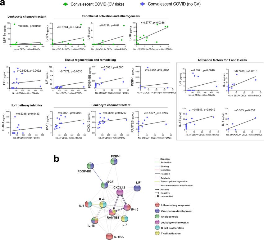
Research article                                                                              Cell Biology Immunology and Inflammation

Table 1. Demographics of patients and healthy controls.
                                                                                         Convalescent COVID-19 individuals
                                                                                         (n = 30)
                         Healthy             Non-COVID-19 patients with cardiovascular
Characteristics, N       participants        risk factors                                Cardiovascular risk factors        No cardiovascular risk
(%)                      (n = 24)            (n = 20)                                    (n = 15)                           (n = 15)
Age*                     46.5 (9.9) years    62.4 (8.7) years                            54 (8.7) years                     42 (13.5) years
Age, male*               46.3 (11.3) years   62.3 (10.4) years                           54 (8.2) years                     36 (6.1) years
Age, female*             46.8 (8.8) years    63 (7.3) years                              54 (9.9) years                     48 (17.1) years
Gender, male             12 (50)             10 (50)                                     8 (53.3)                           8 (53.3)
Gender, female           12 (50)             10 (50)                                     7 (46.7)                           7 (46.7)
Ethnicity                Chinese 22 (91.7) Chinese 17 (85)                               Chinese 11 (73.3)                  Chinese 13 (86.6)
                         Filipino 2 (8.3)    Indian 3 (15)                               Malay 2 (13.3)                     Indian 1 (6.7)
                                                                                         Bangladeshi 1 (6.7)                Caucasian 1 (6.7)
                                                                                         Filipino 1 (6.7)
Days post-symptom        N.A.                N.A.                                        34 (27–46) days
onset
(median [IQR])
COVID-19 severity        N.A.                N.A.                                        Mild 5 (33.3)                      Mild 5 (33.3)
                                                                                         Moderate 5 (33.3)                  Moderate 5 (33.3)
                                                                                         Severe 5 (33.3)                    Severe 5 (33.3)
Comorbidities
Hypertension             0 (0)               20 (100)                                    10 (66.7)                          0 (0)
Hyperlipidemia           0 (0)               10 (50)                                     11 (73.3)                          0 (0)
Diabetes mellitus        0 (0)               9 (45)                                      7 (46.7)                           0 (0)
Fatty liver              0 (0)               Information not available                   2 (13.3)                           0 (0)
Chronic liver disease    0 (0)               Information not available                   1 (6.7)                            0 (0)
Coronary artery          0 (0)               6 (30)                                      Information not available          Information not available
disease
Myocardial infarction 0 (0)                  0 (0)                                       2 (13.3)                           0 (0)

All values are reported as N (%) where N indicates the number of observations.
*
Values are expressed as mean (± standard deviation).

                                        COVID-19 and cardiovascular risk factors contribute to endothelial
                                        dysfunction
                                        There are two major types of blood endothelial cells (Hebbel, 2017). CECs are shed from damaged
                                        vessels and constitute a cell-based biomarker for vascular dysfunction (Blann et al., 2005). On the
                                        other hand, endothelial progenitor cells (EPCs) originate from the bone marrow and are mobilized
                                        into the bloodstream in response to vascular injury (Ackermann et al., 2020a,bAckermann et al.,c;
                                        Li and Li, 2016; Mancuso et al., 2020). Identification of the CEC population from individual periph-
                                        eral blood mononuclear cells (PBMCs) samples was carried out with high stringency using a panel of
                                        established CEC immunophenotypic markers (Figure 1a). We first gated for the CD45 /CD31+ pop-
                                        ulation to isolate CECs along with bone marrow-derived EPCs and platelets, while ruling out CD45+
                                        leukocytes that also expressed the endothelial marker CD31 (PECAM1) (Duda et al., 2007). This
                                        population was further subtyped based on the progenitor marker, CD133, to rule out EPCs. Finally,
                                        a nucleic acid stain was used to distinguish anucleate platelets from nucleated CECs, which were
                                        defined here by a combined immunophenotypic profile of CD45 /CD31+/CD133 /DNA+
                                        (Burger and Touyz, 2012; Duda et al., 2007; Yu et al., 2013). As a comparison, putative EPCs
                                        were identified by CD133+/CD45 /DNA+ population (Ingram et al., 2004; Mancuso et al., 2020;
                                        Rafii and Lyden, 2003). To further analyze endothelial cell activation, CECs were characterized for
                                        the expression of activation markers, namely, intercellular adhesion molecule 1 (ICAM1), P-selectin
                                        (SELP), and fractalkine (CX3CL1) (Figure 1a), which are integral for the processes of leukocyte

Chioh, Fong, et al. eLife 2021;10:e64909. DOI: https://doi.org/10.7554/eLife.64909                                                              3 of 23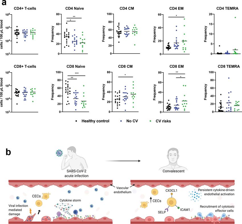
Research article                                                                                Cell Biology Immunology and Inflammation

Figure 1. Enumeration and characterization of circulating endothelial cells (CECs) from COVID-19 convalescents (n = 30), non-COVID-19 patients
(n = 20), and healthy participants (n = 24). (a) Using flow cytometry, CEC populations were gated from peripheral blood mononuclear cells (PBMCs)
using a strategy involving positive (nuclear stain and CD31) and negative (CD45 and CD133) markers before characterization with three separate
markers of endothelial activation. (b) and (c) Scatterplot visualization of the number of CECs and endothelial progenitor cells (EPCs) per million PBMCs
identified from each sample with mean and standard error of mean for each group shown. (d) and (e) Boxplots extending from 25th to 75th percentile
with bar showing mean and whiskers indicating the minimum and maximum number of CECs per million PBMCs from each group staining positive for
endothelial activation markers ICAM1, SELP, or CX3CL1. Kruskal—Wallis test was performed (b—e) to test for difference between the groups with
Dunn’s multiple comparison test carried out for pairwise testing post hoc. (f) and (g) Cumulative analysis of patient frequency data of CECs staining
positive for endothelial activation markers, ICAM1, SELP, and/or CX3CL1. Chi-squared goodness-of-fit test was performed to assess for difference in
frequencies between the groups in (f) and (g).

Chioh, Fong, et al. eLife 2021;10:e64909. DOI: https://doi.org/10.7554/eLife.64909                                                                4 of 23

Research article                                                                       Cell Biology Immunology and Inflammation

                                   adhesion, platelet aggregation, and trans-endothelial migration, respectively (Goncharov et al.,
                                   2017; Johnson and Jackson, 2013).
                                       We enumerated and compared the mean CEC and EPC numbers per million PBMCs between
                                   healthy participants, convalescent COVID-19 patients, convalescent COVID-19 patients with cardio-
                                   vascular risks, and patients with cardiovascular risk but no history of COVID-19. The mean CEC
                                   counts were significantly increased in all the convalescent COVID-19 groups and non-COVID-19 car-
                                   diovascular risk group compared to the healthy participants, while no significant difference was
                                   noted for EPCs (Figure 1b). Between the two convalescent COVID-19 groups, patients with cardio-
                                   vascular risk had higher mean CEC numbers (M = 153.75 ± 51.55) than patients without cardiovascu-
                                   lar risk (M = 116.21 ± 28.17). These observations are consistent with our hypothesis that COVID-19
                                   patients, especially those with pre-existing cardiovascular risk, may display persistent signs of vascu-
                                   lar dysfunction even after recovery from COVID-19.
                                       To ascertain the consequence of COVID-19 on endothelial dysfunction, we regrouped the conva-
                                   lescent COVID-19 patients by their disease severity during acute infection (Figure 1c). We found
                                   that both convalescent COVID-19 patients who recovered from moderate symptoms (p=0.0009) and
                                   non-COVID-19 patients with cardiovascular risks (p
Research article Cell Biology Immunology and Inflammation Figure 2. Plasma cytokine levels of COVID-19 patients with and without cardiovascular risk factors during the acute and convalescent phases of infection. Concentrations of 45 immune mediators were quantified using a 45-plex microbead-based immunoassay. (a) Heatmap of immune mediator levels in plasma samples of patients with (n = 15) and without (n = 15) cardiovascular risk factors at both acute (median 10 days post-illness onset), convalescent (0—90 days post-hospital discharge) phases of SARS-CoV-2 infection, and non-COVID-19 healthy controls. Each color represents the relative concentration of a particular analyte. Blue and red indicate low and high concentrations, respectively. (b) Profiles of significant immune mediators of COVID-19 patients with and without cardiovascular risk factors during acute and convalescent phases are illustrated as scatter plots. Cytokine levels in plasma fraction samples from first collection time point during hospital admission (acute, median 10 days post-illness onset) and discharge (convalescent, median 7 days post-hospital discharge) were compared. Mann—Whitney U tests were performed on the logarithmically transformed concentration (*p
Research article                                                                                   Cell Biology Immunology and Inflammation

                                      CEC attributes from convalescent patients without prior cardiovascular risk correlated significantly
                                      with a greater number of cytokines than those with cardiovascular risk (Figure 3a). This may suggest
                                      that persistent cytokine production contributed primarily to endothelial injury (CEC counts) and acti-
                                      vation hallmarks (CX3CL1+ and SELP+) in convalescent patients without previously known risk

Figure 3. Significantly correlated cytokines with CEC attributes in COVID-19 convalescent patients. (a) Spearman rank correlation coefficients were
calculated to assess the associations between the level of cytokines and CEC attributes in terms of mean numbers of CECs, SELP+ CECs, or CX3CL1+
CECs. Spearman’s correlation coefficient r and p values (two-tailed test) were shown in plots. Source data relating to this figure is available. (b) Network
analysis of CEC-associated cytokines in the convalescent COVID-19 patients without prior cardiovascular risk factors. Interactive relationships between
the cytokines or chemokines were determined by Search Tool for the Retrieval of Interacting Genes/ Proteins (STRING) analysis, with a confidence
threshold of 0.4.
The online version of this article includes the following source data for figure 3:
Source data 1. Significantly correlated cytokines with CEC attributes in COVID-19 convalescent patients.

Chioh, Fong, et al. eLife 2021;10:e64909. DOI: https://doi.org/10.7554/eLife.64909                                                                   7 of 23
Research article Cell Biology Immunology and Inflammation
factors. On the other hand, more pronounced endothelial injury and activation in those with cardio-
vascular risk would have been attributed to their pre-existing cardiovascular risk factors, on top of
the aftermath of COVID-19. Among the positively correlated cytokines in the cardiovascular risk
group (Figure 3a), MIP-1a (CCL3) related to a chemoattractant for leukocytes, along with IL-17A
(Williams et al., 2019), IL-8 (Apostolakis et al., 2009), and IL-18 (Gerdes et al., 2002), known to
evoke activation of endothelial cells during atherogenesis, may suggest chronic development of ath-
erosclerotic plaques already in these individuals.
For the convalescent patients without prior risk factors, the measures of CEC attributes reflected
greater sensitivity to cytokine-driven endothelial dysfunction that could signify a more direct conse-
quence of COVID-19. Further protein—protein interaction network analyses with Search Tool for the
Retrieval of Interacting Genes/Proteins (STRING) highlighted the direct and indirect interactions
between the significant cytokines and their functional associations with biological processes involved
in inflammatory response, vasculature development, angiogenesis, leukocyte chemotaxis, B cell pro-
liferation, and T cell activation (Figure 3b). Among the positively correlated cytokines (Figure 3a),
we observed growth factors—associated tissue regeneration and remodeling, namely EGF, LIF,
PDGF-BB, and PIGF-1 (PGF), indicating possibility of adaptive angiogenesis taking place as a
response to preceding damages caused by viral infection and cytokine overdrive. IL-1RA, an IL-1
pathway inhibitor, could be an inflammation resolving mediator post infection. Conversely, IP-10
(CXCL10) is known to limit angiogenesis (Bodnar et al., 2006). This collective turnover of endothelial
cells during blood vessel remodeling may result in elevated number of CECs. A number of signifi-
cantly correlated cytokines are chemokines known to induce endothelial activation and promote che-
motaxis of leukocytes to vascular endothelium. Chemotactic factors such as IP-10 (CXCL10),
CXCL12, and RANTES (CCL5) regulate adhesion and transmigration of T lymphocytes, monocytes,
and/or neutrophils through endothelial barrier (Sokol and Luster, 2015). Moreover, RANTES was
specifically correlated with SELP+ CEC (r = 0.5677, p=0.0295), indicating that activated platelet-
derived RANTES may work in concert with endothelial SELP to mediate platelet aggregation and
trigger coagulation cascade. In partial correlation analyses, a majority of these CEC—cytokines cor-
relations were sustained even after accounting for age (Supplementary file 1).
Overall, CEC attributes of convalescent COVID-19 patients correlated with a vast majority of dif-
ferentiation and activation factors associated with T cells and B cells (IL-4, IL-5, IL-7, IL-17A, IL-18,
MIP-1a, and RANTES) (Turner et al., 2014), implicating a broad adaptive immune response with
endothelial dysfunction. Besides being a target of the immune mediators, we recognize that endo-
thelial cells, once activated, could also be a source of these cytokines, which in turn could activate
their immune counterparts.
Endothelial—immune crosstalk
Our ability to measure activation markers directly on CECs motivated us to better understand the
receptor—receptor and chemokine—receptor interactions between activated endothelial cells with
putative immune subpopulations in COVID-19. We performed data mining of published single-cell
transcriptomic datasets on PBMCs from healthy participants, mild and severe COVID-19 patients
(Wilk et al., 2020), and COVID-19 patients with or without cardiovascular disease (Schulte-
Schrepping et al., 2020). Then, we re-analyzed the expressions of counter receptors (i.e., ITGAL,
SELPLG, and CX3CR1) to our endothelial activation markers (i.e., ICAM1, SELP, and CX3CL1, respec-
tively), in order to identify the potential immune interactors with activated endothelial cells. We
found that those counter receptors were most pronouncedly expressed by CD8+ T cells, natural killer
(NK) cells, and to some extent monocytes (Figure 4a and Figure 4—figure supplement 1). In Wilk
et al.’s dataset, the expression of CX3CR1 was intensified in mild and severe COVID-19 patients
than healthy participants (Figure 4a). The relationship between counter receptors expression and
COVID-19 disease was even more marked in Schulte-Schrepping et al.’s dataset with a higher pro-
portion of NK or both CD8+ and NK cells expressing all three counter receptors found in mild and
severe COVID-19 samples, respectively, regardless of comorbidity with cardiovascular disease (Fig-
ure 4—figure supplement 1). As the presence of those counter receptors generally marks cytotoxic
effector lymphocytes in peripheral blood (Nishimura et al., 2002), we correspondingly found that
CD8+ T cells and NK cells were the major cytotoxic populations expressing perforin and granzymes
(Figure 4b), especially in COVID-19 patients (Figure 4—figure supplement 1).
Chioh, Fong, et al. eLife 2021;10:e64909. DOI: https://doi.org/10.7554/eLife.64909 8 of 23Research article Cell Biology Immunology and Inflammation Figure 4. Immune interactors of activated endothelial cells. (a) UMAP representations of immune cell populations from healthy participants and COVID- 19 patients annotated by cell types (left) and differential expressions of counter receptors ITGAL, SELPLG, and CX3CR1, which are known to interact with surface molecules of activated endothelial cells (right). (b) Distribution of the expressions of cytotoxic genes GZMA, GZMB, and PRF1 across immune cell populations. Figure 4 continued on next page Chioh, Fong, et al. eLife 2021;10:e64909. DOI: https://doi.org/10.7554/eLife.64909 9 of 23
Research article                                                                         Cell Biology Immunology and Inflammation

Figure 4 continued
The online version of this article includes the following figure supplement(s) for figure 4:
Figure supplement 1. COVID-19 samples with or without cardiovascular comorbidity have a higher proportion of immune cells expressing counter
receptor and cytotoxicity-associated genes.

                                         We hypothesize that persistent cytokine production activates endothelial cells in convalescent
                                      COVID-19 patient, which may promote interactions with cytotoxic effector cells. Therefore, we ana-
                                      lyzed T cell features by immunophenotyping in our cohort (Carissimo et al., 2020) and found that
                                      convalescent COVID-19 patients, in particular those with underlying cardiovascular risk, had signifi-
                                      cantly higher frequencies of effector CD8+ and CD4+ T cells (EM), and central memory CD8+ T cells
                                      (CD8 CM) than healthy controls (Figure 5a), while there were no notable differences across the
                                      groups for absolute numbers of circulating CD8+/CD4+ T cells. There is a possibility that individuals
                                      recovered from COVID-19 could be susceptible to vascular injury due to effector T cell cytotoxicity
                                      (Figure 5b). However, the results herein remain associative, and further experiments are required to
                                      establish cytotoxicity-induced endothelial injury in COVID-19 convalescents.

                                      Discussion
                                      The rapid rise of COVID-19 ‘long-haulers’ with lingering symptoms post-infection or recovered indi-
                                      viduals who suffer from sudden cardiovascular events, would continue to place tremendous strain on
                                      our healthcare systems. About 2.5% of convalescent COVID-19 patients are found with thrombosis
                                      (including arterial and venous events), 30 days post discharge (Patell et al., 2020). As endothelial
                                      dysfunction often precedes serious thrombotic complications and ischemic damages to organs, we
                                      performed vascular phenotyping through analysis of CECs in convalescent COVID-19 patients. CECs
                                      are dysfunctional endothelial cells shed from damaged vessels, hence a surrogate marker of vascular
                                      injury (Farinacci et al., 2019; Hill et al., 2003). Our findings reveal that COVID-19 convalescents
                                      had significantly higher CEC count than non-COVID-19 healthy participants. Beyond enumeration of
                                      CECs, we further measured ICAM1, SELP, and CX3CL1 on CECs, expressions of which would sug-
                                      gest proinflammatory and procoagulant state of the endothelial cells originating from sites of vascu-
                                      lar injury. In comparison with non-COVID-19 patients with cardiovascular risk (i.e., hypertension,
                                      diabetes, hyperlipidemia), the compounded effects of COVID-19 and prior cardiovascular risk factors
                                      rendered the most pronounced endothelial activation markers in COVID-19 convalescents. Activated
                                      endothelial cells are likely to release cytokines, which trigger extrinsic coagulation pathway (Coc-
                                      cheri, 2020), suggesting that recovered individuals may be susceptible to risk of thrombotic
                                      complications.
                                          The sequence of events leading to post-COVID-19 complication has not been fully elucidated,
                                      but likely a vicious cycle caused by vascular leakage, activation of coagulation pathway, and inflam-
                                      mation (Teuwen et al., 2020). In this study, our cytokine profiling confirmed that cytokine produc-
                                      tion remained heightened post-infection. Of interest, the CEC attributes of our COVID-19
                                      convalescents correlated significantly with a number of proinflammatory cytokines. We also found
                                      significantly higher proportions of effector CD8+ and CD4+ T cells in our COVID-19 convalescents,
                                      especially those with pre-existing cardiovascular risk, than healthy participants. Collectively, these
                                      suggest that prolonged overactive state of the immune system could be implicated with endothelial
                                      dysfunction. Published immunophenotyping studies on recovered COVID-19 patients are emerging.
                                      In many COVID-19 convalescents, broad CD8+ T-cell response against multiple SARS-CoV-2 proteins
                                      mediated by effector and memory T cell populations were detected, suggesting a key role for T cells
                                      in protective immunity in recovered patients (Dan et al., 2021; Grifoni et al., 2020;
                                      Oxford Immunology Network Covid-19 Response T cell Consortium et al., 2020). It was reported
                                      that majority of these SARS-CoV-2 reactive T cells in COVID-19 convalescents had phenotypic
                                      markers of TEMRA (Dan et al., 2021) known to be more differentiated and cytotoxic in nature.
                                      Here, our analysis of published scRNA-seq datasets demonstrated that CD8+ T cells in COVID-19
                                      patients were associated with cytotoxic genes, as well as receptors that might potentially interact
                                      with counter receptors/ligands on activated endothelial cells. This led us to hypothesize that acti-
                                      vated endothelial cells in COVID-19 convalescents could become a target of cytotoxic effector T

Chioh, Fong, et al. eLife 2021;10:e64909. DOI: https://doi.org/10.7554/eLife.64909                                                      10 of 23
Research article Cell Biology Immunology and Inflammation Figure 5. CD4+ and CD8+ T cells in COVID-19 patients with and without cardiovascular risk factors during the convalescent phase of infection. (a) Flow cytometry was performed on whole blood obtained from COVID-19 patients with (n = 12) and without (n = 16) cardiovascular risk factors at convalescent (11—50 days post hospital discharge) phase of SARS-CoV-2 infection, and non-COVID-19 healthy controls (n = 19). Naı̈ve, terminally differentiated effector memory cells (TEMRA), central memory (CM), and effector memory (EM) T cells were characterized based on CD45RA and CD27 expressions. Absolute counts of CD4/CD8+ T cells and individual frequencies of CD45RA vs. CD27 differentiation stage of T cells are illustrated as scatter plots. One-way ANOVA with post hoc tests were performed to compare the profiles across the groups (*p
Research article                                                                      Cell Biology Immunology and Inflammation

                                    inflammation could induce apoptosis of infected endothelial cells, causing vascular leakage
                                    (Claser et al., 2019; van de Berg et al., 2012; Wolf et al., 2015; Yang et al., 2000). Likewise, in
                                    non-infectious diseases such as acute coronary syndrome and systemic sclerosis, cytotoxic CD4+ T
                                    cells were capable of causing damages to endothelial cells (Maehara et al., 2020; Nakajima et al.,
                                    2002). It is also possible that some endothelial cells affected by SARS-CoV-2 infection had elicited
                                    pattern recognition receptors such as toll-like receptors, activating interferon pathway and inflamma-
                                    tory cytokine production that persist to convalescent phase. Antigen-experienced T cells preferen-
                                    tially adhere to activated endothelial cells and undergo transendothelial migration (Carman and
                                    Martinelli, 2015; Marelli-Berg et al., 2004), triggering vascular remodeling due to endothelial dam-
                                    age (Cuttica et al., 2011). Therefore, we postulate that activated and infected endothelial cells
                                    could be susceptible to direct cytotoxic T cell-mediated killing due to persistent immune activation
                                    in COVID-19 convalescents. However, in vitro experimentation with activated endothelial and
                                    immune cells is needed to confirm this hypothesis. How persistent cytokine response and effector T
                                    cell populations could translate into endothelial dysfunction during convalescence warrant further
                                    functional investigation.
                                        We are mindful of the limitations with this current study. First, we noted our small sample size
                                    and other unmeasured confounding conditions, besides the current determinants of cardiovascular
                                    risks, which could contribute to some heterogeneity in our CEC and immune profiling. Second, con-
                                    valescent blood samples were collected at various time points due to logistical constraints in recall-
                                    ing recovered patients on the same day post-infection. Finally, to understand post-COVID-19
                                    complications, long-term phenotyping of vascular functions and immune profiles of convalescent
                                    patients is still lacking due to short-term horizons from when these individuals were first infected.
                                    There is a critical need to monitor prospective cohorts of recovered individuals in order to establish
                                    the full spectrum of clinical courses of complications. Global efforts in this aspect are evident and
                                    are underway.
                                        Hematologic assessment (i.e., thrombin generation, platelet activation studies, von Willebrand
                                    factor) is commonly performed to help manage convalescent patients who have been re-admitted
                                    for acute thrombosis. It would be valuable to risk stratify convalescent patients before adverse
                                    events happen. Our profiling of CECs may serve as a form of vascular surveillance to complement
                                    hematologic assessment in identifying early molecular and endothelial changes in high-risk individu-
                                    als. Analysis of CECs may detect subclinical changes in the vasculatures and be able to provide accu-
                                    rate and easily transferable endpoints in clinical assessment.
                                        In summary, managing the aftermath of COVID-19 is an imperative. Endothelial instability may be
                                    a key mechanism underpinning the development of post-infection vascular complications. Clinical tri-
                                    als in preventive therapy for vascular complications may be needed.

                                    Materials and methods
Key resources table
Reagent type
(species) or resource Designation                     Source or reference                     Identifiers            Additional information
Biological             Peripheral blood               Singapore Immunology Network,                                  Frozen PBMCs from healthy
sample (human)         mononuclear cells              National Heart Centre, Singapore                               participants, convalescent
                                                                                                                     COVID-19 patients,
                                                                                                                     non-COVID-19
                                                                                                                     patients with
                                                                                                                     cardiovascular risk
Antibody               Anti-mouse Ig,                 BD Bioscience, San Jose, California,    Cat# 552843            To optimize fluorescence
                       k/negative control             United States                           RRID: AB_10051478      compensation settings for
                       compensation particles set                                                                    Flow Cytometry
Antibody               Hoechst 33342 Ready            Invitrogen, Thermo Fisher Scientific,   Cat# R37165            Flow Cytometry (5:100)
                       Flow Reagent                   Waltham, Massachusetts, United States
Antibody               PE/cyanine7 anti-human       Biolegend, San Diego, California,         Cat# 303118            Flow Cytometry (4:100)
                       CD31 antibody                United States                             RRID: AB_2247932
                       (mouse IgG1, k, clone# WM59)
Continued on next page

Chioh, Fong, et al. eLife 2021;10:e64909. DOI: https://doi.org/10.7554/eLife.64909                                                 12 of 23
Research article Cell Biology Immunology and Inflammation
Continued
Reagent type
(species) or resource Designation Source or reference Identifiers Additional information
Antibody APC anti-human Biolegend, San Diego, California, Cat# 372806 Flow Cytometry (4:100)
CD133 antibody United States RRID: AB_2632882
(mouse IgG1, k,
clone# clone 7)
Antibody Human CX3CL1/fractalkine Research And Diagnostic Systems, Inc. Cat# IC365G-100UG Flow Cytometry (6:100)
chemokine domain Alexa (R&D Systems, Inc.), Minneapolis, Minnesota, RRID: AB_2885194
Fluor 488- United States
conjugated antibody
(monoclonal mouse
IgG1 clone# 51637)
Antibody Alexa Fluor 488 Biolegend, San Diego, California, Cat# 304916 Flow Cytometry (5:100)
anti-human CD62P United States RRID: AB_10719839
(P-selectin) antibody
(mouse IgG1, k, clone# AK4)
Antibody PE anti-human CD45 Biolegend, San Diego, California, Cat# 368510 Flow Cytometry (3:100)
antibody (mouse IgG1, United States RRID: AB_2566370
k, clone# 2D1)
Antibody BV711 mouse anti-human BD Bioscience, San Jose, California, Cat# 564078 Flow Cytometry (5:100)
CD54 (mouse BALB/c United States RRID: AB_2738579
IgG1, k, clone# HA58 (RUO))
Commercial Cytokine/ chemokine/ Invitrogen, Cat# EPX450-12171-901 Luminex assay
assay or kit growth factor 45-Plex Thermo Fisher Scientific,
Human Procarta Waltham, Massachusetts, United States
Plex Panel 1
Software, algorithm Bio-Plex Manager Bio-Rad Laboratories, Hercules, California, Data analysis of
6.1.1 software United States multiplex assay
Software, algorithm GraphPad Prism, GraphPad Software Data analysis, statistics,
version 8.3.1 and graphing
Software, algorithm FACSDiva software, BD Bioscience, San Jose, California, Flow data acquisition
version 8.0.1 United States
Software, algorithm FlowJo software, BD Bioscience, San Jose, California, Flow analysis
version 10.7.1 United States
Software, algorithm STRING (Search Tool for ELIXIR Core Data Resources, Europe Protein-Protein interaction
the Retrieval of networks and functional
Interacting Genes/Proteins) enrichment analysis
Software, algorithm Cellxgene The Chan Zuckerberg Initiative Analysis and visualization
Wilk et al of scRNA-seq data
dataset:
https://cellxgene.cziscience.com
/d/Single_cell_atlas_of_peripheral_
immune_response_to_SARS_
CoV_2_infection-25.cxg/
Schulte-Schrepping et al
dataset:
https://beta.fastgenomics.org/
datasets/detail-dataset-952687f71
ef34322a850553c4a24e82e#Cellxgene
Study design, participants, and clinical data collection of convalescent
COVID-19 patients
Convalescent COVID-19 individuals were recalled from the PROTECT study, which is a prospective
observational cohort study at three public hospitals in Singapore (the National Centre for Infectious
Diseases, National University Hospital, and Changi General Hospital). Written informed consent was
obtained from participants who provided clinical data and biological samples. Study protocols were
approved by ethics committees of the National Healthcare Group (2012/00917).
Chioh, Fong, et al. eLife 2021;10:e64909. DOI: https://doi.org/10.7554/eLife.64909 13 of 23Research article Cell Biology Immunology and Inflammation
Biological sample collection and processing for convalescent COVID-19
patients
The electronic medical records of patients enrolled in the PROTECT study were reviewed and their
data entered onto a standardized collection form adapted from the International Severe Acute
Respiratory and Emerging Infection Consortium’s case record form for emerging severe acute respi-
ratory infections. Serial blood samples were collected during hospitalization and post-discharge.
Blood samples were processed as previously reported (Singapore 2019 Novel Coronavirus Out-
break Research Team et al., 2020b; Young et al., 2020c). PBMCs were isolated from whole blood
collected in Cell Preparation Tubes (BD, #362761) for downstream CEC characterization. Plasma
samples were separately stored for subsequent cytokine profiling.
Study design, participants, and clinical data collection of healthy
participants and non-COVID-19 patients with cardiovascular risk factors
Healthy participants and non-COVID-19 patients with cardiovascular risk factors were obtained from
the Cardiac Ageing Study (Koh et al., 2018), which is a prospective observational cohort study per-
formed at the National Heart Centre Singapore. The current study sample consisted of healthy par-
ticipants who had no known cardiovascular disease or cerebrovascular disease or cancer. We
included non-COVID-19 patients with cardiovascular risk factors for this comparison. All participants
were examined and interviewed on one study visit by trained study coordinators. Participants com-
pleted a standardized questionnaire that included medical history and coronary risk factors. Sinus
rhythm status was ascertained by resting electrocardiogram. Clinical data were obtained on the
same day as biological sample collection.
Biological sample collection and processing for healthy participants and
non-COVID-19 patients with cardiovascular risk factors
Antecubital venous blood samples were taken from participants on the same day. After collection,
the blood samples were immediately placed on ice for transportation and were processed within 6
hr to obtain buffy coat samples, which were subsequently cryopreserved.
Vascular phenotyping by flow cytometry analysis
Study protocols were approved by ethics committees of the Nanyang Technological University Sin-
gapore (IRB-2020-09-011). PBMC samples were washed with Dulbecco’s phosphate-buffered
saline (DPBS; Hyclone, SH30028.02) + 1% BSA (Bovine Serum Albumin, Hyclone, SH30574.02), and
then resuspended in 100 mL of DPBS + 1% BSA for antibody staining (Antibody panel
in this section). Staining was carried out in the dark for 10 min at room temperature, followed by 20
min at 4˚C on an analog tube rotator. After staining, cells were rinsed and resuspended in DPBS +
1% BSA for downstream flow cytometry analysis. Flow cytometry was performed using BD LSRFor-
tessa X-20 (BD Bioscience, San Jose, California, United States ) and data acquisition was performed
on FACSDiva software, version 8.0.1 (BD Bioscience, San Jose, California, United States ). Spectral
overlap between INDO-1, APC, PE, PE-Cy7, AF488, and BV711 channels was calculated automati-
cally by the FACSDiva software after measuring single-color compensation controls from pooled
PBMCs. Optimal compensation was achieved using compensation control beads (anti-mouse Ig, k/
negative control compensation particles set, 552843) together with corresponding conjugated anti-
bodies. Acquired data were analyzed using FlowJo software, version 10.7.1. Analysis of each patient
typically included between 50,000 and 2,00,000 PBMCs depending on sample availabilities. CECs
were detected by a combined immunophenotypic profile of CD45 /CD31+/CD133 /DNA+ and
were further characterized for the expressions of ICAM1, SELP, and CX3CL1. Attributes such as
CECs or "activated" CECs were represented as cells per million of PBMCs in Figure 1.
Antibodies Cat. number (Manufacturer)
Hoechst 33342 Ready Flow Reagent R37165 (Invitrogen,
Thermo Fisher Scientific,
Waltham, Massachusetts, United States)
Continued on next page
Chioh, Fong, et al. eLife 2021;10:e64909. DOI: https://doi.org/10.7554/eLife.64909 14 of 23Research article Cell Biology Immunology and Inflammation
Continued
Antibodies Cat. number (Manufacturer)
APC-conjugated monoclonal 372806 (Biolegend, San Diego, California, United States )
antibody against human CD133
PE-conjugated monoclonal 304008 (Biolegend, San Diego, California, United States )
antibody against human CD45
PE-Cy7-conjugated monoclonal 303118 (Biolegend, San Diego, California, United States )
antibody against human CD31
AF488-conjugated monoclonal IC365G-100UG (Research And Diagnostic Systems, Inc.
antibody against human (R&D Systems, Inc.), Minneapolis, Minnesota, United States
CX3CL1/fractalkine chemokine domain
BV711-conjugated monoclonal 564078 (BD Bioscience, San Jose, California, United States)
antibody against human C54
AF488-conjugated monoclonal 304916 (Biolegend, San Diego, California, United States )
antibody against human CD62P
Data mining of published immune single-cell transcriptomes of COVID-
19
Single cell transcriptomic datasets published by Wilk et al., 2020 (Figure 4) were re-analyzed using
the cellxgene platform (https://cellxgene.cziscience.com/d/Single_cell_atlas_of_peripheral_immu-
ne_response_to_SARS_CoV_2_infection-25.cxg/). Briefly, the uniform manifold approximation and
projection (UMAP) visualization was annotated using the metadata included using the "cell_type_-
fine" taxonomy. The expression data were obtained by interrogating the individual expression of the
genes in the UMAP (CX3CR1, ITGAL, and SELPLG) or across the various immune population
included in the "cell_type_fine" taxonomy (PRF1, GZMA, and GZMB). The single cell datasets pub-
lished by Schulte-Schrepping et al., 2020 were re-analyzed as described in the Figure 4—figure
supplement 1.
Cytokine analysis by multiplex microbead-based immunoassay
Plasma samples were treated with 1% Triton X-100 solvent-detergent mix for virus inactivation
(Darnell and Taylor, 2006). Cytokine levels in COVID-19 patient plasma across different acute and
convalescent time points were measured with the Luminex assay using the cytokine/chemokine/
growth factor 45-plex Human ProcartaPlex Panel 1 (ThermoFisher Scientific). The Cytokine/Chemo-
kine/Growth Factor 45-plex Human ProcartaPlex Panelone panel included granulocyte-macrophage
colony-stimulating factor (GM-CSF), epidermal growth factor (EGF), brain-derived neurotropic factor,
beta-nerve growth factor (bNGF), basic fibroblast growth factor (FGF-2), hepatocyte growth factor
(HGF), monocyte chemoattractant protein (MCP) 1, macrophage inflammatory protein (MIP) 1a,
MIP-1b, RANTES (regulated on activation, normal T cell expressed and secreted), chemokine (C-X-C
motif) ligand (CXCL) 1 (GRO-a), stromal cell-derived factor 1 (SDF-1a), interferon (IFN) gamma-
induced protein 10 (IP-10), eotaxin, IFN-a, IFN-g, interleukin (IL) IL-1a, IL-1b, IL-1RA, IL-2, IL-4, IL-5,
IL-6, IL-7, IL-8, IL-9, IL-10, IL-12p70, IL-13, IL-15, IL-17A, IL-18, IL-21, IL-22, IL-23, IL-27, IL-31, leuke-
mia inhibitory factor (LIF), stem cell factor (SCF), tumor necrosis factor (TNF)-a and -b, vascular endo-
thelial growth factors A and D (VEGF-A, VEGF-D), platelet derived growth factor (PDGF-BB), and
placental growth factor (PLGF-1). Standards and plasma from COVID-19 patients and healthy con-
trols were incubated with fluorescent-coded magnetic beads pre-coated with respective antibodies
in a black 96-well clear-bottom plate overnight at 4˚C. After incubation, plates were washed five
times with wash buffer (PBS with 1% BSA [Capricorn Scientific GmbH, Ebsdorfergrund, Germany])
and 0.01% Tween (Promega Corporation, Madison, Wisconsin, United States). Sample—antibody—
bead complexes were incubated with Biotinylated detection antibodies for 1 hr and washed five
times with wash buffer. Subsequently, Streptavidin-PE was added and incubated for another 30 min.
Plates were washed five times again before sample—antibody—bead complexes were re-suspended
in sheath fluid for acquisition on the FLEXMAP 3D (Luminex) using xPONENT 4.0 (Luminex) software.
Internal control samples were included in each Luminex assays to remove any potential plate effects.
Readouts of these samples were then used to normalize the assayed plates. A correction factor was
Chioh, Fong, et al. eLife 2021;10:e64909. DOI: https://doi.org/10.7554/eLife.64909 15 of 23Research article Cell Biology Immunology and Inflammation
obtained from the differences observed across the multiple assays and this correction factor was
then used to normalize all the samples. Standard curves were generated with a 5-PL (5-parameter
logistic) algorithm, reporting values for mean florescence intensity (MFI) and concentration data. The
concentrations were logarithmically transformed to ensure normality. Patient samples with a concen-
tration out of measurement range were assigned the value of the logarithmic transformation of the
limit of quantification. Data analysis was done with Bio-Plex Manager 6.1.1 software.
T cells phenotyping by flow cytometry analysis
Whole blood was stained with antibodies for 20 min in the dark at room temperature, as reported
previously (Carissimo et al., 2020). Samples were then supplemented with 0.5 mL of 1.2 BD FACS
lysing solution (BD 349202). Final 1 concentration taking into account volume in tube before addi-
tion is ~1% formaldehyde to fix viruses as well as lyse the red blood cells. Samples were vortexed
and incubated for 10 min at room temperature. About 500 mL of PBS (Gibco, #10010–031) was
added to wash the samples and spun at 300 g for 5 min. Washing step of samples were repeated
with 1 mL of PBS. Samples were then transferred to polystyrene FACS tubes containing 10 mL
(10,800 beads) of CountBright Absolute Counting Beads (Invitrogen, Thermo Fisher Scientific, Wal-
tham, Massachusetts, United States, #36950). Samples were then acquired using BD LSRII five laser
configuration using automatic compensations. For analysis of flow cytometry data, FlowJo version
10.6.1 was used for analysis of flow cytometry data. T cell populations were identified by previously
reported gating strategies (Carissimo et al., 2020).
Statistics
Due to inter-individual heterogeneities of flow cytometry and cytokine data, nonparametric tests of
association were preferentially used throughout this study unless otherwise stated. For flow cytome-
try data on CEC attributes, statistical differences between groups were calculated using Kruskal—
Wallis test with Dunn’s multiple comparison post-test. Significant p values (Research article Cell Biology Immunology and Inflammation
Study approval
This study was approved by the Local Ethics Committee of the National Healthcare Group (2012/
00917) and Nanyang Technological University Singapore Institutional Review Board (IRB-2020-09-
011).
Acknowledgements
We thank all clinical and nursing staff who provided care for the patients; staff in the Singapore
Infectious Disease Clinical Research Network and Infectious Disease Research and Training Office of
the National Centre for Infectious Diseases for coordinating patient recruitment. We also wish to
thank Ms Siti Naqiah Amrun, Ms Rhonda Sin-Ling Chee, Mr Nicholas Kim-Wah Yeo, Mr Anthony
Torres-Ruesta, Drs Chek Meng Poh, Cheryl Yi-Pin Lee, Matthew Tay, and Zi-Wei Chang from the Sin-
gapore Immunology Network (SIgN), for their help in isolating PBMCs and plasma fractions from the
blood of COVID-19 patients. We are also grateful to Dr Danielle Anderson and her team at Duke-
NUS, for their technical assistance in virus inactivation procedures with Triton-X-100; Dr Olaf
Rötzschke, Dr Bernett Lee, Wilson How, and Norman Leo Fernandez from the Singapore Immunol-
ogy Network (SIgN) Multiplex Analysis of Proteins (MAP) platform, for their assistance in running
multiplex microbead-based immunoassay. We also thank Ms Manisha Cooray, Dr Chun-Yi Ng, and
Ms Natalie Yeo for their technical help during experimental optimization.
Additional information
Competing interests
Barnaby E Young: Barnaby E. Young declares no direct competing interests with this work, but has
received honorarium outside this work from Sanfoi and Roche. Paul A Tambyah: Paul A. Tambyah
declares no direct competing interests with this work but has received research support outside this
work from Roche, Sanofi-Pasteur, Johnson and Johnson, AJ Biologicals and Shionogi. The other
authors declare that no competing interests exist.
Funding
Funder Grant reference number Author
National Medical Research COVID19RF-001 Siew-Wai Fong
Council Barnaby E Young
Yi-Hao Chan
Guillaume Carissimo
Seow-Yen Tan
Paul A Tambyah
Laurent Renia
Lisa FP Ng
David C Lye
National Medical Research COVID19RF-060 Siew-Wai Fong
Council Barnaby E Young
Yi-Hao Chan
Guillaume Carissimo
Seow-Yen Tan
Paul A Tambyah
Laurent Renia
Lisa FP Ng
David C Lye
Biomedical Research Council, H20/04/g1/006 Siew-Wai Fong
A*STAR Yi-Hao Chan
Guillaume Carissimo
Laurent Renia
Lisa FP Ng
National Research Foundation NRF2017_SISFP09 Siew-Wai Fong
Singapore Yi-Hao Chan
Guillaume Carissimo
Laurent Renia
Lisa FP Ng
Chioh, Fong, et al. eLife 2021;10:e64909. DOI: https://doi.org/10.7554/eLife.64909 17 of 23Research article Cell Biology Immunology and Inflammation
Nanyang Technological Uni- Nanyang Assistant Christine Cheung
versity Professorship Start-Up Anthony Siau
Grant Shuba Krishnan
Ministry of Education - Singa- MOE2018-T2-1-042 Christine Cheung
pore Florence WJ Chioh
Kan-Xing Wu
Agency for Science, Technol- H18/01/a0/017 Christine Cheung
ogy and Research
National Medical Research NMRC/TA/0031/2015 MOH- Louis LY Teo
Council 000153 NMRC/OFIRG/ Fei Gao
0018/2016 NMRC/BnB/ Ru San Tan
0017/2015 MOH-000358 Liang Zhong
Angela S Koh
The funders had no role in study design, data collection and interpretation, or the
decision to submit the work for publication.
Author contributions
Florence WJ Chioh, Data curation, Formal analysis, Validation, Investigation, Visualization, Methodol-
ogy, Writing - review and editing; Siew-Wai Fong, Data curation, Formal analysis, Investigation, Visu-
alization, Methodology, Writing - original draft, Writing - review and editing; Barnaby E Young,
Resources, Data curation, Formal analysis, Funding acquisition, Writing - review and editing; Kan-
Xing Wu, Formal analysis, Methodology, Writing - original draft, Writing - review and editing;
Anthony Siau, Formal analysis, Visualization, Writing - original draft, Writing - review and editing;
Shuba Krishnan, Methodology; Yi-Hao Chan, Data curation, Formal analysis; Guillaume Carissimo,
Data curation, Formal analysis, Methodology; Louis LY Teo, Fei Gao, Ru San Tan, Liang Zhong,
Resources, Funding acquisition, Writing - review and editing; Angela S Koh, Resources, Data cura-
tion, Funding acquisition, Writing - review and editing; Seow-Yen Tan, Paul A Tambyah, Resources,
Data curation; Laurent Renia, Conceptualization, Supervision, Funding acquisition, Writing - review
and editing; Lisa FP Ng, David C Lye, Conceptualization, Resources, Supervision, Funding acquisi-
tion, Writing - review and editing; Christine Cheung, Conceptualization, Formal analysis, Supervision,
Funding acquisition, Investigation, Visualization, Methodology, Writing - original draft, Project
administration, Writing - review and editing
Author ORCIDs
Yi-Hao Chan http://orcid.org/0000-0003-0329-238X
Seow-Yen Tan http://orcid.org/0000-0001-7870-2879
Laurent Renia http://orcid.org/0000-0003-0349-1557
Christine Cheung https://orcid.org/0000-0001-7127-9107
Ethics
Human subjects: This study was approved by the Local Ethics Committee of the National Healthcare
Group (2012/00917) and Nanyang Technological University Singapore Institutional Review Board
(IRB-2020-09-011). Written informed consent was received from participants prior to inclusion in the
PROTECT study.
Decision letter and Author response
Decision letter https://doi.org/10.7554/eLife.64909.sa1
Author response https://doi.org/10.7554/eLife.64909.sa2
Additional files
Supplementary files
. Supplementary file 1.
. Transparent reporting form
Chioh, Fong, et al. eLife 2021;10:e64909. DOI: https://doi.org/10.7554/eLife.64909 18 of 23Research article Cell Biology Immunology and Inflammation
Data availability
All data generated or analysed during this study are included in the manuscript and supplemental
files. Source data files have been provided for Figure 3.
The following previously published datasets were used:
Database and Identifier
Author(s) Year Dataset title Dataset URL
Wilk AJ, Rustagi A, 2020 A single-cell atlas of the https://www.ncbi.nlm. NCBI Gene Expression
Zhao NQ, Roque J, peripheral immune response nih.gov/geo/query/acc. Omnibus, GSE150728
Martinez-Colon GJ, to severe COVID-19 cgi?acc=GSE150728
McKechnie JL,
Ivison GT,
Ranganath T,
Vergara R, Hollis T,
Simpson LJ, Grant
P, Subramanian A,
Rogers AJ, Blish CA
Schulte-Schrepping 2020 ScRNA-seq of PBMC and https://www.ebi.ac.uk/ ebi, EGAS00001004571
J, Reusch N, Paclik whole blood samples reveals a ega/studies/EGA
D, Bassler K, dysregulated myeloid cell S00001004571
Schlickeiser S, compartment in severe
Zhang B, Kramer B, COVID-19
Krammer T,
Brumhard S,
Bonaguro L,
De Domenico E,
Wendisch D,
Grasshoff M,
Kapellos TS,
Beckstette M, Pecht
T, Saglam A,
Dietrich O, Mei HE,
Schulz AR, Conrad
C, Kunkel D,
Vafadarnejad E, Xu
CJ, Horne A,
Herbert M, Drews
A, Thibeault C,
Pfeiffer M,
Hippenstiel S,
Hocke A, Muller-
Redetzky H, Heim
KM, Machleidt F,
Uhrig A,
Bosquillon de Jarcy
L, Jurgens L,
Stegemann M,
Glosenkamp CR,
Volk HD, Goffinet
C, Landthaler M,
Wyler E, Georg P,
Schneider M,
Dang-Heine C,
Neuwinger N,
Kappert K, Tauber
R, Corman V,
Raabe J, Kaiser KM,
Vinh MT, Rieke G,
Meisel C, Ulas T,
Becker M, Geffers
R, Witzenrath M,
Drosten C, Suttorp
N, von Kalle C,
Kurth F, Handler K,
Schultze JL,
Aschenbrenner AC,
Li Y, Nattermann J,
Sawitzki B, Saliba
AE, Sander LE,
Deutsche Covid-
Omics Initiative
Chioh, Fong, et al. eLife 2021;10:e64909. DOI: https://doi.org/10.7554/eLife.64909 19 of 23Research article Cell Biology Immunology and Inflammation
References
Ackermann M, Mentzer SJ, Kolb M, Jonigk D. 2020a. Inflammation and intussusceptive angiogenesis in COVID-
19: everything in and out of flow. European Respiratory Journal 56:2003147. DOI: https://doi.org/10.1183/
13993003.03147-2020
Ackermann M, Stark H, Neubert L, Schubert S, Borchert P, Linz F, Wagner WL, Stiller W, Wielpütz M, Hoefer A,
Haverich A, Mentzer SJ, Shah HR, Welte T, Kuehnel M, Jonigk D. 2020b. Morphomolecular motifs of pulmonary
neoangiogenesis in interstitial lung diseases. European Respiratory Journal 55:1900933. DOI: https://doi.org/
10.1183/13993003.00933-2019
Ackermann M, Verleden SE, Kuehnel M, Haverich A, Welte T, Laenger F, Vanstapel A, Werlein C, Stark H,
Tzankov A, Li WW, Li VW, Mentzer SJ, Jonigk D. 2020c. Pulmonary vascular endothelialitis, thrombosis, and
angiogenesis in Covid-19. New England Journal of Medicine 383:120–128. DOI: https://doi.org/10.1056/
NEJMoa2015432
Alomari MA, Khabour OF, Maikano A, Alawneh K. 2015. Vascular function and brain-derived neurotrophic factor:
the functional capacity factor. Vascular Medicine 20:518–526. DOI: https://doi.org/10.1177/
1358863X15598390, PMID: 26285588
Apostolakis S, Vogiatzi K, Amanatidou V, Spandidos DA. 2009. Interleukin 8 and cardiovascular disease.
Cardiovascular Research 84:353–360. DOI: https://doi.org/10.1093/cvr/cvp241, PMID: 19617600
Basak SK, Bera A, Yoon AJ, Morselli M, Jeong C, Tosevska A, Dong TS, Eklund M, Russ E, Nasser H, Lagishetty
V, Guo R, Sajed D, Mudgal S, Mehta P, Avila L, Srivastava M, Faull K, Jacobs J, Pellegrini M, et al. 2020. A
randomized, phase 1, placebo-controlled trial of APG-157 in oral Cancer demonstrates systemic absorption
and an inhibitory effect on cytokines and tumor-associated microbes. Cancer 126:1668–1682. DOI: https://doi.
org/10.1002/cncr.32644, PMID: 32022261
Blann AD, Woywodt A, Bertolini F, Bull TM, Buyon JP, Clancy RM, Haubitz M, Hebbel RP, Lip GY, Mancuso P,
Sampol J, Solovey A, Dignat-George F. 2005. Circulating endothelial cells biomarker of vascular disease.
Thrombosis and Haemostasis 93:228–235. DOI: https://doi.org/10.1160/TH04-09-0578, PMID: 15711737
Bodnar RJ, Yates CC, Wells A. 2006. IP-10 blocks vascular endothelial growth factor-induced endothelial cell
motility and tube formation via inhibition of calpain. Circulation Research 98:617–625. DOI: https://doi.org/10.
1161/01.RES.0000209968.66606.10, PMID: 16484616
Brown DM, Hong SP, Farrell CL, Pierce GF, Khouri RK. 1995. Platelet-derived growth factor BB induces
functional vascular anastomoses in vivo. PNAS 92:5920–5924. DOI: https://doi.org/10.1073/pnas.92.13.5920,
PMID: 7597054
Burger D, Touyz RM. 2012. Cellular biomarkers of endothelial health: microparticles, endothelial progenitor cells,
and circulating endothelial cells. Journal of the American Society of Hypertension 6:85–99. DOI: https://doi.
org/10.1016/j.jash.2011.11.003, PMID: 22321962
Carissimo G, Xu W, Kwok I, Abdad MY, Chan YH, Fong SW, Puan KJ, Lee CY, Yeo NK, Amrun SN, Chee RS, How
W, Chan S, Fan BE, Andiappan AK, Lee B, Rötzschke O, Young BE, Leo YS, Lye DC, et al. 2020. Whole blood
immunophenotyping uncovers immature neutrophil-to-VD2 T-cell ratio as an early marker for severe COVID-19.
Nature Communications 11:5243. DOI: https://doi.org/10.1038/s41467-020-19080-6, PMID: 33067472
Carman CV, Martinelli R. 2015. T Lymphocyte-Endothelial interactions: emerging understanding of trafficking
and Antigen-Specific immunity. Frontiers in Immunology 6:603. DOI: https://doi.org/10.3389/fimmu.2015.
00603, PMID: 26635815
Carmeliet P, Moons L, Luttun A, Vincenti V, Compernolle V, De Mol M, Wu Y, Bono F, Devy L, Beck H, Scholz D,
Acker T, DiPalma T, Dewerchin M, Noel A, Stalmans I, Barra A, Blacher S, VandenDriessche T, Ponten A, et al.
2001. Synergism between vascular endothelial growth factor and placental growth factor contributes to
angiogenesis and plasma extravasation in pathological conditions. Nature Medicine 7:575–583. DOI: https://
doi.org/10.1038/87904, PMID: 11329059
Claser C, Nguee SYT, Balachander A, Wu Howland S, Becht E, Gunasegaran B, Hartimath SV, Lee AWQ, Theng
Theng Ho J, Bing Ong C, Newell EW, Goggi J, Guan Ng L, Renia L. 2019. Lung endothelial cell antigen cross-
presentation to CD8+T cells drives malaria-associated lung injury. Nature Communications 10:4241.
DOI: https://doi.org/10.1038/s41467-019-12017-8, PMID: 31534124
Coccheri S. 2020. COVID-19: the crucial role of blood coagulation and fibrinolysis. Internal and Emergency
Medicine 15:1369–1373. DOI: https://doi.org/10.1007/s11739-020-02443-8, PMID: 32748128
Corrales-Medina VF, Alvarez KN, Weissfeld LA, Angus DC, Chirinos JA, Chang CC, Newman A, Loehr L, Folsom
AR, Elkind MS, Lyles MF, Kronmal RA, Yende S. 2015. Association between hospitalization for pneumonia and
subsequent risk of cardiovascular disease. Jama 313:264–274. DOI: https://doi.org/10.1001/jama.2014.18229,
PMID: 25602997
Cuttica MJ, Langenickel T, Noguchi A, Machado RF, Gladwin MT, Boehm M. 2011. Perivascular T-cell infiltration
leads to sustained pulmonary artery remodeling after endothelial cell damage. American Journal of Respiratory
Cell and Molecular Biology 45:62–71. DOI: https://doi.org/10.1165/rcmb.2009-0365OC, PMID: 20813993
Dan JM, Mateus J, Kato Y, Hastie KM, Yu ED, Faliti CE, Grifoni A, Ramirez SI, Haupt S, Frazier A, Nakao C,
Rayaprolu V, Rawlings SA, Peters B, Krammer F, Simon V, Saphire EO, Smith DM, Weiskopf D, Sette A, et al.
2021. Immunological memory to SARS-CoV-2 assessed for up to 8 months after infection. Science 371:
eabf4063. DOI: https://doi.org/10.1126/science.abf4063, PMID: 33408181
Darnell MER, Taylor DR. 2006. Evaluation of inactivation methods for severe acute respiratory syndrome
coronavirus in noncellular blood products. Transfusion 46:1770–1777. DOI: https://doi.org/10.1111/j.1537-
2995.2006.00976.x
Chioh, Fong, et al. eLife 2021;10:e64909. DOI: https://doi.org/10.7554/eLife.64909 20 of 23You can also read