Neovascularization: The Main Mechanism of MSCs in Ischemic Heart Disease Therapy - Frontiers
←
→
Page content transcription
If your browser does not render page correctly, please read the page content below
REVIEW
                                                                                                                                            published: 26 January 2021
                                                                                                                                       doi: 10.3389/fcvm.2021.633300

                                            Neovascularization: The Main
                                            Mechanism of MSCs in Ischemic
                                            Heart Disease Therapy
                                            Weili Shi 1,2 , Qiqi Xin 1,2 , Rong Yuan 1,2 , Yahui Yuan 1,2 , Weihong Cong 1,2* and Keji Chen 1,2
                                            1
                                                Laboratory of Cardiovascular Diseases, Xiyuan Hospital, China Academy of Chinese Medical Sciences, Beijing, China,
                                            2
                                                National Clinical Research Center for Chinese Medicine Cardiology, Beijing, China

                                            Mesenchymal stem cell (MSC) transplantation after myocardial infarction (MI) has been
                                            shown to effectively limit the infarct area in numerous clinical and preclinical studies.
                                            However, the primary mechanism associated with this activity in MSC transplantation
                                            therapy remains unclear. Blood supply is fundamental for the survival of myocardial
                                            tissue, and the formation of an efficient vascular network is a prerequisite for blood flow.
                                            The paracrine function of MSCs, which is throughout the neovascularization process,
                                            including MSC mobilization, migration, homing, adhesion and retention, regulates
                                            angiogenesis and vasculogenesis through existing endothelial cells (ECs) and endothelial
                                            progenitor cells (EPCs). Additionally, MSCs have the ability to differentiate into multiple cell
                          Edited by:
                Sarawut Kumphune,           lineages and can be mobilized and migrate to ischemic tissue to differentiate into ECs,
       Chiang Mai University, Thailand      pericytes and smooth muscle cells in some degree, which are necessary components of
                       Reviewed by:         blood vessels. These characteristics of MSCs support the view that these cells improve
                          Junjie Yang,
University of Alabama at Birmingham,
                                            ischemic myocardium through angiogenesis and vasculogenesis. In this review, the
                        United States       results of recent clinical and preclinical studies are discussed to illustrate the processes
                       Parinya Noisa,
                                            and mechanisms of neovascularization in ischemic heart disease.
               Suranaree University of
                  Technology, Thailand      Keywords: mesenchymal stem cells, ischemic heart disease, neovascularization, angiogenesis, vasculogenesis
                  *Correspondence:
                       Weihong Cong
                   congcao@188.com          INTRODUCTION
                    Specialty section:      Ischemic heart disease (IHD) is characterized by reduced blood supply to the heart and is the
          This article was submitted to     leading cause of death and disability worldwide. Long-term myocardial ischemia and acute massive
         Cardiovascular Therapeutics,       myocardial infarction often result in decreased left ventricular function. Although the development
                a section of the journal    of new drugs and the use of stent implantations have benefited numerous patients with coronary
  Frontiers in Cardiovascular Medicine
                                            heart disease, some patients still have no effective treatment due to issues associated with diffuse
       Received: 25 November 2020           coronary artery lesion, postoperative restenosis and heart failure after myocardial infarction (MI).
        Accepted: 05 January 2021              The foundation of IHD treatment is the reconstruction of vessels and the recovery of blood
        Published: 26 January 2021
                                            flow. Over the past decades, with the introduction of the concept of therapeutic angiogenesis, more
                             Citation:      and more studies have demonstrated that neovascularization can effectively improve the blood
Shi W, Xin Q, Yuan R, Yuan Y, Cong W        supply of ischemic myocardium. There are two primary mechanisms by which neovascularization
                   and Chen K (2021)
                                            occurs: vasculogenesis and angiogenesis. Vasculogenesis is the in situ assembly of endothelial
         Neovascularization: The Main
     Mechanism of MSCs in Ischemic
                                            progenitors into capillaries, while angiogenesis is a process through which new blood vessels form
               Heart Disease Therapy.       from pre-existing vessels through sprouting and intussusception (1). Cytokine-based therapeutic
   Front. Cardiovasc. Med. 8:633300.        angiogenesis from the bench to clinical trials has been a major focus of medical research, and
     doi: 10.3389/fcvm.2021.633300          the efficacy of vascular endothelial growth factor (VEGF) blockers has led to the approval of

Frontiers in Cardiovascular Medicine | www.frontiersin.org                          1                                          January 2021 | Volume 8 | Article 633300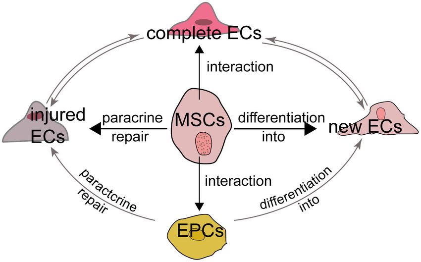
Shi et al.                                                                                                            Neovascularization of MSCs in IHD

anti-angiogenesis drugs for cancer and eye disease. Conversely,           TABLE 1 | Comparison of MSCs from different sources.
the use of angiogenesis factors, such as VEGF and basic
                                                                                                        BMSCs              AMSCs              UCMSCs
fibroblast growth factor (bFGF), has been shown to promote
notable increases in collateral vessel and myocardial perfusion in        Differentiation capacity
ischemic myocardium, reduced infarct size and improved cardiac            Osteogenesis                    ++                  +                +++
function (2), demonstrating the theoretical and experimental              Chondrogenesis                  ++                  +                +++
promise of this approach in treating ischemic diseases.                   Adipogenesis                     +                 ++                +++
Unfortunately, despite the exciting results obtained using                Endothelial cells                +                  +                   +
angiogenesis factors to treat IHD, gene therapy is also limited by        Pericytes                        +                  +                   +
its restricted efficacy and resistance (3). For example, VEGF also        Smooth muscle cells              +                  +                   +
accelerates angiogenesis in atherosclerotic plaques and promotes
                                                                          Proliferation capacity           +                 ++                +++
plaque growth, which may eventually lead to plaque instability,
                                                                          Migration capacity            +++                   +                  ++
while it promotes angiogenesis in ischemic tissue, an observation
                                                                          Tube formation                   +                  +                   +
referred to as the famous Janus phenomenon (4). Angiogenesis
greatly improves blood flow in myocardial ischemia, but the
safety of growth factor-based angiogenesis therapy is an issue that
remains to be overcome. Thus, how to avoid the risks associated           most popular MSCs in clinical and preclinical experiments
with angiogenesis therapy is a problem that must be considered.           and trials, and some of their capabilities are compared below
    Stem cell-based therapies provide a promising new method for          (Table 1).
the formation of new blood vessels. MSCs have become the most
promising seed cells for the treatment of IHD, with advantages            Differentiation Capacity
of rapid self-renewal, multidifferentiation potential, and weak           MSCs have the ability to differentiate into adipocytes, osteoblasts
immunogenicity in autologous transplantation. Clinical and                and chondroblasts. The amount of calcium deposits and
preclinical studies have shown that MSCs therapy effectively              sulfated proteoglycans stained by Alizarin red and Alcian blue,
limits the infarcted area and improves heart function. However,           respectively were both higher in BMSCs than that observed in
the mechanisms associated with the activities of MSCs in                  AMSCs, indicating that BMSCs have a higher capacity toward
IHD therapy remain controversial. We primarily attribute the              osteogenic and chondrogenic differentiation than AMSCs.
cardiac protective effect of MSCs to their ability to promote             While similar adipogenic differentiation potential was observed
neovascularization for the following two reasons. First, MSCs             between these two types of cells (6), some studies have reported
secrete soluble paracrine factors that contribute to angiogenesis         that AMSCs are more prone to adipogenic differentiation
and vasculogenesis. Second, MSCs are able to differentiate into           than BMSCs (7). Baksh et al. (8) observed that compared to
ECs, pericytes and smooth muscle cells (SMCs), which form                 BMSCs, UCMSCs underwent osteogenic differentiation more
the foundation of vessels, processes that both participate in the         rapidly, exhibited higher alkaline phosphatase activity, and
protective ability of MSCs toward IHD. In this review, we focus           generated significantly more fat-containing cells when grown
on the mechanisms and clinical applications of MSCs in IHD                under adipogenic conditions by day 21. The differentiation
therapy through neovascularization to provide reference for the           ability of stem cells is affected by donor sex, age, isolation and
application of stem cells in IHD.                                         culture conditions, etc. (9). Thus, which types of MSCs have a
                                                                          greater ability to differentiate into adipocytes, osteoblasts and
                                                                          chondroblasts remains disputed. In addition, MSCs also have the
COMPARISON OF MSCS FROM                                                   ability to differentiate into ECs, pericytes and SMCs, which are
DIFFERENT SOURCES                                                         necessary components of blood vessels (10–13). Lu et al. (14)
                                                                          showed that MSCs from adipose tissue may have significantly
MSCs can be isolated from bone marrow, adipose tissue,                    greater ability to promote angiogenesis both in vitro and in vivo
umbilical cord blood, peripheral blood and almost every tissues           than UCMSCs and endometrial MSCs.
in adults. Although MSCs can be harvested from different
sources, regardless of their origin, they all have the capability         Proliferation Capacity
of differentiating into adipocytes, osteoblasts and chondroblasts         MSCs from different tissue sources do not have the same
in vitro under specific conditions and can adhere to plastic              proliferative ability in vitro. Choudhery et al. (15) observed that
under culture conditions. Furthermore, the surface of MSCs                UCMSCs have higher population doublings than AMSCs (33.0
displays CD73, CD90, and CD105 but lack CD34, CD45, HLA-                  ± 1.5 vs. 25.8 ± 0.6), with the doubling time being longer for
DR, CD14 or CD11b, CD79a or CD19. The International Society               AMSCs (2.7 ± 0.03 days) than UCMSCs (2.0 ± 0.04 days).
for Cell Therapy proposed the three criteria described above              Moreover, after prolonged passaging (30 times), the proliferative
as identification standards for MSCs (5). Although MSCs from              ability of UCMSCs did not change significantly, while BMSCs
different sources share many of the same biological features, there       showed decreased proliferation after 6 passages (8), indicating
are also some differences between distinct MSC populations.               that UCMSCs have a stronger proliferative ability than BMMSCs
Bone marrow-derived MSCs (BMSCs), adipose-derived MSCs                    and AMSCs. Under human platelet lysate-supplemented culture
(AMSCs) and umbilical cord-derived MSCs (UCMSCs) are the                  conditions, AMSCs were observed to have greater proliferative

Frontiers in Cardiovascular Medicine | www.frontiersin.org            2                                         January 2021 | Volume 8 | Article 633300
Shi et al. Neovascularization of MSCs in IHD
potential than BMSCs (6). Therefore, the proliferative ability of after MSCs intervention. Which suggested the efficacy of MSCs
UCMSCs is the strongest, followed by AMSCs and BMSCs. did not benefited from themselves in some degree.
Paracrine hypothesis was firstly advanced by Gnecchi et al.
Migration Capacity (27). They found genetically modified BMSCs overexpressing
MSCs play an important role in posttraumatic tissue repair and the Akt1 released paracrine factors that exert cytoprotective
cell therapy, and their migration ability is a key factor affecting effects on cardiomyocytes exposed to hypoxia and limited infarct
their therapeutic efficacy. The migration capacity of BMSCs size and improved ventricular function (27, 28). Furthermore,
and placenta-derived MSCs (PMSCs) was observed to be 5.9- high VEGF, bFGF, IGF-1 and SDF-1 expression in hypoxia-
and 3.2-fold higher than that of UCMSCs, respectively. These preconditioned MSCs medium was examined, the results of
results were consistent with the observed levels of migration- which indicated that the paracrine function of MSCs may
enhancing proteins in UCMSCs, including cathepsin B, cathepsin play more important role than their differentiation ability (26).
D and prohibitin, which were significantly lower than those Recently, it has been reported that MSCs secreted a wide array
observed in BMSCs and PMSCs, while the levels of migration- of cytokines that exerted beneficial angiogenesis in ischemic
inhibiting proteins such as plasminogen activator inhibitor-1 and tissue, including PDGF, thrombopoietin, and angiogenin (29, 30).
manganese superoxide dismutase were higher (16). Vimentin These factors are all involved in the neovascularization process,
also contributed to the higher migration capability of BMSCs including MSC mobilization, migration, homing, adhesion and
than UCMSCs (17). In contrast, UCMSCs exhibited an enhanced retention, and the differentiation of ECs. Especially VEGF and
migration capacity toward factors released by hepatocellular bFGF, which both have high affinity toward heparin and
carcinoma compared with BMSCs (18). participate in angiogenic processes such as migration and
amplification of ECs, are also necessary substances to induce
Capacity of MSCs to Promote Tube the transformation of stem cells into ECs (10, 31). MSCs
Formation of ECs overexpressing Akt and angiopoietin-1 showed higher Flk1
Tube formation is the last step in the formation of vessels and and Flt1 positivity and promoted intrinsic Flk1+ and Flt1+
is necessary to supply blood for ischemia. Pill et al. (19) showed cell mobilization into the infarcted heart (32). Huang et al.
that AMSCs and BMSCs are both promising cell types to induce (33) observed that overexpression of miR-126 promoted the
vascularization with ECs in vitro and are promising candidates differentiation of MSCs toward ECs through activation of
to support in vivo vascularization. Nevertheless, Kim et al. (20) the PI3K/Akt and MAPK/ERK pathways and the release
observed that conditioned medium from human AMSCs showed of VEGF and bFGF factors. Therefore, paracrine factors
better tube formation-promoting effects than that from BMSCs secreted by MSCs may have pivotal functions throughout the
in vitro, and AMSC group showed better recovery of blood flow neovascularization process. The role of various secretory factors
than BMSC group in hindlimb ischemia model of nude mice. in the neovascularization process will be discussed below.
Furthermore, young AMSCs may have a higher tube formation
capacity than old ones (21). UCMSCs are also capable of forming INVOLVEMENT OF MSCS IN THE
tubular networks (22). Du et al. (23) reported tube numbers of
11.65 ± 2.92, 0.91 ± 0.76 and 0.41 ± 0.20 for BMSC, AMSC NEOVASCULARIZATION PROCESS
and UMSC groups, respectively, indicating that BMSCs may have
The process by which MSCs promote neovascularization involves
better angio-vasculogenic capacities than UMSCs and AMSCs.
in a number of steps. First, once ischemia occurs which also
In contrast, Panepucci et al. (24) thought that UCMSCs would
follows stress change, MSCs can perceive the associated changes
be more committed to angiogenesis and BMMSCs would be
and are mobilized from their niches to migrate and adhere
more committed to osteogenesis. Above all, there is still no
to ischemic tissue to proliferate and differentiate. Notably,
consensus on which cell type has the greater capacity to promote
MSCs secrete various factors, including chemokines and growth
tube formation.
factors, and this paracrine function is carried out throughout
the neovascularization process (Figure 1). The completion of
PARACRINE FUNCTION OF MSCS all biological processes depends on the cooperation of different
THROUGHOUT THE types of cells, and the neovascularization process requires the
collaboration of ECs, endothelial progenitor cells (EPCs) and
NEOVASCULARIZATION PROCESS
pericytes. In addition, exosomes derived from MSCs act as a
The mechanism of MSC therapy is still controversial, because few messenger that participate in cell-to-cell communication.
MSCs can be found in myocardium after injection in vivo study.
Wang et al. (25) showed that most intravenously injected MSCs Environmental Perception by MSCs
remain in the lungs and liver, with only a small portion reaching Perception of Hypoxia by MSCs
the myocardial tissue. Similarly, Uemura et al. (26) observed Despite the benefits of MSC transplantation in cardiac
only a few GFP-labeled MSCs in the periinfarct myocardium. tissue, detailed in vivo observations have shown that MSCs
Even so, clinical and preclinical studies still indicated the cardiac only survive for a brief period after engraftment due to
function of ischemic heart was improved, and infarct size and the harsh microenvironmental conditions (including ischemia,
number of apoptotic cardiomyocytes were significantly reduced inflammation and anoikis) in the infarcted myocardium (34).
Frontiers in Cardiovascular Medicine | www.frontiersin.org 3 January 2021 | Volume 8 | Article 633300Shi et al. Neovascularization of MSCs in IHD
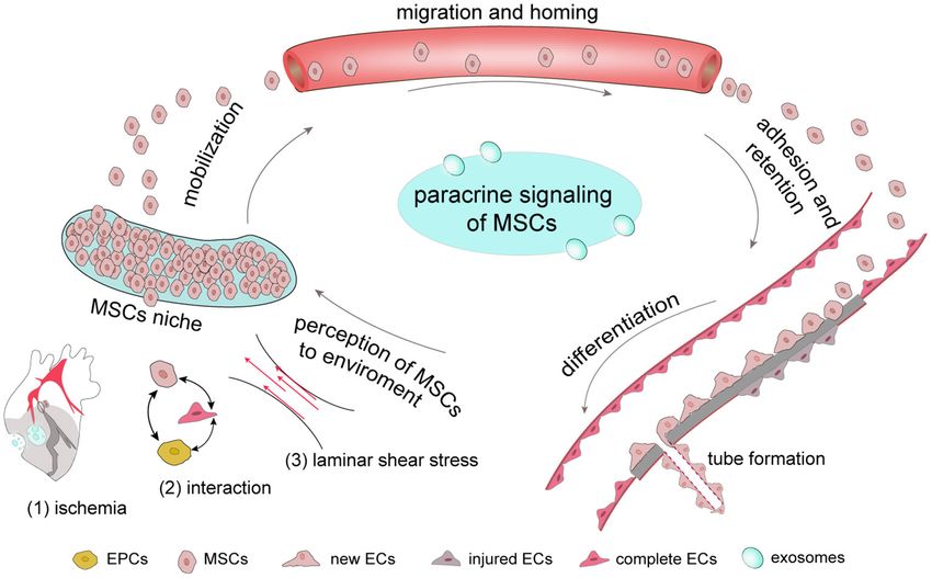
FIGURE 1 | The complete process by which MSCs respond to ischemia In general, MSCs are stored in their niches, which retain adult stem cell in a dormant state.
Once tissue is damaged, signals, including those involve in ischemia-associated pathways, cell-cell interaction and stress mobilize stem cells to migrate from the
stem-cell niche to damaged tissues, where they adhere, self-renew and differentiate. Once ischemia occurs, MSCs have the ability to secrete a number of growth
factors through their paracrine function to promote new tube formation of ECs to provide new blood for ischemic tissue.
However, this environment contributes to the mobilization of in myocardial neovascularization after myocardial infarction in
MSCs from their niches. rats (39).
MSCs originated from the bone marrow microenvironmental
niche exhibit low oxygen tension. O2 is a necessary factor Cell-Cell Interactions
in the maintenance of cell life as the final receptor in the In 1997, Asahara et al. (42) identified and named a small
intracellular aerobic respiration electron transport chain and is a population of CD34+ cells as “EC progenitors.” Indeed, EPCs
substrate of some enzymes. Once the supply of O2 is insufficient, are involved in a number of processes during angiogenesis,
the hypoxia signal will be rapidly transmitted to nucleus and including mobilization, differentiation into ECs, homing,
initiate related gene expression to maintain oxygen homeostasis paracrine function and others (43, 44). Coculture of EPCs
and the balance of energy metabolism between the cells and and MSCs significantly increased the transcription levels
organism. Hypoxia inducible factor 1 (HIF-1), which has a of endothelial specific markers, including vWF, CD31, VE-
dimeric complex composed of HIF-1a and HIF-b subunits, is cadherin, Flk-1 and Flt-1 (45) and enhanced tube-like formation
oxygen-sensitive and the most important transcription factor (46) through platelet derived growth factor (PDGF), Notch
affecting gene regulation under hypoxia (35). Once ischemia and TACE/TNF alpha signaling (45, 47). Joensuu et al. (48)
occurs, HIF-1 increases the expression of angiogenesis-associated noted that in cocultures of human MSCs and peripheral blood
genes, including VEGF, its receptors Flk-1 and Flt-1, bFGF and mononuclear cells, the previously nonadherent cells attached
the fibrinogen system (36, 37). At the same time, HIF-1 improves and started to elongate and form tube-like structures within
the expression of proteases, such as membrane type matrix 1 week concomitant with VEGFR1 upregulation, and platelet
metalloproteinases, which hydrolyzes extracellular protein to endothelial cell adhesion molecule 1 (PECAM-1) and endoglin-
promote cell migration, matrix reconstruction and the formation positive vessel-like structures were observed after 20 days.
of tubule-like structures (38). In addition, MSC-EC interactions were observed to decrease
Hypoxia is also a basic aspect of the microenvironment endothelial permeability induced by lipopolysaccharide through
that determines the differentiation of MSCs. Compared with hepatocyte growth factor (HGF) by restoring the integrity of
a normoxia group, VEGF expression in embryonic and MSCs endothelial monolayers and remodeling endothelial intercellular
under hypoxia was observed to be significantly increased (39– junctions (49). VEGF secreted by stem cells from apical papilla is
41). Likewise, the in vivo administration of hypoxia-inducible also used by human umbilical vein endothelial cells to increase
VEGF-engineered MSCs was shown to induce ischemia- the number of endothelial tubules, tubule lengths, and branching
responsive VEGF production and lead to a significant increase points (50).
Frontiers in Cardiovascular Medicine | www.frontiersin.org 4 January 2021 | Volume 8 | Article 633300Shi et al. Neovascularization of MSCs in IHD
Laminar Shear Stress and Pulsatile Stress myocardial and vascular tissues secrete SDF-1 to attract CXCR-
There are many force-sensitive molecules on the cell surface, 4-expressing cells, particularly their therapeutic progenitors.
such as cilia, integrins, ion channels and plaque proteins. Yu et al. (62) showed that SDF-1/CXCR-4 may mediate the
Integrins connect the cytoskeleton and extracellular matrix migration of BMSCs toward heart MI through activation of
through adhesive plaque and transform the force signals into PI3K/Akt signaling. Growth factors play an important role in
intracellular biological signals through this plaque (51, 52). the process of MSC migration. The stimulation of SDF-1α
Considering the key role of shear force in the differentiation of expression in infarcted hearts by VEGF-overexpressing MSCs
ECs, researchers reported that such mechanical stimulation in was observed to result in the massive mobilization and homing
cell culture in vitro was equally effective for the differentiation of of BMSCs (59). TGF-β1, HGF, IGF-1 and endothelial nitric
stem cells into ECs (53). MSCs are highly reactive to mechanical oxide synthase (eNOS) also promoted the migration and homing
stimuli in the environment, and different types of stress on the BMSCs to the ischemic myocardium (63–65). Schmidt et al.
same MSC population will lead to different differentiation results (31) showed that low concentrations of bFGF attracted cells,
(54). After generating canine BMSCs under shear stress provided indicating that bFGF may direct the migration of MSCs. In
by a pulsatile bioreactor for 4 days, the expression of endothelial addition, Yan et al. (66) observed that C1q/tumor necrosis factor-
cell markers, such as PECAM-1, VE-cadherin and CD34 was related protein-9 (CTRP9) enhances AMSC proliferation and
observed to be significantly increased (55). Fisher et al. (56) noted migration through the ERK1/2-MMP-9 signaling pathway and
that AMSCs could form cords but failed to take up acetylated also promotes anti-apoptotic/cell survival via ERK-Nrf2/anti-
low density lipoprotein (acLDL) or express molecular markers oxidative protein expression. MiRNA, like miR-206 also involved
after being cultured in endothelial cell growth supplement. Only in migration of MSCs by targeting Pim-1 (67).
the subsequent exposure of stem cells to shear stress did the
cells exhibit realignment, acLDL uptake and CD31expression, Proliferation and Survival
indicating that stem cells differentiation to ECs requires the Although MSCs transplantation is a promising therapeutic
synergism of biochemical and shear force. approach for IHD, the low viability of MSCs after transplantation
needs to be improved. Hypoxic preconditioning may improve
the functional survival and therapeutic efficiencies of engrafted
Dynamic Process of MSCs to Repair BMSCs, at least in part through autophagy regulation (68).
Ischemic Tissue Some growth factors, including increased VEGF, TGF-β, IGF-1,
Mobilization SDF-1a and angiogenin were shown to enhance MSC survival
MSC mobilization is key for its involvement in tissue repair and vasculogenesis in an MI model (69). Preconditioning with
following their sensing of hypoxia, stress or other signals. An other factors, such as protein kinase C epsilon (εPKC), CTRP9,
anoxic environment is one of the factors that induces stem dimethyloxalylglycine and connexin-43 improves the retention
cells to migrate out of their niches. Prolyl hydroxylase (PHD) and survival of transplanted MSCs in rat MI through the SDF-
and factor inhibiting HIF-1 (FIH) are key oxygen sensors in 1/CXC and PI3K/AKT pathways (66, 70–72). Qu et al. (73)
MSCs. HIF-1α upregulation by double knockdown of PHD showed that atorvastatin, a hypolipidemic agent, has a protective
and FIH synergistically increases stem cell mobilization and effect on cardiomyocytes against apoptotic cell death in infarct
myocardial angiogenesis and improves cardiac function (57). and peri-infarct areas and could also increase the survival rate of
The high concentration of growth factors outside of stem-cell implanted BMSCs in acute myocardial ischemia.
niches may be another factor causing MSCs to mobilize from
their original niches. Stromal cell-derived factor-1 (SDF-1 α)/Cxc Adhesion and Retention
chemokine receptor 4 (CXCR-4) are part of the most important MSCs need to stay and adhere to ischemic tissue to play their
chemotactic axis regulating MSC mobilization and migration. important role in ischemic tissue repair. Molecular imaging
VEGF and insulin-like growth factor-1 (IGF-1)-overexpressing studies have shown thatShi et al. Neovascularization of MSCs in IHD
PI-3K/AKT/m-TOR/eNOS and p38/MAPK (83, 84). Hypoxia
also influences the interactions between the endothelium and
MSCs (85).
In addition to the interactions between MSCs and ECs,
studies showed MSCs had the ability to differentiate into ECs
to promote angiogenesis in some degree although it was still
controversial. For example, Otto et al. (86) did not observe
MSC transdifferentiation into cardiomyocytes, ECs or SMCs and
that the transdifferentiation of MSCs into cardiomyocytes or
vascular cells did not significantly contribute to the improvement
of cardiac function. Conversely, Silva et al. (87) showed that
BMSCs promoted the angiogenesis of dog ischemic myocardium
by differentiating into ECs, which accelerated the establishment
of collateral circulation. Studies support MSCs own the potential
to differentiated into ECs according to the below reasons. First,
FIGURE 2 | MSCs and endothelial cells The relationship between MSCs and
ECs can be summarized as follows. First, MSCs can repair the injured but not MSCs are multipotent stem cells derived from the mesoderm.
dead ECs through their paracrine function to induce the release of growth Theoretically, MSCs can be differentiated into all mesoderm
factors. Second, MSCs have the potential to differentiate into new ECs derived cells, and since ECs are mesoderm-derived cells, MSCs
although it is controversial. Third, the interaction and crosstalk between MSCs have the potential to differentiate into ECs. Second, MSCs
and existing ECs promotes the formation of new ECs and the repair of injured
ECs. Last, MSCs also interact with EPCs to influence EC formation and
express molecular markers of early ECs, such as VEGF receptor
function. 2 (VEGFR-2/Flk-1/KDR) and bFGF, indicating that ECs can
be derived from mesenchymal colonies and that MSCs arise
from precursors with angiogenic potential (31, 88). Last, a
series of in vivo and in vitro experiments proved that MSCs
(79). Bortolotti et al. (80) demonstrated that cardiotrophin-1 can differentiate into ECs. Oswald et al. (10) successfully used
(CTF1) increases the retention and adhesion of BMMSCs to 2% fetal bovine serum supplemented with 50 ng/mL VEGF to
protect BMSCs from apoptosis, identifying it as a new powerful induce BMSCs to differentiate into ECs in vitro. They observed
cytokine promoting cell engraftment. The retention and survival that differentiated cells increased the expression of endothelial-
of transplanted MSCs was also shown to be improved by the specific markers, such as KDR and VEGF receptor 1 (VEGFR-
overexpression of εPKC in AMI rats through the SDF-1/CXC and 1/Flt-1), and formed capillary-like structures. Furthermore, the
PI3K/AKT pathways (70). process of MSC differentiation into ECs may require the synergy
of bFGF, IGF, epidermal growth factor (EGF) (89, 90). ERK
INTERACTIONS AMONG MSCS AND ECS, signaling may also involve in the differentiation of porcine
EPCS AND PERICYTES AMSCs into ECs (90).
Furthermore, MSCs also function with EPCs to promote
Multiple cell types are known to be involved in the processes of tissue repair. As a precursor of ECs, EPCs also differentiate
angiogenesis and vasculogenesis, including MSCs, ECs, EPCs and into ECs and promote ischemia angiogenesis through their
pericytes. In particular, ECs are indispensable for angiogenesis paracrine function (91, 92). MSCs could attract and promote
and the relationship between MSCs and ECs mainly attribute to the migration and vascularization of EPCs, which may depend
the following aspects. First, MSCs secret growth factors which on a positive feedback loop between CXCR-2 and CXCR-
can repair the injured but not dead ECs through their paracrine 4 (93, 94). The viability and ability of MSCs to promote
function. Second, the interaction and crosstalk between MSCs nerve regeneration is also improved by EPCs through PDGF-
and existing ECs promotes the formation of new ECs and BB/PDGFR-β signaling (95). Rossi et al. (96) found MSCs
the repair of injured ECs. Third, MSCs have the potential to and EPCs into the hind limbs of ischemia model together
differentiate into new ECs although it is controversial. Last, MSCs accelerated ischemic muscle recovery through an endoglin-
also interact with EPCs and pericytes to influence EC formation dependent mechanism. Consequently, MSCs, ECs and EPCs may
and function (Figure 2). have a synergistic effect in ischemic tissue repair (Figure 2).
The ability of MSCs to limit infarct size may attributed to Pericytes, also known as mural cells, wrap around ECs in
their pro-angiogenesis activity through existing ECs (Figure 2). arterioles, capillaries and venules to regulate the maturation of
The stimulated angiogenic activity of ECs is associated with ECs, stabilize the microvascular wall and promote angiogenesis.
the secretion of various growth factors and cytokines, including Although pericytes are surrounded by a basement membrane,
VEGF, HGF, IL-6, TGF-β1 and monocyte chemoattractant they contact the ECs with through a “peg and socket” mechanism
protein-1 (81). Lu et al. (82) showed that nestin(+) BMSC through holes in the basement membrane. Studies have shown
transplantation improved cardiac function in a mouse AMI that pericytes also communicate with ECs via paracrine signaling
model by recruiting resident cardiac ECs to the infarcted border to improve tissue repair (97, 98). It is notable that pericytes
region. BMSCs also rescued injured ECs through modulation also have stem cell-like properties and exhibit the morphology,
of mitophagy or activation of signaling pathways such as mitotic activity and surface antigens of MSCs (99) and are
Frontiers in Cardiovascular Medicine | www.frontiersin.org 6 January 2021 | Volume 8 | Article 633300Shi et al. Neovascularization of MSCs in IHD
seemingly able to differentiate into adipocytes, chondrocytes, APPLICATION OF MSCS IN IHD CLINICAL
osteoblasts, neurons, astrocytes and oligodendrocytes, leading AND PRECLINICAL PRACTICE
them to be identified as MSCs (100–102). However, it is still
debated whether pericytes are MSCs. Guimaraes-Camboa et al. The results of numerous clinical and preclinical studies have
(103) challenged this concept and suggested that mural cells do indicated that MSC transplantation is safe, significantly improves
not intrinsically behave as MSCs during aging and repair in cardiac function and decreases infarct size and fibrosis in
multiple adult organs using a transgenic cell line. Over 2 years, ischemic patients, which may be associated with the survival,
the study showed that Tbx18 lineage-derived cells maintained retention, angiogenesis, paracrine action and the anti-apoptosis
their perivascular identity in the brain, heart, muscle and fat, activities of MSCs (Tables 2, 3). Although cellular therapies hold
indicating that mural cells do not exhibit an overt potential great promise for the treatment of human IHD and have good
to give rise to other cell types. In contrast, MSCs can serve safety, the efficacy of MSCs remains disputable, especially when
as a potential source of pericytes and induce vasculogenesis as used in clinical trials. Meta-analyses of randomized clinical trials
mentioned previously (13, 104, 105), but similar to the multi- showed that the transplantation of BMSCs resulted in limited
differentiation potential of MSCs, there needs to be standard improvement on cardiac function for MI patients (171, 172).
guidelines for assessing pericyte differentiation in future studies. As it was showed in Table 2, some clinical trials proved MSC
Furthermore, MSCs secrete various growth factors, including transplantation did not improve LVEF although it may limit
PDGF, which serves as a biomarker and crucial factor controlling infarct size. Different from clinical trials, MSC transplantation in
the differentiation and recruitment of pericytes (106–108). These animal experiments showed significantly elevated LVEF in most
findings indicate that MSCs may regulate the recruitment of studies (Table 3).
pericytes to injured tissue to participate in angiogenesis, but the Clinical patients are different from animal ischemia models,
associated mechanisms between MSCs and pericytes need to be and the efficacy of MSCs in clinical practice is influenced by
further elucidated. many different factors, such as (1) disease etiology and severity
of patients, and (2) the type, number, delivery route and time,
retention, survival, proliferation and differentiation of MSCs.
MSCS AND MSC-DERIVED EXOSOMES Another meta-analysis showed that MSCs are more effective
in patients with lower baseline left ventricular ejection fraction
Exosomes are a type of extracellular microvesicle secreted by (LVEF) (≤50%), and the effects of cells that were transferred
multiple eukaryotes. Compared with cell therapy, MSC-derived at 3–7 days post-AMI was superior to those transferred within
exosomes (MSC-exos) have lower immunogenicity and are 24 h or more than 7 days in improving LVEF and decreasing
safer and more efficient, providing a new strategy for tissue LV end-systolic and diastolic dimensions (173), which suggested
regeneration via cell-free therapy (109, 110). MSC-exos are a transplantation time was a key factor to influence cardiac
type of message carrier that harbor a modifiable content of function. Compared with clinical trials, animal experiments are
microRNAs, mRNAs and proteins, mediating communication easier to obtain positive results because of their simplicity, such
between cells and functioning as key mediators of the paracrine as the MI model can be established uniformly by ligation of the
effect of MSCs (111, 112). The pro-angiogenesis function of left anterior descending coronary artery. Compared with MSCs
MSC-exos has been demonstrated in a number of studies. For intervention alone, pretreated MSCs with some growth factors
instance, exosomes from MSCs overexpressing Akt, HIF-1α or together may get more efficacy (Table 3).
CXCR-4 were shown to accelerate EC proliferation, migration It is notable except for growth factors, more attention has
and tube-like structure formation in vitro, as well as blood been paid to natural botanical medicines. EGb761, an extract
vessel formation to improve cardiac function in an MI model of Ginkgo biloba, was shown to exhibit a biphasic effect on
(113–115). MSC-exosomes may also have anti-inflammatory hypoxia/serum deprivation-induced BMSC apoptosis, and its
activities in MI model (116). Currently, the application of MSC- effect was closely associated with the PI3K/Akt and caspase-9
exos primarily focuses on preclinical experiments. One of the signaling pathways (174). Salvia miltiorrhiza is a widely used
key problems for exosome clinical therapy is how to collect traditional Chinese medicine in cardiovascular diseases, and its
and purify enough exosomes so that they can be used safely. constituent Tanshinone IIA was observed to decrease infarct size
Andriolo et al. (117) developed a GMP-class method for the mass by increasing the recruitment of BMSCs to the infarct region by
preparation of stem cell-derived exosomes to enable them to be upregulating the SDF-1/CXCR-4 axis in a rat MI model (175). In
used in future clinical applications. Indeed, as they are secreted addition to botanical medicines, chemicals such as statins, as the
by MSCs, MSC-exos have similar biological properties to MSCs most commonly used lipid-lowering agents, exert activity toward
to some extent. MSC-exos also have paracrine functions and a wide spectrum of cellular functions in addition to their lipid-
mediate communication between MSCs and ECs, and they are lowering effects, including anti-inflammatory, anti-apoptotic,
also influenced by microenvironmental stress conditions, such as anti-fibrotic and pro-angiogenesis effects (176, 177). The results
hypoxia and irradiation (118, 119). Furthermore, as they harbor of multiple studies have suggested that atorvastatin has the ability
a part of and not the entirety of MSC contents, MSC-exos are not to increase the survival rate of implanted BMSCs in an MI model,
an MSC “mini-me” and cannot replace MSCs in some respects, and combined with MSCs, it also ameliorated the cardiac milieu
including their multiple differentiation and proliferation abilities. by reducing inflammatory cell infiltration, myeloperoxidase
Frontiers in Cardiovascular Medicine | www.frontiersin.org 7 January 2021 | Volume 8 | Article 633300Frontiers in Cardiovascular Medicine | www.frontiersin.org
Shi et al.
TABLE 2 | Completed clinical trials using MSCs to treat ischemic cardiovascular diseases registered at clinicaltrials.gov.
NCT Conditions Interventions Phase Patients No/ Endpoint LVEF Study Cell quantity Method Data References
follow-up improved type
NCT00950274 MI CD133+ ABMSCs III 82/6 mon LVEF, LVEDV, LVESV, scar size, Yes R,PA,D 0.5-5x10⁶ IM 2009.7–2016.3 (120)
LV mass, NT-proBNP
NCT00669227 AMI ABMCs II 42/6 mon LVEF, LVEDV, LVESV, infarct size No R,PA,D 381 × 106 ICA 2005.10–2009.1 (121)
42/36 mon LVEF, LVEDV, LVESV, infarct size, Yes R,PA,D 324 × 106 (122)
MO
NCT00893360 MI/LVD Autologous I 25/6 mon Safety, LVEF, scar mass, systolic No R,PA,N 12.5–25 × 106 ICA 2009.5–2012.2 (123)
cardiosphere-derived wall thickening
stem cells 25/13.4 mon Safety, LVEF, LVEDV, LVESV, scar Yes (124)
mass, scar size,
NCT01033617 MI/HF ABMCs(CD133+ ) – 40/ 6 mon LVEF, LVEDV,LVESV, No R,PA,Q 0.5–10 × 106 IM 2009.10–2016.6 (125)
NCT00279175 AMI ABMCs III 204/12 mon LVEF, safety Yes R,PA,D 236 × 106 ICA 2004.4–2010.10 (126, 127)
NCT00114452 MI Allogeneic hMSCs I 53/6 mon Safety, LVEF,LV remodeling Yes R,PA,D 0.5, 1.6, 5.0 × ICV 2005.2–2009.2 (128)
106 /kg
NCT01291329 STEMI Human umbilical II 116/18 mon Safety, LVEF, perfusion, Yes R,PA,Q 6 × 10⁶ ICA 2011.2–2012.7 (129)
WJ-MSC
NCT00883727 MI Allogeneic BMSCs I/II 20/2 year Safety, LVEF, perfusion, infarct No R,PA, D 2 × 10⁶ /kg ICV 2009.4–2012.8 (130)
volume
NCT00765453 MI ABMCs II 100/12 mon LVEF, infarct size, NT-proBNP No R,PA,T 59.8 (1.9CD34+ ) ICA 2008.3–2018.3 (131)
× 106
8
NCT00264316 STEMI ABMCs II 67/4 mon LVEF, LVEDV, LVESV, infarct size, No R,SA,D 304 (72MNCs) × ICA 2003.5–2005.12 (132)
systolic wall thickening 106
NCT00199823 AMI ABMMCs II 100/12 mon Prothrombotic markers, LV No R,PA,S 68 × 106 ICA 2003.9–2006.5 (133, 134)
function (0.7 × 10⁶
CD34+ )
NCT00313339 STEMI ABMSC CD34+ I 31/6 mon Safety, LVEDV, LVESV, LVEF, No R,FA,N 5, 10, 15 × ICA 2006.3–2013.3 (135)
infarct size, perfusion 106 CD34+ cells
NCT00684060 IHD /LVD ABMSC II 80/6 mon LVEF, wall motion, LV volumes, Yes R,PA,D 150 × 106 ICA 2008.7–2012.2 (136–138)
infarct size, relationship of Ratio
of CD133+ ,CD34+ cells on LVEF
NCT00684021 STEMI/ LVD ABMSC II 120/6 mon Safety, LVEF, wall motion, LV No R,PA,D 150 × 106 ICA 2008.7–2012.11 (139)
function
95/12 mon LVEF,LV volumes, infarct size No (140)
January 2021 | Volume 8 | Article 633300
NCT00355186 STEMI ABM-MNCs II 200/4 mon LVEF, LVEDV, LVESV No R,FA,N 153 (119) × 106 ICA 2006.8–2012.11 (141)
Neovascularization of MSCs in IHD
200/ 12 mon LVEF, LV volumes, scar size, No (142)
N-BNP
NCT01167751 MI BM-MNCs; CD133+ II/III 90/6,18 mon Safety, LVEF, systolic wall Yes R,PA,D MNCs:564.63 × IM 2008.1–2012.7 (143)
cells thickening 106 ;
CD133+ cells:8.19
× 106
(Continued)Frontiers in Cardiovascular Medicine | www.frontiersin.org
Shi et al.
TABLE 2 | Continued
NCT Conditions Interventions Phase Patients No/ Endpoint LVEF Study Cell quantity Method Data References
follow-up improved type
NCT00268307 AMI ABMCs I 40/6 mon LVEF, LVEDV Yes R, CA, D 100 × 106 ICA 2005.12–2010.9 (144)
NCT02439398 AMI Allogeneic hCSCs I/II 49/12 mon Safety, infarct size – R,PA,D 35 × 106 ICA 2014.6–2016.11 (145)
NCT00395811 CABG ABMMNC I/II 60/6 mon safety, LVEF, LVEDV, LVESV, Yes R,PA,D 100 × 106 Graft 2007.1–2009.6 (146)
NCT00418418 Ischemic HF ABMMC II 39/12 mon LVEF, LVEDV, LVESV, infarct size, No R,FA,Q 5–1,000 × 106 ICA 2006.10–2010.12 (147, 148)
safety,
NCT00363324 STEMI BMSCs II/III 80/6 mon LVEF, LVEDV, LVESV Yes R,PA,D 360(2.6 ICA 2005.1-2009.11 (149, 150)
CD34+)x106
NCT01495364 STEMI CD34+ II 161/ 12 mon Safety, LVEF, infarct size, No R,PA,D 10x106 ±20% ICA 2011.12-2016.4 (151)
NCT00289822 IHD BMCs/CPCs II 75/ 3 mon LVEF Yes R,CA,N BMCs: 205 × ICA 2002.1–2005.1 (152)
106 ;CPCs:22 ×
106
NCT01234181 MI Hypoxia BMSCs – 34/1 year Safety, LVEF, LVEDV, LVESV, wall No R,PA,N 10 × 106 ICA 2010.11–2012.12 (153)
motion, perfusion
NCT01076920 CMI/LVD ABMSCs I/II 10/2 year Safety, LVEF, myocardial viability Yes SA;N 61.5 × 10⁶ IM 2009.10–2014.9 (154)
and contraction
NCT01087996 ICM BMSCs I/II 30/13 mon Safety, LVEF, LVEDV, LVESV, – R,PA,N 20, 100, 200 × TE 2010.4–2011.9 (155)
immunologic monitoring, quality 106
of life, pulmonary function
9
R, Randomized; PA, Parallel Assignment; FA, Factorial Assignment; CA, Crossover Assignment; SA, Single Group Assignment; DR, Double Randomized Controlled Trial; Q, Quadruple (Participant, Care Provider, Investigator, Outcomes
Assessor); T, Triple (Participant, Care Provider, Investigator); D, Double blind; S, Single (Outcomes Assessor); N, None (Open Label); ABMMCs, Autologous Bone Marrow Mononuclear Cells; ABMCs, autologous bone-marrow
cells; hCSCs, human cardiac stem cells; LVEDV, Left ventricular end-diastolic volume; LVESV, left ventricular end-systolic volume; MO, microvascular obstruction; IHD, ischemic heart disease; LVD, left ventricular dysfunction; ICM,
ischemic cardiomyopathy.
January 2021 | Volume 8 | Article 633300
Neovascularization of MSCs in IHDFrontiers in Cardiovascular Medicine | www.frontiersin.org
Shi et al.
TABLE 3 | Representative animal studies performed using MSCs in ischemic models.
Disease Animal source cell source/ Dose Method/ Treatment/end Preclinical outcome Mechanisms Specific References
model type No. of sites time treatment
MI/IR Mice mice/ADSCs 2 × 10⁵ IM/3 0 h/- ↑LVEF, ↓fibrosis ↑ADSCs survival/retention, N-cadherin (156)
migration, angiogenesis;
↑cardiomyocyte adhesion,
proliferation;
↑MMP-10/13 and/or HGF
IR Adult male SD rats SD rat within 6–7 d/ ADSCs 2 × 10⁶ Iv/- 0, 24 ↑LVEF, ↓infarct size ↓Neutrophil number by – (157)
h/24 h,72 h,28 d enhanced
M2 macrophage and
macrophage efferocytosis
IR Female Gottingen Male Gottingen 2 × 10⁷ IM/- 1.5–2 h/3 d, 7 d, ↑LVEF, ↓infarct size ↑Macrophage and T-cell – (158, 159)
mini-swine mini-swine/Cortical bone 3 mon populations;
stem cells ↓cardiomyocyte apoptosis
MI Male C57 mice Human umbilical cord 2 × 10⁵ IM/4 0/- Protects cardiac ↓Apoptosis and autophagy Exo-SDF1 (160)
blood-MSCs function (↓infarct size) of
myocardial cells; ↑tube
formation of ECs
MI Male C57/BL6 mice -/MSCs 2 × 10⁵ IM/5 30 min/1, 28 d ↑LVEF, ↓LVEDV, LVESV, ↑Autophagic flux through Exo- MSCs (161)
scar size, exosome containing mainly
miR-125b-5p
IR MI Female Göttingen Male Yorkshire swine 200 × 106 /1 TE/10 -/3 mon ↑LVEF, perfusion, ↑Cardiac regeneration – (162)
10
swine MSCs/CSCs × 106 ↓LVEDV, LVESV, scar
size, remodeling
MI C57/BL6 mice; CTRP9 EGFP-TG mice with 1 × 10⁵ IM/3 1, 3, 7, 14 d/3 d, ↑LVEF, fibrotic area ↑Proliferation/ CTRP9 (66)
knock-out mice C57BL/6J 4w migration by
background/ADSCs ERK1/2-MMP-9, ↓poptotic
/oxidant via ERK-Nrf2
MI C57/BL6 Mice male C57/BL6/BM-MSCs, 2 × 10⁵ IM/1 -/IM after 1, 7, ↑LVEF, LVFS ↑BM-MSC adhesion, CTF1 (80)
ATMSCs 14, and 21 d, LV ↓apoptosis, ↑focal
after 21, 60 d adhesion kinase
I/R Female Large White male Large White 10 × 106 ICA/- 15 min after ↑Myocardial perfusion Pro-angiogenic factors: – (163)
pigs pigs/ATMSCs reperfusion/2 (vascular density), not VEGF,SDF-1a, GM-CSF;
days, 60 d LV Anti-apoptotic, inflammatory
Function and collagen deposition
MI SD rats Aged and young male 10 × 106 IM/5 30 min/28 d ↑Angiogenesis/ survival; SRT1720 (164)
January 2021 | Volume 8 | Article 633300
↑LVEF, ↓fibrosis/scar
hMSCs size against apoptosis
Neovascularization of MSCs in IHD
MI C57BL/6 mice Synthaetic hBMSCS 1 × 10⁵ IM/- 0/15 d Mitigated LV ↑Angiogenesis – (165)
remodeling
MI Male SD Rats SD rats /BMSCs 22 × 106 Tail vein 2 d/IF: weeks 1, ↑Cardiac function ↑Angiogenesis by the VEGF- (166)
2, 4; LV: 3 d, (LVEF, LVDD, LVSD) tropism of MSCs to the MI encapsulated
1,3,6 w area through SDF-1
AMI Female Large White Pigs /ATMSCs 50 × 106 IM /7-8 0/2, 15, 30 d Cardiac function not ↓Inflammation, ↑angiogenic IGF-1 /HGF (167)
pigs significantly improved process
(LVEF)
(Continued)Frontiers in Cardiovascular Medicine | www.frontiersin.org
Shi et al.
TABLE 3 | Continued
Disease Animal source cell source/ Dose Method/ Treatment/end Preclinical outcome Mechanisms Specific References
model type No. of sites time treatment
MI Male Corriedale sheep Corriedale male sheep/ 20 × 106 IM/- 30 min/30, 60 d ↓Infarct volume; ↑LVEF ↑Angio-/arteriogenesis, HIF1-a (168)
BMSCs ↓apoptosis by
HIF1-mediated
overexpression of EPO,
iNOS, VEGF, and ANG-1
CMI Female pigs Human umbilical 30 × 106 ICA/ICV/- 4, 5, 6 w/ 4 w ↑LVEF, perfusion; ↑Angiogenesis by VEGF – (169)
cord-derived MSCs ↓apoptosis, fibrosis and Ang
MI Male Cynomolgus Cynomolgus 10 × 106 IM/5 0/3, 28, 90, 180, ↑LV function, ↓infarct ↑Cardiomyocyte Hypoxia (170)
monkeys monkey/BMSCs 270 d size proliferation,
vascular density, myocardial
glucose uptake,
engraftment, paracrine
activity(EPO, HIF1-α,
ANG-1);
↓endogenous cell apoptosis
AMI SD rat Male SD rat/BMMSCs 1 × 10⁶ IM/5 0/1 day, 4 w ↑LVEF, ↓LVEDD, ↑Retentionl:SDF-1/CXC and PKCε (70)
LVESD), remodeling PI3K/AKT; ↑survival:VEGF,
improved bFGF, TGFβ, cTnI, vWF,
SMA and factor VIII
ICA, intra-coronary artery infusion; ICV, intra-coronary vein infusion; IM, intramyocardial; TE, transendocardial; ↑, indicators increased or improved; ↓, indicators decreased or worsened.
11
January 2021 | Volume 8 | Article 633300
Neovascularization of MSCs in IHDShi et al. Neovascularization of MSCs in IHD
activity and cardiac fibrosis (178, 179). Furthermore, activation Hypoxia or other growth factors used to precondition stem
of the JAK-STAT pathway may play important role in the ability cells may allow MSC survival and retention to be improved, but
of rosuvastatin to increase the efficacy of transplanted MSCs additional comparisons and a set of standards are needed to
(178). Other factors, including glucagon-like peptide-1-eluting identify the most powerful factors.
(GLP-1), lipopolysaccharide and lysophosphatidic acid also exert Another important factor limiting the clinical application
pro-angiogenesis effect by promoting the enhanced expression of of stem cells is the shortage of effective monitoring methods
cytokines and growth factors (180–183). for stem cells. The successful implementation of cell therapies
requires a better understanding of cell fate after transplantation.
Currently, there are three primary labeling methods for
STRATEGIES AND FUTURE DIRECTIONS
stem cells, including reporter genes, fluorescent dyes and
MSCs display robust reparative properties through their nanoparticles, which require optical imaging, MRI and
paracrine and differentiation abilities that can limit apoptosis, radionuclide imaging to trace the transplanted stem cells,
enhance neovascularization and direct positive tissue respectively or in combination, with each technique having
remodeling. However, some problems with MSCs remain its advantages and disadvantages (74, 184). Thus, there
and must be solved before they can have widespread use. is an urgent need to develop a nontoxic and noninvasive
MSCs are important infiltrating cells that are also drived by tracer technology that exhibits long term stability and that
blood and vasoconstriction. So the first problem is the low can also be used to dynamically monitor the survival status of
survival and retention of transplanted cells in vivo which limits transplanted cells with respect to processes such as migration and
their overall effectiveness in clinical usage. Consequently, differentiation in vivo.
identifying strategies to improve cell survival and retention
in vivo is a priority. However, cell transplantation is affected
by many factors, each of which may have an impact on the
AUTHOR CONTRIBUTIONS
survival of transplanted cells, and there is still no consistent WS contributed to the design and manuscript writing. QX,
recommendation for each factor. The microenvironment of RY, and YY collected and assembled data. WC contributed
transplanted cells directly affects the survival of stem cells. The the review design and financial support. KC was responsible
blood supply in the marginal area of myocardial infarction is for proofreading and final approval of the review. All authors
well known to directly affect the survival rate and recovery of contributed to the article and approved the submitted version.
cardiac function after cell transplantation. One important goal
of cell transplantation is to promote angiogenesis in the ischemic
area and reduce the generation of myocardial scars. Studies have FUNDING
shown that hypoxia-induced stem cells release a variety of factors
to improve the microenvironment through anti-inflammatory This work was supported by the National Natural Science
and anti-fibrosis effects and by promoting angiogenesis (170). Foundation of China (No. 81803771).
REFERENCES donors display distinct immunophenotypic profiles. J Cell Physiol. (2011)
226:843–51. doi: 10.1002/jcp.22408
1. Carmeliet P, Jain RK. Molecular mechanisms and clinical applications of 8. Baksh D, Yao R, Tuan RS. Comparison of proliferative and multilineage
angiogenesis. Nature. (2011) 473:298–307. doi: 10.1038/nature10144 differentiation potential of human mesenchymal stem cells derived
2. Zachary I, Morgan RD. Therapeutic angiogenesis for cardiovascular from umbilical cord and bone marrow. Stem Cells. (2007) 25:1384–92.
disease: biological context, challenges, prospects. Heart. (2010) 97:181–9. doi: 10.1634/stemcells.2006-0709
doi: 10.1136/hrt.2009.180414 9. Zhu M, Kohan E, Bradley J, Hedrick M, Benhaim P, Zuk P. The effect
3. Henry TD, Annex BH, McKendall GR, Azrin MA, Lopez JJ, Giordano of age on osteogenic, adipogenic and proliferative potential of female
FJ, et al. The VIVA trial: vascular endothelial growth factor in adipose-derived stem cells. J Tissue Eng Regen M. (2009) 3:290–301.
ischemia for vascular angiogenesis. Circulation. (2003) 107:1359–65. doi: 10.1002/term.165
doi: 10.1161/01.CIR.0000061911.47710.8A 10. Oswald J, Boxberger S, Jorgensen B, Feldmann S, Ehninger G, Bornhauser
4. Epstein SE. Janus phenomenon: the interrelated tradeoffs inherent M, et al. Mesenchymal stem cells can be differentiated into endothelial cells
in therapies designed to enhance collateral formation and those in vitro. Stem Cells. (2004) 22:377–84. doi: 10.1634/stemcells.22-3-377
designed to inhibit atherogenesis. Circulation. (2004) 109:2826–31. 11. Gu W, Hong X, Le Bras A, Nowak WN, Issa BS, Deng J, et al. Smooth
doi: 10.1161/01.CIR.0000132468.82942.F5 muscle cells differentiated from mesenchymal stem cells are regulated by
5. Dominici M, Le Blanc K, Mueller I, Slaper-Cortenbach I, Marini FC, Krause microRNAs and suitable for vascular tissue grafts. J Biol Chem. (2018)
DS, et al. Minimal criteria for defining multipotent mesenchymal stromal 293:8089–102. doi: 10.1074/jbc.RA118.001739
cells. The International Society for Cellular Therapy position statement. 12. Jeon ES, Park WS, Lee MJ, Kim YM, Han J, Kim JH. A Rho
Cytotherapy. (2006) 8:315–7. doi: 10.1080/14653240600855905 kinase/myocardin-related transcription factor-A-dependent mechanism
6. Li C, Wu X, Tong J, Yang X, Zhao J, Zheng Q, et al. Comparative analysis underlies the sphingosylphosphorylcholine-induced differentiation
of human mesenchymal stem cells from bone marrow and adipose tissue of mesenchymal stem cells into contractile smooth muscle cells.
under xeno-free conditions for cell therapy. Stem Cell Res Ther. (2015) 6:55. Circ Res. (2008) 103:635–42. doi: 10.1161/CIRCRESAHA.108.
doi: 10.1186/s13287-015-0066-5 180885
7. Pachón-Peña G, Yu G, Tucker A, Wu X, Vendrell J, Bunnell BA, et al. Stromal 13. Wang HH, Cui YL, Zaorsky NG, Lan J, Deng L, Zeng XL, et al. Mesenchymal
stem cells from adipose tissue and bone marrow of age-matched female stem cells generate pericytes to promote tumor recurrence via vasculogenesis
Frontiers in Cardiovascular Medicine | www.frontiersin.org 12 January 2021 | Volume 8 | Article 633300Shi et al. Neovascularization of MSCs in IHD
after stereotactic body radiation therapy. Cancer Lett. (2016) 375:349–59. 31. Schmidt A, Ladage D, Schinköthe T, Klausmann U, Ulrichs C, Klinz F, et al.
doi: 10.1016/j.canlet.2016.02.033 Basic fibroblast growth factor controls migration in human mesenchymal
14. Lu H, Wang F, Mei H, Wang S, Cheng L. Human adipose mesenchymal stem cells. Stem Cells. (2006) 24:1750–8. doi: 10.1634/stemcells.2005-0191
stem cells show more efficient angiogenesis promotion on endothelial 32. Lai VK, Afzal MR, Ashraf M, Jiang S, Haider HK. Non-hypoxic stabilization
colony-forming cells than umbilical cord and endometrium. Stem Cells Int. of HIF-Iα during coordinated interaction between Akt and angiopoietin-1
(2018) 2018:7537589. doi: 10.1155/2018/7537589 enhances endothelial commitment of bone marrow stem cells. J Mol Med.
15. Choudhery MS, Badowski M, Muise A, Harris DT. Comparison of human (2012) 90:719–30. doi: 10.1007/s00109-011-0852-1
mesenchymal stem cells derived from adipose and cord tissue. Cytotherapy. 33. Huang F, Fang Z, Hu X, Tang L, Zhou S, Huang J. Overexpression
(2013) 15:330–43. doi: 10.1016/j.jcyt.2012.11.010 of miR-126 promotes the differentiation of mesenchymal stem cells
16. Li G, Zhang X, Wang H, Wang X, Meng C, Chan C, et al. Comparative toward endothelial cells via activation of PI3K/Akt and MAPK/ERK
proteomic analysis of mesenchymal stem cells derived from human bone pathways and release of paracrine factors. Biol Chem. (2013) 394:1223–33.
marrow, umbilical cord, and placenta: Implication in the migration. doi: 10.1515/hsz-2013-0107
Proteomics. (2009) 9:20–30. doi: 10.1002/pmic.200701195 34. Lu W, Xie Z, Tang Y, Bai L, Yao Y, Fu C, et al. Photoluminescent mesoporous
17. Huang L, Niu C, Willard B, Zhao W, Liu L, He W, et al. Proteomic analysis silicon nanoparticles with siCCR2 improve the effects of mesenchymal
of porcine mesenchymal stem cells derived from bone marrow and umbilical stromal cell transplantation after acute myocardial infarction. Theranostics.
cord: implication of the proteins involved in the higher migration capability (2015) 5:1068–82. doi: 10.7150/thno.11517
of bone marrow mesenchymal stem cells. Stem Cell Res Ther. (2015) 6:77. 35. Ramirez-Bergeron DL, Simon MC. Hypoxia-inducible factor and the
doi: 10.1186/s13287-015-0061-x development of stem cells of the cardiovascular system. Stem Cells. (2001)
18. Bayo J, Fiore E, Aquino JB, Malvicini M, Rizzo M, Peixoto E, et al. 19:279–86. doi: 10.1634/stemcells.19-4-279
Human umbilical cord perivascular cells exhibited enhanced migration 36. Kanichai M, Ferguson D, Prendergast PJ, Campbell VA. Hypoxia promotes
capacity towards hepatocellular carcinoma in comparison with bone marrow chondrogenesis in rat mesenchymal stem cells: A role for AKT and
mesenchymal stromal cells: a role for autocrine motility factor receptor. hypoxia-inducible factor (HIF)-1α. J Cell Physiol. (2008) 216:708–15.
Biomed Res Int. (2014) 2014:1–9. doi: 10.1155/2014/837420 doi: 10.1002/jcp.21446
19. Pill K, Hofmann S, Redl H, Holnthoner W. Vascularization mediated 37. Koong AC, Denko NC, Hudson KM, Schindler C, Swiersz L, Koch C,
by mesenchymal stem cells from bone marrow and adipose tissue: a et al. Candidate genes for the hypoxic tumor phenotype. Cancer Res.
comparison. Cell Regener. (2015) 4:4–8. doi: 10.1186/s13619-015-0025-8 (2000) 60:883–7. Available online at: https://cancerres.aacrjournals.org/
20. Kim Y, Kim H, Cho H, Bae Y, Suh K, Jung J. Direct comparison of human content/60/4/883.long
mesenchymal stem cells derived from adipose tissues and bone marrow in 38. Kajita M, Itoh Y, Chiba T, Mori H, Okada A, Kinoh H, et al. Membrane-type
mediating neovascularization in response to vascular ischemia. Cell Physiol 1 matrix metalloproteinase cleaves CD44 and promotes cell migration. J Cell
Biochem. (2007) 20:867–76. doi: 10.1159/000110447 Biol. (2001) 153:893–904. doi: 10.1083/jcb.153.5.893
21. Efimenko A, Starostina E, Kalinina N, Stolzing A. Angiogenic properties of 39. Kim SH, Moon HH, Kim HA, Hwang KC, Lee M, Choi D. Hypoxia-
aged adipose derived mesenchymal stem cells after hypoxic conditioning. J inducible vascular endothelial growth factor-engineered mesenchymal stem
Transl Med. (2011) 9:10. doi: 10.1186/1479-5876-9-10 cells prevent myocardial ischemic injury. Mol Ther. (2011) 19:741–50.
22. Arutyunyan I, Fatkhudinov T, Kananykhina E, Usman N, Elchaninov A, doi: 10.1038/mt.2010.301
Makarov A, et al. Role of VEGF-A in angiogenesis promoted by umbilical 40. Han Y, Kuang S, Gomer A, Ramirez-Bergeron DL. Hypoxia influences
cord-derived mesenchymal stromal/stem cells: in vitro study. Stem Cell Res the vascular expansion and differentiation of embryonic stem cell cultures
Ther. (2016) 7:46. doi: 10.1186/s13287-016-0305-4 through the temporal expression of vascular endothelial growth factor
23. Du WJ, Chi Y, Yang ZX, Li ZJ, Cui JJ, Song BQ, et al. Heterogeneity receptors in an ARNT-dependent manner. Stem Cells. (2010) 28:799–809.
of proangiogenic features in mesenchymal stem cells derived from bone doi: 10.1002/stem.316
marrow, adipose tissue, umbilical cord, and placenta. Stem Cell Res Ther. 41. Prado-Lopez S, Conesa A, Armià à N A, Martà Nez-Losa M, Escobedo-
(2016) 7:163. doi: 10.1186/s13287-016-0418-9 Lucea C, Gandia C, et al. Hypoxia promotes efficient differentiation of
24. Panepucci RA, Siufi JL, Silva WJ, Proto-Siquiera R, Neder L, Orellana M, human embryonic stem cells to functional endothelium. Stem Cells. (2010)
et al. Comparison of gene expression of umbilical cord vein and bone 28:799–809. doi: 10.1002/stem.295
marrow-derived mesenchymal stem cells. Stem Cells. (2004) 22:1263–78. 42. Asahara T, Murohara T, Sullivan A, Silver M, van der Zee R, Li T, et al.
doi: 10.1634/stemcells.2004-0024 Isolation of putative progenitor endothelial cells for angiogenesis. Science.
25. Wang C, Cheng L, Xu H, Liu Z. Towards whole-body imaging at the (1997) 275:964–7. doi: 10.1126/science.275.5302.964
single cell level using ultra-sensitive stem cell labeling with oligo-arginine 43. Hristov M, Erl W, Weber PC. Endothelial progenitor cells: mobilization,
modified upconversion nanoparticles. Biomaterials. (2012) 33:4872–81. differentiation, and homing. Arterioscler Thromb Vasc Biol. (2003) 23:1185–
doi: 10.1016/j.biomaterials.2012.03.047 9. doi: 10.1161/01.ATV.0000073832.49290.B5
26. Uemura R, Xu M, Ahmad N, Ashraf M. Bone marrow stem cells prevent left 44. Laurenzana A, Fibbi G, Margheri F, Biagioni A, Luciani C, Del
ventricular remodeling of ischemic heart through paracrine signaling. Circ RM, et al. Endothelial progenitor cells in sprouting angiogenesis:
Res. (2006) 98:1414–21. doi: 10.1161/01.RES.0000225952.61196.39 proteases pave the way. Curr Mol Med. (2015) 15:606–20.
27. Gnecchi M, He H, Liang OD, Melo LG, Morello F, Mu H, et al. doi: 10.2174/1566524015666150831131214
Paracrine action accounts for marked protection of ischemic heart 45. Xu J, Liu X, Chen J, Zacharek A, Cui X, Savant-Bhonsale S, et al. Cell-cell
by Akt-modified mesenchymal stem cells. Nat Med. (2005) 11:367–8. interaction promotes rat marrow stromal cell differentiation into endothelial
doi: 10.1038/nm0405-367 cell via activation of TACE/TNF-α Signaling. Cell Transplant. (2010) 19:43–
28. Gnecchi M, He H, Noiseux N, Liang OD, Zhang L, Morello F, et al. Evidence 53. doi: 10.3727/096368909X474339
supporting paracrine hypothesis for Akt-modified mesenchymal stem cell- 46. Aguirre A, Planell JA, Engel E. Dynamics of bone marrow-derived
mediated cardiac protection and functional improvement. FASEB J. (2006) endothelial progenitor cell/mesenchymal stem cell interaction in co-culture
20:661–9. doi: 10.1096/fj.05-5211com and its implications in angiogenesis. Biochem Bioph Res Co. (2010) 400:284–
29. Danieli P, Malpasso G, Ciuffreda MC, Cervio E, Calvillo L, Copes F, et al. 91. doi: 10.1016/j.bbrc.2010.08.073
Conditioned medium from human amniotic mesenchymal stromal cells 47. Liang T, Zhu L, Gao W, Gong M, Ren J, Yao H, et al. Coculture of endothelial
limits infarct size and enhances angiogenesis. Stem Cell Transl Med. (2015) progenitor cells and mesenchymal stem cells enhanced their proliferation
4:448–58. doi: 10.5966/sctm.2014-0253 and angiogenesis through PDGF and Notch signaling. Febs Open Bio. (2017)
30. Yoon BS, Moon J, Jun EK, Kim J, Maeng I, Kim JS, et al. Secretory 7:1722–36. doi: 10.1002/2211-5463.12317
profiles and wound healing effects of human amniotic fluid–derived 48. Joensuu K, Paatero I, Alm JJ, Elenius K, Aro HT, Heino TJ, et al.
mesenchymal stem cells. Stem Cells Dev. (2010) 19:887–902. doi: 10.1089/scd. Interaction between marrow-derived human mesenchymal stem
2009.0138 cells and peripheral blood mononuclear cells in endothelial cell
Frontiers in Cardiovascular Medicine | www.frontiersin.org 13 January 2021 | Volume 8 | Article 633300You can also read


