Stem cells: Concepts and prospects
←
→
Page content transcription
If your browser does not render page correctly, please read the page content below
Stem cells: Concepts and prospects

                                       SAVNEET KAUR1 and C C KARTHA2
 1
  Division of Cellular & Molecular Cardiology, Sree Chitra Tirunal Institute for Medical Sciences & Technology
                                     Thiruvananthapuram 695 011, India.
2
  Disease Biology and Molecular Medicine, Rajiv Gandhi Center for Biotechnology, Thiruvananthapuram, India.
                         e-mail: cckartha@rgcb.res.in         preetisukhija@yahoo.com

                   1. Introduction                              therapy effort. Companies are viewing at multi-
                                                                billion dollar markets in cell therapy in the future.
Regenerative therapy for organ dysfunction is a                    This review summarizes the current concepts in
rapidly growing domain and involves application of              stem cell biology and important advancements and
multiple enabling technologies incorporating stem               limitations with respect to their prospective use in
cells, genes and growth factors that can acceler-               regeneration therapies in various human diseases.
ate the recovery of a failing organ through cell
and tissue regeneration within the organ. Several
strategies are currently being evaluated for rege-                           2. Historical perspective
neration of damaged organs. These are aimed
at ‘reviving’ existing malfunctioning cells, re-                Rudolf Ludwig Karl Virchow (1821–1902), the
populating the organ by new cells from exogenous                founder of cellular pathology and the one who pio-
or endogenous sources, altering the extracellular               neered the modern concept of cell theory (Omnis
matrix, or increasing blood supply by enhancing                 cellula e cellula) originally proposed the concept
vasculogenesis. Stem cells with their unique and                of ‘stemness of each cell from another cell’. His
facile potentialities, offer building blocks for organ           student, Julius Friedrich Cohnheim (1839–1884)
development and tissue repair.                                  studied the cells appearing in wounds and con-
   Over the years, a number of preclinical and                  cluded that they originate from the bloodstream
small clinical trials have shown that tissue regen-             and, by implication, from the bone marrow. The
eration can be induced when stem cells of various               use of human stem cells for treatment dates
types – embryonic stem cells, stem cells from cord              back to the 1950s. A team led by Nobel lau-
blood and bone marrow, and adult stem cells –                   reate E Donnall Thomas at the Fred Hutchin-
are injected into injured or degenerated tissue.                son Cancer Research Center demonstrated that
Several small clinical trials have reported vary-               bone marrow cells infused intravenously could re-
ing degrees of functional improvement. There are                populate the bone marrow and produce new blood
also attempts to use the progenitor cells to deliver            cells in patients who had bone marrow depres-
functional genes as well as to seed the progeni-                sion following chemotherapy. The first quantita-
tor cells onto implants to improve the biocom-                  tive descriptions of the self-renewing activities of
patibility of implants. The promising results from              transplanted mouse bone marrow cells were doc-
many centers involved in the treatment of end-                  umented a decade later by Canadian researchers
stage diseases using stem cells highlight the wide              Ernest A McCulloch and James E Till. Studies
range of possibilities in this field. Despite its very           by McCullough, Till and colleagues demonstrated
early stage, almost every major medical center                  for the first time the clonal nature of haemato-
across the globe is involved in at least one cell               poietic development. They showed that single bone

Keywords. Stem cells; progenitor cells; cell transplantation; diseases; repair; regenerative therapy.

                                                                                                                 437
438 SAVNEET KAUR AND C C KARTHA
marrow-derived cells (now called hematopoietic clonogenic and capable of unlimited self-renewal, a
stem cells) could give rise to colonies of differen- process during which a stem cell can divide sym-
tiated blood cells [1,2]. In 1976, Friedenstein and metrically and give rise to two daughter stem cells.
coworkers isolated the multipotent mesenchymal It is this capacity to self-renew over a prolonged
stem cells from the bone marrow and discovered period of time that ensures that stem cell popu-
their ability to develop into a mature bone tissue lations last throughout the life of an organism.
in vivo [3]. A plethora of strategies and technolo- Further, it must also be able to divide asymmetri-
gies are available now for isolation and expansion cally and give rise to one daughter stem cell and
of adult stem cells (or the somatic stem cells) from the other daughter cell that in response to appro-
various sources. The normal physiologic function priate signals can differentiate into multiple types
of the stem cells in an adult organism seems to of differentiated cells of all three primitive embry-
be maintenance and repair of their tissue of origin. onic germ layers (the ectoderm, mesoderm, and
The general contention is that adult tissue-derived endoderm).
stem cells are developmentally restricted to the tis-
sue where they reside. Recent studies however pro-
vide evidences to suggest that under appropriate 4. Sources of stem cells
conditions, some populations of adult stem cells
are endowed with the capacity to transdifferenti- Searches for adult stem cells have relied on infor-
ate into cells similar to pluripotent embryonic stem mation derived primarily from studies of stem cells
cells. in the bone marrow. Two important heteroge-
Interest in pluripotent embryonic stem cells was neous populations of stem cells in the bone marrow
stimulated by the isolation of stem cells from mouse include hematopoietic stem cells (HSCs) and mes-
teratocarcinomas, gonadal tumours containing dif- enchymal stem cells (MSCs). HSCs give rise to all
ferentiated somatic tissues such as nerve, bone, the blood cell types including erythrocytes, mono-
muscle, etc., sometimes, embryoid bodies, and also cytes, neutrophils, eosinophils, basophils. MSCs
undifferentiated elements composed of embryonic provide stromal support to the HSCs and are
carcinoma (EC) cells (the key malignant pluripo- progenitor cells for types of cells such as osteo-
tent stem cell of these tumours). The recogni- clasts, chondrocytes, myocytes, etc. These cells
tion that EC cells are the malignant counterparts give rise to intermediate precursor- or progenitor-
of embryonic inner mass cells (ICM) eventually cell populations that partially differentiate and
resulted in the experiments of Evans and Kaufman commit to various cell lineages. Recently various
and also Martin in 1981, who showed that it is subsets of HSCs and MSCs have been isolated from
possible to derive permanent lines of cells directly bone marrow and blood. Besides the bone marrow
from mouse blastocysts, which closely resemble and the peripheral blood, multipotent adult stem
the EC cells derived from teratomas [4,5]. They cells can be isolated from other tissues of the body
termed these cells as mouse embryonic stem cells (figure 1). For example, multipotent MSCs have
(ESCs). The potential therapeutic applications and been isolated from fetal liver, umbilical cord and
the unique opportunity to study early mammalian adipose tissue [14]. A naturally rejuvenating tissue
development exemplified by murine experiments such as the skin comprises a rich source of epider-
motivated the researchers to establish human ESC mal stem cells. Neural stem cells have been isolated
(hESC) lines in a similar manner [6,7]. According from specific regions of the brain, cardiac stem cells
to a recent review, more than 300 cell lines have from atrial biopsies and retinal stem cells from the
been reported worldwide [8]. The most exciting eye. Although, the origin of stem cells in the bone
properties of the hESCs lie in their potential to marrow is well established, the location of stem
differentiate in vitro to cell derivatives of all three cells in other tissues remains elusive. It is hypothe-
embryonic germ layers. Since the initial report of sized that our bodies have retained a population
the derivation of hESCs, using both spontaneous of reserve stem cells; perhaps set-aside during ges-
and induced in vitro differentiation systems, these tation and that these cells might be coerced into
cells have been shown to transform into virtually renewed regeneration later in life.
any type of tissue such as cardiac tissue [9], neu- Early hematopoiesis occurs simultaneously in
ronal tissue [10], hematopoietic progenitors [11], multiple organs, which includes the yolk sac and
keratinocytes [12], endothelial cells [13], etc. aorta-gonad-mesonephros region. These regions are
critical in establishing the blood system in the
embryos and lead to the eventual movement of
3. Defining the ‘stem cell’ stem cells into the fetal liver. Stem cells iso-
lated from these fetal tissues are known as fetal
A ‘true stem cell’ must comply with the follow- stem cells. Since isolation of fetal stem cells from
ing stringent criteria. It must be unspecialized, human fetuses is highly controversial, various otherSTEM CELLS: CONCEPTS AND PROSPECTS 439
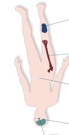
Figure 1. Various stem cell types from different sources.
sources of fetal stem cells have been identified. cells that largely mimic the abilities of ESCs
For example, hematopoietic and mesenchymal fetal [18,19]. Meng et al have discovered a population of
stem cells with pluripotent cell-like properties from stem cells in the menstrual blood [20]. These cells
the cord blood have been identified [15]. There is named as the ‘Endometrial Regenerative Cells’
also the potential to harvest fetal stem cells from are capable of differentiating into nine tissue lin-
discarded placental tissues [16]. eages namely, cardiomyocytic, respiratory epithe-
ESCs are derived from the ICM of preimplanta- lial, neurocytic, myocytic, endothelial, pancreatic,
tion blastocyst stage; the ICM is separated from hepatic, adipocytic and osteogenic.
the trophectoderm (which would develop into the While ethical debate on the propriety of iso-
extra-embryonic tissues) using immunosurgery and lation of ESCs from human embryos continues,
mechanical dissection. These ICM cells are then induced pluripotent stem cell lines have success-
plated onto a feeder layer of cells that supply both fully been derived by inducing the expression of
soluble factors and contact-mediated support. The pluripotency related genes in the somatic cells.
ICM cells attach and over time expand to form These cells designated as induced pluripotent stem
an ESC line [6]. Initially, murine embryonic fibro- cells (iPS) exhibit morphology of embryonic stem
blasts were used as feeder cells for the hESCs, and cells and express ES cell markers [21,22].
they still remain a good option for many research
applications [6]. Newer protocols are however rely-
ing more on mechanical separation of the ICM and 5. Types of stem cells
many new feeder layers, including human fibrob-
lasts, human fetal tissue and non-cellular layers Depending on their regenerative potency, stem cells
made up of basement membrane proteins are being are classified as totipotent, pluripotent, or multi-
used [17]. potent stem cells.
More recently, new sources of pluripotent stem Totipotent stem cells have the potential to
cells have been discovered. Guan et al have suc- become any kind of cell in the body. After an egg
cessfully produced several lines of pluripotent is fertilized, it undergoes a series of divisions to
stem cells from spermatogonial (sperm-producing) become an embryo and later a fetus. The cells440 SAVNEET KAUR AND C C KARTHA
that are formed during these first few divisions have been identified to affect stem cell number
are totipotent, i.e., each cell can form a complete or function and participate in several stem cell
organism. niches [29–31]. Soluble mediators of cellular func-
Pluripotent stem cells result after totipotent tion in the stem cell niche have also been defined.
stem cells undergo the first few divisions. Pluripo- Bone morphogenetic proteins (BMPs), wingless-
tent stem cells include cells from the blastocyst related proteins (WNTs) and their antagonists,
stage of the embryo. Given the right signals, a soluble notch modulators, fibroblast growth fac-
pluripotent stem cell could turn into any cell in tors (FGFs) and Hedgehog (HH) contribute their
an organism (except placenta), potentially growing inputs in a paracrine manner. They have varying
into tissue for a heart, a kidney or bone. capacity to induce proliferation or impair differ-
Multipotent cells can be isolated from many tis- entiation [32,33]. Metabolic products such as cal-
sues of the body and function as a repair system cium, oxidative stress and levels of reactive oxygen
for damaged tissue. As compared to the totipo- species are also known to markedly affect stem-
tent and pluripotent stem cells, they possess a limi- cell function [34,35]. It is expected that further
ted ability to differentiate into other cell types. understanding of the role of the stem cell niche
Adult stem cells from the blood, nervous system would pave way for novel therapies to enhance
and heart represent multipotent stem cells. and improve the regenerative capacity of stem
cells.
6. The stem cell niche
7. Stem cell therapy for regeneration
Stem cells are defined by their function in complex
multidimensional environments termed as stem cell 7.1 Hematological
niches. The simple location of stem cells is not
sufficient to define a niche. The niche must have Allogenic HSC transplantation or bone marrow
both anatomic and functional dimensions, specifi- transplantation (BMT) has now become an effec-
cally enabling stem cells to reproduce or self- tively established curative treatment of genetic and
renew. Adult stem cells generally have limited malignant hematologic disorders. Significant and
function without the niche. For example, HSC, sometimes even substantial improvements have
which regenerates the entire blood and immune in fact been achieved, albeit to different degrees
system, circulate freely, but seem to have little for different diseases, with allogeneic transplant
function outside specific anatomic locations. It is of HSC over the last 10–15 years. The signifi-
specific cues from specific sites that allow stem cant impact can be deduced from data related
cells to persist, and to change in number and fate. to pediatric patients affected by severe combined
Importantly, it is also the niche that provides the immune deficiency (SCID) and severe aplastic ane-
modulation in stem-cell function needed under con- mia (SAA) [36,37]. In the former group, the cumu-
ditions of physiologic challenge. The ability of the lative probability of survival in patients treated
niche to impose functions on stem cells makes by BMT from an identical sibling, estimated as
them relevant in disease conditions. The concept roughly 60% until 1982, has risen above 95%
of a niche as specialized microenvironment hous- since 1983. For patients with SAA, the increase
ing stem cells was first proposed by Schofield [23]. in disease-free survival has been from 49% in the
Experimental evidence was first provided in the period 1970–1980 to 70% in the period 1981–1983
invertebrate models, thirty years later [24,25]. In and to 81% over the next five years (1984–1988).
invertebrate models, it has been demonstrated that There have been less significant improvements in
niche is composed of heterologous cell types. This patients with acute lymphoblastic leukemia (ALL)
has led to search for niche components in mam- given an allogeneic BMT from an HLA-compatible
malian tissues and identification of the osteoblast relative. In these patients, according to the data
in the bone marrow, and the endothelium in the provided by the AIEOPBMT registry, the cumula-
brain, and possibly in the bone marrow [26–28]. tive probability of leukemia free survival was 42%
Cells, matrix glycoproteins and the three- in the period between 1985 and 1990 and has
dimensional spaces form a stem-cell niche. The increased only to 50% in the period 1991–1995 [38].
contact between these elements allows molecular Besides BMT, the two other most widely prac-
interactions that are critical for regulating stem- tised transplantation techniques include transplan-
cell function. Secreted proteins offer a paracrine tation of circulating progenitor cells (CPCs) from
measure of control, but non-protein components peripheral blood and umbilical cord blood cells
of the local microenvironment also affect stem-cell (UCBC) (table 1).
function. Among the matrix proteins, β-1 integrins Autologous CPCs are increasingly being used
in the skin and tenascin C in the nervous system following high-dose therapy for malignant disease,STEM CELLS: CONCEPTS AND PROSPECTS 441
Table 1. Status of clinical trials using different types of stem cells in human diseases.
Types of stem cells in use
Human diseases (Experimental and clinical studies) Status of clinical trials
Hematological Bone-marrow derived stem cells Phase III using HLA-matched HSCs∗ from all
Peripheral blood stem cells three stem cell sources, Phase-II/III allogenic,
Cord blood stem cells haploidentical HSCs in malignant and non-
malignant hematological disorders
Corneal and Corneal stem cells Phase II/III using ex vivo cultured human lim-
Retinal Adult epithelial stem cells or bal epithelial stem cells for patients with lim-
Retinal pigment epithelial cells bal stem cell deficiency Phase I/II using retinal
Embryonic and Adult Neural Stem pigment epithelial cells in patients with retinal
Cells degeneration
Cardiovascular Hematopoietic stem cells Phase I/II using autologous skeletal
Mesenchymal stem cells myoblasts, bone marrow stem cells, periph-
Endothelial progenitor cells eral blood and adipose-derived stem cells in
Cardiac stem cells patients with myocardial ischemia, myocar-
Embryonic stem cells dial infarction, coronary artery disease, heart
Fetal cardiomyocytes failure
Skeletal myoblasts
Neurological Mesenchymal stem cells Phase I/II using autologous bone marrow stem
Embryonic and Adult Neural Stem cells in patient with ischemic stroke, multiple
Cells sclerosis, spinal cord injury
Embryonic stem cells
Muscoskeletal Mesenchymal stem cells Phase I/II using autologous bone marrow stem
Muscle-derived stem cells cells in patients with critical limb ischemia,
Bone marrow-derived side population Phase I using muscle-derived stem cells in
cells patients with Duchenne muscular dystrophy,
Vascular wall stem cells Phase I/II and II/III using MSCs# in patients
with long bone defects and articular cartilage
defects
Renal Hematopoietic stem cells Clinical trials using MSCs yet to be initiated
Mesenchymal stem cells
Endothelial progenitor cells
Embryonic renal cells
∗ #
HSCs: hematopoeitic stem cells; MSCs: mesenchymal stem cells
because of the ease of collection and the markedly of HSC using the cord blood of a healthy,
faster kinetics of engraftment in comparison with HLA-compatible sibling [43]. Wagner et al has
bone marrow [39]. In childhood autologous trans- demonstrated the applicability of the procedure
plantation, CPCs have been mobilized into peri- even in adults [44]. The cells obtained from a
pheral blood and collected on a large scale by cord may however be quantitatively insufficient
leukapheresis after treatment with hematopoietic for quick engraftment of the transplant in most
growth factors [40,41]. Recently, CPCs have been adult patients, a great limitation in terms of
considered as an alternative to bone marrow routine use of the cord cells [45]. UCBC trans-
for allogeneic transplantation and this procedure plants are currently performed, both from an HLA-
is also being used increasingly in adults [42]. compatible family donor and from an unrelated
Although there is no definitive proof from con- donor. Improvements in the methods used for cell
trolled clinical studies, allogenic transplant of collection, manipulation and freezing have allowed
CPCs has some undisputed advantages in compari- a rapid increase in the use of UCBC. Reported low
son with BMT; for the recipient the duration of incidence of acute and chronic graft versus host dis-
neutropenia and of thrombocytopenia is reduced, ease has promoted the establishment of large cord
and for the donor the trauma of harvesting mar- blood banks in Europe and the USA, where at
row from the bone, with associated inevitable present more than 12,000 cord blood units have
anesthesia, is eliminated. been collected and typed for the HLA system [46].
The other most significant alternative to BMT Significant expansion of CB progenitor cells in vitro
(which is now used routinely) is UCBC transplan- is also possible now with the use of a combina-
tation, which was introduced by Gluckman et al tion of cytokines and chemokines [47]. Despite the
in a 5-year-old child affected by Fanconi’s anemia. impressive effect seen in vitro, clinical benefit with
The child was given an allogeneic transplant the expanded cells however do not seem to differ442 SAVNEET KAUR AND C C KARTHA
much from that obtained with the unmanipulated indefinitely in culture, and there are additional
cells [48]. issues related to tissue availability and repro-
ducibility [57,58]. Adult neural stem cells from the
7.2 Corneal and retinal hippocampus have also been reported to incorpo-
rate into the retina and adopt the morphologies
Repair of degenerative diseases of the eye is a prime and positions of bipolar, horizontal, photoreceptor,
example of stem cell therapy in routine, effective and astroglial cells [59].
clinical practice. Both corneal and retinal stem A breakthrough has been achieved with the dis-
cell populations have been identified in the adult covery of retinal stem cell (RSC) population from
human eye. The corneal epithelial cells have a finite human adult ciliary epithelium [60]. These RSCs
life span and are continuously renewed by prolifer- can be expanded from single cells and differentiate
ating stem cells in the limbus located at the junc- into a variety of retinal cell types. Human RSCs
tion between the cornea and the conjunctiva [49]. have also been shown to integrate and differentiate
The corneal stem cells constitute between 0.5 and into photoreceptors after transplantation into the
10% of the total cell population in the epithelial host neonatal retina [61]. However strategies for in
tissue. Under certain conditions, however, the lim- vitro expansion of RSCs and photoreceptor devel-
bal stem cells may be partially or totally depleted opment still need to be optimized.
resulting in varying degrees of stem cell deficiency Recent studies demonstrate that hESCs can
with accompanying abnormalities in the corneal also be induced to generate retinal progeni-
surface. Such deficiency leads to conjuctivalization tor cells, which in culture can differentiate into
of the cornea with vascularization, appearance of photoreceptors [62,63].
goblet cells and irregular and unstable epithelium.
Stem cells can be delivered by limbal auto or allo 7.3 Cardiovascular
grafts depending on the source of the donor tis-
sue [50–53]. Transplantation of ex vivo expanded Cardiovascular diseases are one of the leading
donor limbal cells is another strategy to provide causes of death and disability worldwide. These
the limbal tissue [54,55]. Amniotic membrane har- diseases lead to loss of cardiac tissue through
vested from human placenta is used as an adjunct, death of the cells by apoptosis and necrosis.
as a substrate for epithelial growth and ocular sur- The average left ventricle contains approximately
face reconstruction [56]. Therapy using limbal stem 4 billion cardiomyocytes and the myocyte deficit in
cells has a reasonable success rate even in patients infarction-induced heart failure is about one billion
with severely diseased cornea. cardiomyocytes [64]. The remaining myocytes
The role of bone marrow stem cells as a source are unable to reconstitute the host tissue, and
of ocular surface tissue is yet to be evaluated. the diseased heart deteriorates functionally with
Retinal transplantation as therapy for retinal time. Current therapeutic approaches available
degenerative diseases such as retinosa pigmentosa including medical therapy, mechanical left ventri-
(RA) and glaucoma has gained interest during the cular assist devices, and cardiac transplantation
past 20 years. Structurally, retina is organized into are primarily focused at limiting disease progres-
three cellular layers: photoreceptor, interneuron sion rather than repair and restoration of healthy
and ganglion cell layers. RA is characterized by tissue and function. The limited efficacy and co-
the widespread degeneration of the rods and cones morbidity of these current treatments have thus
in the photoreceptor layer. In glaucoma, ganglion increased the interest to investigate other alterna-
neurons are the major targets of degeneration. tive and additional long-term therapeutic strate-
Hence cell-based therapies for these degenerative gies. In this context, a cell-based therapy for
diseases are directed towards replacing the missing myocardial regeneration seems to be a potentially
neurons with new ones, thereby hoping to restore beneficial approach to achieve cardiac repair.
vision. Since retina to some extent is an immuno- Several cell types that might replace necrotic
logically privileged site, allogenic transplantion tissue and minimize scarring have been consi-
is highly feasible. Various types of cells and tis- dered (table 1). Initial cardiac cell transplantation
sues are being investigated for treating retinal efforts have utilized skeletal myoblasts (SMBs),
regeneration (table 1). Transplantation of retinal adult stem cells isolated from skeletal muscle biop-
pigment epithelial (RPE) cells in animal models of sies [65]. Based on their utility in animal studies,
RPE degeneration has been reported to improve SMBs have been utilized in several clinical trials
photoreceptor survival and visual outcome. Some in patients with severe post-infarction left ventric-
clinical benefits have been observed in patients ular dysfunction [66–68]. Follow-up studies have
with macular degeneration after autologous trans- shown a moderate, but significant increase in the
plants of RPE to the fovea [57]. Embryonic retinal left ventricular ejection fraction (LVEF), as mea-
progenitor cells are not easy to be maintained sured by echocardiography. Similar to SMBs, anSTEM CELLS: CONCEPTS AND PROSPECTS 443 improvement in cardiac function has also been and clonally expanded in vitro [79]. Human observed in rats after coronary artery ligation fol- cardiosphere-derived cells (CDCs) when injected lowed by transplantation of fetal cardiomyocytes as into the border zone of myocardial infarcts engraft compared to non-engrafted infarcted hearts [69]. and migrate into the infarct zone. Injected CDCs In the bone marrow, three populations of stem have also been shown to result in an increased per- cells: HSCs, MSCs and endothelial progenitor cells centage of viable myocardium and improve LVEF (EPCs) have been reported to contribute to heart [80]. However, lack of sufficient numbers of cells muscle repair. In animal models of heart disease, that can be isolated from biopsies from patients administration of bone marrow derived stem cells hinders the clinical utility of cardiac stem cells. has shown to cause an increase in tissue perfusion, Exciting new advances in cardiomyocyte a reduction in apoptosis, reduction in infarct size, regeneration are also being made in human embry- and improvements in global and regional cardiac onic stem cell research. Studies by Itskovitz-Eldor function [70,71]. The first randomized trial called et al and Kehat et al have shown that hESCs can BOOST trial (bone marrow transfer to enhance reproducibly differentiate in culture into embryoid ST-elevation infarct regeneration) was performed bodies and the cells have structural and func- by Helmut Drexler’s group in Hannover, Germany tional properties of early stage cardiomyocytes [72]. The study demonstrated that intracoronary [9,81]. However, if hESCs are to have a future transfer of autologous bone marrow cells 4.8 days in cell-based cardiac repair, substantial improve- after percutaneous coronary intervention enhanced ment in the efficiency by which cardiomyocytes LVEF primarily in myocardial segments adja- can be generated from hESCs has to be achieved. cent to the infarcted area. Another multicenter Until quite recently, the typical method for obtain- trial (reinfusion of enriched progenitor cells and ing hESC-CMs was to form embryoid bodies (in infarct remodeling in acute myocardial infarction), medium including a relatively high fraction of fetal REPAIR-AMI showed that compared to placebo calf serum) and then harvest the resultant spon- treatment, intracoronary infusion of bone marrow taneously contractile cardiomyocytes by either cells resulted in improved left ventricular func- mechanical dissection [9] or enzymatic methods tion at 4 months and reduction in combined clini- [82]. Embryoid bodies contain an admixture of cal end points of death, recurrence of AMI, and many differentiated cell types, and so cardiogen- any revascularization procedure at 1 year [73]. esis is inefficient through this approach. Recent The benefit was greatest in patients with poor efforts are directed at identifying defined factors left ventricular function. However, other groups to enhance the differentiation of cardiomyocytes from Belgium and Norway, had been unable to from hESC [82,83]. Nonetheless, in experimental detect a difference in outcome between bone mar- studies, the transplantation of mESC-derived car- row cell treated group and controls in AMI set- diomyocytes into the uninjured hearts of immuno- ting [74,75]. Different cell isolation protocols as compatible mice has resulted in the formation of well as dosage, degree of cell viability and func- stable intracardiac grafts [84–86]. In 2004, Kehat tion prior to delivery may contribute to the het- et al reported the first human cardiomyocyte trans- erogeneous clinical results in randomized trials. plantation into the uninjured swine myocardium In the (transplantation of progenitor cells and [87]. Since then, transplantation of ESC-derived recovery of LV function in patients with chronic cardiomyocytes into normal and injured heart in ischemic heart disease) TOPCARE-CHD trial, the animals has been shown to improve the global absolute change in LVEF at 3 months, was signifi- myocardial function, although for a short period cantly greater among patients receiving the bone of time [88,89]. marrow cells than among those receiving circula- Besides cardiomyocytes, two other cell types ting progenitor cells [76]. An alternative approach that are important to a properly functioning heart used includes the mobilization of endogenous stem are the vascular endothelial cells, which forms the or progenitor cells in vivo from the bone mar- inner lining of new blood vessels, and the smooth row, to the damaged heart using specific cytokines muscle cell, which forms the wall of blood ves- and growth factors. Recent meta-analysis includ- sels. The heart has a large demand for blood flow ing 8 randomized controlled trials has demon- and these specialized cells are important for devel- strated that, granulocyte-colony stimulating factor oping a new network of arteries to bring nutri- therapy increased LVEF by 1.09% in patients with ents and oxygen to the cardiomyocytes after heart AMI [77]. tissue has been damaged. The potential capa- There is now accumulating evidence that the bility of both embryonic and adult stem cells heart itself contains resident stem cells with the to develop into these cells types is also being capacity to differentiate into cardiac myocytes [78]. explored as part of a strategy to restore car- In humans, autologous cardiac stem cells can be diovascular function via the processes of ther- isolated from surgical or endomyocardial biopsies apeutic angiogenesis and arteriogenesis. In this
444                                     SAVNEET KAUR AND C C KARTHA

regard, bone-marrow derived EPCs isolated from          but to achieve cell engraftment with electro-
peripheral blood and/or bone marrow have shown          mechanical integration into the heart, arrest
incorporation into sites of physiological and patho-    adverse myocardial remodeling and improve con-
logical neovascularization in the endothelium after     tractility of the diseased heart.
either systemic injection or direct intramyocar-
dial transplantation in animal models of peripheral                      7.4 Neurological
limb ischemia and myocardial infarction [90–92].
Several clinical studies have however reported an       Despite the protection of the central nervous
inverse correlation between the number and activ-       system (CNS) by the skull and vertebral column,
ity of circulating EPCs and risk factors for coro-      it remains susceptible to several insults and neu-
nary artery disease [93,94]. In this regard, genetic    rodegenerative diseases. The hallmark of sev-
engineering of EPCs with growth factors offers a         eral degenerative disorders in the CNS such as
useful approach to developing these cells into effi-      amyotrophic lateral sclerosis (ALS), Parkinson’s
cient therapeutic tools. Iwaguro et al have demon-      disease (PD) and Alzheimer’s disease (AD) is the
strated that the transfer of VEGF in ex vivo            massive loss of one or several types of neuronal
expanded EPCs enhances EPC proliferation, adhe-         populations. There is evidence both in humans
sion, and impaired neovascularization in an ani-        and in experimental animal models of neurode-
mal model of experimentally induced limb ischemia       generative diseases for spontaneous neurogenesis
[95]. Gene modified EPCs have also been shown to         involving endogenous neural stem cells (NSCs)
serve as cellular vehicles for the delivery of ther-    [102–105]. This putative endogenous repair process
apeutic genes such as eNOS to the reconstituted         appears to be insufficient to compensate neu-
endothelium [96]. The use of autologous EPCs            ronal loss and to ensure functional recovery. These
seeded onto a scaffold has also been reported for the    observations have raised interest in the use of
tissue engineering of heart valves [97,98]. hESCs       exogenous embryonic and adult stem cells in
have also been demonstrated to differentiate into        substitution therapies with the hope that these
EPCs and then to mature endothelial cells leading       cells could generate new neurons after they are
to vascular network structures in three dimensional     grafted into lesioned nervous tissues [106]. Stem
culture models [99].                                    cells used for applications in neurological diseases
   A final issue worth considering is the mecha-         are from four different sources: NSCs from the
nism by which the implanted cells mediate the           embryonic or the adult brain, stem cells from
beneficial effects on contractile function. Several       other tissues such as the bone marrow and ESCs
lines of evidence support the concept that new          (table 1).
endogenous or exogenous cells can incorporate and          Adult NSCs exist within multiple regions of the
become functional within the heart. Early stud-         CNS (subventricular zone, hippocampus, etc.), and
ies with bone marrow derived HSCs in mice have          it is possible to isolate and expand these cells to
suggested that they differentiate into cardiomy-         give rise to progenitor cells restricted to defined
ocytes after transplantation to induce the repair of    neural lineages such as neuronal and glial cells
damaged myocardium [71]. However, more recent           [107]. Neural stem cells that proliferate in the ven-
studies with genetically marked cells indicate that     tricular region and later in the subventricular zone
the transplanted cells do not transdifferentiate into    of the developing brain give rise to three neural
cardiomyocytes [100,101]. It is possible that the       lineages of the CNS, i.e., neurons, astrocytes, and
stem cells confer their beneficial effects, possibly      oligodendrocytes [108]. The identification of NSCs
by secreting paracrine factors that are cardiopro-      and progenitor cells has completely challenged the
tective or angiogenic.                                  past notion that adult brain is an organ with no
   Cellular cardiomyoplasty, although appears           ability for self-renewal.
promising in pre-clinical studies, its safety and effi-      Demyelinating diseases of the brain are attrac-
cacy have not been adequately evaluated. Its future     tive targets for cell-based therapeutic strategies,
will depend on conducting carefully controlled, ran-    since these diseases are caused by the loss of a
domized clinical trials with appropriate selection      single cell type, the oligodendrocyte. In experi-
of end points. Controversies exist over the spe-        mental models of focal demyelination in rodents,
cific cells to be used, the dosages needed for tissue    it has been shown that endogenous cells in the
repair, route of administration and how the trans-      CNS have the potential for regenerating oligoden-
planted cells would affect the electrical activity of    drocytes and myelin [109,110]. Injection of adult
the myocardium. Whether the cells can improve           NSCs has been demonstrated to induce recovery
myocardial function after transplantation over long     in a chronic model of multiple sclerosis [111,112].
term is also not yet clear.                             Transplantation of NSCs of various origins has also
   The challenge in regenerative therapy in cardiac     resulted in the improvement of clinical outcome in
diseases is not simply to arrest cardiac dysfunction    experimental models of spinal cord trauma and the
STEM CELLS: CONCEPTS AND PROSPECTS                                       445

therapy improves myelinating properties as well          damaged brain, these cells survive well, integrate
[113–115].                                               into host tissues, and differentiate into both neu-
   Neurodegenerative diseases such as PD disease         rons and glial cells [138–144].
involve continuous loss of dopaminergic neurons.            Of the various cell types, NSCs have the most
Stem cell therapy for PD is aimed at the induc-          potential for use for treating the broad spectrum
tion and renewal of dopaminergic neurons. In neu-        of neurological disorders. However before embark-
rodegeneration models of PD, ex vivo expanded            ing into routine clinical use, further studies are
NSCs efficiently decrease parkinsonian symptoms            warranted to identify the signals for proliferation,
by rescuing dopaminergic neurons through produc-         differentiation, and integration of NSCs and also
tion of specific growth factors [116,117]. Likewise,      to determine the environment conditions of the
transplantation of NSCs into the lumbar spinal           host brain favorable for implanted NSCs to survive,
cord of rodents with ALS has been shown to post-         prosper, and restore damaged tissue.
pone the disease onset, to preserve the viability
of motor neurons, and to prolong animal survival                         7.5 Musculoskeletal
[118,119].
   Alteration in the local blood flow is believed         Since the pathbreaking studies of Friedenstein et al
to participate in the progression of neuronal            who isolated bone-forming progenitor cells from
death after stroke. Accumulating evidence suggests       rat marrow, the ability of these cells, designated
that after transplantation, NSCs migrate toward          the MSCs to differentiate into various cell types
ischemic boundary regions in embolic stroke and          of mesenchymal tissues, including cartilage, bone,
that the engrafted cells increase angiogenesis [120].    fat, muscle, tendon, has been widely recognized.
   In addition to adult NSCs, two prototypic stem        Although MSCs represent only a very small frac-
cell populations from the adult bone marrow, viz,        tion of the total population of nucleated cells in
HSCs and MSCs can also transdifferentiate into            the marrow, they can be easily isolated and exten-
neural cells. Indeed, both cell types have been          sively expanded or specifically differentiated under
shown to migrate efficiently towards the site of           appropriate in vitro conditions. Besides the bone
injury within the CNS [121]. Mimicking NSCs,             marrow, the multipotential MSCs for bone regen-
MSCs also promote functional recovery after brain        eration have also been isolated from other sources
injury in several experimental models [122–125].         such as the adipose tissue and skeletal muscle. An
For example, it has been shown that intraventricu-       added advantage of using MSCs is that they do
lar as well as intravenous transplantation of MSCs       not elicit alloreactive lymphocyte proliferation and
into mice with multiple sclerosis, result in signifi-     modulate the immune responses [145,146].
cant clinical improvement [126]. Similarly, cerebral        The ability of MSCs to form bone was one among
neovascularization, restoration of cerebral blood        the first properties to be evaluated. In animal mod-
flow, and reconstitution of the blood–brain barrier       els of bone defects, implantation of MSCs adsorbed
in animal models of stroke have been obtained with       onto appropriate scaffolds, resulted in a signifi-
HSCs [127–129] and MSCs through enhanced pro-            cant increase in the rate of bone formation and
duction of VEGF [130] and FGF-1 [131]. Further,          also improvement in the physical properties of the
in vivo experiments suggest that MSCs can induce         bone [147–149]. Success in animal studies paved
the proliferation of endogenous NSCs [132] and           way for initiation of the first clinical trial. Quarto
their differentiation into oligodendrocytes [133] or      et al reported repair of large segmental defects in
astrocytes [134].                                        humans using autologous MSCs on hydroxypatite
   Besides adult stem cells, studies have shown          scaffolds [150]. In animal models, MSCs delay graft
that undifferentiated ESCs grafted into lesioned          rejection, and in children with osteogenesis imper-
brain develop into normal dopaminergic neurons           fecta, allogenic bone marrow transplantation result
and express neuronal and dopaminergic markers in         in the engraftment of donor derived MSCs and new
vivo [10,135]. Nevertheless, despite the promising       bone formation [151]. Recently, genetically engi-
results obtained with ESCs in experimental models        neered MSCs with potent osteogenic genes such
of nervous insults [136,137], the risk of transplanted   as BMPs have been used to repair fracture repair
cells evolving into teratomas [135] combined with        and rapid bone formation has been observed in
ethical issues limit the use of ESCs in cellular         vivo [152]. MSCs have also been evaluated as a
therapies.                                               substitute for chondrocytes in the cartilage repair
   Recently, continuously dividing immortalized          process. In a few animal studies, implantation of
cell lines of NSCs have been generated by the intro-     MSCs has been seen to differentiate both into car-
duction of oncogenes. These immortalized NSC             tilage and subchondral bone [153,154]. Ongoing
lines have emerged as a highly efficient source of         investigations have now been focusing to engineer
cells for genetic manipulation and gene transfer         MSCs into soft tissues, tendons and ligaments that
into the CNS ex vivo. Once transplanted into the         play a major role in the movement of joints.
446                                      SAVNEET KAUR AND C C KARTHA

   Besides MSCs, autologous articular chondro-           which contributed to improved integration of the
cytes have also been in use for local cartilage repair   engineered muscle when transplanted to immunod-
in both animal and clinical studies.                     eficient mice [171].
   Stem cell therapy to repair and replace damaged          The potential for stem cell regeneration of mus-
skeletal muscle cells in chronic, debilitating muscle    culoskeletal tissues seems immense. One of the
diseases such as muscular dystrophies has shown          major challenges of any orthopedic application
great promise. Different stem cell populations, both      would be to identify the proper biocompatible
of embryonic and adult origins appear to have            matrix, one that will withstand the immediate
the potential to generate skeletal muscle cells and      structural forces, provide for cell differentiation
have been studied in animal models of muscular           along appropriate lineage paths and be resorbed
dystrophy (table 1). Caplan and colleagues first          at rates proportional to the rate of increase in
investigated in vitro differentiation of bone marrow      strength of the newly formed matrix.
derived MSCs into muscle [155]. More recently,
Cossu, Mavillo and co-workers have demonstrated                               7.6 Renal
active muscle regeneration in vivo with bone
                                                         The kidney has a remarkable capacity to regenerate
marrow-derived cells [156]. Several stem cell popu-
                                                         after injury, as it is not a terminally differenti-
lations have recently been recognized in skeletal
                                                         ated organ. This regenerative potential is somehow
muscle [157,158]. Satellite cells are dormant
                                                         incomplete and as the insult continues progressive
progenitors often referred to as ‘muscle stem cells’
                                                         and irreversible scarring results in chronic renal
and located beneath the basal lamina of mature
                                                         disease. End stage renal disease is a deadly dis-
skeletal muscle fibers. These cells are considered
                                                         ease unless supportive treatment is given in the
to be monopotential stem cells capable of giving         form of hemodialysis, peritoneal dialysis or kidney
rise only to cells of the myogenic lineage. Among        transplantation. An acute shortage of compatible
other progenitor cells found in skeletal muscle are      organs, coupled with limited adaptability of cur-
side-population (SP) cells, mesoangioblasts, and         rent dialysis techniques has spurred a sense of
pericytes [159]. SP cells have a tremendous abi-         urgency to investigate newer alternatives such as
lity to proliferate and provide myoblasts for muscle     cell-therapy.
regeneration. They also appear to be able to differ-         Three stem cell lineages of the bone marrow:
entiate into additional lineages [160]. Gussoni and      HSCs, MSCs, and EPCs have the potential to
colleagues demonstrated the restoration of dys-          promote repair in various forms of kidney disease
trophin expression in the mdx mouse (an animal           (table 1). Bone marrow-derived stem cells seem to
model of Duchenne muscular dystrophy) by using           have a high capacity for transdifferentiation and
SP population from donor marrow [161]. The inher-        therefore are able to replace damaged renal tis-
ent vascularity of the muscle makes it a useful          sue with tubular epithelial cells, mesangial cells,
depot to deliver secreted proteins via gene therapy.     endothelial cells, and even podocytes [172,173].
Genetically engineered myoblasts, or muscle-             Injection of MSCs protects the kidney from toxin
derived stem cells, have been used for replacing         or ischemia/reperfusion injury and attenuates lost
degenerating muscle in Duchenne Muscular Dys-            renal function, whereas injected HSC do not have
trophy [162,163] or in bone defects [164]. As a gene     the same effect [174]. The first phase of clini-
delivery vehicle, myoblasts have been employed           cal trials using bone marrow MSCs for protection
to deliver growth hormone, VEGF, Factor                  against acute kidney injury may begin shortly. This
IX, erythropoietin and several other molecules           study hopefully would enable further exploration
[165–168].                                               of stem cell therapy in renal patients with multiple
   The myogenic potential of ESCs has been               cormorbidities.
well demonstrated in the in vitro models [169].             Participation of circulating EPCs in renal
A recent study has reported the transforma-              endothelial repair has been demonstrated in several
tion of hESCs into satellite-like myogenic stem          experimental studies [175,176]. Transplantation
cells with remarkably high engraftment effi-               of ex vivo expanded EPCs from a muscle stem cell
ciency compared to myoblast transplantation in a         pool has shown to locally engraft, and improve
muscle injury model [170]. Levenberg et al have          renal function in rats with acute renal ischemia
described a method for the in vitro expansion            [177]. Animal studies have also provided evi-
of engineered skeletal muscle tissue developed by        dence that EPCs contribute to glomerular capil-
means of co-seeding the myoblasts with hESCs-            lary repair [178,179]. In the clinical setting, renal
derived endothelial cells and embryonic fibrob-           diseases in concert with cardiovascular risk factors
lasts on a porous biodegradable scaffold. The             have been reported to significantly influence the
co-culture of myoblasts in the presence of hESC-         number and function of EPCs [180,181].
derived endothelial cells resulted in neovasculari-         Multipotent resident renal stem cells have not
zation in the construct prior to implantation,           yet been discovered in the kidney. However, Oliver
STEM CELLS: CONCEPTS AND PROSPECTS 447
et al have demonstrated the existence of resident should not only support attachment, spreading
stem-cell pools in the renal papilla [182]. Iwatani growth and differentiation of cells but also control
and colleagues have suggested that renal stem cells inflammation and foreign body reaction. It should
may reside in the bone marrow and take up resi- be biodegradable into non-toxic products, steriliz-
dence in the kidney when needed [183]. able and manufacturable. It should offer options
Whether human ESCs can be used as a starting to deliver drugs, cytokines and genes. The set
material for renal regeneration still remains to be of criteria would appear demanding, but has to
determined. be met for the tissue-engineered scaffolds to be
effective.
8. Stem cells and tissue engineering
9. Stem cell research in India
Since stem cells are highly regulated by their
microenvironment or the niche in which they Stem cell research has gained considerable impe-
reside, efforts are on to provide constructs that tus in India in the recent years. Draft guidelines
can mimic the cell milieu through development for stem cell research in the country have been
of tissue-engineered scaffolds [184]. These scaffolds formulated jointly by the Department of Biotech-
also temporarily provide biomechanical support nology and Indian Council for Medical Research.
for cells until they are able to produce their own Several groups are actively and enthusiastically
extra-cellular matrix [184]. Better control of the pursuing the field with reasonably good results.
tissue formation process is an additional advan- According to a recent review, for haematologi-
tage. Scaffolds are typically fabricated by natural cal disorders, a total of 1540 bone marrow trans-
materials, which are inherently bioactive but lack plants have been performed in a country of over
mechanical strength, or synthetic materials, which one billion population [191]. At Christian Medical
lack inherent bioactivity but could be mechanically College (CMC), in Vellore, a total of 626 trans-
strong and can be fabricated with the desirable plants have been performed in 595 patients, with
macro- (shape) and microarchitecture (pore size, 28 patients having more than one transplant from
porosity). Numerous types of biomaterials both October 1986 to December 2006 [191]. Besides,
man-made or from natural sources are continually CMC Vellore, autologous and allogenic bone mar-
being discovered [185]. Efforts are being carried row or blood stem cell transplantation is being
out to modify the surface of these materials, to performed at other hospitals such as All India
guide, and enhance stem cell differentiation. Ini- Institute of Medical Sciences (AIIMS), New Delhi
tially, scaffolds were designed to be bioinert. Cur- and Tata Memorial Hospital, Mumbai [192–194].
rently, biomaterials are made to interact with the AIIMS has also set up the country’s first cord
cells that release growth factors, genes, or other sig- blood bank for isolation of cord blood stem cells
nals in a time-dependent manner [185–187]. Based for in-house patients. At the L V Prasad Eye
on these active bio-materials, the conventional two- Institute, Hyderabad, transplantation of autolo-
dimensional (2-D) culture models have now paved gous cultivated limbal stem cells in patients with
the way for three-dimensional (3-D) culture envi- limbal stem cell deficiency, has shown a success-
ronments that mimic the in vivo environments ful outcome with a stable ocular surface with-
more closely and hence are more conducive to out conjunctivalization [195]. Small scale phase-I
regulating stem cell proliferation and differentia- clinical trials using bone marrow stem cells have
tion [188]. Elements of the extracellular matrix been reported for the treatment of diabetes at
and stromal MSCs have gained increasing atten- Dr. H L Trivedi Institute of Transplantation Sci-
tion as potentially crucial mediators in developing ences, Ahmedabad [196], acute myocardial infarc-
and maintaining the characteristics of 3-D cell cul- tion at Nizam’s Institute of Medical Sciences,
tures. Fibrin alone or in combination with other Hyderabad [197], Sir H N Hospital and Research
materials has emerged as an important biological Centre, Mumbai [198] and nonischemic dilated car-
scaffold for stem cells to regenerate adipose tissue, diomyopathy at AIIMS, New Delhi [199]. At Sree
bone, cardiac tissue, cartilage, liver, nervous tissue, Chitra Tirunal Institute for Medical Sciences and
ocular tissue, skin, tendons, and ligaments [189]. Technology (SCTIMST), Trivandrum, procedures
Culture on fibrous biodegradable scaffolds that for the isolation and expansion of EPCs from
mimic basement membrane texture has resulted peripheral blood of patients with CAD have been
in an increased expansion of both HSCs and optimized [200]. Recent strategies are now directed
ESCs [184]. Similarly, the immobilization of cell- towards augmenting the angiogenic potency of
associated Notch ligands has shown to increase the these cells by modulation with endothelial nitric
self-renewal of HSCs [190]. A perfect tissue engi- oxide synthase gene transfer. Besides EPCs, ckit-
neered scaffold is elusive at present. The scaffold positive stem cells have been isolated from atrial448 SAVNEET KAUR AND C C KARTHA
biopsies of CAD patients and also induced to dif- ethical issues. The biggest hurdle for the clinical
ferentiate into beating cardiospheres [201]. At the use of adult stem cells is the small number of cells
biomedical technology wing of SCTIMST, recent that can be isolated from any adult tissue. The
studies have reported that platelet rich plasma in identification of cells and factors in the so called
combination with goat bone marrow-derived MSCs ‘stem cell niche’ affecting the growth and differ-
cultured on bioactive ceramic scaffolds leads to a entiation of resident adult stem cells may be one
much faster sequence of healing events in large seg- possible answer. For example, the bone marrow
mental bone defects in a goat femur model [202]. stromal cells are known to promote proliferation
Stem cell research at the Centre for Cellular and and differentiation of HSCs in long-term cultures
Molecular Biology, Hyderabad has been focusing [214]. The other approach is based on introduc-
on the genetic and epigenetic mechanisms govern- tion of genes in the supporting feeder layer of cells
ing the transient dormancy and activation of satel- that inhibits differentiation of target cells. The up-
lite cells, the stem cells in adult muscle tissues regulation of notch ligands such as Jagged-1 and
[203,204]. Delta in the stromal cells by gene modification
Vanikar et al have reported the generation of strategies has been demonstrated to promote the
30 healthy hESC lines from 33 voluntary oocyte expansion of stem cells without inducing differenti-
donors using a donor somatic cell nuclear trans- ation [26,27,190]. Another technique actively pur-
fer technique on 190 oocytes [205]. Researchers sued is the usage of modified stem cells. Based
at National Brain Research Centre, Gurgaon and on our understanding of the molecular pathways
National Centre of Cell Sciences, Pune are working responsible for self-renewal and proliferation of
towards the differentiation of hESCs into neural stem cells as well as discoveries of new genes that
stem cells [206–208]. Very recently, Jagatha et al control stem cell proliferation and differentiation,
have demonstrated the potential of FGF2-induced novel strategies have come up. For example, HOX
ES cell derived neural progenitors (ES-NPs) to gen- genes that are expressed during early development
erate retinal ganglion-like cells in vitro upon dif- and which govern various processes including body-
ferentiation [209]. At the Reliance Life Sciences, part patterning have been shown to increase the
Mumbai, functional dopaminergic precursor neu- self-renewal potential of HSCs [215].
rons from human embryonic stem cells (hESCs) Destruction of life in the form of an embryo
have been recently reported. Transplantation of has been a major ethical objection in embryonic
these precursor neurons into the lesioned rat model stem cell derivation and research in several west-
of Parkinson’s disease has also shown to elicit sig- ern countries. One way that has been suggested to
nificant reversal of lesion induced motor deficits circumvent the objection is to fuse existing hESCs
sustained up to the end of 1 year long study with an adult somatic cell, generating a cell line
period [210]. Researchers at the Reliance Life Sci- that retains ESC specific properties and yet has the
ences have also demonstrated the generation of genotype of the somatic cell [216]. There is however
spontaneously beating cardiomyocytes using FGF no technology available at present to selectively
from ESCs [211]. Studies at the Manipal Institute remove all the ESC chromosomes while retaining
of Regenerative Medicine, Bangalore are directed the somatic cell chromosomes. Development of such
towards the optimization of culture conditions of a technology is potentially expensive and will pre-
human MSCs with an attempt to obtain large sumably take many more years. Other approach
numbers, preserve their characteristics and multi- is the generation of induced pluripotent cell lines
lineage differentiation potential for therapeutic from induced somatic cell dedifferentiation. In this
uses [212]. They have also reported the derivation method, the adult somatic cells are genetically
of FGF2 expressing germ layer derived fibroblast modified and reprogrammed to undergo a process
cells from hESC lines for use as a feeder layer of dedifferentiation [22].
for culture of hESCs. These feeders could support Availability of methods for growth and main-
the pluripotency, karyotypes and proliferation of tenance of ESC in culture present another major
hESCs with or without FGF2 in prolonged cul- obstacle to their potential clinical use. Conven-
tures as efficiently as that on mouse embryonic tionally, hESC lines are grown in a medium con-
fibroblasts [213]. taining animal serum as a source of nutrients and
growth factors and then on mouse-derived fibrob-
last as feeder layers. The use of any cell based
10. Current challenges and future therapeutic agent in humans must however be
possibilities free of animal contamination. In this direction,
some laboratories have successfully cultured hESCs
Besides the overwhelming promise of stem cells in in a serum-free defined medium on human cell-
various cellular therapies, their clinical and prac- derived feeders or even in feeder free conditions
tical use is constrained by several technical and [217,218].You can also read