Reflections on an unconventional neuropathology career
←
→
Page content transcription
If your browser does not render page correctly, please read the page content below
Free Neuropathology 1:18 (2020)                                                                                                                                         Margaret Miriam Esiri
doi: https://doi.org/10.17879/freeneuropathology-2020-2903                                                                                                                      page 1 of 16

                                                                                                                                                                            Reflections

Reflections on an unconventional neuropathology career
Margaret Miriam Esiri

Nuffield Department of Clinical Neurosciences, Oxford University, John Radcliffe Hospital, Oxford, OX3 9DU, UK

Corresponding author:
Margaret Miriam Esiri · Neuropathology Department · Level 1, West Wing · John Radcliffe Hospital · Oxford OX3 9DU · United Kingdom
margaret.esiri@ndcn.ox.ac.uk
The author´s CV has been attached as electronic supplementary material: CV supplementary material

Submitted: 06 July 2020                   ·        Accepted: 07 July 2020                    ·           Copyedited by: Deanna Fang                         ·         Published: 13 July 2020

Keywords: Neuropathology, Oxford University, Personal reflections

     I have no idea if this short account will be of                                                       from the UK Medical Research Council to have
any interest to others but in writing it I have a                                                          training in research methods as the next stage of
strong sense of good fortune for having had the                                                            my education. The training was undertaken under
career I have had as a neuropathologist.                                                                   the supervision of Professor Graham Weddell in the
                                                                                                           Department of Anatomy. My brief was to spend a
      My interest in joining the medical profession
                                                                                                           year learning the relatively new technique of elec-
emerged in my teens and I found myself in 1960, at
                                                                                                           tron microscopy and applying it to a study of dis-
the age of 18, commencing a degree in Physiology
                                                                                                           eased muscle in mice which had been infected with
at St Hugh’s College, Oxford University, as the first
                                                                                                           the leprosy bacillus, Mycobacterium leprae, after
stage towards attaining this ambition. This 3-year
                                                                                                           being immunosuppressed by whole body irradia-
course was to be followed by three years of clinical
                                                                                                           tion. Graham Weddell was an elderly, avuncular
training and a supervised year of hospital practice
                                                                                                           character with a kind but rather vague air. He was
before full registration as a medical doctor. Women
                                                                                                           an expert in the structure of peripheral nerves, a
made up about 10% of the medical students at
                                                                                                           site that the leprosy bacillus was known to damage.
Oxford at that time and, in due course, we were
                                                                                                           By reducing the immune response to the bacillus by
distributed in a wide range of medical specialties.
                                                                                                           subjecting the experimentally infected mice to irra-
                                                                                                           diation, it was thought that light might be cast on
The early stages                                                                                           how nerves were damaged in the naturally occur-
                                                                                                           ring human disease.
     My professional development was complicat-
ed by meeting and marrying a fellow medical stu-                                                                This was a completely new field of endeavour
dent from Nigeria and embarking on bringing up a                                                           for me. I knew nothing about electron microscopy,
family while still a clinical student. This influenced                                                     nor leprosy, nor muscle. My first task was to learn
my choice of specialty because it was almost im-                                                           how to prepare the tissues, sent to us from a col-
possible to specialise part-time and front line clini-                                                     laborating laboratory in London, for light and elec-
cal specialties demanded very long hours of train-                                                         tron microscopy. There were other people in the
ing which would have been incompatible with                                                                laboratory who were using the electron microscope
bringing up a family. Therefore, after completing                                                          for other purposes and they could share their ex-
my clinical studentship, I took up a scholarship                                                           pertise with a newcomer such as myself. It took

Copyright: © 2020 The author(s). This is an open access article distributed under the terms of the Creative Commons Attribution 4.0 International License (https://creativecommons.org/licenses/by/4.0/),
which permits unrestricted use, distribution, and reproduction in any medium, provided the original author and source are credited, a link to the Creative Commons license is provided, and any changes
are indicated. The Creative Commons Public Domain Dedication waiver (https://creativecommons.org/publicdomain/zero/1.0/) applies to the data made available in this article, unless otherwise stated.
Free Neuropathology 1:18 (2020)                                                                         Margaret Miriam Esiri
doi: https://doi.org/10.17879/freeneuropathology-2020-2903                                                      page 2 of 16

some time to become familiar with the hugely                     published with permission from Esiri MM et al. 1972; 106: 73-
magnified features of the cells being examined with              80.
the electron microscope – it was another world                         Some of the muscle samples that I studied had
that was being displayed before one’s eyes. I went               been prepared for examination using a light micro-
to the books and scientific papers to get help in                scope. This was to enable me to get more of a
deciphering the images I was seeing. I had never                 ‘bird’s eye’ view of what I was studying under the
felt so much need to follow every letter of the mes-             electron microscope. I was frustrated to find that
sage in papers and books about these ultrastruc-                 for quite a long time I could not find any leprosy
tural features. My attention was much more rivet-                bacilli in my electron microscope preparations de-
ed on what I was reading than it had been when I                 spite the fact that I could clearly see clusters of
was reading papers for my tutorials as an under-                 them with the light microscope. This happened
graduate. Now the details were needed to help me                 because the tissue samples used for electron mi-
make sense of what I could see.                                  croscopy were tiny compared with those viewed
                                                                 with the light microscope and it was easy to miss
                                                                 such minute structures as bacilli. I began to wonder
                                                                 if I would ever find any bacilli at the ultrastructural
                                                                 level, but then came a red letter day when a whole
                                                                 cluster of them, appearing large and black, were
                                                                 suddenly there in front of me in a new sample of
                                                                 tissue (Fig. 1). I fell on them, photographing them
                                                                 with abandon to preserve a record of their struc-
                                                                 ture and their localization in the muscle fibres.
                                                                       For the electron microscopy work I clearly
                                                                 needed to be present in the Anatomy Department
                                                                 but for the light microscopy I was given the loan of
                                                                 a microscope to take home so that some of my
                                                                 work could be carried on there at times that were
                                                                 the most convenient to me. It was typical of Gra-
                                                                 ham Weddell that he went out of his way to enable
                                                                 my work to fit in with my home life. From time to
                                                                 time we would have a discussion about the work so
                                                                 that he could satisfy himself that it was progressing
                                                                 satisfactorily but most of the time he just left me to
                                                                 get on with it. In due course I wrote a short thesis
                                                                 and we prepared a paper describing our findings
                                                                 which was submitted to a scientific journal.
                                                                      I was aware as I carried out my practical work
                                                                 in the Anatomy Department that Professor Weddell
                                                                 had regular visits from someone from the local
                                                                 teaching hospital, the Radcliffe Infirmary, who
                                                                 wished to familiarise himself with the electron mi-
                                                                 croscopy of muscle and nerve. He wished to be
Fig. 1. Mycobacterium leprae in muscle                           able to extend to the ultrastructural level the ex-
Colony (C) of Mycobacterium leprae in a foot-pad striated-       amination he made of muscle and nerve biopsies
muscle fibre. The bacillary membranes can be clearly distin-     from patients that were taken for diagnostic pur-
guishedinthose sectioned transversely. The organisms are lying   poses. His name was Dr Trevor Hughes (Fig. 2) and
freely in a sarcoplasmic matrix. Myofibrils (mf) and mitochon-
dria (M) are also present. Thymectomised-irradiated mouse
                                                                 he worked at the Neuropathology Department of
          7
given 10 Myco. leprae i.v. 11 mth previously. UA, LC, EM         the Radcliffe Infirmary. Trevor told me that he was
x36,000.                                                         hoping to obtain funds to support a Junior Re-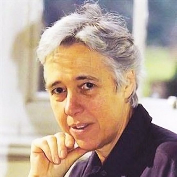
Free Neuropathology 1:18 (2020)                                                                  Margaret Miriam Esiri
doi: https://doi.org/10.17879/freeneuropathology-2020-2903                                               page 3 of 16

search Officer post in his department that would be          numerary pre-registration house job that would
devoted to muscle disease research. Would I be               not pay me but would give me the experience nec-
interested in applying? I explained that I needed to         essary to secure full registration. This dealt with the
obtain my full General Medical Council registration          6 months’ medical experience. To complete the
by occupying a year of preregistration House Of-             additional 6 months’ surgical experience that I
ficer posts, but we agreed to keep in touch.                 needed, I managed to gain approval to work as a
                                                             House Officer in Obstetrics, a specialty that did not
                                                             have routine pre-registration jobs. The advantage
                                                             of taking this route was that it entailed a 1-in-3 on
                                                             call rota at night, whereas in surgery this rota
                                                             would have been 1-in-2. It was also critical that I
                                                             had the offer of such an obstetric post from the
                                                             head of the Obstetric service, Professor John Stall-
                                                             worthy, whereas none of the surgeons was willing
                                                             to offer me a post. Thus, I had secured the possibil-
                                                             ity of getting my full medical registration, albeit
                                                             with an unconventional set of posts. I also needed
                                                             to secure the home situation and here we were
                                                             very lucky to find a very good young nanny, Jenny,
                                                             who was willing to live in so that I could do my
                                                             nights on call. I enjoyed this year of clinical work
                                                             but I knew it would be impossible for me to carry
                                                             on with this type of work in the future.
                                                                   At the end of the year, I therefore applied for,
                                                             and obtained, the Junior Research Officer post that
                                                             Trevor Hughes had secured. The post was for 3
                                                             years and it would enable me to work for a DM
                                                             research degree while allowing me at the same
                                                             time to see what I thought about a career in Neu-
                                                             ropathology. I recognised that this was a very small
                                                             specialty with only about 20 departments in the UK
                                                             and those confined to major medical centres. I had
                                                             always kept at the back of my mind the possibility
Fig. 2. Dr J Trevor Hughes                                   that I might end up working in Nigeria. If I trained in
                                                             Neuropathology I would need to re-train if I were
     While I was carrying out this year of leprosy           going to Nigeria. But since our young family had
research, I had kept my hand in at dealing with              appeared, Nigeria had been experiencing a bloody
patients by helping out once a week in an endocri-           civil war, ruling it out for settling in for the time
nology clinic run by one of the consultants, Dr              being. The prospect of three further years living
Derek Hockaday, whose clinical firm I attended for           and working in Oxford helped us to decide that it
my second medical attachment as a student. Derek             would be best if the children were to be brought up
was aware of my difficulty in obtaining pre-                 with their roots in the UK. We did not think it
registration house jobs while responsible for a              would be a good idea to move the children to and
young family. It was necessary to obtain the full            fro between Nigeria and the UK even if the civil war
GMC registration to enable me to progress to any             had ended.
subsequent clinical specialty. Derek was sympa-                    So, in 1970, I started as a Junior Research Of-
thetic to my plight. He had a wife who worked part-          ficer in Neuropathology. I needed to decide on a
time as a paediatric neurologist and they had 3              topic to research on muscle disease. Perusal of the
young children. He eventually offered me a super-            textbooks about human muscle disease seemed to
Free Neuropathology 1:18 (2020)                                                                 Margaret Miriam Esiri
doi: https://doi.org/10.17879/freeneuropathology-2020-2903                                              page 4 of 16

suggest that although there was a great variety of           nosing the nature of kidney disease from studying
rare diseases, the two categories of disease that            renal biopsies. I was able to use this electron mi-
were more common were inflammatory myopa-                    croscope soon after it was installed and it turned
thies and muscular dystrophies. I chose to study             out to be very useful for diagnosing some of the
inflammatory myopathies because I had become                 rare muscle diseases.
interested in the role of the immune system in dis-
                                                                   Just as I had initiated these studies and satis-
ease during my experience of studying leprosy.
                                                             fied myself they were running well, I was overtaken
There were also not then the tools readily available
                                                             by an unexpected event – a massive fire in March
to carry out genetic studies of muscular dystrophy
                                                             1971 that destroyed the whole of the Neuropa-
that are now very effectively in play.
                                                             thology Department and the adjacent hospital li-
     Inflammatory myopathies can occur at any                brary at the Radcliffe Infirmary. Luckily no one was
age but are most common in late middle age. To               injured. The fire occurred at night and I came in the
make a definitive diagnosis, a small sample of mus-          next morning to find the department, including all
cle – a biopsy – needs to be removed and studied             my records, razed to the ground. The hospital au-
under the microscope to see if inflammation is               thorities had to scurry round searching for some-
present. The cause of the inflammation is not cer-           where to put us. The Professor of Surgery gener-
tain but it was thought likely that it was due to an         ously offered an office and there was some recent-
immune reaction to some component of muscle,                 ly vacated animal house accommodation, rather
triggered by division and activation of subsets of           dark and damp, into which we could move. Having
lymphocytes. I decided to investigate blood lym-             scavenged a few chairs and a desk or two from
phocytes taken from people with recently diag-               sympathetic colleagues, we sat ourselves in the
nosed inflammatory myopathies and compare their              animal house to draw breath when who should
reactions in culture to a small concentration of             appear but a former neuropathologist who had
homogenised muscle with the reactions of healthy             moved a few years earlier to London, Dr Sabina
people (myself and my colleagues). To do this I              Strich. She had heard the news of the fire on the
needed to find an immunology laboratory that                 radio and promptly come to visit and commiserate
could accommodate me and my lymphocyte cul-                  and to provide us with a new electric kettle, mugs,
tures. There was such a laboratory in the Nuffield           coffee and chocolate wholemeal biscuits. It was
Department of Medicine. It was headed by Dr Ian              such a welcome gesture! It raised all our spirits and
McLellan. Although the laboratory was already full           made us determined to make the best of the situa-
of researchers, he very generously allowed me to             tion. Fortunately, my lymphocyte studies could
join them. I carried out my human lymphocyte                 continue in the Immunology Laboratory which was
studies there and also studied rats in which I at-           in a different part of the hospital, even though the
tempted to produce a similar condition of inflam-            records of the first experiments were lost. There
matory myopathy by injecting them with muscle                were also negatives from the electron microscope
homogenate combined with an adjuvant. While the              studies that could be retrieved and reprinted. Some
lymphocytes were being cultured, I also examined             of the brain specimens from earlier years were still
the muscle biopsies from the same patients with              retained in a hut in the grounds of another hospital
light and electron microscopy. Although there                and those that were most instructive for teaching
would be no foreign organisms expected to be pre-            purposes were retrieved and sections re-cut for
sent in the muscle if this inflammatory myopathy is          microscopy. Some second hand equipment such as
an autoimmune disease, it still seemed worthwhile            microscopes and microtomes, needed to cut thin
to check for any invading organisms, such as virus-          sections, were rapidly acquired and the elements of
es, which might be detectable with an electron               a re-formed department thus assembled. Eventual-
microscope. To start with, I used the electron mi-           ly the whole destroyed floor of the hospital was re-
croscope in the Anatomy Department that I was                built, though this was not to happen for several
already familiar with but the Radcliffe Infirmary            years.
soon acquired one of its own in the Pathology De-
                                                                  The three years of the junior research officer
partment where it was particularly useful in diag-
                                                             post passed quickly with my energies divided be-
Free Neuropathology 1:18 (2020) Margaret Miriam Esiri
doi: https://doi.org/10.17879/freeneuropathology-2020-2903 page 5 of 16
tween the laboratories and the home. We now had disease, to include control samples as well as sam-
a new nanny, Jane, who stayed with us until all the ples from the disease under investigation. It was
children were at full time school. The combination important for the investigator, whenever possible,
of research and pathology was much more compat- to be unaware of which samples were from con-
ible with family commitments. My husband was trols and which from disease so that there was no
pursuing a surgical training and holding a succes- chance of bias creeping into the observations being
sion of short term posts all over the country as was made. It seemed to me that if careful quantitative
the usual practice in those days. He returned home studies were performed they could almost always
whenever he was able to. yield useful information even if they showed that
the ideas that prompted the experiments turned
The lymphocyte studies yielded modest evi-
out to be incorrect. In short, science satisfied my
dence of lymphocyte sensitivity to muscle in in-
urge to ask questions and seek answers to them
flammatory myopathies. It was not a dramatic ef-
and having commenced my efforts in that direction
fect and could have been the result, rather than the
I was going to continue if I could.
cause, of the condition. It was difficult to provoke
any more than a slight inflammatory reaction in the
muscle of the sensitised rats. In the electron micro- The next stage
scopic studies, there was only one biopsy that con-
tained what might have been virus-like particles. To become a neuropathologist in the 1970s I
Thus, the fruits of my studies were far from conclu- needed first to gain experience of other specialties
sive. I believe it remains the case today, more than of clinical pathology. I became a trainee registrar
40 years after these studies were performed, that with a post that rotated through haematology,
inflammatory myopathies are really little better bacteriology, virology and histopathology. The
understood. However, the thesis I wrote describing longest time (2 years) was spent in histopathology
these studies earned me a DM degree and I pub- (also known as cellular pathology). There were ex-
lished a couple of papers summarising the findings. ams to take for membership of the Royal College of
I had found satisfaction from this work but now felt Pathologists once the practical experience had
increasingly curious about the central nervous sys- been obtained. It was essential to gain this qualifi-
tem and its diseases that I had started to become cation before applying for a post as consultant in
familiar with. However, before I was certain that the NHS. The exam had a heavy component of his-
neuropathology should become my chosen special- topathology, hence the longer time spent in that
ty, I needed to become familiar with other branch- specialty.
es of pathology as part of my training. I didn’t find the haematology or bacteriology
What did these two short periods of research very interesting. The numbers of samples to be
teach me? First they reinforced my admiration for analysed in each of these departments were so
science. I particularly admired the way science was overwhelming that much of the work had been
prepared to embrace the contributions of people automated. Many samples would typically be sent
wherever they came from and whatever their to each of these departments during the course of
background provided they worked with integrity each patient’s hospital admission and, in addition,
and had innovative ideas. I admired the way sci- many samples were sent in each day from local
ence relied on reproducibility of findings to give general practitioners. The departments were pri-
them credence and not on the advertising ability of marily concerned with delivering a diagnostic ser-
their perpetrators to give validity to their findings. I vice and there was relatively little that I, as a new-
came to appreciate the importance of Karl Popper’s comer, could contribute. They did not undertake
dictum that scientists should seek to refute their active research. Histopathology was of greater in-
ideas, not confirm them. Scientific ‘facts’ are al- terest, being more directly relevant to neuropa-
ways provisional and can be revised if subsequent thology. I learnt how to perform post mortem ex-
experiments are incompatible with them. I learnt to aminations (in the process giving myself the only
recognise the importance of quantitation in record- nightmares I’ve ever had, in which the corpse came
ing results of experiments and the need, in studying to life while I was busy and I attempted to replaceFree Neuropathology 1:18 (2020) Margaret Miriam Esiri
doi: https://doi.org/10.17879/freeneuropathology-2020-2903 page 6 of 16
the larynx so that it could speak!) and to report on This was research I undertook when I had
surgical specimens of tissue. Because of the large moved back from the Histopathology Department
variety of specimens and of the conditions they to be a registrar in the Neuropathology Depart-
exhibited, it took a fair time to be able to make ment. My experience in more general pathology
correct judgements on the nature of the disease, had not seduced me into getting more interested in
often cancer. Samples were examined under the that field. Rather, I thought there were important
microscope, first by myself and then by a consult- things I had learnt there, particularly this new sub-
ant who was able to share his or her experience ject of immunohistology, that I could usefully apply
and pass on to me the critical visual clues that in neuropathology. Back in the Neuropathology
yielded the essential evidence about the nature of Department, I was also gaining experience of diag-
the disease process. nostic aspects of the specialty by examining biop-
sies removed from brain tumours by the neurosur-
The head of the Histopathology Department
geons as well as other biopsies such as those from
was Dr Robb-Smith, a slightly shy and enigmatic
muscle and peripheral nerves. I was also perform-
character whom I had encountered as a student at
ing post mortem examinations on cases of neuro-
post mortem demonstrations. He was interested in
logical disease and brain trauma. This diagnostic
attracting women to his specialty and had secured
work I found still left time for a limited amount of
a trainee post that could be held part-time. This
research in which I applied the antibody techniques
enabled me to have two free afternoons each
I had been lucky enough to learn about from Ian
week. I found the children were all occupied at
Burns to nervous system diseases. This was an ex-
these times so I used this time to write up my DM
ample of the wonderful way that collaboration can
thesis, a task I had been putting off while I dealt
be developed in science and I was to become a
with more immediate concerns at home and at
great advocate of such collaboration, prizing it far
work.
above competition which, in some fields or de-
One of the great advantages for me of work- partments of medical science sadly seemed to pre-
ing in the Histopathology Department was that I dominate.
not only gained essential diagnostic experience but
I was lucky enough to obtain the modest funds
I was also able to participate in research. There was
needed to enable me to employ a technician to
a research officer there, Dr Ian Burns, who was
assist with the application of immunohistology in
experimenting with the use of antibodies to detect
neuropathological material. I started by studying
specific proteins in tissue sections. If antibodies
the lesions or areas damaged in the central nervous
could be produced towards a purified protein of
system in multiple sclerosis, most generally consid-
interest and tagged with a marker substance such
ered as an autoimmune disease1. This was a condi-
as a coloured dye or a fluorescent dye, this could
tion that was of interest to the other consultant in
be used to detect specific proteins in tissue sec-
the Neuropathology Department, Dr David Oppen-
tions with much greater specificity than could be
heimer (Fig. 3), who had written a DM thesis on it.
provided with the traditional tinctorial stains. This
He had seen many examples of this condition in
was the principle behind the work that Ian Burns
post mortem studies and the tissues from some of
was carrying out and which has now become the
these cases were still available in the department
hugely successful branch of pathology, immuno-
despite the fire that had destroyed much material
histology or immunocytochemistry. When Ian was
in 1971. One of the beauties of the immunohisto-
working in the early 1970s, he was concentrating
logical technique was that it could be used on tis-
on the use of antibodies to detect immunoglobu-
sue that had been preserved in the department
lins, themselves antibody proteins produced by
after being fixed in formalin and then embedded in
plasma cells. I was able to make use of Ian’s rea-
paraffin wax to enable thin sections to be cut. Alt-
gents to study plasma cells containing different
hough some proteins that may be of interest in
types of immunoglobulins in sections of central
tissues are destroyed by such treatment, meaning
nervous system tissue affected by different diseas-
that fresh frozen sections are needed to study
es.
them, there are many others that survive thisFree Neuropathology 1:18 (2020) Margaret Miriam Esiri
doi: https://doi.org/10.17879/freeneuropathology-2020-2903 page 7 of 16
treatment. The advantage of being able to use for- Hughes had a particular interest in muscle disease
malin-fixed, paraffin-embedded sections is that this and spinal cord disease. He had moved into neuro-
tissue shows better preservation of its structure pathology from histopathology. David Oppenhei-
than can usually be seen in frozen sections. mer had a less conventional background. He had
started his academic career with degrees in Philos-
ophy, Politics and Economics and also Music. Dur-
ing the Second World War, he had been a conscien-
tious objector and worked with an ambulance team
in London rescuing injured people in the blitz. This
experience awakened his interest in medicine and
he read for his medical degree immediately after
the war. Initially he worked on neuroanatomy and
it was from there that he moved to neuropatholo-
gy. He was initiated into diagnostic work by his
predecessor, Sabina Strich, over a matter of a few
months. He recognised that his lack of experience
in histopathology and other pathology specialties
was a handicap but his knowledge of neuroanato-
my was most valuable. It made him particularly
interested in the many important diseases of the
nervous system in which there is a neuroanatomi-
cal basis for the way the disease is expressed or, in
some cases, seems to spread. I had been intro-
duced to the Neuropathology Department by Tre-
Fig. 3. Dr David Oppenheimer
vor Hughes but as time went on I became more
interested in the conditions that interested David
I followed up the work on multiple sclerosis Oppenheimer, starting with multiple sclerosis.
with studies of diseases that did have a foreign
organism in the damaged tissues, starting with One of the first opportunities that arose from
poliomyelitis of which we had cases dating from an the diagnostic work that I carried out was a chance
epidemic of polio in the UK in the late 1950s. Later I to study a rare case of herpes zoster (shingles) in an
extended the work to a study of cases of the rare elderly woman who happened to die just after the
complication of measles, subacute sclerosing eruption developed. It wasn’t the herpes zoster
panencephalitis. I wanted to know if these diseases that killed her but an advanced state of multiple
showed a similar distribution of plasma cells con- myeloma which had rendered her susceptible to
taining immunoglobulins as I saw in multiple scle- developing herpes zoster. My patient had herpes
rosis. Although this turned out to be the case, the zoster affecting the region supplied by part of the
difficulty in interpreting the significance of the find- trigeminal nerve. Herpes zoster is caused by the
ing was that the arrangement of plasma cells might same virus that causes chickenpox, varicella-zoster
be just the same in autoimmune disease as in one virus. The theory had been developed that in the
with a foreign organism. While these studies were natural history of infection with this virus, chicken-
proceeding, more antibodies for use in immuno- pox is the first manifestation of infection. As the
histology were being produced, including some virus is overcome by the immune response that
directed to viruses. I was to make use of these for a enables the person with chickenpox to recover,
later study when I was a Research Fellow. some virus survives and lies latent in the nerve cells
of one or more sensory nerves. Circulating antibody
The Neuropathology Department when I which persists after recovery from chickenpox
joined it was a small one. There were 2 consultants, makes any reactivation of the virus uncommon
Drs Hughes and Oppenheimer, a registrar (myself), until later in life when the level of antibody wanes
3 technicians and a laboratory assistant. Trevor or when the person has their immunity compro-Free Neuropathology 1:18 (2020) Margaret Miriam Esiri
doi: https://doi.org/10.17879/freeneuropathology-2020-2903 page 8 of 16
mised by disease. When the virus reactivates in the that could not survive formalin fixation. This obser-
sensory nerve cells of a particular nerve, it travels vation made me think that there could be more we
along the nerve to the skin where it causes the could learn from our post mortem and surgical
eruption of herpes zoster. Although this theory was specimens if we thought carefully about whether
widely accepted, the reactivation of the virus in we should preserve a portion of tissue by snap
nerve cells had never been demonstrated. It was freezing it in liquid nitrogen and then storing it in a
this reactivation that I was able to show by electron -70◦C freezer. Snap freezing in liquid nitrogen pre-
microscopy which revealed the virus particles in the served the tissue structure better than slowly freez-
nerve cells of the trigeminal ganglion (Fig. 4). A ing tissue in a freezer but only if the tissue sample
colleague in the Virology Department, Dr Albert was small. The opening of our minds to the poten-
Tomlinson, carried out immunofluorescence using tial importance of deep freezing some tissue was to
an antibody to the varicella-zoster virus on frozen prove important in years to come, not only to ena-
sections of the ganglion and the nerve and was able ble immunohistology to be performed for detection
to show the virus at both sites. of antigens that were sensitive to formalin but also
to enable biochemical and genetic studies to be
carried out. It led to Oxford being early in the field
of brain banking for research (for a history of brain
banking in the UK see 2).
The highly trained technicians that we worked
with in neuropathology played an absolutely essen-
tial role in all that we did. Unlike in histopathology,
where most tissue sample sections were subjected
to a single stain, in neuropathology it was common
to have consecutive thin sections from the same
block of tissue stained with several different stains,
some of which were quite difficult to perform. Each
technician had their special expertise in carrying
out a selection of these stains. Some were histo-
chemical stains that enabled enzymes to be dis-
played in frozen sections. These histochemical
stains were mainly used on muscle biopsies. Each
of the tinctorial stains picked out a particular fea-
ture of the nervous system to highlight. Nowadays
the need for these special tinctorial stains is much
reduced because immunohistology can convey the
same information using antibodies that are more
specific and more straightforward to apply. Some
of the reagents used in the tinctorial reactions are
quite toxic and it is far preferable that technicians
can now avoid them.
Fig. 4. Herpes zoster viral particles in the cytoplasm (C) of a
trigeminal ganglion neuron. My closest colleagues were those in the Neu-
ropathology Department. Trevor and David took it
From Esiri MM, Tomlinson AH J Neurol Sci 1982; 15(1); 35—48.
in turns, several months at a time, to supervise my
diagnostic work and I had the close assistance of
The varicella-zoster virus that Dr Tomlinson the technicians in the preparation of the sections I
was able to show in frozen sections of the nerve needed to do this work. I liaised closely with the
and ganglion of our case could not be seen when neurosurgeons and neurologists over the results. At
we tried to detect the virus in formalin fixed tissue times there was another trainee doing a project in
– the viral protein was an example of an antigen the department or a visitor making use of the ma-Free Neuropathology 1:18 (2020) Margaret Miriam Esiri
doi: https://doi.org/10.17879/freeneuropathology-2020-2903 page 9 of 16
terial we had available for them to study. I remem- someone interested in collaborating with him to
ber particularly a very pleasant, recently retired, study the neuropathology of dementia, particularly
Canadian professor of anatomy, Richard Saunders, Alzheimer’s disease. He was himself interested in
who came to spend a few winters in Oxford with learning some neuropathology and he had many
his wife to escape the worst of the winter in Nova patients under his care in the geriatric medicine
Scotia. They had a son who was an Oxford Rhodes wards on whom, if they agreed, he had carried out
Scholar. Richard had an intimate knowledge of the a simple cognitive test to determine whether or not
structure of the hippocampus which he did his best they had dementia. A considerable number of them
to impart to me. It turned out to be useful had dementia but there were others who did not,
knowledge when I became interested in Alz- including quite a number who had had strokes. If
heimer’s disease. any of these patients subsequently died, and their
next-of-kin was willing for a post mortem to be
I made regular visits to local district general performed, we were able to see what abnormali-
hospitals where I provided a diagnostic service on ties there were in the brain that contributed to
neurological post mortem cases. I discovered that making them demented. It was known that in Alz-
histopathologists in these hospitals had to deal heimer’s disease there were two distinct micro-
with quite a lot of neurological cases as the hospi- scopic abnormalities: the presence of plaques and
tals had consultant neurologists working in them. tangles. Gordon and I wanted to see if we could
This visiting service yielded many interesting cases relate either plaques or tangles (or both) to the
from which I brought back to our department se- presence of dementia in his cognitively tested pa-
lected samples for expert processing. I would then tients.
send reports on these cases back to the histo- We had the impression that the distribution of
pathologists. tangles in Alzheimer’s disease affected parts of the
brain that were becoming known to be anatomical-
Once or twice a year there were meetings of ly linked together. We took to discussing the neu-
the UK neuropathology community. Trainee and roanatomy of Alzheimer’s disease on Friday after-
consultant neuropathologists could apply to join noons with experts on these cortico-cortical and
the British Neuropathological Society, which organ- cortico-hippocampal connections, Drs Tom Powell
ised these meetings, after they had delivered one and his former DPhil student, Carl Pearson. Tom
sponsored paper or poster at one of the meetings. had spent his career on meticulous studies of the
Some overseas neuropathologists also joined the cerebral cortex and was greatly pleased to find that
Society. My first paper to the Society, sponsored by some of his work might have relevance to an un-
Trevor, described the work I had done on experi- derstanding of such an important disease as Alz-
mental leprosy infection in the Anatomy Depart- heimer’s disease. From these discussions came a
ment. It was interesting to get to know neuropa- study of the detailed distribution of plaques and
thologists working in other centres and to compare tangles in the different layers of the cerebral cor-
notes on our respective research projects. I found tex3. The findings fitted well with what was known
them to be a very congenial group. Collaboration of the manner in which one part of the cortex was
with some of them became an important asset in connected to others and to the hippocampus.
devising some research projects for which more Those regions most closely linked to the hippocam-
cases of a particular disease might be needed than pus had many tangles while those parts of the cor-
were available in one centre. It was also helpful at tex which were most remote from the hippocam-
times to be able to send a puzzling diagnostic spec- pus had the fewest tangles. This work was my in-
imen to someone in another centre to obtain a troduction to trying to understand Alzheimer’s
second opinion on the diagnosis. disease from a neuropathological perspective.
Towards the end of my time as a registrar I Another influence in fuelling my new interest
made the acquaintance of a recently appointed in dementia was a publication by a neurochemist at
consultant in geriatric medicine, Gordon Wilcock. London’s Institute of Neurology, Dr David Bowen.
Gordon had come to enquire if there might be In the mid-1970s, he published a paper showingFree Neuropathology 1:18 (2020) Margaret Miriam Esiri
doi: https://doi.org/10.17879/freeneuropathology-2020-2903 page 10 of 16
that in post mortem brain specimens that had been cruit patients into clinic-pathological studies of
frozen at -70◦C immediately after removal from the human disease.
body there were many enzymes whose activity
By the mid-1970s, I had acquired enough ex-
could be reliably measured. He furthermore
perience to take the Royal College of Pathologists
showed that in Alzheimer’s disease the enzyme,
examination in Histopathology slanted to Neuropa-
choline acetyl transferase, required for the synthe-
thology. The written examinations were taken in
sis of acetyl choline, was deficient. A similar finding
London and for the practical exam I was instructed
was reported from two other laboratories at about
to go to Glasgow. I remember casting my eye hasti-
the same time. This paper was a real eye-opener
ly over my notes on the long train journey to Glas-
for me. I had previously assumed that after death
gow and thinking that to be taking examinations at
all enzyme activity would cease. Gordon and I had
the age of 35 years was too old! Fortunately, I
discussions with David about providing him with
passed this last one and was then in a position to
frozen brain samples from the patients Gordon had
think about the next step in my career. For the sake
tested for cognition on the wards and we were
of our children who were happily settled in schools
soon adopting the policy of deep freezing one side
in Oxford, I wanted to remain there but there was
of the brain for David’s studies and carrying out our
only one academic post in Neuropathology and
microscopic studies on the other side. This led to a
that would be occupied by David Oppenheimer for
realisation that it is the burden of neurofibrillary
another few years. One of my colleagues in the
tangles that is held within the brain that deter-
Histopathology Department made a most helpful
mines the extent of dementia in Alzheimer’s dis-
move at this point. He wrote to the Medical Re-
ease, much more so than the amyloid load4. I have
search Council on my behalf and asked if there
watched with dismay the dominance of amyloid in
might be a prospect of getting support from this
thinking about Alzheimer’s disease and how to
source to fund research for a few years in Oxford.
prevent it that has lasted for decades. Hopefully,
An encouraging reply to this enquiry led to me ap-
now that may change5.
plying for a Senior Clinical Fellowship from the
Not long after we had set up this programme Medical Research Council.
to study dementia, Gordon was offered a profes-
sorship in care of the elderly at Bristol University Becoming a Senior Clinical Fellow
and left Oxford but there was an interested con-
sultant in geriatric psychiatry at the large Little- I put together an application for this Senior
more mental hospital near Oxford, Dr John Robin- Clinical Fellowship to study a disease that had long
son, who was able to play a similar role in supplying been of interest to me – herpes simplex encephali-
cases of dementia, though he was not in a position, tis. This is a rare condition but one that had a con-
as Gordon had been, to supply cases that did not siderable mortality at that time and which I had the
have dementia. chance to study post mortem. There were several
This collaboration with clinicians in our re- enigmatic aspects to this disease which intrigued
search on dementia was the first time I had had the me. It seemed to affect people of a wide range of
opportunity to work directly with doctors looking ages in whom it developed quite unexpectedly. Yet
after patients to devise a research strategy. Neuro- it was caused by a virus that infected some 90% of
pathologists are too remote from direct patient people in some of whom it caused recurrent cold
care to be in a position to recruit patients to a sores. Like its cousin, varicella-zoster virus, which I
study themselves. Some neuropathologists devel- had already encountered, it had the capacity to lie
oped interests in brain tumours that they could latent in sensory ganglia (for this virus, usually the
follow using the biopsy specimens they received. trigeminal ganglion, because it usually causes a
Others developed experimental models of diseases facial infection) from which it could reactivate un-
that could be studied alongside human post mor- der a range of conditions such as fever, exposure to
tem material. But I think the most valuable contri- strong sunlight or the common cold. What was the
bution that clinical neuropathologists can make is process by which it could occasionally cause a dev-
to work closely with their clinical colleagues to re- astating encephalitis? I knew I would be able toFree Neuropathology 1:18 (2020) Margaret Miriam Esiri
doi: https://doi.org/10.17879/freeneuropathology-2020-2903 page 11 of 16
study the distribution of the virus in the brain using To take this work further I teamed up again
immunohistology and an anti-herpes simplex anti- with Albert Tomlinson, the virologist who had
body, and I had tissue from enough cases, which helped me examine the case of herpes zoster. We
had survived for varying periods of time after dis- developed a mouse model of herpes simplex en-
ease onset, to enable me to try to piece together cephalitis and showed that inoculation of the face
the pathological process. Armed with this intention led to the virus ascending to the brain via both the
I was lucky enough to get the Fellowship for 5 years trigeminal and olfactory routes and the develop-
at consultant level salary plus some part-time tech- ment of both brain stem and temporal lobe in-
nical help and costs of consumables. flammation. Others have pursued the possibility
that latent infection of the brain with herpes sim-
This period of full-time research reinforced my
plex virus may predispose to the development of
passion for investigating brain diseases of which
Alzheimer’s disease but the opportunity to further
there seemed so many without an effective treat-
investigate the human acute encephalitis was lost
ment, in part a consequence of inadequate under-
on account of the fortunate development of an
standing of the disease process – hardly surprising
effective treatment which reduced the fatality of
given the complexity of the nervous system and the
the disease and resulted in many people affected
difficulty of accessing brain tissue during the course
recovering at least partially.
of many of these diseases. At the time, brain imag-
ing was still rudimentary compared with how it is
today and the post mortem examination, for those A change of direction?
diseases that were fatal, gave the best lead to in-
crease understanding. While I was intent on this research, my chil-
dren had grown into their teens and my husband
Herpes simplex encephalitis has a very intri- was exploring the possibility of returning to Nigeria
guing distribution of damage in the brain with an to provide medical care there. This posed a dilem-
emphasis on the temporal lobes but with the dam- ma for me. Should I abandon my neuropathological
age usually asymmetrically distributed6. The focus career and re-train in tropical medicine or could I
of damage overlaps with that in Alzheimer’s dis- carry on with my neuropathology in Oxford part-
ease with the hippocampus and sites connected to time while taking periods of unpaid leave to join my
it badly affected. From my visits to the Anatomy husband in Nigeria during school terms when our
Department to discuss Alzheimer’s disease with two younger children were at boarding school? The
Tom Powell and Carl Pearson, I knew these areas other alternative - to pursue neuropathology in
had close connections with the olfactory system so Nigeria, in the very remote region in which my hus-
I studied the olfactory bulbs in these cases of her- band decided to set up a medical practice - would
pes simplex encephalitis because it seemed possi- have been impossible. I was able to negotiate to
ble that the virus, with its propensity to travel carry on part-time in Oxford for the last part of my
along axons, might reach the brain along this route. research project, working full-time while in Oxford
I could indeed detect the virus there but the olfac- and visiting Nigeria between times. Strange though
tory bulbs were not as damaged as sites in the such an arrangement might seem, it actually
amygdala and piriform cortex to which the olfacto- worked well and, by the time the research was
ry tracts projected. Thus it seemed possible that completed, David Oppenheimer had retired, giving
the virus reached the olfactory bulbs by moving me the expectation that I might apply for his (now
centrifugally, not centripetally, perhaps from a site vacant) post.
of latency within the brain, perhaps the hippocam-
pus. The asymmetry of damage to the temporal That expectation was thrown into confusion
lobes, which was more marked in those dying early by the government of the day deciding to impose
in the course of the disease, might be explicable if drastic cuts to university financial support. As a
it reactivated from inside one hippocampus and result, all re-filling of posts was frozen and many
then travelled to the contralateral one via anatomi- posts were lost. I remained in a form of profession-
cal connections between the two. al limbo with short-term Medical Research Council
support lasting 3 months at a time until a new pro-Free Neuropathology 1:18 (2020) Margaret Miriam Esiri
doi: https://doi.org/10.17879/freeneuropathology-2020-2903 page 12 of 16
fessor, John Newsom-Davis, was appointed in the cover or, at worst, return the next day for a re-
Clinical Neurology Department on the retirement think about the diagnosis, I thoroughly enjoyed this
of the previous postholder. John offered to attempt experience. With no diversions such as newspapers
to reinstate the lectureship in Neuropathology or television to occupy the time later in the day I
which had previously been held in the Cellular Pa- was able to think and write without interruption,
thology Department, but only if it could be held in although, in the evenings, any activity had to be
the Clinical Neurology Department. The post was carried out by the dim illumination offered by a
rescued in this way and I was appointed to it on a ‘tilly’ light, fuelled by kerosene.
part-time basis that allowed me to continue with
To follow up after publication of Viral Enceph-
my visits to Nigeria.
alitis, I approached David Oppenheimer, who had
My husband’s choice of where to establish his recently retired, to see if he would be willing to
clinic was partly determined by his wish to live in a write a book on diagnostic neuropathology with
part of the country that is decidedly cooler than the me. I felt greatly honoured to have had much tui-
rest. He hated the tropical heat of most of the tion from David during my training. He was a truly
country and he knew I would too. His clinic was inspiring person and I felt it would be good if other
established in a town, Gembu, on the Mambilla people could benefit from his teaching even if it
Plateau, some 1,500 metres high and not far from had to be in the form of a textbook. I knew he
the Cameroon border which lies to the east of Ni- wrote superbly so could convey his knowledge in
geria. The countryside is made up of grass-covered that way. He thought about it and agreed to write a
hills, interspersed with deep, fertile, valleys and the ‘pamphlet’ in which we would divide up the topics
climate is really perfect, with temperate, warm, evenly between us. He had kept the photographic
sunny weather and abundant rainfall. The roads slides that he had collected over his career so was
were rudimentary in the extreme and getting to the able to make use of these and entered into the
place took some 12-15 hours of uncomfortable writing of the ‘pamphlet’ with enthusiasm. I con-
land rover motoring after a domestic air flight to tinued to learn from him as we wrote and it was a
the nearest place with an airport, Yola. Over the complete pleasure, at the end of a working day
years, a tarred road of sorts was created that when I was in Oxford, to visit him and his wife on
mounted the steep escarpment to the plateau but my way home and to share the new text he had
it was soon full of potholes that were hardly ever written during the day. I think he also enjoyed be-
repaired. ing able to extend his teaching of neuropathology
in this way. The result was a textbook of nearly 400
Spending 2 months at a time in Gembu in the
pages that was published as Diagnostic Neuropa-
spring and autumn of each year became my routine
thology8.
for about 20 years from the early 1980s. Because I
couldn’t continue with neuropathological research The third book that I initiated was one on the
while I was away, I decided to turn my attention to neuropathology of dementia9, which was co-edited
writing about the diseases I was interested in. My with my colleague James Morris, who had stepped
aim was to inform those coming after me about into the post that Trevor Hughes had vacated when
what I had learnt so far. My first book was written he retired. That all these three books went in to 2nd
with John Booss, a neurologist from Yale, who editions pleased me as it suggested they had found
came to Oxford on a sabbatical to study multiple useful homes among those in the neuroscience
sclerosis. Together we wrote a book on viral en- community.
cephalitis7. To write my parts, I took piles of refer-
By this time, a major longitudinal study of age-
ences with me to read and digest in Gembu, mainly
ing and dementia, the Oxford Project to Investigate
in the afternoons and evenings after I had helped in
Memory and Dementia (OPTIMA,) had been started
the mornings at my husband’s clinic. My offerings
by David Smith, Professor of Pharmacology in Ox-
there were decidedly amateurish but one felt that
ford, and we were benefitting from the opportunity
in such a place, where medical care barely existed,
to study the brains of dementia sufferers and
one was justified in doing one’s best. Once I had
healthy elderly controls who had been followed up
gained a little confidence from seeing people re-Free Neuropathology 1:18 (2020) Margaret Miriam Esiri
doi: https://doi.org/10.17879/freeneuropathology-2020-2903 page 13 of 16
in a detailed way during life and had consented to may be changing now. There were also superb op-
donating their brain for research after their death. portunities in Oxford to collaborate and learn from
This study grew slowly, as such longitudinal studies neuroscientists in the basic science departments
do, but we eventually collected over 500 brains from which we benefitted enormously.
that helped to increase understanding of dementia,
A great boost to our research on schizophre-
and still continue to do so.
nia came from the generous provision of a substan-
tial group of brains donated for research in Belfast,
Forging ahead Northern Ireland. Professor Ingrid Allen, the senior
neuropathologist in Belfast, and her colleague in
Once I had been appointed to an academic psychiatry, Dr Steven Cooper, were instrumental in
position in neuropathology in the mid-1980s, I creating this initiative with Tim Crow. Tim had a
started to supervise graduate students who wished fascinating hypothesis about schizophrenia: that it
to study the brain and its diseases. I always insisted was somehow related to subtle differences in cere-
that students should have two co-supervisors be- bral asymmetry, that differs normally between men
cause of the periods I spent in Nigeria and out of and women, but which he thought might be dis-
contact with the department. Two months was the turbed in those with schizophrenia. This led to our
maximum that I considered possible to remove studies of the brain in schizophrenia being carried
myself from all the activities that go on in a de- out having regard to sex and cerebral hemisphere
partment that has diagnostic, teaching and re- side. We also studied the corpus callosum and oth-
search interests. Sometimes I would return to find er tracts that connect the two hemispheres. We
a pile of problems to solve but usually everything found tantalising findings on this basis that seemed
had continued smoothly and I had the pleasure of to indicate, among other things, that schizophrenic
hearing about the new findings that had emerged males have cerebral hemispheres more akin to
while I was away. Because of these periods of leave normal females than occurs normally in males and
I kept my other absences from the department to a vice versa for female schizophrenics. We should not
minimum which meant attending very few confer- have found these differences if we had studied
ences and meetings – a sacrifice that I was willing schizophrenia without distinguishing between the
to make in return for the privilege of being able to sex of subjects whose brains we studied. Although
continue with my professional work in my rather it has become fashionable in psychology to down-
unusual personal circumstances. play differences in brain structure between males
The department was expanding at this time and females I think these differences are important
(the late 1980s) with the arrival in Oxford of Profes- and may have significant roles in psychiatric disease
sor Tim Crow, a psychiatrist with a deep interest in that we are not yet able to understand.
biological aspects of psychiatric disease, particular- While these attempts to understand psychiat-
ly schizophrenia. We were able to expand into a ric disease were launched, we were also continuing
new small laboratory and recruit an experienced to investigate dementia and ageing with the OPTI-
technical manager and a small group of research MA resource. A graduate student, Zsuzsa Nagy,
students to work on psychiatric disease. We were who remained for a while in the department as a
also joined by a research group created by a local postgraduate research scientist, discovered that
academic psychiatrist, Dr Paul Harrison. I was never neurons expressed antigens indicating they had re-
in any doubt that study of the brain was needed as entered the cell division cycle in elderly subjects.
much to understand these diseases as it was for This discovery, which has been repeatedly con-
dementia and ageing and for neurological diseases. firmed by others, led to studies of lymphocytes and
In the neuropathology department, I had wonder- the suggestion that properties of these cells in cul-
ful collaborations with psychiatrists as well as neu- ture might have diagnostic value for Alzheimer’s
rologists and neurosurgeons. It seemed very odd to disease. Although this work has been followed up,
me that there was little direct communication be- we are still without a simple method of diagnosing
tween clinical neurologists and psychiatrists and this disease, particularly in its earliest phase, when
little shared training for the two disciplines but this symptoms are absent or minimal, but at the bestFree Neuropathology 1:18 (2020) Margaret Miriam Esiri
doi: https://doi.org/10.17879/freeneuropathology-2020-2903 page 14 of 16
time for any intervention to be made to slow or panied it. But we soon realised that because there
stop its development. is much redundancy in the CNS, progression of
damage would only occur when a threshold of axon
My interest in multiple sclerosis was re-ignited
loss and other forms of neurodegeneration was
at about this time by two developments. The first
reached, leaving room for recovery between epi-
was provoked by a paper that came out in The Lan-
sodes of inflammation before that point was ar-
cet linking episodes of clinical expression at the
rived at.
start and in relapse in multiple sclerosis to episodes
of previous sinusitis. The author, Frederick Gay,
was a general practitioner and he made his obser-
vations using general practitioner records. He
thought it possible that bacterial antigens derived
from microorganisms in the paranasal sinuses
might reach the CNS compartment in some individ-
uals and lead to an immune response that created
the demyelinating, inflammatory lesions typical of
this condition. We worked together on this idea
but, given its unfashionable nature at a time when
most interest lay in viruses having a role in the dis-
ease (if, indeed, any foreign antigens were in-
volved), funding to make progress was hard to ac-
quire. The idea, however still has great attractions,
in my opinion, and it is still being actively pursued.
The second development that brought back
my active interest in multiple sclerosis was applying
immunohistology yet again but this time using an
antibody to amyloid precursor protein as a marker Fig. 5. Graphs to illustrate the profile of axonal injury (APP
of damaged axons. Although axons have long been stippled areas) and the number of macrophages (black areas)
known to be damaged in MS, the extent of that in a typical acute multiple sclerosis lesion (A), active chronic
damage, its timing and its role in progression of the multiple sclerosis lesion (B) and chronic multiple sclerosis
lesion (C).
disease were little appreciated largely because
there had been no sensitive method of assessing its From ref. 10
extent. Being able to detect damaged axons with
great sensitivity, which detection of amyloid pre- This reassessment of multiple sclerosis and, in
cursor protein provided, changed this situation and particular, its neurodegenerative aspects, made me
when we applied the technique to multiple sclero- acutely aware of how progress in understanding
sis lesions of differing age I was greatly surprised by disease depends on new techniques being applied
the result. I had expected that we should see dam- to enable new knowledge to be acquired. The spec-
aged axons in chronic lesions which are common in tacular developments in neuroimaging over the
progressive disease but it was actually in the most past few decades have enabled much progress to
recent lesions that axon damage was most prolific10 be made in understanding CNS diseases and this
(Fig. 5). This was despite the fact that recent lesions will continue. Nevertheless, there is still a place for
are more common at earlier phases of the disease pathological studies because these are still needed
when recovery from relapses is common. Initially it to penetrate to the cellular and molecular levels of
seemed hard to understand how damage to axons, change that accompany disease. Without
which we assumed was irreversible, was compati- knowledge at these levels rational treatment or
ble with a history of relapse followed often by re- prevention cannot be achieved. The new frontier
mission. This pattern of illness seemed more com- relating to knowledge that has grown up in the last
patible with pathology that was reversible, likely two decades is about genetics. Here there is such a
demyelination and the inflammation that accom- wealth of new information that new ways of man-You can also read