Coronavirus Infections in Companion Animals: Virology, Epidemiology, Clinical and Pathologic Features - MDPI
←
→
Page content transcription
If your browser does not render page correctly, please read the page content below
viruses
Review
Coronavirus Infections in Companion Animals:
Virology, Epidemiology, Clinical and
Pathologic Features
Christine Haake 1, * , Sarah Cook 2 , Nicola Pusterla 3 and Brian Murphy 4
 1    School of Veterinary Medicine, University of California, Davis, CA 95616, USA
 2    Graduate Group Integrative Pathobiology, School of Veterinary Medicine, University of California,
      Davis, CA 95616, USA; sestevens@ucdavis.edu
 3    Department of Medicine & Epidemiology, School of Veterinary Medicine, University of California,
      Davis, CA 95616, USA; npusterla@ucdavis.edu
 4    Department of Pathology, Microbiology, and Immunology, School of Veterinary Medicine, University of
      California, Davis, CA 95616, USA; bmurphy@ucdavis.edu
 *    Correspondence: cjhaake@ucdavis.edu

 Received: 28 July 2020; Accepted: 11 September 2020; Published: 13 September 2020

 Abstract: Coronaviruses are enveloped RNA viruses capable of causing respiratory, enteric,
 or systemic diseases in a variety of mammalian hosts that vary in clinical severity from subclinical
 to fatal. The host range and tissue tropism are largely determined by the coronaviral spike
 protein, which initiates cellular infection by promoting fusion of the viral and host cell membranes.
 Companion animal coronaviruses responsible for causing enteric infection include feline enteric
 coronavirus, ferret enteric coronavirus, canine enteric coronavirus, equine coronavirus, and alpaca
 enteric coronavirus, while canine respiratory coronavirus and alpaca respiratory coronavirus result in
 respiratory infection. Ferret systemic coronavirus and feline infectious peritonitis virus, a mutated
 feline enteric coronavirus, can lead to lethal immuno-inflammatory systemic disease. Recent human
 viral pandemics, including severe acute respiratory syndrome (SARS), Middle East respiratory
 syndrome (MERS), and most recently, COVID-19, all thought to originate from bat coronaviruses,
 demonstrate the zoonotic potential of coronaviruses and their potential to have devastating impacts.
 A better understanding of the coronaviruses of companion animals, their capacity for cross-species
 transmission, and the sharing of genetic information may facilitate improved prevention and control
 strategies for future emerging zoonotic coronaviruses. This article reviews the clinical, epidemiologic,
 virologic, and pathologic characteristics of nine important coronaviruses of companion animals.

 Keywords: feline infectious peritonitis; coronavirus; canine; ferrets; spike glycoproteins; SARS Virus;
 COVID-19; zoonoses

1. Introduction
     Coronaviruses are spherical, enveloped, single-stranded, positive-sense RNA viruses within the
family Coronaviridae, named for the ultrastructural “crown-like” (corona) appearance of the spike
proteins on the virion surface. Coronaviruses infect humans as well as many other mammalian
and avian species, generally causing variably severe intestinal, respiratory, neurologic, or systemic
disease syndromes [1–4]. Genomically, coronaviruses are among the largest of the RNA viruses,
with genomes spanning 27.6 to 31 kilobases (kb) in length [5], approximately three times the size of most
retroviruses. On the basis of comparative genome sequence analyses, coronaviruses are subdivided
into four genera: alphacoronavirus, betacoronavirus, gammacoronavirus, and deltacoronavirus.
Alpha- and betacoronaviruses originate from bats and predominantly infect mammals, while gamma-

Viruses 2020, 12, 1023; doi:10.3390/v12091023                                   www.mdpi.com/journal/viruses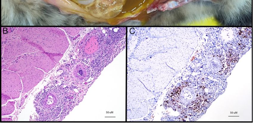
Viruses 2020, 12, x FOR PEER REVIEW 2 of 22
Viruses 2020, 12, 1023 2 of 22
Alpha- and betacoronaviruses originate from bats and predominantly infect mammals, while
gamma- and deltacoronaviruses originate from birds and are capable of infecting both bird and
and deltacoronaviruses
mammal originateanimals
species [6]. Companion from birds and are
presently capableinclude
considered of infecting both ferrets,
cats, dogs, bird and mammal
horses, and
species [6]. Companion animals presently considered include cats, dogs, ferrets,
alpacas. While not universally recognized as companion animals, alpacas and horses are considered horses, and alpacas.
While
by the notauthors
universally
to berecognized
companion as animals
companion andanimals, alpacasincluded
are therefore and horses are considered
in this by the
review. Notable
authors to be companion animals and are therefore included in this review.
coronaviruses of companion animals include feline enteric coronavirus (FECV), feline infectious Notable coronaviruses of
companion animals include feline enteric coronavirus (FECV), feline infectious peritonitis
peritonitis virus (FIPV), canine enteric coronavirus (CCoV), ferret enteric coronavirus (FRECV), ferret virus (FIPV),
canine enteric
systemic coronavirus
coronavirus (CCoV),
(FRSCV), andferret
alpaca enteric coronavirus
respiratory (FRECV),
coronavirus, ferret
which aresystemic coronavirus
alphacoronaviruses,
(FRSCV),
and canine and alpaca respiratory
respiratory coronavirus coronavirus,
(CRCoV), whichequineare alphacoronaviruses,
enteric coronavirus (ECoV), and and
canine respiratory
alpaca enteric
coronavirus
coronavirus,(CRCoV),
which areequine enteric coronavirus
betacoronaviruses (ECoV), and
[7]. Phylogenetic alpaca enteric
relationships coronavirus,
of these whichare
coronaviruses are
betacoronaviruses
shown in Figure [7]. Phylogenetic
1, while clinical andrelationships
pathologicoffeatures
these coronaviruses
are summarized are shown
in Table in 1.
Figure
Other1,
coronaviruses
while belonging
clinical and pathologic to the betacoronavirus
features are summarized genusininclude
Table 1.SARS-CoV-1, MERS-CoV,
Other coronaviruses and SARS-
belonging to the
CoV-2, zoonoticgenus
betacoronavirus coronaviruses that have recently
include SARS-CoV-1, transferred
MERS-CoV, from animal zoonotic
and SARS-CoV-2, to humancoronaviruses
populations and that
are capable
have recentlyof causing severe
transferred diseaseto
from animal and deathpopulations
human [8]. The ability
and of areSARS-CoV-2 to initiate
capable of causing infections
severe disease
in companion
and death [8]. animals is currently
The ability poorly understood,
of SARS-CoV-2 although preliminary
to initiate infections in companion studies
animalshaveisindicated
currently
that ferrets
poorly and cats
understood, are permissive
although for SARS-CoV-2
preliminary studies haveinfection
indicatedand thatreplication,
ferrets andwhile thepermissive
cats are virus has
been
for shown to replicate
SARS-CoV-2 infectionpoorly in dogs, pigs,
and replication, whilechickens,
the virusandhas
ducks
been[9]. SARS-CoV-2
shown infection
to replicate poorlyofin
horses
dogs,
and camelids has not been reported.
pigs, chickens, and ducks [9]. SARS-CoV-2 infection of horses and camelids has not been reported.
Figure 1. Phylogenetic relationships of coronaviruses of companion animals. The 30 portions of the
Figure 1. Phylogenetic relationships of coronaviruses of companion animals. The 3′ portions of the
coronaviral genomes encoding the spike and other non-structural proteins (~9 kb) were compared
coronaviral genomes encoding the spike and other non-structural proteins (~9 kb) were compared
and plotted as a “guide tree” using MacVector software (ClustalW Multiple Sequence Alignment).
and plotted as a “guide tree” using MacVector software (ClustalW Multiple Sequence Alignment).
Betacoronavirus
Betacoronavirussequences
sequencesare are
highlighted in yellow,
highlighted while alphacoronavirus
in yellow, sequencessequences
while alphacoronavirus are highlighted
are
inhighlighted
blue; the zoonotic SARS CoV-2 coronavirus is surrounded by a red box. GenBank submission
in blue; the zoonotic SARS CoV-2 coronavirus is surrounded by a red box. GenBank
numbers are numbers
submission indicatedare
forindicated
each sequence.
for each sequence.
Coronavirus genomes encode three classes of proteins: structural, accessory, and non-structural
Coronavirus genomes encode three classes of proteins: structural, accessory, and non-structural
proteins. Major structural proteins of coronaviruses include the nucleocapsid (N), spike (S), membrane
proteins. Major structural proteins of coronaviruses include the nucleocapsid (N), spike (S),
(M), and envelope (E) proteins [5]. The S protein is the primary viral binding protein and mediator
membrane (M), and envelope (E) proteins [5]. The S protein is the primary viral binding protein and
ofmediator
membrane fusion and
of membrane viral and
fusion entry. The
viral N protein,
entry. in closeinassociation
The N protein, with genomic
close association viral viral
with genomic RNA
(gRNA), forms the helical nucleocapsid, which is stabilized via binding to the M
RNA (gRNA), forms the helical nucleocapsid, which is stabilized via binding to the M protein (Figure protein (Figure 2).
The viral genome and helical nucleocapsid are surrounded by a host-derived
2). The viral genome and helical nucleocapsid are surrounded by a host-derived lipid bilayer, inlipid bilayer, in which
the S, E,the
which andS,M E,proteins are anchored.
and M proteins The transmembrane
are anchored. The transmembraneE and M proteins
E and are involved
M proteins in virion
are involved in
assembly and budding [10]. In addition to the four structural proteins, coronavirus
virion assembly and budding [10]. In addition to the four structural proteins, coronavirus genomes genomes also
encode a number
also encode of accessory
a number proteins.
of accessory WhileWhile
proteins. the roles
theof most
roles of of the of
most accessory proteins
the accessory remainremain
proteins poorly
understood
poorly understood and may be dispensable for virus replication in vitro, certain accessory proteinsto
and may be dispensable for virus replication in vitro, certain accessory proteins appear
enhance
appear toviral virulence
enhance viralinvirulence
vivo; for example, theexample,
in vivo; for SARS coronavirus
the SARS encodes
coronavirusaccessory proteins
encodes that
accessory
antagonize
proteins thattheantagonize
development the of type I interferon
development of type(IFN) responses
I interferon (IFN)[11].
responses [11].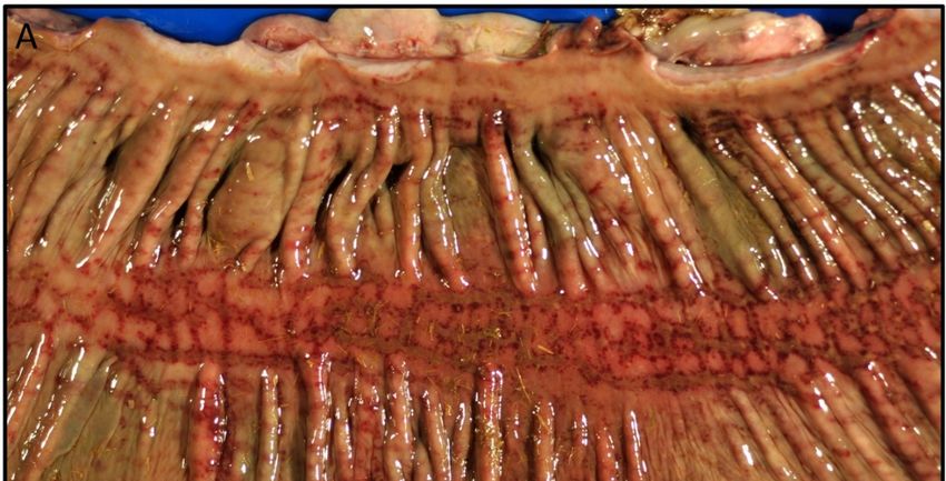
Viruses 2020, 12, 1023                                                                                   3 of 22
Viruses 2020, 12, x FOR PEER REVIEW                                                                      3 of 22

                                      Figure 2. Coronavirus structural proteins.
                                      Figure 2. Coronavirus structural proteins.
      Unlike alphacoronaviruses, a subset of betacoronaviruses are more structurally complex and
 haveUnlike    alphacoronaviruses,
       additional                     a subset of
                    membrane glycoproteins,        betacoronaviruses
                                                 called               are more (HE)
                                                        hemagglutinin-esterase   structurally
                                                                                       proteins, complex
                                                                                                   encoded andby
have
 an additional gene roughly 1.2 kb in size [12]. Coronavirus HEs are thought to be acquired from by
       additional  membrane     glycoproteins,  called  hemagglutinin-esterase  (HE)   proteins,   encoded    an
an  additional
 influenza  virusgene  roughly
                   C-like  gene 1.2 kb in size
                                encoding       [12]. Coronavirus HEs fusion
                                           a hemagglutinin-esterase    are thought  to in
                                                                              protein   be aacquired   from
                                                                                              relatively     an
                                                                                                         recent
influenza   virus  C-like  gene encoding   a hemagglutinin-esterase   fusion  protein  in a  relatively
 horizontal gene transfer event [13]. While coronavirus HEs are able to bind to sialic acid, they are    recent
horizontal
 reported togene
              servetransfer  event
                     primarily      [13]. While coronavirus
                               as receptor-destroying        HEs
                                                        enzymes   are able
                                                                (RDE),      to bind
                                                                        which        to sialic
                                                                               facilitates      acid, they are
                                                                                            the reversibility of
reported to serve primarily as receptor-destroying enzymes (RDE), which facilitates the reversibility
 the virus-host cell attachment. For all coronaviruses, the S protein is thought to be the primary binding
of the virus-host cell attachment. For all coronaviruses, the S protein is thought to be the primary
protein, responsible for attachment of coronavirus to the cell surface. However, the contribution of
binding protein, responsible for attachment of coronavirus to the cell surface. However, the
 HEs to virion attachment and their role in tissue tropism and pathogenesis are currently not well
contribution of HEs to virion attachment and their role in tissue tropism and pathogenesis are
 understood [14].
currently not well understood [14].
      The molecular events of the coronavirus replication cycle are complex and begin with virion
      The molecular events of the coronavirus replication cycle are complex and begin with virion
 attachment to the host cell, accomplished by binding of the viral S protein to a unique target receptor
attachment to the host cell, accomplished by binding of the viral S protein to a unique target receptor
 on the host cell surface. As the primary binding protein and mediator of virus-host cell membrane
on the host cell surface. As the primary binding protein and mediator of virus-host cell membrane
 fusion and subsequent virus entry into the cell, the S protein is critical in determining the host species
fusion and subsequent virus entry into the cell, the S protein is critical in determining the host species
 and tissue and cell tropism for each coronavirus [15]. Upon receptor binding, conformational changes
and tissue and cell tropism for each coronavirus [15]. Upon receptor binding, conformational changes
 in the S protein expose the fusion peptide, facilitating fusion of the viral and host cell membranes and
in the S protein expose the fusion peptide, facilitating fusion of the viral and host cell membranes and
 subsequent release of the viral nucleocapsid into the host cell cytoplasm [10,16]. Upon cytoplasmic
subsequent release of the viral nucleocapsid into the host cell cytoplasm [10,16]. Upon cytoplasmic
 release of the viral nucleocapsid, the positive sense genomic RNA (+gRNA) serves as viral messenger
release of the viral nucleocapsid, the positive sense genomic RNA (+gRNA) serves as viral messenger
 RNA (mRNA) for the direct translation of the replicase gene complex utilizing the host cell’s ribosomal
RNA (mRNA) for the direct translation of the replicase gene complex utilizing the host cell’s ribosomal
 machinery. The replicase gene complex consists of two large open reading frames (ORF) approximately
machinery. The replicase gene complex consists of two large open reading frames (ORF)
 20 kb in total size [17], ORF1a and ORF1b, the latter transcribed via a ribosomal frameshift. The ORF1a
approximately 20 kb in total size [17], ORF1a and ORF1b, the latter transcribed via a ribosomal
 and ORF1b mRNA are translated into polypeptides 1a or 1ab, which are subsequently cleaved by viral
frameshift. The ORF1a and ORF1b mRNA are translated into polypeptides 1a or 1ab, which are
proteases to create sixteen nonstructural proteins (nsps). These nonstructural proteins reassemble
subsequently cleaved by viral proteases to create sixteen nonstructural proteins (nsps). These
 to form a viral replicase-transcriptase complex, consisting of the RNA-dependent RNA polymerase
nonstructural proteins reassemble to form a viral replicase-transcriptase complex, consisting of the
(RdRp, nsp12), helicase (nsp13), nsps with accessory functions, such as the nsp14 exoribonuclease,
RNA-dependent RNA polymerase (RdRp, nsp12), helicase (nsp13), nsps with accessory functions,
 as well as multiple membrane-spanning proteins that are thought to provide a membrane-associated
such as the nsp14 exoribonuclease, as well as multiple membrane-spanning proteins that are thought
 scaffold for the assembly of the replicase-transcriptase complex [18–20]. As eukaryotic cells typically
to provide a membrane-associated scaffold for the assembly of the replicase-transcriptase complex
 do not encode an RdRp, that is, they lack the ability to catalyze the formation of RNA using RNA as a
[18–20]. As eukaryotic cells typically do not encode an RdRp, that is, they lack the ability to catalyze
 substrate, the viral RdRp enzyme provides a useful target for antiviral therapeutics [21,22]. Within this
the formation of RNA using RNA as a substrate, the viral RdRp enzyme provides a useful target for
 group of nonstructural proteins is an exonuclease with proof-reading function, unusual for RNA
antiviral therapeutics [21,22]. Within this group of nonstructural proteins is an exonuclease with
viruses but perhaps important for ensuring the fidelity of the very large coronaviral RNA genome
proof-reading function, unusual for RNA viruses but perhaps important for ensuring the fidelity of
 during replication [23].
the very large coronaviral RNA genome during replication [23].
      The viral polymerase synthesizes complementary full-length negative-sense RNA copies of the
genome, which serve as templates for full length positive-sense RNA genomes, generated via RdRp’s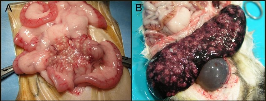
Viruses 2020, 12, 1023                                                                                                      4 of 22

     The viral polymerase synthesizes complementary full-length negative-sense RNA copies of
the genome, which serve as templates for full length positive-sense RNA genomes, generated via
RdRp’s replicase function. In addition to replicase activity, RdRp also has transcriptase activity;
by discontinuous RNA synthesis directed by transcriptional regulatory sequences, RdRp creates a set of
subgenomic RNAs (sgRNA) of different sizes [24], which are then copied by RdRp into positive-sense
mRNAs, serving as templates for translation of viral proteins necessary for virion assembly, including
the structural proteins S, E, M, and N. Translated viral proteins are inserted into the cell’s endoplasmic
reticulum and then transported to the site of viral assembly, the endoplasmic reticulum-Golgi
intermediate compartment (ERGIC). Viral genomes (+gRNA) encapsidated by N proteins bud into the
ERGIC membrane, forming fully assembled virions surrounded by a host-derived lipid bilayer [25].
Assembled virions are subsequently transported in vesicles to the plasma membrane, where they
are released from the infected cell via exocytosis [26]. In some coronaviruses, the accumulation of
S proteins on the surface of infected cells can result in fusion of adjacent cells and the formation of
syncytia, facilitating rapid cell-to-cell spread of the virus [27].
     The genetic diversity of coronaviruses is a consequence both of polymerase error-driven point
mutations, as well as of genetic recombination between different strains and species of coronaviruses
during coinfection within the same host cell [5,28]. Relative to other single-stranded RNA viruses,
coronavirus mutation rates are moderate to high [29], despite the proof-reading function of the viral
exonuclease [30]. Genetic recombination is a direct result of the discontinuous transcriptional activity
of the coronaviral polymerase and likely contributes to the emergence of new viruses with altered
virulence, novel host species range, and novel tissue tropism [18].

         Table 1. Clinical and pathologic features of major coronavirus infections of companion animals.

                          Primary Target                                                                            Cellular
         Virus                                  Primary Host Cell   Disease/Symptoms          Transmission
                             Organ(s)                                                                               Receptor
                                                     Genus Alphacoronavirus
                                                                                                                   Serotype I:
     Feline enteric                                                 Asymptomatic to            Direct contact;
                                                                                                                    unknown
      coronavirus             GI tract             Enterocyte       mild gastroenteritis    fecal-oral, maternal
                                                                                                                   Serotype II:
        (FECV)                                                         and diarrhea              shedding
                                                                                                                      APN
                             Omentum,
                                                                    Peritonitis, thoracic                          Serotype I:
   Feline infectious      serosal/pleural                                                        Rare to no
                                                   Monocyte,          and abdominal                                 unknown
   peritonitis virus       surfaces, liver,                                                      horizontal
                                                  macrophage        effusions, CNS and                             Serotype II:
        (FIPV)            kidneys, lymph                                                       transmission
                                                                        ocular signs.                                 APN
                         nodes, eyes, brain
     Ferret enteric
                                                                    Epizootic catarrhal
      coronavirus             GI tract             Enterocyte                                   Fecal-oral         Unknown
                                                                         enteritis
       (FRECV)
                                                                       Weight loss,
                         Spleen, mesenteric
    Ferret systemic                                                 anorexia, diarrhea,
                             lymph nodes,
     coronavirus                                   Unknown              abdominal               Unknown            Unknown
                         intestines, kidneys,
       (FRSCV)                                                      granulomas/masses,
                          liver, lungs, brain
                                                                        CNS signs
                                                                           Mild
                                                                                                                   Serotype I:
    Canine enteric                                                  gastroenteritis and
                                                                                                                    unknown
     coronavirus              GI tract             Enterocyte         diarrhea; rarely,         Fecal-oral
                                                                                                                   Serotype II:
      (CECoV)                                                       severe enteritis and
                                                                                                                      APN
                                                                      systemic signs
                                                   Respiratory
  Alpaca respiratory                                                  Mild to severe             Aerosol
                          Respiratory tract          epithelia                                                     Unknown
     coronavirus                                                    respiratory disease        (presumed)
                                                   (presumed)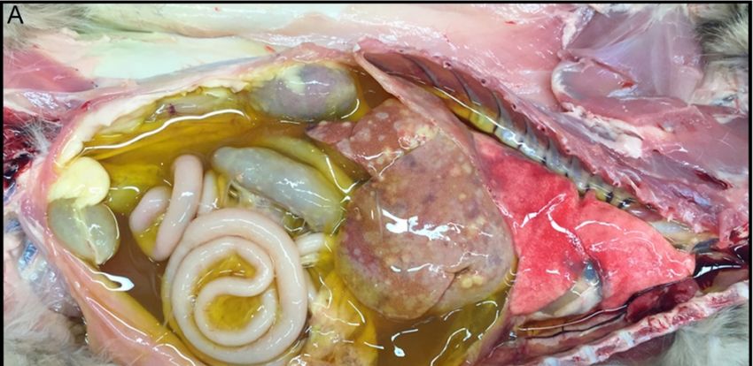
Viruses 2020, 12, 1023                                                                                      5 of 22

                                                      Table 1. Cont.

                         Primary Target                                                               Cellular
         Virus                               Primary Host Cell   Disease/Symptoms      Transmission
                            Organ(s)                                                                  Receptor
                                                   Genus Betacoronavirus
  Canine respiratory
                                                Respiratory          Mild upper
    coronavirus          Respiratory tract                                               Aerosol      Unknown
                                                 epithelia       respiratory disease
      (CRCoV)
                                                                  Fever, anorexia,
                                                                   lethargy; less
  Equine coronavirus
                             GI tract           Enterocyte           frequently,        Fecal-oral    Unknown
       (ECoV)
                                                                   diarrhea, colic,
                                                                  neurologic signs
    Alpaca enteric                               Enterocyte       Enteritis, severe     Fecal-oral
                             GI tract                                                                 Unknown
     coronavirus                                (presumed)           diarrhea          (presumed)

2. Feline Enteric Coronavirus and Feline Infectious Peritonitis Virus

2.1. Epidemiology, Clinical and Pathologic Features
      Feline coronaviruses are separated into two distinct biotypes: feline enteric coronavirus (FECV)
and feline infectious peritonitis virus (FIPV). FECV is endemic in domestic cat populations worldwide
and primarily infects intestinal enterocytes, typically resulting in either mild enteric disease or a lack
of clinical signs (subclinical infections). Experimental studies have demonstrated consistent shedding
of FECV in the feces of infected cats from 2 days to 2 weeks post-infection, followed by a decrease in
viral loads and intermittent shedding for up to 20 weeks after this period [31,32]. Subclinical carriers
of FECV play an important role in shedding and transmitting the virus to other cats via the fecal-oral
route, especially in those animals housed indoors in multi-cat environments [33]. FECV primarily
infects the apical epithelial cells of the intestinal villi (enterocytes), from the distal duodenum to the
cecum. Villous atrophy of the lining mucosa and sloughing and degeneration of epithelial cells at the
villous tips occur in severe infections [34]. Shortening and fusion of intestinal villi and hyperplasia of
crypt epithelia are also common pathological findings [3].
      In contrast to the mild enteric disease or absence of clinical signs associated with FECV infection,
the closely related FIPV biotype generally results in a highly inflammatory, systemic, and nearly 100%
fatal disease once clinical signs develop. This clinical syndrome is called feline infectious peritonitis
(FIP). The origin of FIPV is thought to arise from a select number of spontaneous mutations in the FECV
genome, which confers a tropism switch from enterocytes to macrophages, facilitating systemic spread.
These mutations are thought to arise de novo within each FECV-infected cat. Male cats, purebred cats,
and those living in multi-cat environments are more likely to develop FIP [35]. Specific cat breeds
at higher risk for the development of FIP include Abyssinian, Bengal, Birmans, ragdoll, and rex cat
breeds [36,37], likely due to inherited genetic factors in these breeds, leading to increased susceptibility
to FIP [38]. FIP seems to preferentially occur in young, very old, or immunosuppressed individuals.
An FIP-like disease has also been documented in a number of wild species of felids infected with
coronaviruses, including African lions, cheetahs, mountain lions, leopards, jaguars, lynx, servals,
caracal, European wild cats, Sand cats, and Pallas cats [39–47].
      Key point mutations proposed to be responsible for the conversion of FECV to FIPV include two
alternative amino acid differences in the gene encoding the fusion peptide of the spike (S) protein [48],
substitutions in the furin cleavage site between receptor-binding (S1) and fusion (S2) domains of
the spike protein [49], and mutations in open reading frame 3abc resulting in a truncated protein 3c
protein [50]. It has subsequently been reported that one of the amino acid differences in the fusion
peptide of the FECV spike protein, specifically, a methionine to leucine substitution at position 1058,
is involved in systemic spread of FCEV from the intestine, rather than with the potential to cause
FIP [51]. Mutations of 3c and S protein genes are often found in combination, but a single mutation in
either S or 3c appears to be sufficient to dramatically alter the tropism of FECV, allowing for enhanced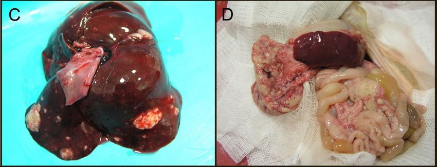
Viruses 2020, 12, 1023                                                                               6 of 22

internalization and replication of the virus within monocytes and macrophages, facilitating systemic,
cell-associated dissemination of the virus [50].
       Clinically, FIP typically manifests as one of two forms: “wet” (effusive) FIP, “dry” (granulomatous)
FIP, or some combination of the two. Effusive FIP is the more common and classical form of disease and
is generally associated with rapid disease progression and the exudation of fluid into the peritoneal or
thoracic body cavities. The “dry”/granulomatous form of FIP generally lacks cavitary effusion and is
instead characterized by multifocal granuloma formation in a variety of organs and a more insidious
disease progression. As a result, the initial clinical signs of FIP are often nonspecific and may include
anorexia, weight loss, and/or chronic fever [52,53]. Clinical neurologic signs, including ataxia, seizures,
nystagmus, hyperesthesia, and/or cranial nerve deficits [54,55], as well as ocular disease may occur in
some cats, with a higher frequency in those with the dry form of FIP than the wet form [56].
       It has been hypothesized that a “strong and focused” cell-mediated immune (CMI) response
directed to the coronavirus may prevent FIP disease development, while animals with a “weak” CMI
in combination with a strong humoral immune response will likely develop wet FIP. Further, it has
been hypothesized that animals with a “moderate” CMI will likely develop the dry form of FIP [57].
       Grossly, the wet/effusive form of FIP is characterized by “straw-colored,” semi-translucent,
protein-rich peritoneal or thoracic effusions and fibrinous and granulomatous serositis/pleuritis with
variable involvement of parenchymal organs (Figure 3A) [56]. Virus-associated pyogranulomatous
inflammation is focused on small and medium sized veins, resulting in vascular injury and
leakage [58]. These vasculo-centric lesions can occur in the omentum and serosal surfaces of the
liver, spleen, intestines, kidneys, and lungs, and are composed primarily of macrophage aggregates in
combination with smaller numbers of neutrophils and lymphocytes (pyogranulomatous inflammation).
Occasionally, pyogranulomatous nodular lesions extend beyond the serosal surfaces into underlying
parenchyma. Coronaviral antigen can often be detected within intralesional macrophages using
immunohistochemistry techniques; coronaviral antigen detection via immunohistochemistry is a
commonly utilized diagnostic method.
       The dry form of FIP is characterized grossly by variably sized parenchymal and serosal
pyogranulomas in affected organs but lacks the exudation archetypal of wet FIP. Granulomatous
lesions of dry FIP may extend from serosal surfaces into the parenchyma of affected organs, and lesions
may be restricted to a single organ, such as the kidney, eye, or brain [59]. Other frequently affected
organs in the dry form of FIP include the mesenteric and mediastinal lymph nodes, omentum, intestine,
and liver. Perivascular inflammatory lesions contain aggregates of macrophages and fewer neutrophils,
which are surrounded by dense infiltrates of primarily B lymphocytes and plasma cells extending into
surrounding tissues, with or without the presence of vasculitis [47,59]. It is not uncommon for affected
animals to demonstrate some combination of both effusive and granulomatous forms of the disease.
       It has been hypothesized that immune-mediated type III and/or type IV hypersensitivity reactions
may play a role in the perivascular granulomatous inflammation characteristic of FIP [60,61]. Type III
hypersensitivity lesions feature the overproduction of immune complexes comprised of antibody bound
to soluble antigen, and subsequent inflammatory pathway activation, deposition into vessel walls
(e.g., vasculitis) and tissue injury [62]. FIPV-associated vascular lesions have been hypothesized as being
caused by type III hypersensitivity based on studies demonstrating antibody, complement, and FCoV
antigen within vascular lesions; however, a definitive connection between type III hypersensitivity and
FIP-associated vasculitis/peri-vasculitis has not been unequivocally confirmed [60,63]. Alternatively,
FIPV-associated vascular injury and subsequent permeability may be a result of virus-induced
activation of monocytes and macrophages. This hypothesis is supported by the finding that vascular
endothelial growth factor (VEGF) produced by FIPV-infected monocytes and macrophages causes
vascular permeability and effusion in cats with FIP [64]. Matrix metalloprotease 9 (MMP-9) has
also been shown to be upregulated in activated monocytes and macrophages in FIP, contributing
to the destruction of type IV collagen and degradation of the basal lamina of affected vessels in FIP
vasculitis [58]. Type IV, or delayed-type hypersensitivity, is mediated by hyperstimulated T cells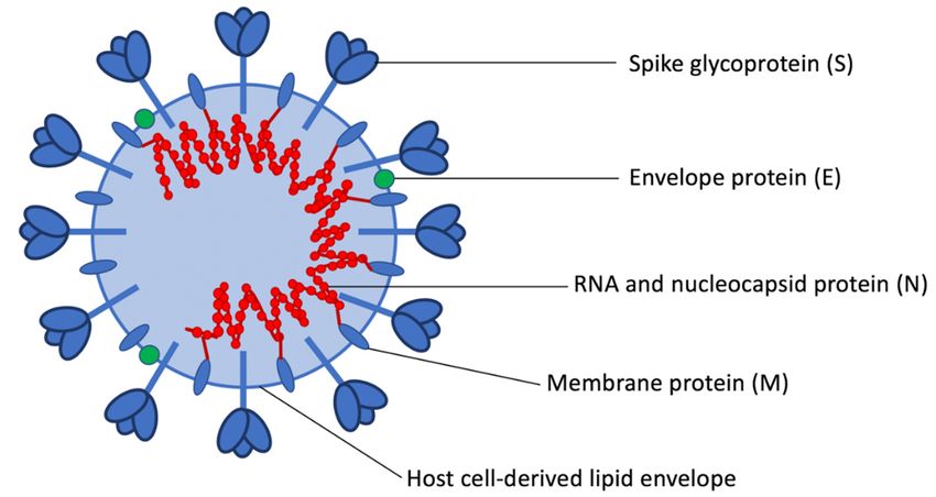
Viruses 2020, 12, 1023                                                                                                       7 of 22

and macrophages, which cause damage to surrounding tissue and may contribute to the granuloma
formation characteristic of the dry form of FIP [65].
     In a subset of wet and dry FIP cases, affected cats may present with ocular and/or neurologic
involvement. In cases with ocular involvement, diffuse and multifocal inflammatory infiltrates
may be found in the ciliary body, retina, and choroid as well as throughout the uvea and in the
sclera, conjunctiva, and optic nerve. Ocular perivascular leukocytes are typically lymphoplasmacytic,
composed mostly of B cells and plasma cells, with fewer numbers of T cells and macrophages [66].
       Viruses 2020, 12, x FOR PEER REVIEW                                                                         7 of 22
Gross lesions of FIP in the central nervous system include ventricular dilation, flattening of cerebral gyri,
and ependymal
       Gross lesions  and of meningeal     congestion
                             FIP in the central   nervous[67].  Mild
                                                            system     to marked
                                                                    include        ventricular
                                                                            ventricular dilation,enlargement     is associated
                                                                                                  flattening of cerebral
with accumulation           of inflammatory        cells within    the  ventricles  [54], with
       gyri, and ependymal and meningeal congestion [67]. Mild to marked ventricular enlargement corresponding      increased
                                                                                                                        is
       associated     with   accumulation    of  inflammatory    cells within the ventricles [54], with
protein, increased cellularity, and presence of virus in cerebrospinal fluid [67]. Histopathological     corresponding
lesionsincreased
         in the brain protein,   increased
                             consist          cellularity, neutrophilic
                                       of perivascular       and presence and of virus  in cerebrospinal infiltrates
                                                                                  lymphoplasmacytic          fluid [67].in the
       Histopathological       lesions  in the  brain  consist  of perivascular neutrophilic  and   lymphoplasmacytic
leptomeninges, the choroid plexus, the periventricular space, and/or the parenchyma of the spinal cord
       infiltrates in the leptomeninges, the choroid plexus, the periventricular space, and/or the parenchyma
and brainstem [68]. Less common (atypical) manifestations of FIP include nodular dermatitis [69–71],
       of the spinal cord and brainstem [68]. Less common (atypical) manifestations of FIP include nodular
rhinitis  [72], orchitis
       dermatitis     [69–71],[73,74],
                                rhinitispriapism     [75],[73,74],
                                          [72], orchitis    and syringomyelia       associated
                                                                   priapism [75], and            with associated
                                                                                       syringomyelia    involvement withof the
fourthinvolvement
        ventricle [76].  of the fourth ventricle [76].

      FigureFigure
                3. (A) 3. (A) image
                        Gross  Gross image    of or
                                        of “wet”  “wet”  or effusive
                                                     effusive         feline infectious
                                                              feline infectious         peritonitis
                                                                                 peritonitis (FIP), (FIP),
                                                                                                    thoracicthoracic and
                                                                                                               and abdominal
              abdominal     cavities,  cat. Abundant    semi-translucent   “straw-colored”,  proteinaceous
      cavities, cat. Abundant semi-translucent “straw-colored”, proteinaceous peritoneal effusion with         peritoneal
              effusion
      fibrinous          with fibrinous and
                   and granulomatous          granulomatous
                                            serositis          serositisgranulomatous
                                                      and multifocal     and multifocal granulomatous
                                                                                         lesions in the lesions    in theimage
                                                                                                           liver. Gross
              liver. Gross image courtesy of Chrissy Eckstrand. (B) FIP, urinary bladder serosal surface, cat,
      courtesy of Chrissy Eckstrand. (B) FIP, urinary bladder serosal surface, cat, hematoxylin and eosin
              hematoxylin and eosin (HE). Severe, necrotizing, pyogranulomatous and lymphoplasmacytic
      (HE). Severe, necrotizing, pyogranulomatous and lymphoplasmacytic serositis and vasculitis. (C) FIP,
              serositis and vasculitis. (C) FIP, urinary bladder serosal surface, cat, FCoV immunohistochemistry.
      urinary    bladder
              Same          serosal
                     lesion tissue    surface,
                                   as 3b         cat, FCoV
                                         with frequent,      immunohistochemistry.
                                                        positive immunoreactivity for FCoV Same    lesion
                                                                                              antigen (browntissue  as 3b with
                                                                                                                pigment).
      frequent, positive immunoreactivity for FCoV antigen (brown pigment).
        2.2. Virology
              Feline coronaviruses are alphacoronaviruses and are divided into two serotypes, type I and type
        II, based on genetic and antigenic properties [77]. Although both serotypes are capable of causing FIP
        [78], serotype I is much more prevalent in nature and is responsible for 80–90% of naturally occurring
        clinical cases [79,80]. Serotype II is comparatively rare, having emerged as a result of recombination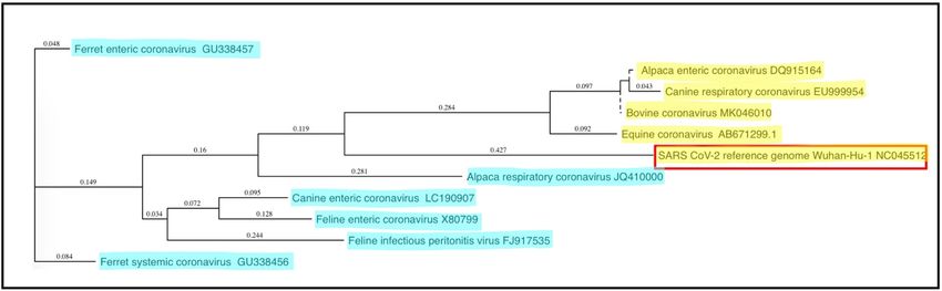
Viruses 2020, 12, 1023                                                                                8 of 22

2.2. Virology
       Feline coronaviruses are alphacoronaviruses and are divided into two serotypes, type I and type II,
based on genetic and antigenic properties [77]. Although both serotypes are capable of causing FIP [78],
serotype I is much more prevalent in nature and is responsible for 80–90% of naturally occurring
clinical cases [79,80]. Serotype II is comparatively rare, having emerged as a result of recombination
events between feline coronavirus serotype I and canine enteric coronavirus serotype II, following
cross-species transmission of CCoV to cats [81–83]. Although the feline serotype I is more prevalent,
it is less well studied due to challenges in propagating this viral serotype in culture-adapted cell lines
in vitro. The molecular events of the serotype II viral lifecycle are better understood due to the relative
ease with which serotype II can be propagated and studied in vitro.
       The target cell membrane receptor of serotype II feline coronaviruses has been identified as feline
aminopeptidase N (fAPN) [84]. Feline APN is a membrane peptidase expressed on the brush border of
small intestine and renal tubule microvilli, as well as by cells of myeloid origin, including monocytes,
macrophages, and granulocytes [85]. Additional non-specific viral receptors, including the lectin
molecule DC-SIGN, have also been proposed [41,78]. The primary cell receptor for the more prevalent
serotype I FIPV has yet to be definitively identified [86]; however, studies have proposed DC-SIGN [87],
as a potential host cell entry co-receptor. The Fc receptor CD16 (FcyRIII) has also been proposed
as a potential cellular receptor [88]. Interestingly, antibodies directed to the spike protein of feline
coronavirus have been shown to enhance virus infection both in vitro [89] and in vivo [63] through
a mechanism known as antibody dependent enhancement (ADE). In ADE, antibodies facilitate the
uptake of virus-antibody complexes by monocytes and macrophages using Fc receptors like CD16,
resulting in more efficient infection than by virus alone [90,91].
       Following binding to the target receptor on the cell surface, FIPV serotype II enters monocytes via
clathrin and caveolae-independent and dynamin-dependent endocytosis [92]. Consistent with the
initial distribution of lesions on serosal surfaces of abdominal organs, FIPV is thought to preferentially
target peritoneal macrophages [88]. Once ensconced within these histiocytic cells, FIPV is able to seed
the abdominal and thoracic cavities and, in some cases, spread to more distant sites, such as the brain
and eye.

3. Ferret Enteric Coronavirus and Ferret Systemic Coronavirus

Epidemiology, Virology, and Clinical and Pathologic Features
     Similar to feline coronaviruses, infection with ferret coronaviruses can result in enteric or systemic
disease [93]. First identified in the United States in 2000, ferret enteric coronavirus (FRECV) is associated
with epizootic catarrhal enteritis (ECE), originally called “green slime disease” due to the development
of profuse, foul-smelling, bright green mucus-laden diarrhea [94]. Clinically, ECE is associated with
lethargy, anorexia, and vomiting. ECE is characterized by high morbidity but low mortality [95].
While juvenile ferrets develop mild to subclinical disease and can be subclinical carriers, ECE can cause
more severe disease in older ferrets [96].
     A genetically distinct coronavirus called ferret systemic coronavirus infection (FRSCV) was
subsequently identified, which causes a systemic, progressive, and fatal pyogranulomatous inflammatory
disease resembling the dry form of feline infectious peritonitis (FIP) in cats. The average age at the time
of FRSCV diagnosis has been reported to be 11 months, and clinical signs include chronic weight loss,
anorexia, diarrhea, palpable abdominal masses, and neurologic disease [97].
     Both the enteric (FRECV) and systemic (FRSCV) ferret coronaviruses are alphacoronaviruses,
related to feline coronavirus and canine enteric coronavirus, and most closely related to mink
coronavirus. Complete genome sequencing of FRSCV and FRECV strains revealed a shared 89%
nucleotide identity, but only 49.9–68.9% nucleotide identity with other known coronaviruses [98].
The pathogenic relationship of these two ferret coronaviruses, and whether FRSCV arises by mutation
within ferrets infected with FRECV, has not been determined. The cellular entry receptors have also
Viruses 2020, 12, 1023 9 of 22
not been identified. Many facets of the pathogenesis of the virulent systemic ferret coronavirus remain
unknown, but as is true for FIPV, macrophages appear to play an important role in the inflammatory
response. Furthermore, similarities in pathologic lesions suggest parallels in the pathogenesis of ferret
Viruses 2020, 12, x FOR PEER REVIEW 9 of 22
systemic coronavirus and FIP.
FRECV is associated with lesions restricted to the gastrointestinal tract, which include lymphocytic
FRECV is associated with lesions restricted to the gastrointestinal tract, which include
enteritis, villous blunting, fusion, and atrophy, as well as vacuolar degeneration and necrosis of apical
lymphocytic enteritis, villous blunting, fusion, and atrophy, as well as vacuolar degeneration and
villous enterocytes, similar to FECV [94].
necrosis of apical villous enterocytes, similar to FECV [94].
In contrast to FRECV, FRSCV is grossly associated with pale to white nodules (granulomatous
In contrast to FRECV, FRSCV is grossly associated with pale to white nodules (granulomatous
inflammation) in multiple organs, including the spleen, kidneys, mesenteric lymph nodes, intestines,
inflammation) in multiple organs, including the spleen, kidneys, mesenteric lymph nodes, intestines,
liver, lungs, and brain (Figure 4) [99,100]. Granulomas have a heterogenous cellular composition
liver, lungs, and brain (Figure 4) [99,100]. Granulomas have a heterogenous cellular composition
including
includingmacrophages,
macrophages, TT and and BB lymphocytes,
lymphocytes, andand plasma
plasma cells. These granulomatous
cells. These granulomatouslesions
lesionsare
are
morphologically similar to FIP both in immune cell composition and presence
morphologically similar to FIP both in immune cell composition and presence of virus within theof virus within the
macrophage
macrophagecytoplasm
cytoplasm [101].
[101]. Interestingly,
Interestingly, cavitary effusions and
cavitary effusions and vasculitis,
vasculitis, characteristic
characteristicfeatures
features
ofofthe
thewet
wetform
form ofof FIP,
FIP,have
have not
not been
been identified
identified in
in the
the majority of ferrets
majority of ferrets infected
infected with
with systemic
systemic
coronavirus. Clearly, much remains to be learned about ferret coronavirus virology
coronavirus. Clearly, much remains to be learned about ferret coronavirus virology and pathology. and pathology.
Figure 4.
Figure 4. Gross lesions
Gross associated
lesions with ferret
associated withsystemic
ferret coronavirus (FRSCV). (A)(FRSCV).
systemic coronavirus Ferret, coronavirus-
(A) Ferret,
associated granulomatous
coronavirus-associated mesenteritis.
granulomatous Numerous,Numerous,
mesenteritis. multifocal multifocal
to coalescing, pale tan, firm
to coalescing, pale nodular
tan, firm
masses masses
nodular (granulomas) distributed
(granulomas) throughout
distributed the mesentery,
throughout often corresponding
the mesentery, to vasculature.
often corresponding (B)
to vasculature.
Ferret, coronavirus-associated serositis and splenitis. Numerous, multifocal to coalescing,
(B) Ferret, coronavirus-associated serositis and splenitis. Numerous, multifocal to coalescing, pale pale tan
nodules
tan nodules(granulomas)
(granulomas) expanding
expanding thethe
serosa
serosa with
withvariable
variableparenchymal
parenchymalinvolvement.
involvement.(C) (C)Ferret,
Ferret,
coronavirus-associatedhepatitis.
coronavirus-associated hepatitis. Multifocal,
Multifocal, pale
pale tan,
tan, expansile
expansilenodular
nodularmasses
massesthroughout
throughoutthetheliver.
liver.
(D)Ferret,
(D) Ferret,coronavirus-associated
coronavirus-associated peritonitis.
peritonitis. Multifocal
Multifocal toto coalescing,
coalescing, pale
pale tan,
tan, nodular
nodularmasses
masses
(granulomas)throughout
(granulomas) throughoutthe theperitoneum.
peritoneum. All
All images
images courtesy
courtesy of
of Jordi
Jordi Jimenez.
Jimenez.
4. Canine Enteric Coronavirus
4.1. Epidemiology and Clinical Features
As is true of FECV, canine enteric coronavirus (CCoV) is a common infection of dogs with
worldwide distribution. While not universally recognized as an important canine enteric pathogen,
multiple independent studies have demonstrated that CCoV is significantly associated with diarrheaViruses 2020, 12, 1023 10 of 22
4. Canine Enteric Coronavirus
4.1. Epidemiology and Clinical Features
As is true of FECV, canine enteric coronavirus (CCoV) is a common infection of dogs with
worldwide distribution. While not universally recognized as an important canine enteric pathogen,
multiple independent studies have demonstrated that CCoV is significantly associated with diarrhea
in dogs [102,103]. CCoV is transmitted via the fecal-oral route, with higher prevalence in dogs housed
in dense populations such as in shelters or kennels [104]. First reported in 1971 in dogs in a canine
military unit in Germany [105], CCoV generally causes mild, self-limiting diarrhea in dogs, especially
in young puppies. More severe hemorrhagic disease associated with higher mortality has also been
reported in combination with other pathogens [106], including canine parvovirus type 2 [107] and
canine adenovirus type I [108]. CCoV infection has a synergistic effect with canine parvovirus type 2,
increasing severity of enteric disease [109]. More virulent strains of CCoV, capable of causing significant
enteric disease in the absence of coinfection have recently been reported [110], as well as pantropic
strains that cause a fatal systemic disease involving lethargy, inappetence, vomiting, hemorrhagic
diarrhea, ataxia, and seizures [111–113]. CCoV has also been detected in a number of wild canids,
including foxes and raccoon dogs in China [114] and wolves in Alaska [115] and Europe. Remarkably,
sequences of the CCoVs found in European wolves were up to 98–99% homologous to known CCoV
sequences isolated from domestic dogs [116].
4.2. Virology
Similar to FECV, two serotypes of the CCoV exist: serotypes I and II. Mixed infections with
strains of both serotypes are common [117]. Like the feline coronavirus serotype II, CCoV serotype
II strains replicate well in tissue culture and use APN as an entry receptor. The cellular receptor for
serotype I viruses has not been determined, as these viruses are much more difficult to propagate in
tissue culture systems. CCoV serotypes I and II share close to 96% nucleotide identity throughout
most of their genome, while the gene encoding the S protein is much more divergent, with only 56%
sequence identity. It is likely that FECV serotype I and CCoV serotype I arose from a common viral
ancestor, while CCoV serotype II arose via recombination with an unknown coronavirus, in the process
acquiring an antigenically distinct S gene [83].
The continuing evolution of canine enteric coronaviruses with altered virulence and tropism
is likely a result of changes in the genome due to random point mutations and periodic genetic
recombination. Genetic recombination between serotype II CCoV and other coronaviruses resulted in
the emergence of canine coronavirus variants with spike protein N-terminal domains that are largely
homologous to transmissible gastroenteritis virus (TGEV), a coronavirus of pigs [118].
4.3. Pathology
Similar to the pathology of other enteric coronaviruses, CCoV infects and replicates in the apical
and lateral enterocytes of the intestinal villi (mature enterocytes), resulting in cellular degeneration
and/or necrosis characterized by atrophy of enterocytes, loss of the cellular brush border, and sloughing
of necrotic cells into the intestinal lumen. Degeneration and destruction of mature enterocytes at the
villous tips can lead to villous atrophy, ultimately resulting clinically in maldigestion, malabsorption
and diarrhea [119].
A more severe form of enteritis in puppies infected with CCoV has also been reported, in the
absence of co-infection. Gross pathology in one case revealed moderate, diffuse, hemorrhagic enteritis,
and in another, severe ileo-cecal intussusception and segmental necrotic enteritis. Histologically, mild,
lymphocytic and plasmacytic enteritis was present in the first case, along with necrosis and enteric and
splenic lymphoid depletion. In the second case, depletion of gut associated lymphoid tissues was also
noted, along with diffuse villous blunting and crypt necrosis [110]. A case report of pantropic CCoV
described lesions in multiple organs, including a fibrinopurulent bronchopneumonia, renal corticalViruses 2020, 12, 1023 11 of 22
infarcts, severe coalescing centrilobular hepatic fatty change, and multifocal hemorrhage in the spleen
with lymphoid depletion. Chronic diffuse enteritis in this case was associated with the presence of
adult ascarids in addition to CCoV [120].
5. Canine Respiratory Coronavirus
5.1. Epidemiology and Clinical Features
First discovered in 2003 in dogs housed at a rehoming kennel in the United Kingdom [121],
canine respiratory coronavirus (CRCoV) is a coronavirus with worldwide distribution [122–125] and a
significant etiologic component of canine infectious respiratory disease (CIRD) or “kennel cough” [126].
CIRD is a highly contagious, polymicrobial respiratory disease syndrome associated with a number of
bacterial and viral agents and is readily transmitted via aerosols between dogs housed in relatively
high-density groups, like shelters or kennels. Pathogens associated with CIRD include CRCoV, canine
adenovirus 2 (CAV-2), canine parainfluenza virus (CPIV), Bordetella bronchiseptica, canine herpesvirus,
canine pneumovirus (CnPnV), Streptococcus equi subsp. zooepidemicus, and Mycoplasma spp [127,128];
infection by one or a combination of these pathogens may result in disease.
CRCoV has also been shown to be capable of causing disease on its own [4] and is thought to
play a role in early CIRD infection by damaging the mucociliary elevator. Affected dogs have an
impaired ability to clear pathogens and foreign material from the lower respiratory tract, predisposing
them to secondary infections and more severe clinical disease [129]. CRCoV is spread by aerosol
transmission and is most commonly associated with mild signs of upper respiratory disease, including
nasal discharge, sneezing, and coughing [4]. As is true of SARS CoV-2, CRCoV can also be associated
with more severe clinical signs, inappetence, and bronchopneumonia. Disease occurs most frequently
in fall to winter months [130], and populations most at risk are dogs densely housed in shelter,
kennel, or group environments [131]. CRCoV has been proposed as a naturally occurring animal
model of SARS-CoV-2 infection in humans, due to parallels in pathogenesis and early host immune
response [132].
5.2. Virology
CRCoV is a betacoronavirus, genetically distinct from the alphacoronavirus, canine enteric
coronavirus. Based on the polymerase gene sequence, the two canine coronaviruses have sequence
identity of 68.5%, but only 21.1% similarity based on the Spike gene [123]. Among betacoronaviruses,
CRCoV has the highest sequence identity with the polymerase gene of bovine coronavirus (BCoV)
(98.8%) and human “common cold” coronavirus OC43 (98.4%), all of which cause mild to moderate
upper respiratory disease in their respective hosts [126]. As is true for other betacoronaviruses, CRCoV
binds initially to sialic acids and heparan sulfate on the cell surface for attachment, prior to cell
entry via caveolin-dependent endocytosis [133]. Human leukocyte antigen class I (HLA-1), a human
transmembrane glycoprotein, has been shown to act as the entry receptor for the in vitro infection of
human airway epithelial cells by both CRCoV and BCoV [134].
5.3. Pathology
Histopathological lesions are most significant in the trachea and nasal cavity, where infection with
CRCoV causes inflammation and damage to the ciliated respiratory epithelium, impairing the clearance
of particulate matter in the lower respiratory tract and predisposing individuals to secondary bacterial
infection of the lungs. Histological examination following experimental infection with CRCoV has
demonstrated that the epithelia of the respiratory tract is disordered and devoid of cilia and goblet cells,
and inflammatory cells infiltrate within the epithelium and subjacent lamina propria [4]. The trachea
and nasal tonsil are the most common sites of CRCoV infection and are reported to have the highest
viral loads, detected by quantitative RT-PCR. Though infrequent, CRCoV has also been detected in the
spleen, mesenteric lymph node, and colon of infected dogs; while this may indicate the potential ofViruses 2020, 12, 1023 12 of 22
CRCoV to display a dual tropism, it is likely that the detection of CRCoV outside the respiratory tract
is a result of passive transport from the respiratory tract through the ingestion of saliva and respiratory
secretions. [135].
6. Equine and Alpaca Coronaviruses
6.1. Epidemiology and Clinical Features
Equine coronavirus (ECoV) is an enteric coronavirus originally reported in 2000 in a neonatal foal
with enterocolitis [136]. Sporadic outbreaks in riding, racing, and show horses have been reported in the
USA, Europe, and Japan with increasing frequency. Clinically, ECoV is associated with anorexia, fever,
and lethargy, and in some cases, diarrhea, colic, and neurologic signs [137]. While ECoV infections are
generally self-limiting, severe damage to the intestinal mucosa and subsequent loss of barrier function
can lead to mortality due to secondary endotoxemia, septicemia, and hyperammonemia-associated
encephalopathy [138]. Signs of encephalopathy associated with ECoV infection have been reported
in 3% of clinical cases and include circling, head pressing, ataxia, proprioceptive deficits, nystagmus,
recumbency, and seizures [138,139]. Hyperammonemia may be caused by increased ammonia
production due to enteric microbiome dysbiosis associated with ECoV infection or increased absorption
of ammonia from the gastrointestinal tract due to breakdown of the normal intestinal mucosal
barrier function [137]. Similar to CRCoV, infections appear to be increased during colder months.
While mainly affecting adult horses, infection in foals is associated with more severe gastrointestinal
disease. Transmission is fecal-oral [140,141], and it is likely that subclinical horses play a role in
transmission of the virus [142].
Alpaca enteric coronavirus is associated with outbreaks of diarrhea in llamas and alpacas;
an Oregon study found alpaca enteric coronavirus to be the most common pathogen causing diarrhea
in unweaned crias. Alpaca enteric coronavirus was noted to cause diarrhea throughout the year and
was involved in outbreaks affecting adult animals as well as unweaned crias ranging in age from 1 to
7 months old [143].
A strong epidemiologic association has been made between alpaca respiratory coronavirus and
an outbreak of alpaca respiratory syndrome (ARS) in alpacas in 2007. ARS is characterized by acute
respiratory signs ranging in severity from mild upper respiratory disease to severe respiratory distress,
high fever, and death [144]. Though all signalments can be affected, ARS is primarily reported in
pregnant alpacas; severe fetal hypoxia in alpacas with ARS can result in abortion [145].
6.2. Virology
Equine coronavirus is a betacoronavirus, classified in the same genus as canine respiratory
coronavirus. The cellular entry receptor has not been identified. Complete genome sequences have
been determined for three ECoV isolates from Japan and one from the USA [146,147]. All three isolates
from Japan were genetically similar to the isolate from the USA (NC99), with a sequence identity between
98.2 to 98.7% [147]. ECoV is phylogenetically related to a wide variety of coronaviruses including
bovine coronavirus, human coronavirus OC43, and porcine hemagglutinating encephalomyelitis
virus. Compared to these three coronaviruses, the ECoV nsp3 protein, a critical component of the
replicase-transcriptase complex, is the most divergent, containing 3 amino acid deletions and 55 amino
acid insertions, though the functional significance of these insertions and deletions has not been
clarified [146].
Similar to equine coronavirus, the enteric alpaca coronavirus is a betacoronavirus, first recognized
as causing severe diarrhea in llamas and alpacas in 1998 [148]. The enteric alpaca coronavirus is
most closely related to bovine coronavirus (>99.5% nucleotide identity), human coronavirus OC43
(>96% identity), and porcine hemagglutinating encephalomyelitis virus (>93% identity), with the
most significant differences present in the spike protein sequences [149]. CRCoV, ECoV, and alpacaViruses 2020, 12, 1023 13 of 22
betacoronavirus are all either thought to descend from BCoV or have a common ancestor, likely a rat
betacoronavirus [7,150].
A novel alpaca coronavirus belonging to the alphacoronavirus genus was isolated in 2007
and associated with acute respiratory disease rather than enteric disease [144]. Complete genome
sequencing revealed less than 50% nucleotide identity with the previously reported enteric alpaca
coronavirus but a much higher 92.2% nucleotide identity with the human coronavirus (HCoV) 229E,
with striking similarity between the HCoV 229E and alpaca respiratory coronavirus spike proteins.
Comparison of spike gene sequences revealed that alpaca respiratory coronavirus is most similar to
HCoV 229E strains isolated between the 1960s and 1980s, suggesting the possibility that a transmission
event may have
Virusesoccurred
2020, 12, x FORbetween alpacas and humans [151].
PEER REVIEW 13 of 22
Comparison of spike gene sequences revealed that alpaca respiratory coronavirus is most similar to
6.3. PathologyHCoV 229E strains isolated between the 1960s and 1980s, suggesting the possibility that a
transmission event may have occurred between alpacas and humans [151].
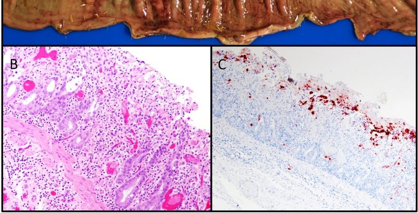
The pathology of equine coronavirus in two horses and one donkey ranging in age from 6 months
to 11 years old
6.3.has been described. The naturally infected equids had severe diffuse necrotizing enteritis
Pathology
characterized byThe marked villous
pathology attenuation,
of equine coronavirusnecrosis of apical
in two horses and oneenterocytes in the
donkey ranging small
in age from intestinal
6 villi,
pseudomembranemonthsformation,
to 11 years oldandhas been described.
hemorrhage andThe naturally infected within
microthrombosis equids had
the severe
mucosa diffuse
and submucosa
necrotizing enteritis characterized by marked villous attenuation, necrosis of apical enterocytes in the
(Figure 5B). In contrast to enteric coronavirus infections in the carnivores, ECoV infection in horses has
small intestinal villi, pseudomembrane formation, and hemorrhage and microthrombosis within the
also been associated
mucosa andwith crypt(Figure
submucosa necrosis.
5B). InIn casestoof
contrast hyperammonemia-associated
enteric encephalopathy,
coronavirus infections in the carnivores,
Alzheimer typeECoVIIinfection in horses
astrocyte has also been associated
hypertrophy with crypt necrosis.
and hyperplasia were In cases of hyperammonemia-
observed diffusely throughout the
associated encephalopathy, Alzheimer type II astrocyte hypertrophy and hyperplasia were observed
cerebral cortex [2].
diffusely throughout the cerebral cortex [2].
Figure 5. (A) Equine coronavirus-associated colitis, colon, horse. Moderate, necrohemorrhagic colitis.
Figure 5. (A) Equine coronavirus-associated colitis, colon, horse. Moderate, necrohemorrhagic
Image courtesy of Silvia Siso. (B) Equine coronavirus-associated enteritis, jejunum, horse. Mixed
colitis. Image courtesyenteritis
inflammatory of Silvia Siso.ectasia
with crypt (B) and
Equine coronavirus-associated
necrosis enteritis,
(crypt “abscesses”) and microvascular jejunum, horse.
thrombi.
(C) Equine coronavirus-associated enteritis, jejunum, horse. Diffuse immunoreactivity
Mixed inflammatory enteritis with crypt ectasia and necrosis (crypt “abscesses”) and microvascular at the tips of
necrotic villi using bovine coronavirus antiserum (immunohistochemistry). Images 5B and 5C
thrombi. (C) Equine coronavirus-associated enteritis, jejunum, horse. Diffuse immunoreactivity at
courtesy of Federico Giannitti.
the tips of necrotic villi using bovine coronavirus antiserum (immunohistochemistry). Figure 5B,C
courtesy of Federico Giannitti.You can also read