Legionellosis Caused by Non-Legionella pneumophila Species, with a Focus on Legionella longbeachae - MDPI
←
→
Page content transcription
If your browser does not render page correctly, please read the page content below
microorganisms

Review
Legionellosis Caused by Non-Legionella pneumophila Species,
with a Focus on Legionella longbeachae
Stephen T. Chambers *, Sandy Slow, Amy Scott-Thomas                            and David R. Murdoch

                                          Department of Pathology and Biomedical Science, University of Otago, Christchurch 8011, New Zealand;
                                          Sandy.slow@otago.ac.nz (S.S.); Amy.scott-thomas@otago.ac.nz (A.S.-T.); David.murdoch@otago.ac.nz (D.R.M.)
                                          * Correspondence: Steve.chambers@otago.ac.nz; Tel.: +64-21-279-8394

                                          Abstract: Although known as causes of community-acquired pneumonia and Pontiac fever, the
                                          global burden of infection caused by Legionella species other than Legionella pneumophila is under-
                                          recognised. Non-L. pneumophila legionellae have a worldwide distribution, although common testing
                                          strategies for legionellosis favour detection of L. pneumophila over other Legionella species, leading
                                          to an inherent diagnostic bias and under-detection of cases. When systematically tested for in
                                          Australia and New Zealand, L. longbeachae was shown to be a leading cause of community-acquired
                                          pneumonia. Exposure to potting soils and compost is a particular risk for infection from L. longbeachae,
                                          and L. longbeachae may be better adapted to soil and composting plant material than other Legionella
                                          species. It is possible that the high rate of L. longbeachae reported in Australia and New Zealand is
                                          related to the composition of commercial potting soils which, unlike European products, contain
                                          pine bark and sawdust. Genetic studies have demonstrated that the Legionella genomes are highly
                                          plastic, with areas of the chromosome showing high levels of recombination as well as horizontal
                                gene transfer both within and between species via plasmids. This, combined with various secretion

                                          systems and extensive effector repertoires that enable the bacterium to hijack host cell functions
Citation: Chambers, S.T.; Slow, S.;
                                          and resources, is instrumental in shaping its pathogenesis, survival and growth. Prevention of
Scott-Thomas, A.; Murdoch, D.R.
                                          legionellosis is hampered by surveillance systems that are compromised by ascertainment bias,
Legionellosis Caused by
                                          which limits commitment to an effective public health response. Current prevention strategies in
Non-Legionella pneumophila Species,
with a Focus on Legionella longbeachae.
                                          Australia and New Zealand are directed at individual gardeners who use potting soils and compost.
Microorganisms 2021, 9, 291.              This consists of advice to avoid aerosols generated by the use of potting soils and use masks and
https://doi.org/10.3390/                  gloves, but there is little evidence that this is effective. There is a need to better understand the
microorganisms9020291                     epidemiology of L. longbeachae and other Legionella species in order to develop effective treatment
                                          and preventative strategies globally.
Academic Editor: Janet E. Stout
Received: 31 December 2020                Keywords: L. longbeachae; Legionella; epidemiology; environment; pathogenesis; prevention
Accepted: 26 January 2021
Published: 31 January 2021

Publisher’s Note: MDPI stays neutral      1. Introduction
with regard to jurisdictional claims in
                                               Following the discovery of Legionella pneumophila as the cause of the Legionnaires’
published maps and institutional affil-
                                          disease outbreak in Philadelphia in 1976 [1], it was quickly realised that there were other
iations.
                                          species within the genus Legionella, some of which could also cause human disease. Nearly
                                          90 different Legionella species have now been reported, of which more than 20 different
                                          species are known to be pathogenic in humans [2,3]. A list of species currently known to
                                          cause human disease is available online [4]
Copyright: © 2021 by the authors.
                                               Despite recognition that a variety of Legionella species and serogroups can cause
Licensee MDPI, Basel, Switzerland.
                                          human disease, L. pneumophila is overwhelmingly reported as the most common cause of
This article is an open access article
                                          legionellosis globally. While the predominance of L. pneumophila may be a true observation,
distributed under the terms and
                                          there is inherent diagnostic bias in current testing strategies for legionellosis that favours
conditions of the Creative Commons
                                          detection of L. pneumophila over other Legionella species. In particular, many countries
Attribution (CC BY) license (https://
creativecommons.org/licenses/by/
                                          rely on the urinary antigen test in cases of suspected Legionnaires’ disease. This test only
4.0/).
                                          reliably detects L. pneumophila serogroup 1, so other Legionella species and serogroups may

Microorganisms 2021, 9, 291. https://doi.org/10.3390/microorganisms9020291                         https://www.mdpi.com/journal/microorganisms
Microorganisms 2021, 9, x FOR PEER REVIEW 2 o
Microorganisms 2021, 9, 291 2 of 17
only reliably detects L. pneumophila serogroup 1, so other Legionella species and serogrou
may not be tested for at all if this is used as the sole, or predominant testing method. A
not be tested for at all if detection
consequence, this is used as the sole,
of infections or predominant
caused by Legionellatesting
speciesmethod.
other than AsL.apneumoph
consequence, aredetection
usually madeof infections caused
by chance, Legionella
andbythe true burdenspeciesandother than L. pneumophila
epidemiology of disease is larg
are usually unknown.
made by chance, and the is
This situation true burden
further and epidemiology
complicated of disease
by the lack is largely
of studies on the microb
unknown. aetiology
This situation is further complicated
of community-acquired by the lack
pneumonia thatof studies on the
systematically testmicrobial
for all species of
aetiology ofgionella
community-acquired
using PCR and/or pneumonia
culture-basedthat methods.
systematically test for all species of
Legionella using PCR and/or culture-based methods.
The one exception is Legionella longbeachae, which was first isolated from patients w
The one exception is
pneumonia inLegionella
the USA longbeachae,
40 years ago which
[5]. Inwas first countries,
some isolated from
such patients with and N
as Australia
pneumoniaZealand,
in the USA 40 years ago [5]. In some countries, such as Australia
where the aetiology of community-acquired pneumonia has been systematica and New
Zealand, where the aetiology
studied, L. longbeachaeof community-acquired
disease has emergedpneumonia has beencause
as the predominant systematically
of legionellosis [6
studied, L. longbeachae disease has emerged
Cases are increasingly as the predominant
being reported from other regions cause as
of well.
legionellosis [6,7]. that ha
The countries
Cases are increasingly
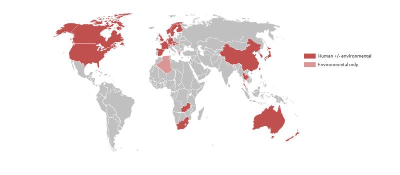
reported at least one case are shown in Figure 1, demonstrating that thethat
being reported from other regions as well. The countries conditions
have reported at leastmay
infection oneoccur
case are shown inwith
in countries Figure 1, demonstrating
widely varying climatesthat and
the conditions
socioeconomic con
for infection mayHowever,
tions. occur in countries with widely
it is still unclear whether varying climateshigh
the relatively and incidence
socioeconomic of Legionnair
conditions. disease
However, caused by species other than L. pneumophila in AustraliaLegionnaires’
it is still unclear whether the relatively high incidence of and New Zealand i
disease caused
trueby species other
geographic than L.or
variation pneumophila in Australia
simply reflects that theseandspecies
New Zealand
are more is arigorously
true tes
geographic forvariation or simply
in this region. reflects that these species are more rigorously tested for in
this region.
Figure 1. Reported global distribution of L. longbeachae isolated from human and/or
environmental sources [5,8–29]. Data from Austria, Norway and Slovakia provided by
Figure 1.European
Julien Beauté, ReportedCentre
global distribution of L. longbeachae
for Disease Prevention and isolated
Control from human
(ECDC); andand/or
for environmen
sources [5,8–29]. Data from Austria, Norway and Slovakia provided by Julien
the Czech Republic by Vladimir Drašar, Head Public Health Institute Ostrava, National Beauté, European
Centre for Disease Prevention and Control (ECDC); and for the Czech Republic by Vladimir
Legionella Reference Laboratory, Czech Republic.
Drašar, Head Public Health Institute Ostrava, National Legionella Reference Laboratory, Czech
Republic.
This paper reviews legionellosis caused by Legionella species other than L. pneumophila.
The primary focus is on legionellosis caused by L. longbeachae, simply because considerably
This paper reviews legionellosis caused by Legionella species other than L. pneumo
more data are available on this species. Indeed, apart from L. pneumophila, L. longbeachae is
ila. The primary focus is on legionellosis caused by L. longbeachae, simply because cons
the only species causing legionellosis for which there are sufficient data to make detailed
descriptionserably
aboutmore data
clinical are available
features on this species. Indeed, apart from L. pneumophila, L. lo
and epidemiology.
beachae is the only species causing legionellosis for which there are sufficient data to ma
detailed descriptions about clinical features and epidemiology.
2. Clinical Features
The clinical spectrum of legionellosis caused by Legionella species other than L. pneu-
2. Clinical
mophila is similar Features
to that caused by L. pneumophila, with pneumonia (Legionnaires’ disease)
The clinical
being the predominant clinical spectrum of legionellosis
presentation. There are caused by Legionella
no clinical species
features that other than L. pn
distinguish
mophila is similar to that caused by L. pneumophila, with pneumonia
Legionnaires’ disease caused by L. pneumophila from L. longbeachae, nor can Legionnaires’ (Legionnaires’ d
disease be itease) being the predominant
distinguished clinically fromclinical presentation.
other causes There are no clinical
of community-acquired features that d
pneumo-
nia [30,31]. tinguish
Increasing Legionnaires’
age, immunedisease causedand
suppression by pre-existing
L. pneumophila from
lung L. longbeachae,
disease have beennor can
regarded asgionnaires’
importantdisease be it distinguished
predisposing conditions clinically from other
for legionellosis. causes of community-acqui
Recognition of these
pneumonia
factors has led [30,31]. Increasing
some researchers to developage, immune
scoring suppression
systems to increaseand pre-existing
recognition lung dise
of Le-
gionnaires’ disease, but none of these have been developed for non-L. pneumophila species
or gained widespread acceptance as a clinical tool [32]. A case series of L. longbeachaeMicroorganisms 2021, 9, 291 3 of 17
disease from New Zealand found that a higher proportion of cases were males (63%),
most cases (59%) were of mild to moderate severity (CURB-65 score 0–1), 25% required
admission to the intensive care unit, and the mortality rate within 30 days of admission
was 5% [31].
Pontiac fever, is transient flu-like illness with a high attack rate, [33] and has been associ-
ated with exposure to a variety of Legionella species, including L. pneumophila,
Legionella feeleii, Legionella micdadei, Legionella anisa, and L. longbeachae [34–39]. Outbreaks
of Pontiac fever caused by species other than L. pneumophila have occurred in a variety of
occupational and recreational settings, including workers in an automobile manufacturing
plant (L. feeleii) [34], users of whirlpool spas (L. micdadei) [35,36,40], workers at a compost
manufacturing site (L. longbeachae) [37] and conference delegates and restaurant patrons ex-
posed to decorative fountains (L. anisa) [38,39]. Reports of Pontiac fever are almost certainly
underestimated because sporadic cases are unlikely to come to attention and be diagnosed.
Non-L. pneumophila has also been identified in cases of skin and soft tissue infection,
septic arthritis and bacterial endocarditis [41–50]. These cases may be severe and difficult
to diagnose as legionellae are unexpected as the infecting organism. A recent example is
that of a 78-year-old gardener who pricked her hand on a rose thorn. L. longbeachae was
cultured on selective media from surgically debrided tissue that would have been missed
with standard investigations [41]. Likewise, diagnosis and treatment of recurrent upper
limb abscesses of L. bozemanii was delayed for weeks in a patient on steroid therapy and
methotrexate for seronegative arthritis; the diagnosis was eventually made by 16S rRNA
PCR and seroconversion [51].
3. Epidemiology
The epidemiology of infection with L. longbeachae and other non-L. pneumophila species
of Legionella is poorly described worldwide, although cases have been reported in many
countries (Figure 1). A major reason for this is the difficulty with diagnosis, with the likeli-
hood that cases are markedly under-diagnosed and under-recognised. In many centres,
testing is primarily based around urinary antigen testing that recognises only L. pneumophila
serogroup 1. Culture of respiratory samples is discouraged by some authorities for nonse-
vere disease, including in hospitalised patients [52]. The results may also be compromised
if specimens are taken after empiric therapy (including antilegionella therapy) has been
administered. Where other techniques are employed, such as serology, specific culture
techniques and PCR, L. longbeachae has emerged as an important cause of Legionnaires’ dis-
ease in community-acquired pneumonia. Other non-L. pneumophila species are recognised
particularly in severely immunocompromised patients [20,53,54]. The non-L. pneumophila
species almost certainly cause many more cases of mild to moderate disease that do not
come to medical attention and remain an ongoing burden of morbidity on the population.
3.1. Incidence
Several high-income countries and regions have well established surveillance systems
but almost all data on legionellosis relies on various forms of passive surveillance (Table 1).
Most of these data are derived from cohort studies or case reports of patients who required
admission to hospital. These reports demonstrate striking differences in the number of
cases identified in Australia and New Zealand compared with Europe and North Amer-
ica [6,55–58]. Most cases are sporadic, although clusters of Legionnaires’ disease caused by
L. longbeachae have been recognised in Scotland, where six cases of Legionnaires’ disease
caused by L. longbeachae were identified over a four-week period in 2013 [59], and in Sweden,
where 30 cases were identified during spring/summer 2018 [60].Microorganisms 2021, 9, 291 4 of 17
Table 1. Number of cases of Legionellosis reported from countries with national surveillance systems.
Countries *
Legionella New Zealand Australia Europe [58] USA [61]
Japan [56]
species [55] (2010–2019) [6] (2014) (2008–2017) (2016–2017)
L. pneumophila 314 (19%) 195 (46%) 4738 (97%) 230 (60%) 679 (98%)
L. longbeachae 902 (54%) 164 (39%) 48 (1%) 1 (0.3%) 6 (0.1%)
L. anisa 3 (0.2%) na 6 (Microorganisms 2021, 9, 291 5 of 17
low and middle-income countries, but a year-long population-based study of the microbio-
logical causes of clinically defined community-acquired pneumonia requiring hospitalisation
in a rural province of Thailand has been reported [27]. Infection with L. longbeachae was
identified by seroconversion using an indirect immunofluorescence assay of combined IgG,
IgA and IgM titres. These cases were not confirmed by culture or PCR testing. The incidence
rate was 5–29 cases per 100,000 of the population, incidence increased with age, and it was
more common among males (rate ratio 1.6 95% CI 1.1–2.3). There was also a single case of
Legionnaires’ disease caused by L longbeachae reported in a child in Zambia [29]. This was
the only case found as part of a year-long study conducted in five locations in Africa and
two in Asia of 2757 cases of childhood community-acquired pneumonia.
The epidemiologic evidence of human infection of other non-L. pneumophila species is
limited. These infections are rarely reported due, at least in part, to diagnostic bias that
precludes cases being identified. There appears to be little difference in the distribution or
reported incidence rates of these species across the world. Of these, L. micdadei is the most
consistently reported across regions (Table 1). Clusters of cases related to a common source
have been described, including 12 cases of Legionnaires’ disease caused by L. micdadei
among 38 renal and cardiac transplant patients in one centre over a three-month period, and
further cases were detected 16 months later [65]. Subsequently, 16 cases of L. micdadei were
reported in another tertiary care centre over a 12 year period, predominantly among im-
munocompromised hospitalised patients [66]. Another cluster of five cases of nosocomial
pneumonia caused by L. bozemanii in a community hospital among immune compromised
patients has been identified [67]. Case series have demonstrated that Legionnaires’ disease
may be caused by a wide variety of non-L. pneumophila species in immune-compromised pa-
tients. The MD Anderson Centre in Houston Texas reported consecutive cases of Legionella
infections in cancer patients from 2002 to 2014. Most of the 33 strains identified were on
specimens obtained at bronchoscopy, and 18 were non-L. pneumophila species [54]. Twenty-
seven patients had underlying hematologic malignancies and 23 were leukopenic. Six
patients were recipients of allogeneic hematopoietic stem cell transplant, with their infec-
tions caused by five Legionella species. Legionnaires’ disease was the immediate cause of
death in six [54]. Some cases may be related to travel but the numbers are difficult to obtain
as the species of infecting Legionella species may not be reported [68,69].
3.2. Environmental Studies
3.2.1. Potting Soils and Gardens
An important epidemiological link has been made between exposure to compost or
potting soils and L. longbeachae infection. An investigation during 1988–1989, into 22 cases
of L. longbeachae infection in South Australia, found cases were among regular gardeners.
L. longbeachae was subsequently isolated from compost and potting soils found at the
homes of four cases, providing a possible link to a natural habitat for this bacterium [70,71].
Further studies have shown that L. longbeachae infection was linked to the use of potting
soils or gardens in other states in Australia, as well as Japan, the United States, Canada, the
Netherlands and Scotland through case-series and laboratory evidence [59,64,72–76]. In the
Scottish cluster all cases were associated with amateur gardeners and required admission
to the intensive care unit [59]. A case control study in South Australia found recent use
of potting mix was associated with illness (OR 4·74, 95% CI 1·65–13·55, p = 0·004) [72].
Similarly, exposure to purchased compost was a risk factor for Legionnaires’ disease
from L. longbeachae in New Zealand (OR 6.2, 95% CI 2.2–17.3), unlike for homemade
compost [77].
Seminal studies by Steele et al. further implicated potting soils as a source of human
L. longbeachae infection. L. longbeachae was recovered on selective media from soil samples
and potting mixes from four patients who suffered Legionnaires’ disease with this organism.
The bacteria were able to persist for seven months in potting soil held at room temperature
but did not grow at 43◦ C or above in soil [70]. Subsequently L. longbeachae was isolated
from 26 (56%) of 45 potting mixes manufactured in Australia but not from imported pottingMicroorganisms 2021, 9, 291 6 of 17
soils [78]. L. longbeachae was found in one of four samples of fresh pine sawdust and
in a soil sample close to the site, but not hammer milled bark. L. longbeachae was also
found at two of six large scale manufacturing plants and in samples from three of 20 home
compost heaps [78]. L. Longbeachae has since been detected in potting soils and gardens
from many other countries in Europe, North America and Japan, but not as frequently as
in Australia [16,79–84]. There is a highly diverse pool of L. longbeachae genotypes within
the samples from potting soils that makes it difficult to identify the source of a clinical
infection [85].
Steele and colleagues also demonstrated that the ecology of Legionella species in
potting soils was extremely complex, as multiple species of pathogenic Legionella were
usually present in Australia-made potting soils [86]. Among 20 Australian potting soils,
L. anisa, L. bozemanii, L. micdadei, L. gormani, L. dumoffi and L. pneumophila serogroups 1–14
were found, in addition to L. longbeachae [78]. A similar range of Legionella species were
found in 20 home composts, but with a lower proportion of L. longbeachae present among
the 29 isolated (L. pneumophila serogroup 2–14, 11 strains (55%); L. pneumophila SG, 1 strain
(5%); L. anisa, 5 strains (25%); L. longbeachae and L. bozemani, three strains each (15%);
unidentified, six (30%)) [86]. UK studies of 24 compost products found 15 (62.5%) samples
tested contained Legionella species either on first testing or after enrichment by coculture
with amoeba when retested 8–10 weeks later. The most common species identified before
enrichment was L. sainthelensis (5/24; 21%), while L. longbeachae (4/24; 17%) was the most
common after amoebal enrichment. Other identified species known to cause human disease
were also isolated (L. anisa, L. bozemanii, L. micdadei, L. gormani, L. dumoffi, L. birminghamensis
and L. pneumophila) [73,83]. A similar range of Legionella species have been identified in
potting soils in Greece, Switzerland and Japan [16,79,82]. These results suggest that potting
soils could be a source of other Legionella infections, including with L. pneumophila species.
L. longbeachae and other non-L. pneumophila species have been found in water contami-
nated by soil, including rain water puddles in Europe, and to be widely distributed among
fresh water and marine waters in Puerto Rico [87,88]. Legionella species were also found
in water collected in rain forests from epiphytes 30 feet above ground [87]. L. longbeachae
was not found in soil or other environmental samples obtained from around houses and
work places of people in Southern Thailand where a high prevalence of L. longbeachae
infection had been found by serological testing [89]. Many other Legionella species were
found, suggesting one or more of these may have been responsible for the infections.
One possible difference of European potting mixes compared to and Australian and
New Zealand products is the range and proportions of the constituents used in the manu-
facturing process. Australian and New Zealand potting soils contain a significant amount
of partially or fully composted pine bark and sawdust in addition to peat, and composted
green waste. Recent studies in New Zealand using qPCR identified L. longbeachae DNA
in pine bark, pine sawdust and potting soil products containing pine products, but did
not identify it in peat or commercially composted green waste [90]. Studies from Europe
indicate that Legionella species, including L. pneumophila and L. bozemanii are common in
composted green waste, but not fresh green waste. L. longbeachae is rarely found in either
fresh or composted green waste [80]. Australian, New Zealand and European studies have
failed to find Legionella species in peat. These studies raise the possibility that composting
facilities are contaminated with Legionella species from other sites [78,83,90]. Studies of bark
from living pine trees in New Zealand found L. longbeachae DNA is common in spring, but
very uncommon on bark from other tree species [91]. L. longbeachae could be brought into
composting facilities with bark, and the load may contribute to the seasonal variation in
clinical cases. In Switzerland, it has been suggested that Legionella species could be brought
into the storage facilities by wind and rain [79].
All of these studies indicate that composted material is an important reservoir of
pathogenic species of Legionella and are a potential source of human infection, but we are
unaware of any substantial data linking cases of Legionnaires’ disease to the manufacture of
compost. There is some evidence that compost facilities can release bioaerosols containingMicroorganisms 2021, 9, 291 7 of 17
Legionella species, and transmission of L. longbeachae aerosol has been suggested as a cause
of a nosocomial infection [92,93].
3.2.2. Water Bodies
There have been occasional reports of nosocomial L. longbeachae Legionnaires’ disease
associated with detection of this species in hospital and residential water supplies and
unpasteurised water tanks [94,95]. L. longbeachae has been identified in samples taken from
a cooling tower, but infection from this source was not proven [96]. It has also been isolated
from a hot spring water in Algeria by culture and molecular methods [8].
In contrast to L. longbeachae, infection with other non-L. pneumophila species have
typically been associated with water bodies, and the epidemiology of water related
non-L. pneumophila species is likely to be similar to L. pneumophila. The environmental
sources of community acquired Legionnaires’ disease has been extensively reviewed [97].
Nosocomial Legionnaires’ disease with L. micdadei, L. bosemani, L. feelii, and L. anisa has been
attributed to infected water distribution systems, including those in hospitals [58,98–100].
L. micdadei has also been identified in ultrasonic nebulisers [101]. L. micdadei has been found
in whirlpool spas in Norway, and L. anisa associated with a decorative fountain [35].
4. Pathogenesis
The pathogenesis of Legionella infection has been most extensively studied for
L. pneumophila, and this has been comprehensively reviewed [102]. Relatively recent
advances in high throughput next generation sequencing technologies has allowed the
genomes of non-L. pneumophila species to be sequenced and examined in greater depth.
As such, comparative genomic analysis has proven to be instrumental in elucidating the
factors that are important for the pathogenicity of Legionella species. This has revealed
both important parallels, and differences between the various pathogenic factors of
Legionella species, which remain under continued investigation.
Analysis of the genomes of up to 58 different Legionella species revealed they are
highly dynamic and diverse, with lengths ranging in size from the smallest at 2.37 Mb in
Legionella adelaidensis to the largest at 4.82 Mb in Legionella santicrucis [103,104]. Indeed, across
the Legionella pan genome only 6% of the genes (1008) constitute the core genome [103,104],
illustrating the high genomic diversity and the importance of horizontal gene transfer and
recombination in shaping the genome of the genus. At around 4.1 Mbp, the single circular
chromosome of L. longbeachae is approximately 500 kb larger than L. pneumophila, but has
a similar GC content at around 37% [104–106]. The gene organization is also markedly
different to L. pneumophila, with limited gene synteny, although there are small clusters
where the gene order is highly conserved [105,106].
There are around 3500 predicted coding sequences in the L. longbeachae genome,
and around 2000 of these have been assigned putative functions [105–109]. Only 65%
of the genes in L. longbeachae are orthologous to those in L. pneumophila [105,110]. The
L. longbeachae genome, as a result of its soil and plant habitat, contains numerous genes
that reflect its ecological niche, with several genes that encode proteins that can degrade
components of plant cell walls, such as cellulases, hemicellulases and pectin lyases [105].
In addition, the genome is highly methylated with m4 C and m6 A epigenetic modifications
throughout the genome, where m6 A is strongly associated with particular sequence
motifs [109]. Although the L. longbeachae genome appears to be more stable than that
of L. pneumophila [105], large-scale rearrangements have occurred in some regions of the
chromosome with evidence of extensive recombination [85].
In the accessory genome, L. longbeachae strains NSW150 and D4968 [106] were both found
to contain a similar plasmid of around 70 kb. It was first thought that L. longbeachae plasmids
only circulated within this species and were distinct from those found in L. pneumophila, which
range in size from around 59 kb to 131 kb [110]. More recently, sequencing data from multiple
clinical and environmental L. longbeachae isolates has shown that most contain at least one
plasmid ranging in size from around 70 kb to 150 kb [85,109]. Despite still relatively limitedMicroorganisms 2021, 9, 291 8 of 17
data, the plasmids have been shown to have a common backbone including conjugational
genes separated by variable regions. Importantly, there is evidence of extensive recombination
and horizontal gene transfer between the plasmids from different species, indicating both
intra and interspecies exchange of genetic material across the genus [85,107–109].
Transcriptome analysis of L. pneumophila and L. longbeachae has shown that both have
a biphasic lifestyle (a replicative/avirulent phase and a transmissive/virulent phase), al-
though this is less pronounced in L. longbeachae [105]. Both also typically exist in biofilm
forms or intracellularly with amoeba or macrophages. Conditions which disrupt the
biofilm may release large numbers of organisms into the environment [111]. Likewise,
the ability to multiply within free-living amoeba may facilitate transmission, as a single
amoebal cyst may contain thousands of Legionella bacteria [93]. The major route of infection
is probably by inhalation of aerosols derived from soil or water, but may also reach the
lung by microaspiration [112]. The infectious dose has not been precisely defined and
may be higher than 1000 organisms [113]. This is almost certainly lower in people with
pre-existing lung disease that disrupts the mucociliary escalator, allowing the bacterium
to reach the alveolar space. Immune deficiencies, particularly cell mediated deficiencies,
such as haematological malignancy, as well as immunosuppressive therapy such as dose
steroid therapy, tumor necrosis factor inhibitors and anti CD-52 agents, are also important
risk factors for developing disease from Legionella species [114–116]. Mucosal associated
invariant T (MAIT) cells that are resident in the lung, also appear to contribute to pro-
tection against fatal infection with Legionella. The mechanism is dependent on the major
histocompatibility complex (MHC) class I-related molecule MR1, interferon-γ (IFN-γ) and
granulocyte macrophage-colony stimulating factor (GM-CSF) [117].
On reaching the alveolar space L. pneumophila is known to attach to alveolar macrophages
using flagella, pili and outer membrane porins [118–123]. The attachment enables phagocy-
tosis to follow, mediated by macrophage infectivity potentiator protein (MIP) and human
complement C3. In contrast L. longbeachae lack flagella but have other effector proteins,
possibly derived from organisms that typically live in soil and plant derived material. The
lack of flagella has been cited as one of the reasons for the different responses to infection in
different strains of mice, where A/J mice are susceptible to L. pneumophila infection and repli-
cation, but C57BL/6 and BALB/c mice are not [110,124]. In contrast, all three mouse strains
are susceptible to L. longbeachae infection and replication. It is thought that the flagellin of
L. pneumophila triggers pyroptosis and cell death in the C57BL/6 and BALB/c mice, leading
to a failure to establish infection [110,124].
Following phagocytosis, Legionella infection, survival and replication within a host cell
is dependent on successfully transforming the phagosome into a distinct membrane bound
vacuole, termed the Legionella containing vacuole (LCV) [102,125]. To do this Legionella
employ various secretion systems to translocate effector proteins into the vacuole, which,
in turn, inhibit phagocyte lysosome fusion [102]. The effector proteins also facilitate the
transformation of the vacuole into a nutrient rich environment, as they enable the host
resources to be hijacked and transported back to the LCV to support legionella replication.
As the nutrients are depleted by bacterial replication, the organism enters stationary phase,
triggers apoptosis of the host cell releasing the organisms to be phagocytosed and enters a
new replicative cycle.
The various secretion systems and the effector proteins of Legionella are the predom-
inant drivers of their pathogenicity. Comparative genomic analysis has shown that all
species analysed to date contain the genes for both a type 2 secretion system (T2SS) and a
type 4B secretion system (T4BSS), also known as the Icm/Dot secretion system [102,103,126].
In addition to these, and similarly to L. pneumophila, L. longbeachae contains the genes for
a type 4A secretion system (T4ASS) but, like all other non-L. pneumophila species, it lacks
a type 1 secretion system [126]. The Icm/Dot T4BSS is crucial for LCV biosynthesis and
intracellular replication. It is also the primary and most well-studied pathogenic secretion
system of Legionella [104,125–127], particularly in L. pneumophila. The components are en-
coded by 25 genes that occur within two distinct pathogenicity regions on the chromosome,Microorganisms 2021, 9, 291 9 of 17
and the gene order and orientation is highly conserved genus-wide [104,126,127]. The
Icm/Dot T4BSS translocates effector proteins into the host cell that interact with the host to
subvert resources and cell function for the benefit of the bacterium and is indispensable
for pathogenicity. Despite the highly conserved nature of the Icm/Dot T4BSS, the effector
repertoire that it translocates into the host has been shown to be highly variable across the
various species examined to date [103,104]. From species to species, the effectors differ
in number and, although they share functional similarities, there is little DNA sequence
homology [128]. Of the over 300 effectors identified in L. pneumophila, only 35% are found
to occur in L. longbeachae [125]. Genus-wide, comparative analysis of 38 species has re-
vealed even greater variability with only seven putative core effectors identified [104],
i.e., effectors that were found to have orthologs in each of the species examined out of a
total of 5885. More recently, Gomez-Valero et al., 2018, after examining 80 strains from
58 different Legionella species showed that the pan-genus pool of putative Icm/Dot T4BSS
effectors could be well over 18,000 proteins, again illustrating the extremely high diversity
of the effector proteins in Legionella. Furthermore, analysis of these putative effector genes
revealed that they encode secreted proteins that contain a large number and variety of
eukaryotic domain-encoding proteins and eukaryotic-like proteins [102,103]. The genes
encoding these proteins are thought to have been acquired via horizontal gene transfer
from their protozoan hosts in a type of coevolutionary arms race that allows Legionella to
mimic the host to establish infection and successfully replicate.
The survival tactics and pathogenic trajectory of Legionella is significantly shaped by
the interactions and exchanges that it has with its protozoan hosts, but not by interactions
it has with its human host [129–131]. This is because human infection by Legionella is an
evolutionary dead-end; either the bacterium dies because of successful response/treatment
or the human host dies because of the infection, which consequently causes the bac-
terium to die as it does not have recourse to the external environment. Therefore, infec-
tivity and pathogenicity to humans is not an evolutionary advantage to Legionella. While
L. pneumophila has been found to exhibit a broad host range that spans several protozoan
phyla [129], little is known about the protozoan hosts of other Legionella species. Nonethe-
less, it has been shown in various coculture studies that the protozoan host species has a
major influence on L. pneumophila’s ability to infect human cells [132], and it is reasonable
to expect that the same is true for other Legionella species. For instance, L. pneumophila
cocultured in Acathamoeba polyphaga is more tolerant to antibiotics, while resistance to
chlorination is higher in those grown in Vermamoeba vermiformis when compared to those
from Acathoamoeba castillanii [132].
5. Prevention and Public Health
The public health aspects most specific to legionellosis caused by Legionella species
other than L. pneumophila relate to L. longbeachae and exposure to gardening activities. The
public health response to the threat posed by potting soils in New Zealand and Australia
has been to place responsibility onto the individual gardener. Gardeners are alerted to the
risk with warnings printed on potting soil containers and information leaflets available
at the point of sale. Information on Legionnaires’ disease can be found on the Ministry
of Health or State Health authorities’ websites. Gardeners, particularly those with other
health conditions or advanced age, are advised to protect themselves by using gloves
and masks and to dampen the potting soil when the bag is opened to minimise aerosol
formation [31,70].
This advice makes intuitive sense as inhalation of aerosols is probably the most
important means of transmission and is the major route of infection of L. pneumophila. This
is supported by results of a case control study among gardeners that found that tipping
or trowelling potting soils was a major risk factor for Legionnaires’ disease (population
attributable fraction 65%) [77]. However, behaviours that involve touching the face, such as
eating or drinking without handwashing, or smoking, may also place gardeners at risk of
infection (population attributable fraction 35%) [77]. These results suggest that eliminatingMicroorganisms 2021, 9, 291 10 of 17
aerosolization activities and effective hand hygiene could reduce the risk of Legionnaires’
disease but, disappointingly, the study did not find any protective effect of wearing a mask
or gloves [77].
There has been little research on the acceptability or effectiveness on the use of per-
sonal protective equipment for preventing Legionnaires’ disease while gardening. Multiple
barriers to the use of face masks have been identified in other settings with serious health
risks, such as tuberculosis and influenza, and contribute to poor acceptability among
gardeners [133–135]. These include lack of perceived risk, discomfort and poor fit. Discom-
fort may be compounded when masks become dampened, as this increases the work of
breathing and the masks subsequently lose their protective effect. Alongside discomfort,
masks may also may give a false sense of security and interfere with communication, and
may also accumulate bacteria and become hazardous prior to disposal [136]. It may be
possible to change the behaviour of gardeners as the COVID-19 pandemic has changed
public perception of the value of masks [135].
Other avenues to change gardeners’ behaviour to reduce Legionnaires’ disease risk
are still to be explored. These could include warning about the risks through major media
outlets during the high seasons, and physicians providing personalised information for
immune compromised patients to help them prevent exposure to all Legionella species.
A more proactive approach in Australia and New Zealand may be to engage with
the potting soil industry to develop low-risk manufactured potting soils. The apparent
differences in epidemiology of Legionnaires’ disease between Europe and Australia and
New Zealand suggests that there is less exposure to L. longbeachae in Europe. Pasteurising
potting soils, or reducing or eliminating pine products from potting soils, and using peat
and well composted green waste, which appear to be inhospitable to L. longbeachae, may
reduce these infections. As peat is a limited resource, supply may become restricted for
environmental reasons so that manufactures may turn to other products such as pine bark
to fill the gap.
The prevention of Legionnaires’ disease in hospitals has been widely discussed, and
water standards have been set by the World Health Organization and many national
agencies [137,138]. These have been developed appropriately with particular reference
to L. pneumophila, but the measures employed to minimise infection with this organism
would be expected to be effective for other waterborne Legionella species. These include
well designed hospital water systems, and maintenance of hot water systems, to ensure
inhibitory temperatures are maintained. Hot water pipes need to be properly insulated
to prevent the warming of adjacent cold-water pipes, stagnation sites such as blind pipe
sections and holding tanks eliminated, and material that supports legionella growth min-
imised [139–142]. A metanalysis has shown that temperatures of 55–59◦ C are associated
with a low probability of Legionella species being detectable in water samples, and in cold
water kept below 20 ◦ C [143,144].
There is a lack of consensus on the need for monitoring water systems in hospitals, with
some organisations recommending against monitoring, others monitoring only those areas
housing immune-compromised patients, and others all hospital water supplies [145,146].
This is in part because it has been difficult to define specific and sensitive target con-
centrations for remediation although recent studies have suggested methods to improve
monitoring techniques [100,147,148].
If Legionella species are detected, methods such as the use of monochloramine, heat,
ultraviolet light, copper-silver ionization and chlorination have been used to reduce the risk
to patients [67,149–153]. Improvement of chlorination of water supplies may reduce the
burden of travel-related legionellosis from non-L pneumophila species [151]. It is important
that detection systems are not solely focused on L. pneumophila, as molecular techniques
may help identify the source of infection. These techniques should also be used for
monitoring water distribution systems that may harbour non-L. pneumophila species [154].Microorganisms 2021, 9, 291 11 of 17
6. Conclusions
The number and distribution of infections caused by L. longbeachae and other
non-L. pneumophila legionellae are almost certainly markedly underestimated globally.
Humans are accidental hosts, where infections are most likely to occur following inter-
actions with the natural environment. It is the relationship between the environmental
conditions and the interactions Legionella has with is protozoan hosts that shape their
pathogenesis, enabling their survival and growth. Favourable conditions are needed
to allow Legionella numbers to increase in the environment, thereby accounting for the
seasonal pattern of human infections.
Legionnaires’ disease is a preventable pneumonia. Resourcing of the public health
response will inevitably be governed by the number of cases identified and how political
figures assign responsibility for controlling Legionnaires’ disease. Multiple factors cur-
rently combine to mitigate against case ascertainment. These include the passive design of
surveillance systems with testing of only respiratory samples submitted for analysis. Many
patients with community-acquired pneumonia are not asked to produce sputum samples,
and up to 40% of patients with community-acquired pneumonia are unable to expectorate
sputum when asked [62]. Investigation of nonsevere cases of community-acquired pneu-
monia is discouraged including for hospitalised cases in many centres. This is exacerbated
by a perception among some clinicians that Legionnaires’ disease is necessarily a severe
infection, and only these cases are investigated, and then testing is limited to only the uri-
nary antigen test for L. pneumophila serogroup 1. Compounding these issues, investigations
may only be done after antimicrobial therapy has been initiated, including agents that
are active against Legionella species, thereby reducing the sensitivity of any testing that is
conducted. There may be significant numbers of cases who present to health practitioners
in the community and are not investigated or are not sufficiently unwell to seek medical
attention. Lastly, financial constraints discourage testing, or at least minimise the resources
allocated to investigating community-acquired pneumonia.
Practice guidelines make pragmatic sense but should not override the need for well
conducted studies that adequately define the causes of community-acquired pneumonia in
both the general public and immune compromised patients. These studies should avoid
the tyranny of urine testing only, to identify pinch points where public health interventions
can minimise infection rates of non-L. pneumophila species.
Author Contributions: Conceptualization, S.T.C., S.S., A.S.-T. and D.R.M.; Methodology, S.S.; Project
administration, A.S.-T.; Supervision, S.T.C.; Validation, D.R.M.; Writing—original draft, S.T.C.;
Writing—review & editing, S.S., A.S.-T. and D.R.M. All authors have read and agreed to the published
version of the manuscript.
Funding: This research received no external funding.
Data Availability Statement: No new data presented.
Conflicts of Interest: The authors declare no conflict of interest.
References
1. McDade, J.E.; Shepard, C.C.; Fraser, D.W.; Tsai, T.R.; Redus, M.A.; Dowdle, W.R. Legionnaires’ disease: Isolation of a bacterium
and demonstration of its role in other respiratory disease. N. Engl. J. Med. 1977, 297, 1197–1203. [CrossRef] [PubMed]
2. Honigsbaum, M. Legionnaires’ disease: Revisiting the puzzle of the century. Lancet 2016, 30, 456–457. [CrossRef]
3. Cunha, B.; Burillo, A.; Bouza, E. Legionnaires’ disease. Lancet 2016, 23, 376–385. [CrossRef]
4. Specials Pathogens Laboratory. Available online: https://specialpathogenslab.com/legionella-species/ (accessed on 21 January 2021).
5. McKinney, R.M.; Porschen, R.K.; Edelstein, P.H.; Bissett, M.L.; Harris, P.P.; Bondell, S.P.; Steigerwalt, A.G.; Weaver, R.E.; Ein, M.E.;
Lindquist, D.S.; et al. Legionella longbeachae species nova, another etiologic agent of human pneumonia. Ann. Intern. Med. 1981, 94,
739–743. [CrossRef]
6. Australia’s notifiable disease status, 2015: Annual report of the National Notifiable Diseases Surveillance System.
Commun. Dis. Intell. 2019, 43. [CrossRef]Microorganisms 2021, 9, 291 12 of 17
7. Priest, P.C.; Slow, S.; Chambers, S.T.; Cameron, C.M.; Balm, M.N.; Beale, M.W.; Blackmore, T.K.; Burns, A.D.; Drinković, D.;
Elvy, J.A.; et al. The burden of Legionnaires’ disease in New Zealand (LegiNZ): A national surveillance study. Lancet Infect. Dis.
2019, 19, 770–777. [CrossRef]
8. Boilattabi, N.; Barrassi, L.; Bouanane-Darenfed, A.; La Scola, B. Isolation and identification of Legionella spp. from hot spring
water in Algeria by culture and molecular methods. J. Appl. Microbiol. 2020. [CrossRef]
9. Steele, T.W. Legionnaires’ disease in South Australia, 1979–1988. Med. J. Aust. 1989, 151, 5–6. [CrossRef]
10. Wright, A.J.; Humar, A.; Gourishankar, S.; Bernard, K.; Kumar, D. Severe Legionnaire’s disease caused by Legionella longbeachae in
a long-term renal transplant patient: The importance of safe living strategies after transplantation. Transpl. Infect. Dis. 2012, 14,
E30–E33. [CrossRef]
11. Wang, J.Y.; Li, X.; Chen, J.Y.; Tong, B. Epileptic Seizure after Use of Moxifloxacin in Man with Legionella longbeachae Pneumonia.
Emerg. Infect. Dis. 2020, 26, 2725–2727. [CrossRef]
12. Svendsen, J.H.; Jønsson, V.; Niebuhr, U. Combined pericarditis and pneumonia caused by Legionella infection. Br. Heart J. 1987, 58,
663–664. [CrossRef] [PubMed]
13. Mentula, S.; Pentikäinen, J.; Perola, O.; Ruotsalainen, E. Legionella longbeachae infection in a persistent hand-wound after a
gardening accident. JMM Case Rep. 2014, 1, e004374. [CrossRef] [PubMed]
14. Desplaces, N.; Nahapetian, N.; Bure, A.; Dournon, E. Legionella longbeachae serogroup 2 in Europe. Lancet 1982, 2, 1400. [CrossRef]
15. Kümpers, P.; Tiede, A.; Kirschner, P.; Girke, J.; Ganser, A.; Peest, D. Legionnaires’ disease in immunocompromised patients: A
case report of Legionella longbeachae pneumonia and review of the literature. J. Med. Microbiol. 2008, 57, 384–387. [CrossRef]
[PubMed]
16. Velonakis, E.N.; Kiousi, I.M.; Koutis, C.; Papadogiannakis, E.; Babatsikou, F.; Vatopoulos, A. First isolation of Legionella species,
including L. pneumophila serogroup 1, in Greek potting soils: Possible importance for public health. Clin. Microbiol. Infect. 2010,
16, 763–766. [CrossRef] [PubMed]
17. Lang, R.; Wiler, Z.; Manor, J.; Kazak, R.; Boldur, I. Legionella longbeachae pneumonia in a patient splenectomized for hairy-cell
leukemia. Infection 1990, 18, 31–32. [CrossRef]
18. Irie, S.; Akagi, K.; Hiraga, T.; Tada, S.; Kimura, I.; Saito, A. The first case of Legionella longbeachae pneumonia in Japan. Nihon Kyobu
Shikkan Gakkai Zasshi 1984, 22, 518–522.
19. van’t Hullenaar, N.G.; van Ketel, R.J.; Kuijper, E.J.; Bakker, P.J.; Dankert, J. Relapse of Legionella longbeachae infection in an
immunocompromised patient. Neth. J. Med. 1996, 49, 202–204. [CrossRef]
20. Yu, V.L.; Plouffe, J.F.; Pastoris, M.C.; Stout, J.E.; Schousboe, M.; Widmer, A.; Summersgill, J.; File, T.; Heath, C.M.;
Paterson, D.L.; et al. Distribution of Legionella species and serogroups isolated by culture in patients with sporadic community-
acquired legionellosis: An international collaborative survey. J. Infect. Dis. 2002, 186, 127–128. [CrossRef]
21. Wolter, N.; Carrim, M.; Cohen, C.; Tempia, S.; Walaza, S.; Sahr, P.; de Gouveia, L.; Treurnicht, F.; Hellferscee, O.; Cohen, A.L.; et al.
Legionnaires’ Disease in South Africa, 2012–2014. Emerg. Infect. Dis. 2016, 22, 131–133. [CrossRef]
22. Lee, H.K.; Woo, M.K.; Ju, Y.I.; Baek, S.J.; Song, H.J.; Choi, J.S.; Kweon, S.S.; Jeon, D.Y.; Kang, Y.H. Prevalence of antibodies in
response to Legionella species, analysis of a healthy population from Jeollanam-do Province, Korea. J. Microbiol. 2008, 46, 160–164.
[CrossRef] [PubMed]
23. García, C.; Ugalde, E.; Campo, A.B.; Miñambres, E.; Kovács, N. Fatal case of community-acquired pneumonia caused by Legionella
longbeachae in a patient with systemic lupus erythematosus. Eur. J. Clin. Microbiol. Infect. Dis. 2004, 23, 116–118. [CrossRef]
[PubMed]
24. Eitrem, R.; Forsgren, A.; Nilsson, C. Pneumonia and acute pancreatitis most probably caused by a Legionella longbeachae infection.
Scand. J. Infect. Dis. 1987, 19, 381–382. [CrossRef] [PubMed]
25. Reynaldos Canton Migotto, T.; Györik Lora, S.; Gaia, V.; Pagnamenta, A. ARDS with septic shock due to Legionella longbeachae
pneumonia in a patient with polymyalgia rheumatica. HeartLung Vessel. 2014, 6, 114–118.
26. Wei, S.H.; Tseng, L.R.; Tan, J.K.; Cheng, C.Y.; Hsu, Y.T.; Cheng, E.T.; Lu, C.S.; Hsiao, Y.C.; Wu, T.H.; Hsu, J.F.; et al. Legionnaires’
disease caused by Legionella longbeachae in Taiwan, 2006–2010. Int. J. Infect. Dis. 2014, 19, 95–97. [CrossRef]
27. Phares, C.R.; Wangroongsarb, P.; Chantra, S.; Paveenkitiporn, W.; Tondella, M.L.; Benson, R.F.; Thacker, W.L.; Fields, B.S.;
Moore, M.R.; Fischer, J.; et al. Epidemiology of severe pneumonia caused by Legionella longbeachae, Mycoplasma pneumoniae,
and Chlamydia pneumoniae: 1-year, population-based surveillance for severe pneumonia in Thailand. Clin. Infect. Dis. 2007, 45,
e147–e155. [CrossRef]
28. Pravinkumar, S.J.; Edwards, G.; Lindsay, D.; Redmond, S.; Stirling, J.; House, R.; Kerr, J.; Anderson, E.; Breen, D.;
Blatchford, O.; et al. A cluster of Legionnaires’ disease caused by Legionella longbeachae linked to potting compost in Scotland,
2008–2009. Eurosurveillance 2010, 15, 19496. [CrossRef]
29. The Pneumonia Etiology Research for Child Health (PERCH) Study Group. Causes of severe pneumonia requiring hospital
admission in children without HIV infection from Africa and Asia: The PERCH multi-country case-control study. Lancet 2019,
394, 757–779. [CrossRef]
30. Puri, S.; Boudreaux-Kelly, M.; Walker, J.D.; Clancy, C.J.; Decker, B.K. Clinical Presentation of Community-Acquired Legionella
Pneumonia Identified by Universal Testing in an Endemic Area. Int. J. Envion. Res. Public Health 2020, 17, 533. [CrossRef]Microorganisms 2021, 9, 291 13 of 17
31. Isenman, H.; Chambers, S.; Pithie, A.; MacDonald, S.; Hegarty, J.; Fenwick, J.; Maze, M.; Metcalf, S.; Murdoch, D. Legionnaires’
disease caused by Legionella longbeachae: Clinical features and outcomes of 107 cases from an endemic area. Respirology 2016, 21,
1292–1299. [CrossRef]
32. Ito, A.; Ishida, T.; Washio, Y.; Yamazaki, A.; Tachibana, H. Legionella pneumonia due to non-Legionella pneumophila serogroup 1:
Usefulness of the six-point scoring system. BMC Pulm. Med. 2017, 17, 211. [CrossRef] [PubMed]
33. Glick, T.H.; Gregg, M.B.; Berman, B.; Mallison, G.; Rhodes, W.W., Jr.; Kassanoff, I. Pontiac fever. An epidemic of unknown etiology
in a health department: I. Clinical and epidemiologic aspects. Am. J. Epidemiol. 1978, 107, 149–160. [CrossRef] [PubMed]
34. Herwaldt, L.A.; Gorman, G.W.; McGrath, T.; Toma, S.; Brake, B.; Hightower, A.W.; Jones, J.; Reingold, A.L.; Boxer, P.A.;
Tang, P.W.; et al. A new Legionella species, Legionella feeleii species nova, causes Pontiac fever in an automobile plant. Ann. Intern.
Med. 1984, 100, 333–338. [CrossRef] [PubMed]
35. Fallon, R.J.; Rowbotham, T.J. Microbiological investigations into an outbreak of Pontiac fever due to Legionella micdadei associated
with use of a whirlpool. J. Clin. Pathol. 1990, 43, 479–483. [CrossRef] [PubMed]
36. Götz, H.M.; Tegnell, A.; De Jong, B.; Broholm, K.A.; Kuusi, M.; Kallings, I.; Ekdahl, K. A whirlpool associated outbreak of Pontiac
fever at a hotel in Northern Sweden. Epidemiol. Infect. 2001, 126, 241–247. [CrossRef]
37. Cramp, G.J.; Harte, D.; Douglas, N.M.; Graham, F.; Schousboe, M.; Sykes, K. An outbreak of Pontiac fever due to Legionella
longbeachae serogroup 2 found in potting mix in a horticultural nursery in New Zealand. Epidemiol. Infect. 2010, 138, 15–20.
[CrossRef]
38. Fenstersheib, M.D.; Miller, M.; Diggins, C.; Liska, S.; Detwiler, L.; Werner, S.B.; Lindquist, D.; Thacker, W.L.; Benson, R.F. Outbreak
of Pontiac fever due to Legionella anisa. Lancet 1990, 336, 35–37. [CrossRef]
39. Jones, T.F.; Benson, R.F.; Brown, E.W.; Rowland, J.R.; Crosier, S.C.; Schaffner, W. Epidemiologic investigation of a restaurant-
associated outbreak of Pontiac fever. Clin. Infect. Dis. 2003, 37, 1292–1297. [CrossRef]
40. Fields, B.S.; Haupt, T.; Davis, J.P.; Arduino, M.J.; Miller, P.H.; Butler, J.C. Pontiac fever due to Legionella micdadei from a whirlpool
spa: possible role of bacterial endotoxin. J. Infect. Dis. 2001, 184, 1289–1292. [CrossRef]
41. Desgranges, F.; Coste, A.T.; Wernly, D.; Dufour, J.; Opota, O.; Meylan, S. Immunosuppressed gardener pricked by roses grows
Legionella longbeachae. Lancet 2020, 395, 604. [CrossRef]
42. Vaidya, T.; Schmidt, E.; Papanicolaou, G.; Hauser, J.; Lezcano, C.; Tang, Y.W.; Markova, A. Cutaneous Legionella infections in
allogeneic hematopoietic cell transplantation recipients. Derm. Online J. 2020, 26, 4.
43. Ibranosyan, M.; Beraud, L.; Lemaire, H.; Ranc, A.G.; Ginevra, C.; Jarraud, S.; Descours, G. The clinical presentation of Legionella
arthritis reveals the mode of infection and the bacterial species: Case report and literature review. BMC Infect. Dis. 2019, 19, 864.
[CrossRef] [PubMed]
44. Banderet, F.; Blaich, A.; Soleman, E.; Gaia, V.; Osthoff, M. Septic arthritis due to Legionella cincinnatiensis: Case report and review
of the literature. Infection 2017, 45, 551–555. [CrossRef] [PubMed]
45. Just, S.A.; Knudsen, J.B.; Uldum, S.A.; Holt, H.M. Detection of Legionella bozemanae, a new cause of septic arthritis, by PCR
followed by specific culture. J. Clin. Microbiol. 2012, 50, 4180–4182. [CrossRef]
46. Compain, F.; Bruneval, P.; Jarraud, S.; Perrot, S.; Aubert, S.; Napoly, V.; Ramahefasolo, A.; Mainardi, J.L.; Podglajen, I. Chronic
endocarditis due to Legionella anisa: A first case difficult to diagnose. Infection 2015, 8, 113–115. [CrossRef] [PubMed]
47. Leggieri, N.; Gouriet, F.; Thuny, F.; Habib, G.; Raoult, D.; Casalta, J.P. Legionella longbeachae and endocarditis. Emerg. Infect. Dis.
2012, 18, 95–97. [CrossRef]
48. Fukuta, Y.; Yildiz-Aktas, I.Z.; William Pasculle, A.; Veldkamp, P.J. Legionella micdadei prosthetic valve endocarditis complicated by
brain abscess: Case report and review of the literature. Scand. J. Infect. Dis. 2012, 44, 414–418. [CrossRef]
49. Pearce, M.M.; Theodoropoulos, N.; Noskin, G.A.; Flaherty, J.P.; Stemper, M.E.; Aspeslet, T.; Cianciotto, N.P.; Reed, K.D. Native
valve endocarditis due to a novel strain of Legionella. J. Clin. Microbiol. 2011, 49, 3340–3342. [CrossRef]
50. Heriot, W.J.; Mack, H.G.; Stawell, R. Ocular involvement in a patient with Legionella longbeachae 1 infection. Clin. Exp. Ophthalmol.
2014, 42, 497–499. [CrossRef]
51. Neiderud, C.J.; Vidh, A.L.; Salaneck, E. Soft tissue infection caused by Legionella bozemanii in a patient with ongoing immunosup-
pressive treatment. Infect. Ecol. Epidemiol. 2013, 3. [CrossRef]
52. Metlay, J.P.; Waterer, G.W.; Long, A.C.; Anzueto, A.; Brozek, J.; Crothers, K.; Cooley, L.A.; Dean, N.C.; Fine, M.J.;
Flanders, S.A.; et al. Diagnosis and Treatment of Adults with Community-acquired Pneumonia. An Official Clinical Practice
Guideline of the American Thoracic Society and Infectious Diseases Society of America. Am. J. Respir. Crit. Care Med. 2019, 200,
e45–e67. [CrossRef] [PubMed]
53. Murdoch, D.R.; Podmore, R.G.; Anderson, T.P.; Barratt, K.; Maze, M.J.; French, K.E.; Young, S.A.; Chambers, S.T.; Werno, A.M.
Impact of routine systematic polymerase chain reaction testing on case finding for Legionnaires’ disease: A pre-post comparison
study. Clin. Infect. Dis. 2013, 57, 1275–1281. [CrossRef] [PubMed]
54. Han, X.Y.; Ihegword, A.; Evans, S.E.; Zhang, J.; Li, L.; Cao, H.; Tarrand, J.J.; El-Kweifi, O. Microbiological and Clinical Studies of
Legionellosis in 33 Patients with Cancer. J. Clin. Microbiol. 2015, 53, 2180–2187. [CrossRef] [PubMed]
55. ESR. Annual Notifiable Disease Tables. Available online: https://surv.esr.cri.nz/surveillance/annual_diseasetables.php
(accessed on 10 December 2020).You can also read