Pore-Forming Proteins from Cnidarians and Arachnids as Potential Biotechnological Tools - MDPI
←
→
Page content transcription
If your browser does not render page correctly, please read the page content below
toxins
Perspective
Pore-Forming Proteins from Cnidarians and
Arachnids as Potential Biotechnological Tools
Esperanza Rivera-de-Torre 1 , Juan Palacios-Ortega 1,2 , José G. Gavilanes 1 ,
Álvaro Martínez-del-Pozo 1, * and Sara García-Linares 3, *
 1    Departamento de Bioquímica y Biología Molecular, Facultad de CC. Químicas,
      Universidad Complutense de Madrid, 28040 Madrid, Spain;
      esperanza.rivera.detorre@gmail.com (E.R.-d.-T.); juan.palaciosb1a@gmail.com (J.P.-O.);
      jggavila@ucm.es (J.G.G.)
 2    Biochemistry, Faculty of Science and Engineering, Åbo Akademi University, 20500 Turku, Finland
 3    Cell Biology Department, Harvard Medical School, Boston, MA 02115, USA
 *    Correspondence: alvaromp@quim.ucm.es (Á.M.-d.-P.); sara_garcialinares@hms.harvard.edu (S.G.-L.)

 Received: 29 May 2019; Accepted: 21 June 2019; Published: 25 June 2019

 Abstract: Animal venoms are complex mixtures of highly specialized toxic molecules. Cnidarians
 and arachnids produce pore-forming proteins (PFPs) directed against the plasma membrane of
 their target cells. Among PFPs from cnidarians, actinoporins stand out for their small size and
 molecular simplicity. While native actinoporins require only sphingomyelin for membrane binding,
 engineered chimeras containing a recognition antibody-derived domain fused to an actinoporin
 isoform can nonetheless serve as highly specific immunotoxins. Examples of such constructs targeted
 against malignant cells have been already reported. However, PFPs from arachnid venoms are less
 well-studied from a structural and functional point of view. Spiders from the Latrodectus genus are
 professional insect hunters that, as part of their toxic arsenal, produce large PFPs known as latrotoxins.
 Interestingly, some latrotoxins have been identified as potent and highly-specific insecticides. Given
 the proteinaceous nature of these toxins, their promising future use as efficient bioinsecticides is
 discussed throughout this Perspective. Protein engineering and large-scale recombinant production
 are critical steps for the use of these PFPs as tools to control agriculturally important insect pests.
 In summary, both families of PFPs, from Cnidaria and Arachnida, appear to be molecules with
 promising biotechnological applications.

 Keywords:    venomics; transcriptomics;           pore-forming proteins;      actinoporin;    latrotoxin;
 immunotoxin; bioinsecticides

 Key Contribution: Pore-forming proteins from Cnidaria and Arachnida venoms are interesting
 toxins to develop biotechnological applications such as immunotoxins or bioinsecticides.

1. Introduction
     Venomous organisms have fascinated humankind throughout history. This has been, in great part,
due to their inherent danger and the fatal consequences exerted by a usually small but envenomed
injury. Venomous animals use their venoms for competitive, defensive, and predatory purposes and
comprise a phylogenetically diverse set of organisms: from vertebrates like amphibians, reptiles,
or even mammals, to invertebrates like insects, arachnids, or cnidarians. Most of these venomous
animals are small in size compared to their natural predators and, in many instances, do not seem to
be dangerous at first sight.
     Venoms are complex mixtures of bioactive compounds such as peptides, proteins, salts,
and neurotoxins [1]. Thus, venoms balance the predator–prey relationship. A continuous race is

Toxins 2019, 11, 370; doi:10.3390/toxins11060370                                www.mdpi.com/journal/toxins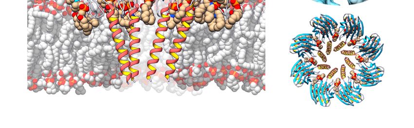
Toxins 2019, 11, 370 2 of 21
established between
Toxins 2019, both
11, x FOR PEERactors:
REVIEWprey are evolutionarily selected for venom resistance, which is achieved
2 of 21
through different mechanisms [1–3]; to work within the new evolutionary landscape, venoms must
adaptachieved
quickly to through different their
keep harming mechanisms
prey. How [1–3];
doto newwork withinappear
venoms the new in aevolutionary
species? One landscape,
hypothesis is
venoms must adapt quickly to keep harming their prey. How do new venoms appear in a species?
that a proto-toxin gene is first duplicated, and then one copy becomes expressed in the organism’s
One hypothesis is that a proto-toxin gene is first duplicated, and then one copy becomes expressed
venom gland. Additional amplification of the venom gland-expressed gene can generate variants
in the organism’s venom gland. Additional amplification of the venom gland-expressed gene can
that produce a toxic protein
generate variants product
that produce or compound.
a toxic protein product Dueortocompound.
this duplication–neofunctionalization
Due to this duplication–
mechanism, it is common to find venom toxins genetically
neofunctionalization mechanism, it is common to find venom toxins structured as genetically
multigene structured
families [2,4–6].
as
When
multigenevenoms are[2,4–6].
families delivered, they must interact with their prey at the molecular level in order to
cause cellular
Whendamage.
venoms are Venoms must
delivered, breach
they must multiple barriers
interact with to reach
their prey at thetheir targets,
molecular including
level in order skin,
to cause network
the intricate cellular damage. Venoms mustmatrix,
of the extracellular breach multiple barriers tothe
and, eventually, reach
celltheir targets, including
membrane. Deliveryskin,
systems
the intricate network of the extracellular matrix, and, eventually, the cell
like chelicerae, nematocysts, and other piercing structures have an initial harmful physical activity, membrane. Delivery
systemsthe
overtaking like chelicerae,
barriers andnematocysts,
bringing toxins and closer
other piercing
to their structures
final targethave an initial1).
[3] (Figure harmful
Withinphysical
the injected
activity, overtaking the barriers and bringing toxins closer to their final target [3] (Figure 1). Within
cocktail, metalloproteases, for example, are very effective in attacking the interstitial space between cells,
the injected cocktail, metalloproteases, for example, are very effective in attacking the interstitial
by digesting extracellular matrix components and facilitating the activity of other toxins on the cells.
space between cells, by digesting extracellular matrix components and facilitating the activity of other
Non-proteinaceous
toxins on the cells. components of the venom,
Non-proteinaceous like serotonin
components and histamine,
of the venom, produce
like serotonin fast and sharp
and histamine,
pain produce
and increasefast andvasodilatation.
sharp pain and As cells are defined
increase by the As
vasodilatation. plasma
cells membrane
are defined [7],by the
the first cellular
plasma
structures
membraneencountered
[7], the by firstthe venomous
cellular cocktail
structures are the transmembrane
encountered by the venomous andcocktail
peripheral are receptor
the
proteins as well as the
transmembrane andlipid membrane
peripheral itself.
receptor Therefore,
proteins as wellvenoms alsomembrane
as the lipid contain proteins that remodel
itself. Therefore,
venoms also contain proteins that remodel and directly attack the
and directly attack the lipid membrane, such as neurotoxins, phospholipases, and pore-forming lipid membrane, such astoxins
neurotoxins, phospholipases, and pore-forming
(PFTs), which finally destroy this key structure defining the cell. toxins (PFTs), which finally destroy this key
structure defining the cell.
Figure 1. Piercing
Figure delivery
1. Piercing systems
delivery systemscross
crossthe
the skin, thefirst
skin, the firstbarrier
barrier encountered
encountered by venom
by venom (A). Specific
(A). Specific
extra-cellular matrix
extra-cellular proteases,
matrix like
proteases, likemetalloproteases, digestscaffold
metalloproteases, digest scaffold proteins
proteins (B).(B). Non-proteinaceous
Non-proteinaceous
components,
components, like serotonin,
like serotonin, promotepromote vasodilatation
vasodilatation (C).plasma
(C). The The plasma membrane
membrane is thewidespread
is the most most
widespread
structure in naturestructure in nature aand,
and, therefore, therefore,
suitable targeta of
suitable
attacktarget of attacklike
by enzymes by phospholipases
enzymes like or
phospholipases
cytolysins or cytolysins
(D). Neurotoxins (D).with
interact Neurotoxins
membrane interact with membrane
receptors receptors
and channels, leading andtochannels,
imbalance in
leading to imbalance in ion distribution across the membrane (E). Some toxins develop their harmful
ion distribution across the membrane (E). Some toxins develop their harmful effects once they are
effects once they are internalized, blocking protein production or oxidative respiration in
internalized, blocking protein production or oxidative respiration in mitochondria (F). This image was
mitochondria (F). This image was created using Servier Medical Art free images database (SERVIER,
created using Servier Medical Art free images database (SERVIER, Paris, France).
Paris, France).
2. Pore-Forming Proteins (PFPs)
2. Pore-Forming Proteins (PFPs)
PFPs are the perfect example of toxins whose activity relies on the disruption of a lipid membrane.
PFPs are the perfect example of toxins whose activity relies on the disruption of a lipid
In general terms, they violate the standard classification of proteins—into water-soluble or membrane
membrane. In general terms, they violate the standard classification of proteins—into water-soluble
proteins—that can be found in any basic Biochemistry textbook. PFPs remain stably folded and soluble
in water but, upon interaction with the membrane of their target cells—and after recognition of a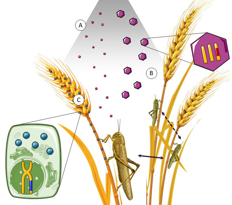
Toxins 2019, 11, 370                                                                                3 of 21

receptor that can be a sugar, a protein, or even a specific lipid—they polymerize into an oligomeric
transmembrane protein that makes a pore. We, and other authors, even speak of a molecular
metamorphosis transforming these toxins from water-soluble to transmembrane proteins, escaping the
aforementioned classification [8–10]. Attachment to the membrane increases the local concentration of
the toxin, reducing protein diffusion to a bidimensional system, and thus facilitating the oligomerization
that leads to pore formation. Depending on the toxin, pore size and permeability selectivity vary,
allowing the passage of species ranging in size from small ions to medium-size proteins. In most
cases, the final outcome of the formation of a pore is cell death by osmotic shock. Based on the protein
structure that forms the final pore, these proteins are classified either as α-PFPs, if the pore walls are
defined by α-helices, or β-PFPs, if the pore walls are β-sheets [11,12].
      PFPs attack the plasma membrane, the primordial structure that defines a cell. As stated, some
of them only need a lipidic receptor, like cholesterol [13] or sphingomyelin [14–18]. But others seem
to need protein receptors, such as the understudied latrotoxin macrocomplexes from black widow
spiders (Latrodectus spp.) [19]. Especially when targeting a particular lipid or lipidic composition, PFPs
are not specific enough. However, since they constitute a component of a complex cocktail (the venom),
they can be used to target a wide range of animals. These toxins act very quickly [10,12,14], so they are
used for both predatory and defensive purposes [1].
      In this Perspective, we focus on PFPs produced by two different groups of animals, which
constitute our major areas of research in the field. Cnidarians, given their privileged evolutionary
position and their wide-range spectrum of attack, and arachnids, whose powerful PFP collection is
usually aimed, with high specificity, at insects. Taking into account the above outlined possibilities on
toxin biotechnological applications, both groups of proteins display different interesting characteristics
to turn them into powerful weapons.

2.1. Pore-Forming Proteins in Cnidaria
     Cnidaria is an ancient clade of animals, whose genetic analysis is interesting from the evolutionary
and phylogenetic point of view because they also are the oldest lineage of venomous animals [2].
Cnidaria includes about 10,000 species, most of them living in saltwater. The Cnidaria phylum is
phylogenetically divided in two subdivisions: the class Anthozoa (anemones and corals) and the
subphylum Medusozoa, which includes the classes Cubozoa (jellyfishes) and Hydrozoa (hydras),
among others. Although they exhibit very simple anatomy, they are able to defend themselves with
high rate of success [2]. This animal group has clinical relevance from the point of view of envenomation,
and its hazard for humans fluctuates from non-hazardous to extremely dangerous like the Australian
box jellyfish (Chironex fleckeri) [20] or the Atlantic Portuguese Man o’ War (Physalia physalis) [21].
Most sea anemones, however, are harmless to humans or just cause skin burning after contact with
tentacles [22].
     Sea anemones inject venom into their prey through nematocysts, specialized penetrant structures
that discharge upon activation of cnidocytes [1,2]. Nematocysts are mostly located in the tentacles,
structures that cover a large area of anemones’ bodies and contain the venomous weapon needed not
only to protect the animal, but also to attack and entrap the prey. However, it is also possible to find
nematocysts surrounding the oral disc, in order to paralyze the prey, or in the ring surrounding the
base of the column, specialized for inter- and intraspecific competition. In fact, Cnidaria is the only
venomous lineage that lacks a centralized venom system [23,24].
     Like most animal venoms, sea anemones’ venoms can be largely classified as non-proteinaceous
toxins, neurotoxins, enzymes, or cytolysins [3,4,6]. Among the latter, actinoporins are a family of α-PFTs
produced by sea anemones as part of their venomous arsenal. They are small (around 20 kDa/175
amino acids), cysteineless, and basic (pI ≈ 9) [10,12,25]. It has been largely accepted that actinoporins
do not need a protein receptor to exert their toxicity, but instead require sphingomyelin as a specific
lipidic receptor [16,17,26–28]. Furthermore, cholesterol, though not indispensable, plays a key role
in their pore-forming mechanism [14,29–34], a mechanism still not fully understood, especially with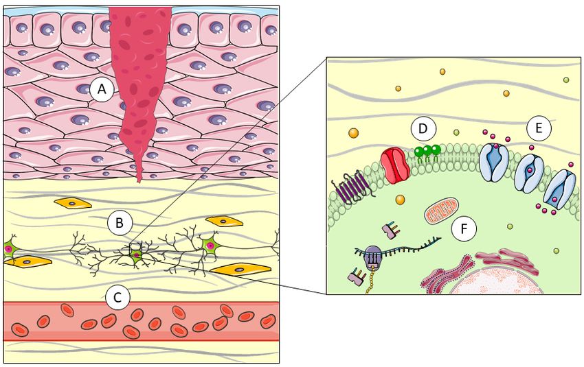
Toxins 2019, 11, 370 4 of 21
Toxins 2019, 11, x FOR PEER REVIEW 4 of 21
regardespecially
to the sequence of events
with regard to the during
sequence pore formation
of events and
during theformation
pore final stoichiometry
and the finalofstoichiometry
the pore [35–42].
Overall,of the pore [35–42].
actinoporins Overall,a actinoporins
represent simple and represent a simple
optimal model to and
studyoptimal model to study
the challenging the
biophysical
challenging biophysical transition from a water-soluble conformation
transition from a water-soluble conformation to an integral transmembrane state. to an integral transmembrane
Sostate.
far, actinoporins have been detected in at least 20 different species of anemones [10,12,25],
So far, actinoporins have been detected in at least 20 different species of anemones [10,12,25],
constituting multigenic families in practically all cases [6,43–45]. All of them show a remarkable
constituting multigenic families in practically all cases [6,43–45]. All of them show a remarkable
sequence identity that reaches values as high as 90% [12,25]. But despite this great structural similarity,
sequence identity that reaches values as high as 90% [12,25]. But despite this great structural
they also show notable
similarity, they alsofunctional
show notable differences
functional[14,46]. This[14,46].
differences identity
Thisofidentity
sequence is logically
of sequence manifested
is logically
in its three-dimensional structure [36,37,42,47–49].
manifested in its three-dimensional All studiedAll
structure [36,37,42,47–49]. actinoporins contain acontain
studied actinoporins core consisting
a core
of a stranded of a strandedwhich
consistingβ-sandwich is flanked
β-sandwich whichby two short
is flanked α-helixes.
by two This sandwich
short α-helixes. contains
This sandwich a motif
contains
that anchors
a motif the
that protein
anchors to
thethe membrane
protein to the and remains
membrane andvirtually
remains unchanged during the
virtually unchanged process
during the of
process of pore formation; in contrast, the N-terminal end, in association with the
pore formation; in contrast, the N-terminal end, in association with the N-terminal ends of adjacent N-terminal ends of
adjacent monomers, is the portion that undergoes a conformational change
monomers, is the portion that undergoes a conformational change and finally pierces through the and finally pierces
membranethrough the(Figure
[50] membrane
2). [50] (Figure 2).
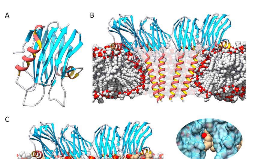
Figure 2. (A) Actinoporin monomers share a common fold: a stranded β-sandwich flanked by two
short α-helixes. With regard to pore structure, two models have been proposed: (B) a tetrameric
Figure 2. (A) Actinoporin monomers share a common fold: a stranded β-sandwich flanked by two
structure in which the lipid membrane adopts a toroidal shape around the pore walls, and (C) an
short α-helixes. With regard to pore structure, two models have been proposed: (B) a tetrameric
octameric lipid-protein
structure in which mixed
the lipidstructure
membranein which
adopts lipids (in tan
a toroidal color)
shape are the
around accommodated in (C)
pore walls, and pore-wall
an
fenestrations
octameric(see inserts onmixed
lipid-protein the right).
structure in which lipids (in tan color) are accommodated in pore-wall
fenestrations (see inserts on the right).
The mechanism by which actinoporins carry out their action has been studied in detail and, while
some aspectsTheremain controversial,
mechanism by which there is at least
actinoporins a general
carry consensus
out their about
action has beenthe stages
studied inleading to pore
detail and,
while[10,12,16,25,51,52].
formation some aspects remainAccording
controversial, there
to the is at least aaccepted
commonly general consensus about
model, the the stages units
monomeric leading
of the
protein bind to its target membrane, initiating an oligomerization process followed by separation and
elongation of the α-helix located at the N-terminal end of the actinoporin [53–55]. It is approximately
the first 30 amino acids that adopt a helical amphipathic structure, insert into the membrane, and,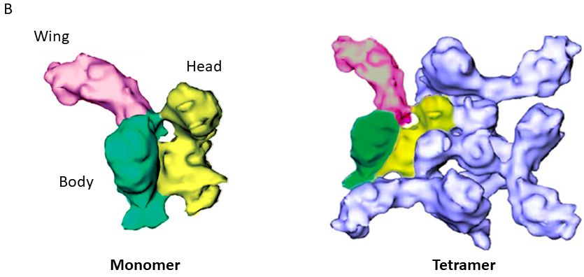
Toxins 2019, 11, 370                                                                                5 of 21

in association with other actinoporin molecules, form a pore in the lumen of which the polar heads of
some lipids would also participate (Figure 2) [23,36,38,42,50,56,57]. However, there are still aspects of
this mechanism to be elucidated: mainly, the order in which these stages occur [8,41,42,51,58], the need
for ‘pre-pore’ type intermediates [38,58], and, above all, the stoichiometry and composition of the final
functionally active structure [36,37,41,42,51].
      At least three different models have been proposed to explain the actinoporins’ transmembrane
pore. The first proposed model suggested the existence of a toroidal tetrameric protein-lipidic
structure [35,38,59]. However, a non-toroidal nonameric pore of fragaceatoxin C (FraC), produced by
the sea anemone Actinia fragacea, was proposed based on a detergent-containing crystalline structure [37].
The most detailed structural study carried out in the field is a crystalline lipid-containing octameric
pore, solved with atomic resolution [42]. In fact, none of these models is fully accepted and, in our
opinion, most probably all of them describe different aspects of the mechanism of pore formation and
cell lysis employed by actinoporins. The different conformations detected are just static images of a
dynamic process (Figure 2).

2.2. Pore-Forming Proteins in Arachnids
      Araneae (spiders) is the largest order within the Arachnida class, which also includes scorpions,
mites, and harvestmen. Most adult arachnids have eight legs and they also count with other mobile
appendages like pedipalps, which are adapted for a wide variety of functions from feeding to
reproduction, and chelicerae, involved in feeding and defense. In most venomous arachnid species
(Latrodectus spp. are just one of the few exceptions), chelicerae are only connected to glands where
venom is produced, as opposed to Cnidaria, where it is usually more homogeneously distributed
within their bodies. Production of the venom is mediated by a process called holocrine secretion,
in which the toxic compounds are produced in the cytosol of the gland cells and, then, these productive
cells disintegrate, dumping the cytosol content into the venom gland lumen.
      Most of natural spiders’ prey are insects. Consequently, their venoms have been selected during
evolution so that they immobilize and kill this particular kind of invertebrate. However, spider venom
can be also harmful for vertebrates, a feature most probably ‘developed’ as a protective weapon against
predation. Species from the Latrodectus genus, commonly known as black widow spiders, constitute a
group of around 40,000 different spider species. Their bite causes acute pain and severe secondary
conditions in humans, called ‘latrodectism’ [60], which involves a complex symptomatology from
nausea to body rigidity and widespread intense pain [61]. Because of the frequency of envenomation
events on humans, severity of clinical symptoms, and the frequent serious clinical consequences of
their bite, black widow spiders are classified as medically important [62].

2.2.1. Latrotoxins
      From the point of view of this Perspective, the most interesting group of toxic proteins from
the venom of species belonging to the Latrodectus genus are latrotoxins. They are high molecular
weight (110–140 kDa) and acidic proteins whose toxic activity relies on the formation of pores through
biological membranes [63–66]. The latrotoxin family includes 3 different subclasses based on their prey
specificity: vertebrates (LTX), crustaceans (LTC), or insects (LIT). LTX and LCT subclasses contain only
one member each, α-latrotoxin (α-LTX) and α-latrocrustaceatoxin (α-LCT), respectively. So far, five
latrotoxins have been identified as insect specific and, consequently, they are known as α, β, γ, δ and
ε-latroinsectotoxins (LITs) [67,68]. Therefore, the main difference between α-LTX and the different LITs
is prey selectivity. From the evolutional point of view, the presence of different LIT isoforms is related
to hunt specialization. Nevertheless, only α-LTX is specific for mammals, which can be associated with
the evolution of a defensive weapon against other attackers, like rodents.
      α-LTX is the best characterized member of the latrotoxin family and neurexins and latrophilins
have been identified as its potential protein receptors [19,63,64]. Although no specific information
about LIT receptors has been reported so far, there are orthologs of the above receptors in insects,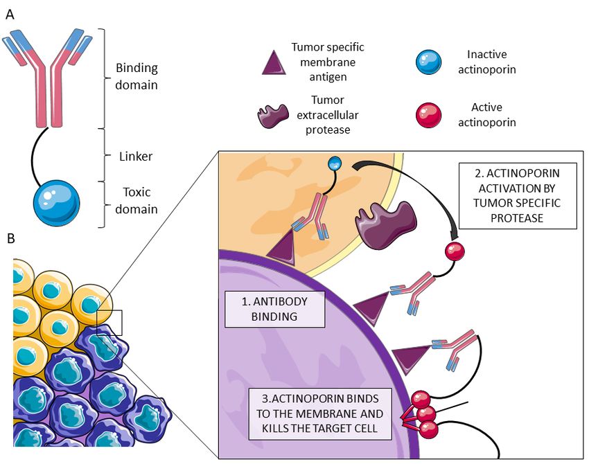
Toxins 2019, 11, 370 6 of 21
Toxins 2019, 11, x FOR PEER REVIEW 6 of 21
suggesting that these similar insect proteins could be involved in recognition. However, both α-LTX
α-LTX is the best characterized member of the latrotoxin family and neurexins and latrophilins
and LITs have been proven to produce pores not only in cells expressing these receptors, but also in
have been identified as its potential protein receptors [19,63,64]. Although no specific information
artificial bilayers [19,69]. Interestingly, although very little is known about the physiological activity of
about LIT receptors has been reported so far, there are orthologs of the above receptors in insects,
it seems tothat
δ-LIT,suggesting be these
receptor-independent, showing a lack of neuronal selectivity.
similar insect proteins could be involved in recognition. However, both α-LTX
LTXs are synthetized as large inactive
and LITs have been proven to produce pores not only inpolypeptide precursors whichthese
cells expressing undergo post-translational
receptors, but also in
processing on bilayers
artificial both their N- and
[19,69]. C-termini although
Interestingly, [63,67,69,70]. The is
very little mature
knownversions
about the show a modular
physiological structure
activity
with three well-differentiated
of δ-LIT, domains: the ‘wing’,
it seems to be receptor-independent, showingthea ‘body’, and theselectivity.
lack of neuronal ‘head’ [19] (Figure 3). The
LTXs areto
wing corresponds synthetized
a unique as large inactive
N-terminal polypeptide
domain which precursors
seems to be which undergo
involved post-translational
in receptor recognition,
processing on both their N- and C-termini [63,67,69,70].
while the C-terminal domain is composed by a high number of consecutive ankyrin The mature versions show a repeats
modular and
structure
comprises the with
bodythreeandwell-differentiated domains: the three-dimensional
the head. A low-resolution ‘wing’, the ‘body’, and the ‘head’
structure was[19] (Figure for
obtained
3). The wing corresponds to a unique N-terminal domain which seems to be involved in receptor
α-LTX by cryo-electron microscopy (cryo-EM). It appeared as a dimer in the absence of cations but,
recognition, while the C-terminal domain is composed by a high number of consecutive ankyrin
in the presence of Ca2+ or Mg2+ , ankyrin-repeats probably mediate protein-protein interactions that
repeats and comprises the body and the head. A low-resolution three-dimensional structure was
led to obtained
the formation of water-soluble homotetramers with a well-defined central channel. This type
for α-LTX by cryo-electron microscopy (cryo-EM). It appeared as a dimer in the absence of
of assembly
cations is known
but, in theaspresence
the ‘four-bladed
of Ca2+ orpropeller’ model [19] probably
Mg2+, ankyrin-repeats (Figure 3). In fact,protein-protein
mediate this four-bladed
structure, which is amphipathic, appears to be inserted into the membrane,
interactions that led to the formation of water-soluble homotetramers with a well-defined forming pores central
that could
also be detected
channel. Thisbytype
cryo-EM [19,63].
of assembly The reconstruction
is known as the ‘four-bladed published had
propeller’ very[19]
model low(Figure
resolution
3). In [19]
fact, but,
thisthe
even so, four-bladed
presencestructure,
of ‘windows’which(fenestrations)
is amphipathic, appears to be inserted
was observed in theinto
porethe membrane,
lumen. forming
The polar heads
of somepores that could also
phospholipids be detected
would presumablyby cryo-EM
show[19,63]. The reconstruction
up through published
those fenestrations, had very stability
providing low
resolution to
and selectivity [19]
thebut, even so,This
channel. the situation
presence of is ‘windows’
far from being(fenestrations)
solved, but wasitobserved
has also in the detected
been pore
lumen. The polar heads of some phospholipids would presumably show up through those
in other PFPs such as the actinoporin FraC [42] (Figure 2). These cryo-EM experiments were carried
fenestrations, providing stability and selectivity to the channel. This situation is far from being solved,
out, however, not only in the absence of the corresponding membrane protein receptors, but also
but it has also been detected in other PFPs such as the actinoporin FraC [42] (Figure 2). These cryo-
using EMpalmitoyl-oleyl-phosphatidylcholine (POPC) as the sole support. Plasma membranes also
experiments were carried out, however, not only in the absence of the corresponding membrane
contain large amounts of other key lipids for the physiological behavior
protein receptors, but also using palmitoyl-oleyl-phosphatidylcholine of a as
(POPC) biological membrane,
the sole support.
such as cholesterol
Plasma membranes(50%), phosphatidylserine
also contain large amounts (7%), sphingomyelin
of other key lipids for (4%), and phosphatidylinositols
the physiological behavior of a
(1%). biological
To date, the role of such
membrane, lipids asin the mechanism
cholesterol of action of LTXs(7%),
(50%), phosphatidylserine is still not fully known.
sphingomyelin (4%), and Thus,
phosphatidylinositols
although the authors of the (1%).
paperTo date, the rolethat
concluded of lipids in theitself
the toxin mechanism of action
was capable of of LTXs ispores
forming still not
in the
fully known.
membrane in the Thus,
absence although
of anythe authors
other of thecomponent
protein paper concluded that the toxin
of neuronal origin itself
it iswas
farcapable of
from proven
forming pores in the membrane in the absence of any other protein
that another component of spider venom, for example, is not needed to carry out this insertion.component of neuronal origin it
is far from proven that another component of spider venom, for example, is not needed to carry out
The structure-function relationship of these proteins is still mostly unknown at the molecular level.
this insertion. The structure-function relationship of these proteins is still mostly unknown at the
Furthermore, there is no three-dimensional structural information about LITs. However, α-LTX and
molecular level. Furthermore, there is no three-dimensional structural information about LITs.
LIT share the same
However, α-LTX genetic
and LIT domain
share thestructure; they domain
same genetic are around 50% identical,
structure; so it can
they are around 50%be assumed
identical, so that
they probably exhibit athat
it can be assumed similar fold.
they probably exhibit a similar fold.
Figure 3. Cont.Toxins 2019, 11, 370 7 of 21
Toxins 2019, 11, x FOR PEER REVIEW 7 of 21
Figure 3. (A)
Figure Latrotoxins
3. (A) Latrotoxinsare
areproduced
produced as as inactive
inactiveprecursor
precursor activated
activated uponupon proteolytic
proteolytic digestion
digestion in
in both
boththetheN-N-and
andC-termini. Thegenetic
C-termini. The genetic structure
structure comprises
comprises a unique
a unique N-terminal
N-terminal domaindomain
and a C-and a
C-terminal
terminal domain
domainrich
richin
in ankyrin repeats.Within
ankyrin repeats. Within a low-resolution
a low-resolution three-dimensional
three-dimensional of α- of
structure
structure
α-LTXLTXobtained
obtained byby
cryo-EM,
cryo-EM,three
three different domainscan
different domains canbebedifferentiated:
differentiated:
thethe wing
wing (pink),
(pink), the body
the body
(green),
(green), and theand head
the head (yellow).
(yellow). (B)(B)The
Thefinal
finalpore
pore is
is composed
composed by byfour α-LTX
four α-LTX monomers,
monomers, combined
combined
incalled
in a so a so called ‘four-bladed
‘four-bladed propeller’.The
propeller’. Thehead
head and
and the
the body
bodyare areresponsible
responsibleforfor
membrane
membrane binding
binding
and pore formation, while the wing seems to be implicated in receptor recognition.
and pore formation, while the wing seems to be implicated in receptor recognition. Reproduced Reproduced fromfrom
Ushkaryov, Y.A., Volynski, K.E., Ashton, A.C., The multiple actions of black widow spider toxins and
Ushkaryov, Y.A., Volynski, K.E., Ashton, A.C., The multiple actions of black widow spider toxins and
their selective use in neurosecretion studies. Toxicon 2004, Elsevier.
their selective use in neurosecretion studies. Toxicon 2004, Elsevier.
The physiological effect observed regarding the toxicity of LTXs or LITs in insect tissues includes
The physiological effect observed regarding the toxicity of LTXs or LITs in insect tissues includes
the increase in the frequency of glutamatergic and GABA-ergic potential at the neuromuscular
the increase in the frequency of glutamatergic and GABA-ergic potential at the neuromuscular junctions
junctions and the asynchronous release of these neurotransmitters [67,69]. The cholinergic sensory
and the asynchronous
nervous system of therelease
insectsof isthese
also neurotransmitters [67,69].
affected. All these events seemThe
to cholinergic sensory
be related with nervous
the major
system molecular event of LTX toxicity: the formation of cation selective pores in membranes. Thus, in theevent
of the insects is also affected. All these events seem to be related with the major molecular
of LTX toxicity: the
presynaptic formation
space, of cation
pore formation selective
leads to Ca2+pores in membranes.
fluctuations, promoting Thus, in the
massive presynaptic space,
neurotransmitter
release. However,
pore formation leads toitCa 2+
is still necessary topromoting
fluctuations, study LITsmassive
in deeper detail in order torelease.
neurotransmitter understand their it is
However,
toxicity and
still necessary to structure-function
study LITs in deeper relationships. Actually,
detail in order all the aforementioned
to understand LITs
their toxicity have
and been tested
structure-function
for toxicityActually,
relationships. against Galleria
all themellonella larvae [71], LITs
aforementioned a model
have system
beenwhere
testedthey
forshowed
toxicityvery different
against Galleria
LD50 values (mg of toxin necessaries to kill 50% of the tested animals expressed per kg of body
mellonella larvae [71], a model system where they showed very different LD50 values (mg of toxin
weight).
necessaries to kill 50% of the tested animals expressed per kg of body weight).
2.2.2. Latrodectins
2.2.2. Latrodectins
Low molecular weight proteins (originally called LMW, or black widow low molecular weight
Low
venom molecular
components) weight proteins
of around (originally
70 amino called
acids long andLMW, or black
with a high widow
content low molecular
of disulfide bridges haveweight
venom components)
also been detected of within
aroundthe 70venomous
amino acids long
black and with
widow spidera high content
cocktail, of disulfide
and even isolated bridges
in smallhave
also been detected
quantities. withinthese
Nowadays, the venomous
proteins are black
knownwidow spider(Ltds)
as latrodectins cocktail,
[72]. and
Theireven
naturalisolated
functioninissmall
quantities.
not yetNowadays,
known, but these the fewproteins are results
available knownsuggest
as latrodectins
that they (Ltds) [72]. Their
are essential natural function
for increasing the
neurotoxic capacity of LTXs, most likely by cooperating with them and increasing
is not yet known, but the few available results suggest that they are essential for increasing their affinity for the
the membrane. Paradoxically, their participation seems to be also key in diminishing
neurotoxic capacity of LTXs, most likely by cooperating with them and increasing their affinity for the the specificity
of this interaction, thus facilitating an insecticidal activity of α-LTX [72], a toxicity which, in the
membrane. Paradoxically, their participation seems to be also key in diminishing the specificity of this
absence of Ltds, seems to be only restricted to vertebrates. The association between LTXs and Ltds is
interaction, thus facilitating an insecticidal activity of α-LTX [72], a toxicity which, in the absence of
presumed to be crucial, given that it seems practically impossible to purify LTXs to homogeneity by
Ltds, conventional
seems to be only restricted to vertebrates. The association between LTXs and Ltds is presumed to
methods [73–75], as they seem to be always contaminated with Ltds. Some authors even
be crucial, given
consider them that it seems
as mere practically
subunits of what impossible to purifymacromolecular
is called the latrotoxin LTXs to homogeneity
complex [74], bygiven
conventional
that,
methods [73–75],they
separately, as they
do notseem to be
appear toalways
be toxiccontaminated
neither againstwith Ltds.
insects Some authors
or vertebrates evenAsconsider
[75–78]. for theirthem
as mere subunits
structure, of what
apart is called
from their the latrotoxin
sequences, there ismacromolecular complexcharacterization,
only one spectroscopic [74], given that,by separately,
means of they
circular dichroism, which suggests that they should have a high α-helix content
do not appear to be toxic neither against insects or vertebrates [75–78]. As for their structure, [79]. Thus, althoughapart
from they
theirare not proven
sequences, to be
there PFPs,one
is only they could be determinant
spectroscopic for the final
characterization, pore formation
by means of circularof dichroism,
LTXs,
which suggests that they should have a high α-helix content [79]. Thus, although they are not proven
to be PFPs, they could be determinant for the final pore formation of LTXs, forming a macromolecular
complex, and their presence should be taken into account when designing biotechnological application
of LTXs.Toxins 2019, 11, 370 8 of 21
3. Biotechnological Applications of PFPs
The obvious benefit of studying venoms in detail is finding the means to block their deleterious
effects. Although envenomation is a neglected public health problem in many of developed
countries [80], it frequently causes intense pain and, when complicated symptoms occur, can even lead
to death of the affected individual. This Perspective, however, focuses on the potential of some venom
components as biotechnological tools.
The study of venoms can then open the gate to not-so-obvious venues. For example, detailed
knowledge about the mechanism of action of many toxins offers the possibility of using them for at
least three different approaches: detecting, inactivating, or modulating different cellular or metabolic
pathways. As mentioned above, toxins co-evolve with their targets. During this intense competition,
toxic compounds become highly specific to their target. Such a great specificity can be used to fight
different pests with the employment of the corresponding venomous natural product. In addition,
venom can interfere with the immune system regulating signaling pathways, tuning cytokine secretion
or promoting cell migration. Understanding how they work can put toxins on the radar of new
immunotherapies with venom or venom-derived products [61,62,81,82].
Given the high specificity and binding affinity of some toxins, they can be also used as molecular
probes, after the appropriate modifications such as conjugation to fluorophores. In fact, one of the most
successful and best systems for cellular in situ detection of sphingomyelin and cholesterol-rich domains
is based on the employment of modified versions of actinoporins, PFTs from sea anemones [83,84].
On the other hand, if the target cellular components or metabolic pathways are dysregulated
during a disease, having a molecule with high binding affinity is a perfect starting point to make the
appropriate modifications and modulate the target activity, regulating its biological function. Our
present knowledge allows molecular mimicry of natural products which appear promising in order to
find new therapeutic treatments.
Another possibility would be providing proteinaceous toxins with the means to change their
specificity without losing their lethal properties, as in the case of immunotoxins (IMTXs). This approach
takes advantage of toxins’ highly specific and potentially deadly activity, but it drives it toward aberrant
cells like cancer, metastatic, or cancer stem cells, for example. IMTXs are chimeric constructions built
from a target domain, which recognizes the harmful cell, and a toxic domain, which kills it [81,85–91].
3.1. The Biotechnological Potential of Cnidaria PFTs as IMTXs
Immunotoxins are hybrid artificial molecules in which the killer action of a toxin is directed to a
target cell through a binding domain (Figure 4). The binding domain can be a monoclonal antibody or,
in order to improve the penetration capacity of the construct, a smaller engineered version such as the
single-chain variable fraction of the selected antibody. Regarding the toxic moiety, most IMTXs use
toxic proteins which, acting intracellularly, lead to cell death by different means. The translocation of
this IMTX is then necessary to achieve the internalization of this toxin moiety into the cytosol. This
approach is especially successful against hematological tumors [92]. However, IMTXs have trouble
attacking solid tumors due to difficult penetration in tumor masses.
One of the advantages of using PFPs as the toxic moiety of IMTXs be is that they do not need
internalization to exert their toxic activity. Some examples can be found in recent scientific literature,
which use actinoporins [39], melittin peptide from bee venom [93], or the N-terminal domain of the
human perforin [94]. Since PFPs increase membrane permeabilization, in addition to their lethal
membrane altering properties, they can also facilitate the action of regular chemotherapies by facilitating
the entrance to the cytosol [95].
The first IMTX built with an actinoporin was developed by Avila et al. [96] conjugating a
monoclonal antibody recognizing IOR-T6, a specific antigen expressed on the surface of immature
T-lymphocytes, and a hemolytic toxin from Stichodacthyla helianthus. Later, the same group developed
a new chimera with a monoclonal antibody against carcinoembryonic antigen (CEA) [97]. One of the
latest approximations was linking, again, an actinoporin from S. helianthus with a monoclonal antibodyToxins 2019, 11, 370 9 of 21
Toxins 2019, 11, x FOR PEER REVIEW 9 of 21
recognizing a colorectal cancer-associated antigen (IOR-C2) [98]. Although the chimeras recognized
lymphocytes, and a hemolytic toxin from Stichodacthyla helianthus. Later, the same group developed
the corresponding antigens preferentially through the monoclonal antibody fraction, the non-specific
a new chimera with a monoclonal antibody against carcinoembryonic antigen (CEA) [97]. One of the
toxicity against cell lines not expressing the targeted antigen was still relatively high. Thus, the greater
latest approximations was linking, again, an actinoporin from S. helianthus with a monoclonal
advantage of these toxins (attacking
antibody recognizing a colorectalthe membrane, which
cancer-associated antigenis (IOR-C2)
the most[98].
widespread
Although target in the living
the chimeras
cells), isrecognized
also theirthe greatest weakness. Further research is required in order to eliminate
corresponding antigens preferentially through the monoclonal antibody fraction, the the off-target
non-specific toxicity against
effects of the actinoporin-based chimeras. cell lines not expressing the targeted antigen was still relatively high.
Thus, the greater
In addition, advantage
once bound, of these toxins
actinoporins need(attacking
to diffusethewithin
membrane, which is the in
the membrane most widespread
order to oligomerize
target in the living cells), is also their greatest weakness. Further research is required in order to
and form a pore. Being attached to an antibody recognizing a surface antigen is probably an obstacle for
eliminate the off-target effects of the actinoporin-based chimeras.
the final poreInformation.
addition, onceHowever,
bound,actinoporins
actinoporins are needvery well studied
to diffuse within from the structure-function
the membrane in order to point
of view, oligomerize
allowing protein engineering to overcome these hurdles. Several research
and form a pore. Being attached to an antibody recognizing a surface antigen is probablystudies delve into the
details ofan
theobstacle
protein-lipid
for theinteractions [10,15,30,31,33,42,99–103],
final pore formation. However, actinoporins makingarepossible
very well thestudied
application
from ofthedifferent
strategiesstructure-function
directed to improve point of view,
toxin allowing protein
specificity: engineering
for example, to overcome
protecting these region
the toxin hurdles.responsible
Several for
research studies delve into the details of the protein-lipid interactions [10,15,30,31,33,42,99–103],
membrane binding or blocking the N-terminal domain directly implicated in pore formation. Actinoporins
making possible the application of different strategies directed to improve toxin specificity: for
can be engineered to protect their key regions with polypeptide domains, which can later be released by
example, protecting the toxin region responsible for membrane binding or blocking the N-terminal
tumor specific proteases. Actinoporins
domain directly implicated in pore areformation.
quite resistant to protease
Actinoporins can beactivity and to
engineered areprotect
cysteineless.
their keyThis last
feature makes
regionsthemwith suitable for site
polypeptide directed
domains, mutagenesis
which can later followed
be releasedby by
modification
tumor specific through conjugation.
proteases.
Actinoporins are quite
Matrix metalloproteases (MMPs)resistant to protease activity
are extracellular and are
proteases thatcysteineless.
remodel the This last feature makes
extracellular matrixthem
and whose
expressionsuitable for site in
is increased directed
the cellsmutagenesis
surrounding followed
tumors by[104],
modification
along withthrough
otherconjugation.
proteinasesMatrix like catepsin
metalloproteases (MMPs) are extracellular proteases that remodel the extracellular matrix and whose
B [105] or furin [106] (Figure 4). High expression of extracellular matrix proteases is correlated with the
expression is increased in the cells surrounding tumors [104], along with other proteinases like
invasioncatepsin
capacityB of theortumor.
[105] Protective
furin [106] (Figuredomains
4). High could be linked
expression to the protein
of extracellular matrixby sites suitable
proteases is for
tumor-specific protease
correlated with thedigestion. Conjugating
invasion capacity the specificity
of the tumor. Protectiveof the antigen
domains could recognition
be linked to the provided
protein by the
monoclonal antibody
by sites suitablemoiety and the proteinase-activated
for tumor-specific toxic activity
protease digestion. Conjugating thecould improve
specificity theantigen
of the specificity of
recognition
cytolysin-based provided
IMTXs by the monoclonal antibody moiety and the proteinase-activated toxic activity
[95,107,108].
could improve the specificity of cytolysin-based IMTXs [95,107,108].
(A) Immunotoxins
Figure 4.Figure 4. (A) Immunotoxins(IMTXs)
(IMTXs)areare
chimeric
chimeric molecules composed
molecules composed of aof a monoclonal
monoclonal antibodyantibody
that that
recognizes
recognizes the malign
the malign cells cells
and and a toxin
a toxin moietythat
moiety that kills
kills the
thetargeted
targetedcells. (B) In
cells. (B)order to improve
In order the
to improve the
specificity
specificity of actinoporin
of actinoporin IMTXs,IMTXs, protease-activated variants
protease-activated variants have
havebeen designed.
been OnceOnce
designed. the chimeric
the chimeric
molecule binds specifically to malign cells (in purple) by recognition of a membrane motif missing
in the healthy ones (in orange) (1), the toxic moiety is activated by a tumor specific protease (2). The
activated actinoporin is then able to bind the membrane, oligomerize with other monomers, and form a
pore, killing the targeted cell by an osmotic shock (3). This image was created using Servier Medical
Art free images database (SERVIER, Paris, France).Toxins 2019, 11, 370 10 of 21
3.2. The Biotechnological Potential of Arachnid PFPs as Bioinsecticides
Although the structural and the associated functional knowledge about LITs is restricted and
further work has to be done to understand the activity of these toxins, the toxicity information
detailed above and the structural characterization of α-LTX make LITs a suitable starting point to
design bioinsecticides.
The world population has been increasing exponentially since 1900. By 2050, it is estimated
to reach 9 billion, according to the United Nations. Obviously, this increase comes together with a
greater need for sustenance. However, crop losses due to plagues are a major problem, worsened
every day by climate change [109]. For decades, approximately since the 1940s, pest control relied
on the use of dichlorodiphenyltrichloroethane (DDT). It was cheap, easy to produce and deliver,
and effective, but its poor selectivity and high toxicity forced many countries to ban it. Within this
context, bioinsecticides have emerged as a promising pest control tool [81], based on the use of natural
toxins. Spiders, scorpions, and many other venomous animals have insects as preferred prey. Therefore,
their toxic cocktails include compounds specifically directed to insects, which appear highly valuable
to fight pests [110,111]. Thus, these biopesticide approaches would include two different strategies:
the development of suitable tools for fumigation or the construction of transgenic variants which
constitutively produce protective toxins to avoid damage caused by pests. Although there is still
skepticism about the consumption of genetically modified organisms, no adverse effects have been
observed so far [112], and proper legislation about their commercialization should be introduced in the
near future to take advantage of their proven beneficial properties [113].
The ideal bioinsecticide should be stable in conditions of extreme humidity and temperature, active
topically or orally, and should be rapidly lethal to pest insects, but innocuous to humans and other
non-pest species like bees, fishes, or birds. In addition, it should not be bioaccumulated or produce
harmful secondary or degradation products [111]. A priori, insecticidal toxins from spiders cover
almost every one of these requirements, even regarding degradation, since proteins are completely
biodegradable into innocuous amino acids.
The most successful example of bioinsecticide is Bacillus thuringiensis insecticidal three-domain
Cry toxin (3d-Cry) [114], which is in fact a PFP. When a susceptible larva ingests the toxin, it gets
activated in the gut, where it binds and leads to pore formation, provoking cell death and compromising
larvae viability. The major issues encountered by this approximation is the UV sensitivity of the
toxin that reduces its availability for spray dispersion and the emergence of insect resistance [115,116].
As introduced at the beginning of this article, venom effectivity/resistance is a constant race for survival
and without insecticidal innovation pest would end up developing resistance.
The toxicity of LTXs, as well as many other components of the spider venom, is exerted at the level
of neuromuscular junction. For spiders, reaching the target cells is easier through piercing chelicerae,
but applying the toxins alone as biopesticides requires identification of an easier administration/delivery.
There are two possible administration routes that imply different technological approaches: intoxication
of the insect through ingestion or reaching the neuromuscular junctions through the spiracles connecting
to the tracheas that transport the gases and connect with tissues for cellular respiration. Some spider
toxins (like disulfide-rich knottin peptides, known as inhibitor-cystine knot (ICK)), have been already
demonstrated to be potent, stable and orally active for insects [111,117–119]. If LITs were orally active,
it would be possible to deliver them in aerosol, which would be the easiest way to commercialize
them. However, big proteins are more prone to degradation, and the resistance to extreme humidity
or temperature conditions is not guaranteed. Designing transgenic crops constitutively producing
the compound is another option that has been successful for other toxins like the aforementioned
B. thuringiensis toxins [120], which have been proven to be safe for human consumption. In addition,
the toxins produced by the transgenic species only affect insects consuming the crop, avoiding the
collateral damage to beneficial insects like bees and other critical species for the pollination maintenance.
Since LTXs are PFPs and they increase the permeability of the target cells, their application in combinationToxins 2019, 11, 370 11 of 21
with other bioinsecticide components whose activity is developed intracellularly [81,111] can increase
the bioinsecticide activity synergistically.
The greatest inconvenience for insecticide application of LTX family raised by different authors is
the high molecular mass of this class of toxins, and therefore, the difficulty of cloning and producing
them in heterologous systems [111,121]. However, successful experience cloning and expressing the
mature form of δ-LIT in bacteria has been already accomplished [69]. Moreover, according to the
preliminary data obtained through the structural information for α-LTX pore, the whole protein is not
necessary to promote pore formation. Given the high identity between α-LTX and LITs and assuming
that the three-dimensional structure would be similar to that of α-LTX, it might be possible to eliminate
the ‘wing’ domain, which is supposed to be implicated in receptor recognition. Although more studies
need to be done on the selectivity of LITs, as well as the structural domain implicated in receptor
recognition, restricting the cloned sequence to the minimum necessary to exert lytic activity would
reduce the specificity of the attack, but it might increase the chances of success. Other approaches to
increase the oral activity of toxins is conjugating them with carrier proteins recognized by insect midgut
transporters like the mannose-specific lectin agglutinin from Galanthus nivalis. Upon ingestion by
insects, it is recognized by a receptor located in the midgut cells. It is receptor-mediated endocytosed,
followed by transportation and accumulation in the hemolymph [122].
Another interesting option is the use of baculoviruses as pest control agents. Natural baculoviruses
have been already used for effective pest control. However, genetic engineering amplifies the
possibilities of this tool, making them an even better alternative [123–126]. Natural baculoviruses kill
pest by themselves within weeks, while recombinant baculoviruses including toxins would shorten
the treatment time. In addition, this system has the advantage of being stable by itself, while also
providing specificity for the host and the possibility of treatment maintenance due to the potential
transmission of the virus both horizontally (among insects of the same species in the same development
state) and vertically (from adult insects to their progeny) [125] (Figure 5). Interestingly, it has been
very recently reported how a transgenic fungus expressing a potent insect-selective spider toxin can be
effective against insecticidal resistant mosquitos [127].
4. Omic Techniques in the Discovery of New Potentially Useful PFPs
One of the most famous examples of toxin-inspired pharmacological drugs is captopril, a drug for
hypertension treatment that inhibits angiotensin-converting enzyme (ACE). Its discovery was derived
from studies with small toxic peptides from the snake Bothrops jararaca [128] and it was approved by the
FDA in 1981. Research in this field is now booming as a consequence of the technological development
of powerful techniques for identification of new bioactive compounds. Identification of new toxins is
associated with a great variety of potential applications.
‘Omic’ techniques are instrumental and informatis-dependent tools that have reached new levels
of performance within the past decade. As stated at the beginning of this article, venomous animals are
usually small and venom yield production is low. These facts were major obstacles in venom research.
The purification of bioactive compounds rendered small yields, such that only the more abundant
compounds were detected and used in research, even if low-abundance molecules had a critical role in
toxicity. Many of these new ‘omic’ approaches have reduced the amount of starting material needed
for the analysis, making it affordable for venom studies [129–132]. In depth analysis of venoms from
long-neglected organisms using these techniques not only increases the knowledge about venom
composition, phylogeny, and evolution, but also allows the identification of underrepresented toxins
which may exert unique biological properties and hold potential applications. These techniques are
excellent for high throughput screening of new bioactive compounds. Venoms are a quite fascinating
material from the evolutionary point of view, and genomic, transcriptomic, and proteomic analysis
reveal quite interesting features about the evolution of venomous species [132,133].You can also read