Molecular underpinnings of ssDNA specificity by Rep HUH-endonucleases and implications for HUH-tag multiplexing and engineering
←
→
Page content transcription
If your browser does not render page correctly, please read the page content below

1046–1064 Nucleic Acids Research, 2021, Vol. 49, No. 2                                                             Published online 7 January 2021
doi: 10.1093/nar/gkaa1248

Molecular underpinnings of ssDNA specificity by Rep
HUH-endonucleases and implications for HUH-tag
multiplexing and engineering
Kassidy J. Tompkins1 , Mo Houtti2 , Lauren A. Litzau1 , Eric J. Aird1 , Blake A. Everett1 ,
Andrew T. Nelson1 , Leland Pornschloegl1 , Lidia K. Limón-Swanson1 , Robert L. Evans, III1 ,
Karen Evans1 , Ke Shi1 , Hideki Aihara1 and Wendy R. Gordon1,*
1
 Department of Biochemistry, Molecular Biology, and Biophysics, University of Minnesota Twin Cities, Minneapolis,
MN 55455, USA and 2 Department of Computer Science and Engineering, University of Minnesota Twin Cities,

                                                                                                                                                              Downloaded from https://academic.oup.com/nar/article/49/2/1046/6067397 by guest on 25 July 2021
Minneapolis, MN 55455, USA

Received October 05, 2020; Revised December 08, 2020; Editorial Decision December 09, 2020; Accepted December 14, 2020

ABSTRACT                                                                        tegration into host genomes (1–3). At the heart of DNA
                                                                                processing of all HUH-endonucleases is a structurally de-
Replication initiator proteins (Reps) from the HUH-                             fined catalytic nickase domain that first recognizes a specific
endonuclease superfamily process specific single-                               sequence/structure of DNA; nicks ssDNA at a ‘nic site’ to
stranded DNA (ssDNA) sequences to initiate rolling                              yield a sequestered 5 end that remains covalently bound to
circle/hairpin replication in viruses, such as crop                             the HUH endonuclease and a free 3 OH that can be used
ravaging geminiviruses and human disease caus-                                  as a primer for DNA replication; and finally, facilitates a
ing parvoviruses. In biotechnology contexts, Reps                               strand transfer reaction to resolve the covalent intermediate
are the basis for HUH-tag bioconjugation and a criti-                           (1). The two major classes of HUH-endonucleases are repli-
cal adeno-associated virus genome integration tool.                             cation initiator proteins (Reps) involved in RCR and RHR
We solved the first co-crystal structures of Reps                               and relaxases involved in bacterial conjugation of plasmids,
complexed to ssDNA, revealing a key motif for con-                              although HUH-endonucleases are also involved in DNA
                                                                                transposition (1).
ferring sequence specificity and for anchoring a
                                                                                   The covalent phosphotyrosine intermediate has recently
bent DNA architecture. In combination, we devel-                                been exploited for biotechnology applications. ‘HUH-tag’
oped a deep sequencing cleavage assay, termed                                   fusion proteins are emerging as a versatile bioconjuga-
HUH-seq, to interrogate subtleties in Rep specificity                           tion platform to covalently link proteins to DNA, com-
and demonstrate how differences can be exploited                                bining the diverse functionality of proteins with the pro-
for multiplexed HUH-tagging. Together, our insights                             grammability of DNA (4). HUH-tag applications have
allowed engineering of only four amino acids in a                               permeated into technologies such as DNA origami scaf-
Rep chimera to predictably alter sequence speci-                                folded protein assembly (5–8), receptor-specific cell tar-
ficity. These results have important implications for                           geting by adeno-associated virus (9), aptamer-based sand-
modulating viral infections, developing Rep-based                               wich detection (10), directed nanoparticle drug-delivery via
genomic integration tools, and enabling massively                               DNA aptamers (11), and CRISPR–Cas9 genome engineer-
                                                                                ing (12,13), mainly due to their ability to form robust co-
parallel HUH-tag barcoding and bioconjugation ap-
                                                                                valent adducts under physiologic conditions. Rather than
plications.                                                                     relying on expensive nucleic acid modifications such as the
                                                                                SNAP-tag (14), CLIP-tag (15) and HALO-tag (16) systems,
INTRODUCTION                                                                    HUH-tags rely on an inherent ssDNA binding moiety that
HUH-endonucleases, so named for a conserved histidine–                          promotes the catalysis of a transesterification reaction re-
hydrophobic residue–histidine (HUH) motif, are diverse en-                      sulting in a stable phosphotyrosine adduct (1).
zymes utilizing common single-stranded DNA (ssDNA)                                 Understanding the molecular basis of DNA recogni-
processing mechanisms that break and join DNA to facil-                         tion by HUH-endonucleases could provide much needed
itate fundamental biological processes such as rolling cir-                     solutions for bacterial antibiotic resistance resulting from
cle replication (RCR), rolling hairpin replication (RHR),                       HUH-endonuclease mediated horizontal gene transfer (17),
bacterial conjugation, DNA transposition, and DNA in-                           as well as in the prevention or treatment of HUH-

* To   whom correspondence should be addressed. Tel: +1 612 301 1196; Email: wrgordon@umn.edu


C The Author(s) 2021. Published by Oxford University Press on behalf of Nucleic Acids Research.
This is an Open Access article distributed under the terms of the Creative Commons Attribution License (http://creativecommons.org/licenses/by/4.0/), which
permits unrestricted reuse, distribution, and reproduction in any medium, provided the original work is properly cited.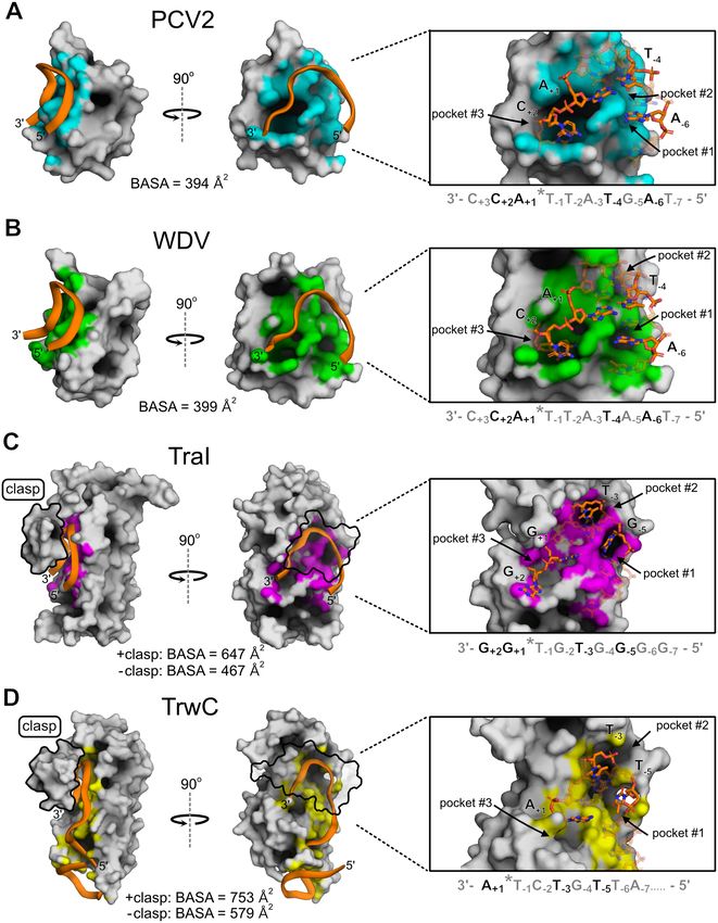
Nucleic Acids Research, 2021, Vol. 49, No. 2 1047
endonuclease mediated viral infections, such as geminivirus Sigma Aldrich), and then grown for 16 hours at 18◦ C. Col-
infections of plants that ravage the agricultural crop indus- lected cell pellets were resuspended in 10 ml of lysis buffer
try (18,19) and parvovirus B19 infections of humans (20) (50 mM Tris pH 7.5, 250 mM NaCl, 1 mM EDTA, cOm-
that are associated with a range of autoimmune diseases plete protease inhibitor tablet (Pierce) and pulse sonicated
(21,22). Moreover, the ability to rationally engineer HUH- for several one minute rounds. The suspension was cen-
endonucleases to recognize a desired DNA sequence has trifuged at 24 000 × g for 25 min, and supernatants were
huge potential in genome engineering (23) and DNA deliv- batch bound for 1 h with 2 ml HisPure Ni-NTA agarose
ery applications as well as in expanding the multiplexibility beads (ThermoFisher) and equilibrated with wash buffer
of HUH-tagging to meet the demand of the recent explo- (50 mM Tris pH 7.5, 250 mM NaCl, 1 mM EDTA, 30 mM
sion of DNA-barcoding applications (24–27). imidazole). After lysate cleared the gravity column, beads
However, while several structures of relaxase HUH- were washed with 30 ml wash buffer, and proteins were
endonucleases in complex with their cognate DNA target eluted from gravity columns with elution buffer (50 mM Tris
sequences have been reported (17,28–30), there are no struc- pH 7.5, 150 mM NaCl, 1 mM EDTA, 250 mM imidazole).
tures of viral Rep HUH-endonucleases in complex with ss- Protein was further purified and buffer exchanged into 50
Downloaded from https://academic.oup.com/nar/article/49/2/1046/6067397 by guest on 25 July 2021
DNA comprising the target sequence at the origin of repli- mM Tris pH 7.5, 150 mM NaCl, 1 mM EDTA using the
cation (ori). Despite structurally superimposable active sites ENrich SEC70 (Bio-Rad) size exclusion column. Aliquots
and a common overall core structure (31), there are several were stored at −20◦ C and −80◦ C at 30 M. SUMO-cleaved
structural elements of the larger relaxase proteins that do recombinant PCV2Y96F and WDVY106F stocks for crystal
not exist in Reps, such as extensions of the C-terminus and screening were prepared in a similar manner as above, how-
internal loops with respect to Reps. These structures form ever Ni-NTA fractions were dialyzed into 50 mM Tris pH
extensive contacts with the target DNA, thus underscoring 7.5, 300 mM NaCl, 1 mM EDTA with the addition of 1
potential differences in DNA recognition mechanisms be- mM DTT and SUMO-cleaving protease ULP-1 at 5 U per
tween Reps and relaxases (32). 1 l of E. coli overnight at 4◦ C. Dialyzed samples were batch
In this study, we determined the structural basis for ss- bound a second time with Ni-NTA beads and were flowed
DNA recognition by viral Rep HUH-endonucleases by through a gravity column to remove cleaved His6-SUMO
solving two Rep-ssDNA co-crystal structures and identi- and His6-ULP-1. Protein was concentrated with spin con-
fied a ssDNA ‘bridging’ motif largely responsible for DNA centrators (Amicon Ultra-15 Centrifugal Filter Unit, 3 kDa
recognition. This bridging motif recognizes specific bases of cut-off) to 16 mg/ml.
bent ssDNA located on either side of the nic site using sur-
face pockets. To further interrogate the ssDNA specificity of
Crystallization, data collection and processing
Reps, we developed HUH-seq, a high-throughput, next gen-
eration sequencing (NGS)-based DNA cleavage assay that An 8-mer oligonucleotide (5 -dAATATTAC-3 ) from part
we used to define ssDNA recognition profiles of a panel of of the geminivirus origin of replication sequence was re-
ten Reps using a ssDNA library containing 16,384 differ- constituted in ddH2 O at 10 mM and mixed with recombi-
ent target sequences. Despite the high similarity of cognate nant WDVY106F . We used Rigaku’s CrystalMation system
nonanucleotide ori sequences and the promiscuous nature to perform a broad, oil-immersion, sitting drop screen of the
of Rep ssDNA recognition we noticed previously (4) and protein–DNA mixture in the presence of either magnesium
further defined in this study, HUH-seq analysis surprisingly or manganese. Crystals were achieved using 8 mg/ml pro-
revealed many examples of orthogonal adduct formation tein solution containing 1.1-fold 8-mer and 5 mM MnCl2
between Reps from different viral families with little or no with a well solution of 12% (w/v) PEG 8000 precipitating
cross-reactivity. Finally, we rationally engineered a chimeric agent, 0.2 mM zinc acetate, and 0.1 M sodium cacodylate
Rep by swapping a few amino acids of the ssDNA ‘bridg- at pH 6.5. The crystals belong to space group P41 21 2 with
ing’ motif of one Rep into the backbone of a related Rep, unit cell dimensions of a = b = 50.63 Å, c = 241.98 Å. Addi-
predictably modulating ssDNA sequence specificity. tion of any cryoprotectant to these crystals resulted in poor
diffraction; the crystals seemed to collapse upon vitrifica-
tion. Our solution to this issue was to collect datasets us-
MATERIALS AND METHODS ing an in-house, X-ray diffractometer (Rigaku Micromax-
007 Rotating Anode, Rigaku Saturn 944 CCD Detector) at
Molecular cloning, protein expression and purification
room temperature. Radiation caused minimal crystal dam-
The N-terminal nickase domain of all Reps (Supplemen- age, and over 100 frames could be obtained from a single
tary Table S1) were synthesized as E. coli codon-optimized crystal. All data was processed with the HKL suite.
gene blocks from Integrated DNA Technologies (IDT) and WDVY106F + 10-mer crystals were also obtained with
designed with 15 nucleotides on each end that were ho- 1:1 protein solution to well solution, where the well so-
mologous to regions of the linearized pTD68/His6-SUMO lution was constant (12% (w/v) PEG 8000 precipitating
parent vector digested with BamHI and XhoI. Final His6- agent, 0.2 mM zinc acetate, and 0.1 M sodium cacody-
SUMO-Rep constructs were created with the In-Fusion HD late at pH 6.5), containing 1mM 10-mer oligonucleotide
Cloning Kit (Takara) and sequence confirmed with Sanger (5 -dTAATATTACC-3 ). Protein and MnCl2 concentra-
sequencing (Genewiz). Purified plasmids were transformed tion, 8 mg/ml and 5 mM respectively, were also held con-
into BL21(DE3) E. coli competent cells (Agilent), initially stant. Crystals were soaked in 25% glycerol, and a dataset
cultured in 1 l LB broth at 37◦ C, then induced at OD600 was collected at the APS Beamline 24 (NE-CAT). Crys-
with 0.5 mM IPTG (isopropyl-D-1-thio-galactopyranoside, tals diffracted to 1.8 Å and belong to the P21 21 21 space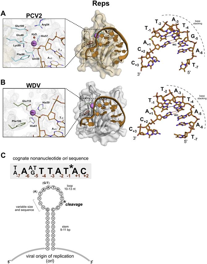
1048 Nucleic Acids Research, 2021, Vol. 49, No. 2
group with unit-cell parameters: a = 45.57 Å, b = 50.01 In vitro HUH cleavage assay
Å, c = 73.44 Å. One complex was present per asymmetric
Cleavage of the synthetic oligos was carried out using fi-
unit.
nal concentrations of 3 M SUMO-Rep and between 4.5
We also used Rigaku’s CrystalMation system’s broad,
and 30 M oligo in 50 mM HEPES pH 8.0, 50 mM NaCl,
sitting drop screen to identify potential conditions for
and 1 mM MnCl2 for 30 min at 37◦ C. The reactions were
PCVY96F + 10-mer crystallization. The protein solution
quenched with 4x Laemmli buffer containing 5% -ME,
contained 8 mg/ml protein, 1 mM 10-mer oligonucleotide
boiled for 5 min at 100◦ C, and run on a 4–12% SDS-PAGE
(5 -dTAGTATTACC-3 ), and 5 mM MnCl2 . Small needle
acrylamide gel. For time course reactions, aliquots were
crystals were obtained with 1:1 protein solution in a well so-
removed from an HUH reaction master mix at specified
lution of 0.1 M ammonium acetate; 25% polyethylene gly-
time intervals and immediately quenched in 4× Laemmli
col 3,350; 0.1 M Bis–Tris pH 7. Crystals were soaked in 25%
buffer containing 5% -ME. Percent covalent adduct for-
glycerol, and a dataset was collected at the APS Beamline
mation was calculated using Bio-Rad ImageLab software.
24 (NE-CAT). Crystals diffracted to 2.03 Å and belong to
The background subtraction function of ImageJ was used
the P64 space group with unit-cell parameters: a = b = 99.53
to process all gel images.
Downloaded from https://academic.oup.com/nar/article/49/2/1046/6067397 by guest on 25 July 2021
Å, c = 73.70 Å. There were three complexes per asymmetric
unit.
HUH-seq ssDNA library cleavage, library preparation, and
sequencing
Structure solution and refinement A 90-nt ssDNA library with a central 7 base randomized re-
gion flanked by conserved regions harboring primer bind-
The WDVY106F + 8-mer structure was solved with the ing sites at either termini (7N ssDNA library) was con-
molecular replacement function in PHENIX using our pre- structed using IDT oPools service consisting of 128 indi-
viously solved structure of apo WDV Rep (PDB ID: 6Q1M) vidually synthesized DNA oligos mixed at equal molarity,
as a model. We visualized the electron density map us- producing a ssDNA library containing 16,384 sequences
ing Coot (33) and modeled the 8-mer into a clear electron (extended data 1). Recombinant Rep cleavage of the 7N ss-
density tunnel (Supplementary Figure S1A). All eight nu- DNA library was carried out in triplicate in 3 M Rep and
cleotides of the oligonucleotide were unambiguously built 300 nM (83.4 ng/l) ssDNA library in 50 mM HEPES pH
into well-defined electron density of each of the two com- 8.0, 50 mM NaCl and 1 mM MnCl2 for 1 h at 37◦ C. The
plexes in the asymmetric unit. Subsequent refinement was Rep enzymes were immediately heat inactivated by boiling
performed with default settings of PHENIX auto.refine at 95◦ C for 3 min. The remaining uncleaved ssDNA library
with NCS applied (34) and alternated with visual inspec- from each Rep in vitro cleavage reaction was diluted 10-fold
tion and model correction. Final R-work and R-free were in water and amplified using 0.5 M TruGrade/HPLC pu-
0.188 and 0.246, respectively. rified primers from IDT containing Nextera adapters and
The WDVY106F + 10-mer, P21 21 21 structure was solved spacer regions with 2× CloneAmp™ HiFi PCR Premix for
with the Phaser molecular replacement function in 30 cycles. The resulting product was a 200 bp dsDNA am-
PHENIX using the previously solved WDVY106F + 8-mer plicon run on a 1.5% agarose gel and stained with Sybr-
structure. The two additional nucleotides were modeled Safe. Each 200 bp product was gel extracted (NucleoSpin
into appropriate density. Again, Coot was used for model Gel and PCR Clean-up kit, Macherey-Nagel) and eluted in
building, and PHENIX auto.refine was used for refine- 30 L NE elution buffer (5 mM Tris–HCl, pH 8.5) resulting
ment. The final R-work and R-free were 0.173 and 0.224 in samples of 30–60 ng/l. All samples were barcoded with
respectively. Illumia dual-indexing sequences via the Nextera adapters
For the PCVY96F + 10-mer structure, a model for molec- (University of Minnesota Genomics Core). Indexed sam-
ular replacement was generated in PyMol by superimpos- ples are were pooled and run on a 1.5% agarose gel; the 270
ing the WDVY106F + 8-mer structure with the Porcine cir- bp barcoded pooled sample was gel extracted and then se-
covirus 2 Rep domain (PDB ID: 5XOR) structure. The 8- quenced using a single Illumina HiSeq lane (350 000 000
mer from the WDV model was added to the PCV Rep do- paired-end reads, Genewiz) spiked with 30% PhiX to pre-
main model and used for Phaser molecular replacement vent molecule clumping to ensure a balanced fluorescent
in Phenix. The two additional nucleotides were modeled, signal. This improves overall run quality due to low library
and the oligonucleotide sequence was corrected using Coot. diversity (i.e. every amplicon has the same constant region
PHENIX auto.refine was used for refinement. Two of the composition).
complexes in the asymmetric unit had well-defined electron
density; density corresponding to the third complex was
HUH-seq read count reduction analysis and sequence logo
poorly defined due to inherent crystal properties as demon-
generation
strated by comparing simulated annealing omit maps of the
active sites from each complex copy (Supplementary Fig- Raw NGS sequence data were processed using R. Non-
ure S1B–D). As a result, R-values are higher than normal randomized portions (e.g. adapter sequences and constant
for this resolution structure. R-work and R-free were calcu- regions) were removed from each read to extract only the
lated to 0.229 and 0.280, respectively. An additional 4 nu- randomized 7-mer (k-mer). 7-mers from reverse reads were
cleotides from a second ssDNA strand was modeled near reverse-complemented, and frequency counts for each of
the surface of the third complex, which seems to be non- the 16 384 unique 7-mers were generated for the refer-
specifically bound (Supplementary Figure S1E). ence library from each of the Rep treatment libraries. Each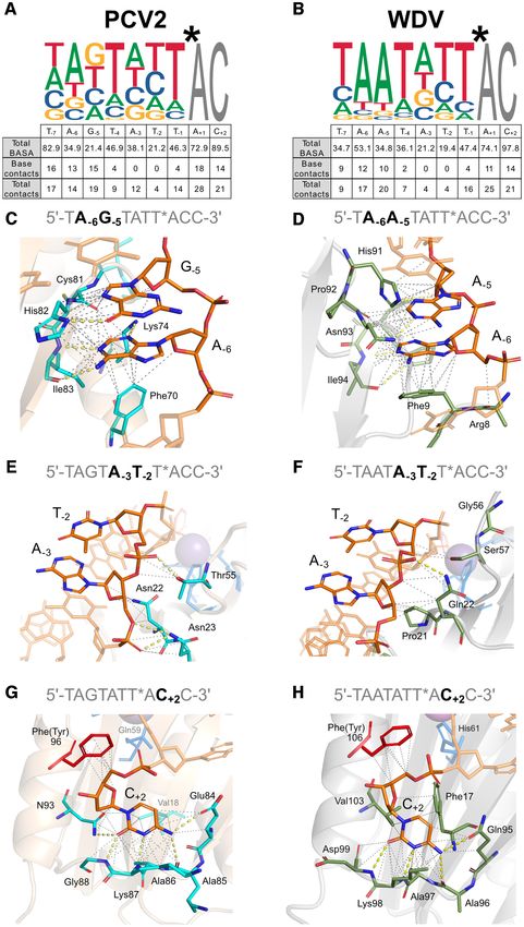
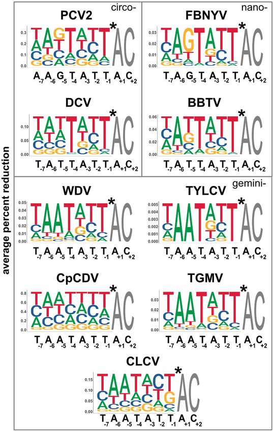
Nucleic Acids Research, 2021, Vol. 49, No. 2 1049

treatment was then compared against the reference to es-            center of the U. The bent conformation of the ssDNA in the
timate a log2 -fold-change and percent reduction (reference         Rep structures is driven by both intermolecular interactions
– treatment/ reference) for each of its 7-mers (extended            with the topological ‘nose’ of the protein and by intramolec-
data 2). The percent reduction data was used to generate            ular Watson–Crick base pairing between T−4 and A+1 along
weighted sequence logos for each Rep using the ggseqlogo            with adjacent hydrogen bonding between N3 of T−1 and N3
package in R. In addition, log counts per million (logCPM),         of A−3 (Figure 1A and B). Moreover, energetically favorable
one-way ANOVA F-test statistics (F), P-values and False             base stacking occurs between 5 nucleotides at positions −6
Discovery Rate (FDR) statistics were generated using the            through −2. These intramolecular, conformation stabiliz-
edgeR package for each k-mer per Rep treatment in trip-             ing interactions, along with protein–nucleotide interactions,
licate (extended data 3). P-value and FDR range are be-             promote the proper orientation needed for catalysis of the
tween 0 and 1, where a value
1050 Nucleic Acids Research, 2021, Vol. 49, No. 2
Table 1. Data collection and refinement statistics
PCVY96F + 10mera WDVY106F + 10mera WDVY106F + 8mera
(PDB 6WDZ) (PDB 6WE0) (PDB 6WE1)
Data collection
Wavelength (Å) 0.979 0.979 1.542
Resolution range (Å) 43.1–2.03 (2.10–2.03)b 41.34–1.8 (1.86–1.8)b 28.55–2.61 (2.71–2.61)b
Space group P64 P21 21 21 P41 21 2
Unit cell (Å) a = b = 99.53, c = 73.70, 90◦ , a = 45.57, b = 50.01, c = 73.44, a = b = 50.63, c = 241.98, 90◦ ,
90◦ , 120◦ 90◦ , 90◦ , 90◦ 90◦ , 90◦
Unique reflections 26261 (2625) 15366 (6674) 9674 (876)
Completeness (%) 97.52 (98.17) 95.23 (98.54) 93.29 (87.60)
Wilson B-factor 25.87 26.51 53.18
Mean I/ 12.49 (3.63) 14.17 (2.05) 7.70 (2.27)
CC1/2 0.996 (0.812) 0.997 (0.571) 0.987 (0.702)
CC* 0.999 (0.947) 0.999 (0.862) 0.997 (0.908)
Downloaded from https://academic.oup.com/nar/article/49/2/1046/6067397 by guest on 25 July 2021
R-meas 0.0821 (0.441) 0.0836 (0.963) 0.112 (0.609)
R-pim 0.0470 (0.248) 0.037 (0.436) 0.071 (0.378)
Refinement
Reflections used in 26 261 (2625) 15 364 (1550) 9674 (876)
refinement
Reflections used for R-free 1228 (91) 818 (92) 486 (40)
R-work 0.229 (0.273) 0.173 (0.220) 0.188 (0.301)
R-free 0.280 (0.287) 0.224 (0.313) 0.246 (0.354)
Number of non-hydrogen 3331 1169 2106
atoms
Macromolecules 3126 1112 2098
ligands 11 4 3
solvent 194 53 5
Protein residues 302 119 229
RMS (bonds) (Å) 0.009 0.006 0.008
RMS (angles) (◦ ) 1.11 1.09 1.06
Ramachandran favored (%) 97.96 98.29 96.31
Ramachandran allowed (%) 2.04 1.71 3.69
Ramachandran outliers (%) 0 0 0
Rotamer outliers (%) 0 0 0.54
Clashscore 10.64 0 6.27
Average B-factor 32.57 30.37 47.17
Macromolecules 32.61 29.89 47.20
Ligands 33.51 34.76 42.20
Solvent 31.84 40.15 34.09
a Data are from one crystal.
b Values in parentheses are for highest resolution shell.
Rep versus relaxase ssDNA interfaces reveal a reminiscent yet however there are striking differences in how the two fam-
distinct recognition mechanism ilies of proteins recognize DNA. Aside from the most ob-
vious difference of a larger size and a more extended DNA
Reps initiate replication of a large number of viruses and
binding interface that includes binding a hairpin structure
plasmids to copy their circular genomes while relaxases
5 to the nic site of relaxase proteins, the most distinctive
catalyze the transfer of one DNA strand of the plasmid
difference is that the relaxase structures contain a protein
genome to the recipient cell during plasmid conjugation
alpha-helical ‘clasp’ that covers the bound DNA (Figure 3C
(1); thus, relaxases are thought to recognize DNA with
and D). This clasp forms extensive contacts with the DNA,
more specificity than Reps. Our structures provide insights
suggesting that it helps anchor the DNA to the protein. This
at the molecular level into different modes of recognition
is underscored by the fact that in the crystal structure of
between Reps and relaxases that should illuminate struc-
NES, the relaxase from Staphylococcus aureus, which does
tural nuances of ssDNA recognition. The two available re-
not contain a ‘clasp’, the 3 end of the DNA has very few
laxase structures that are the most comparable to the Rep
contacts with the protein (17).
co-crystal structures are TraI (PDB ID: 2A0I) and TrwC
Moreover, the DNA in relaxase proteins is embedded in
(PDB ID: 2CDM), which are both complexed with ssDNA
a much deeper channel than in Rep proteins. Indeed, cal-
and have at least one nucleotide bound on the 3 side of
culations of buried solvent accessible surface area (BASA)
the nic site (Figure 3C and D). Structurally, Reps and re-
between protein and DNA reveal a more substantial buried
laxases share a similar central 5-stranded antiparallel beta-
surface area in the binding of DNA to relaxases even when
sheet core displaying the HUH motif, though the relaxases
accounting for the surface area buried by the clasps (Fig-
are circularly permuted with respect to the Reps such that
ure 3). Both Rep and relaxase structures have obvious struc-
the catalytic tyrosine is near the C-terminus of Reps and the
turally conserved pockets in the ssDNA docking interface
N-terminus of relaxases (31). Relaxases have similar active
in which individual nucleotide bases are bound. In all struc-
sites and U-shaped ssDNA architectures to Reps (28,30),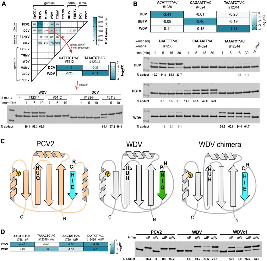
Nucleic Acids Research, 2021, Vol. 49, No. 2 1051
Downloaded from https://academic.oup.com/nar/article/49/2/1046/6067397 by guest on 25 July 2021
Figure 1. PCV and WDV Rep co-crystal structures complexed with ssDNA target sequence. (A) Semi-transparent surface and cartoon representation of
PCV2Y96F colored in beige and (B), WDVY106F colored in gray bound to manganese as a sphere in magenta and DNA 10-mers as sticks colored orange by
element. PCV2Y96F is bound to 10-mer (5 -dTAGTATTACC-3 ), and WDVY106F is bound to 10-mer (5 -dTAATATTACC-3 ) both adopting a U-shaped
conformation. Nucleotides are labeled as single letter abbreviations and positions, indicated as subscripts, relative to the scissile phosphate in yellow. A
dashed gray curve indicates the base stacking chain that occurs between positions –6 through –2. Intramolecular Watson–Crick (WC) base pairing between
A+1 and T−4 are indicated by red dashed lines as well as a non-canonical hydrogen bond between T−1 and A−3 are indicated as a black dashed line. Active
site side chains are indicated as sticks, PCV2Y96F in cyan and WDVY106F in green by element. The PCV2Y96F active site coordinates the manganese in
an octahedral geometry using Glu48, His57, and Gln59 with a water and two oxygens of the scissile phosphate completing the coordination shown as
black dashed lines. The WDVY106F active site coordinates the manganese in an octahedral geometry using Glu110, His59, and His61 with two oxygens
of the scissile phosphate completing the coordination shown as black dashed lines. The active site is displayed within the 2mFo-DFc map mesh at =
2. (C) Consensus cognate nonanucleotide ori sequence of 10 different Reps from Circoviridae, Nanoviridae and Geminiviridae (Supplementary Table S1).
The origin of replication (ori) from these ssDNA viruses contains a stem-loop hairpin with Rep cleavage occurring between position –1 and +1 within the
nonanucleotide sequence. The viral ori contains a stem that varies in sequence and between 9–11 base pairs in length while the loop contains the cognate
nonanucleotide sequence and varies between 10 and 13 nucleotides in length.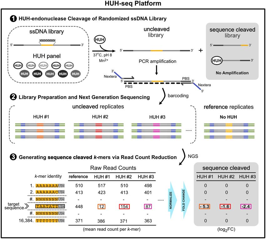
1052 Nucleic Acids Research, 2021, Vol. 49, No. 2

                                                                                                                                                            Downloaded from https://academic.oup.com/nar/article/49/2/1046/6067397 by guest on 25 July 2021

Figure 2. Cartoon depiction and structural alignment of specific Rep protein–DNA interactions: (A) PCV2 and (B) WDV Rep structures depicted as
2D cartoons with relative positions of residues (green) involved in binding ‘U-shaped’ ssDNA within 4 Å. The catalytic tyrosine 106 is indicated in red
with the adjacent phosphate in yellow, the ion coordinating triad is indicated in blue, and the 2+ ion in purple. The single Watson–Crick (WC) base pair
is indicated as a dashed red line, and the ssDNA intramolecular hydrogen bond is indicated as a black dashed line. (C) Structural alignment of Reps
using PROMALS3D including available PCV2 (PDB: 6WDZ), WDV (PDB: 6WE0), TYLCV (1L2M) and FBNYV (6H8O) structures as templates with
conserved residues highlighted - high or absolute conservation (≥90%) indicated in red; moderately conserved (≥70%) indicated in orange; low conservation
(≥50%) indicated in yellow; and no conservation (
Nucleic Acids Research, 2021, Vol. 49, No. 2 1053
Downloaded from https://academic.oup.com/nar/article/49/2/1046/6067397 by guest on 25 July 2021
Figure 3. Structural comparison of ssDNA recognition by Reps and relaxases: (A–D), PCV2 (6WDZ), WDV (6WE0), TraI36 (2A0I), TrwC (2CDM),
are illustrated in gray surface display where DNA interactions within 4 Å are highlighted in cyan, green, magenta, and yellow, respectively. Bound single-
stranded DNA is represented as a cartoon backbone in orange or as sticks with carbons and phosphates colored in orange. Nucleotides bound inside
pockets are solid; other bound nucleotides are transparent. Relaxase ‘clasps’ (TraI36 residues 231–271 and TrwC residues 237–262) are either solid or
transparent and outlined in black. Total buried solvent accessible surface area (BASA; Å2 ) for ssDNA bound to the docking interface was calculated for
each structure including values for with, or without, contribution from relaxase clasps.1054 Nucleic Acids Research, 2021, Vol. 49, No. 2
tures, the sDBM is a major contributor to the formation of indices and sequenced using the HiSeq platform to obtain
these pockets, which is part of 1 in relaxases and 4 in read counts for every sequence in the library (k-mer). Read
Reps. TraI and TrwC bury nucleotides –5 and –3 in strik- counts from reference replicates (no Rep added to the reac-
ingly deep pockets, #1 and #2, respectively (Figure 3). Reps tion) were used to calculate log2 -fold-change (FC) and read
have pockets in this structural region, yet they are much count percent reduction based on the difference between the
more shallow and only minimally bury nucleotides at −4 normalized reference library read counts and normalized
and −6 positions. A−6 is bound in the deepest of these Rep ‘uncleaved’ read counts for each Rep treatment (Figure 4).
pockets, yet it is still oriented in a configuration that fa- We generated weighted sequence logos based on a k-mer
vors base stacking with neighboring nucleotides rather than reduction analysis with a threshold value of 0.3 or greater
a ‘knob-in-pocket’ interaction as seen in both TrwC and to reduce noise based on high confidence data guided by
TraI structures. Conversely, both Reps have a deep pocket, calculated adjusted P-values (FDR) (Figure 5, Supplemen-
#3, where the +2 cytosine base is buried. The only relaxase tary Figure S5B). Percent reduction for each k-mer was cal-
structure that contains the +2 base is TraI, however the base culated by comparing the normalized k-mer read counts
is not bound in the same conserved pocket (Figure 3). for each Rep treatment in triplicate to k-mer read counts
Downloaded from https://academic.oup.com/nar/article/49/2/1046/6067397 by guest on 25 July 2021
from the reference library. For each position in a Rep se-
quence logo, individual characters were scaled by the aver-
HUH-seq uncovers subtle differences in Rep ssDNA recogni-
age percent reduction of all k-mers containing that charac-
tion specificity
ter and position. Because every sequence permutation 5 of
Structural analysis of the Rep protein–DNA contact maps the nic site is present in the 7N ssDNA library, sequence
point to subtle differences that contribute to recognition of logos reveal Rep preferences for nucleotides relative to one
nearly identical nonanucleotide sequences, suggesting that another. The most obvious result is that the most preferred
Reps may differentially tolerate substitutions in the target nucleotides in the first seven positions of sequence logos are
DNA sequence. Thus, we developed a NGS-based cleavage nearly identical to the cognate nonanucleotide ori sequence
assay approach, HUH-seq, to examine both ssDNA speci- found in each respective viral genome (Figure 5). While it is
ficity and to explore expansion of the use of Reps in multi- not surprising that the preferred target sequence is the same
plexed HUH-tag applications. As a first step in assessing the as the cognate nonanucleotide ori sequence cleaved in vivo,
ssDNA recognition specificity of Reps, we asked whether it also gives high confidence that HUH-seq can be used to
viral Rep proteins from different families and genera (Ta- quantitatively rank the k-mers cleaved by each Rep, analyze
ble 2) differentially tolerate mutations in the target nonanu- patterns that dictate these ssDNA recognition profiles, and
cleotide sequence by measuring covalent adduct formation further characterize differences between individual Reps.
with an in vitro HUH cleavage assay (Supplementary Note Within each sequence logo, there are differentially pre-
S2, Supplementary Figure S4A). However, it became imme- ferred nucleotide positions. Positions T−4 and T−1 are al-
diately evident that a low-throughput assay would insuffi- most unanimously the most preferred, while there is only
ciently characterize specificity due to widespread toleration slight preference for A and T at the −3 and −2 positions,
of variable target sequences. A large number of truncations respectively. There are also discernible trends between Reps
and substitutions within the nonameric sequence resulted in from different families. For example, geminivirus Reps have
negligible effects on adduct formation in many cases (a full a strong preference for adenine at the -5 position, whereas
analysis of the small oligo library screen is provided, Sup- Reps from other families prefer thymine or guanine there
plementary Figure S4B and C). This realization prompted (Figure 5). The y-axis scale of the weighted sequence logos
us to devise a high-throughput method that would reveal also indicates the relative overall cleavage efficiency between
ssDNA recognition profiles for each Rep. Reps. For instance, PCV2 has a maximum average percent
To this end, we developed HUH-seq, an NGS-based reduction of about 0.35 and cleaves roughly 10-fold more
approach used to establish comprehensive ssDNA recog- sequences than FBNYV, which has a maximum value of
nition profiles of the Reps contained within a random- about 0.035. This indicates that PCV2 ssDNA recognition
ized ssDNA library containing 16,384 sequences, or k- is more promiscuous than that of FBNYV. CpCDV has the
mers. In brief, the first seven positions of the nonanu- highest maximum average percent reduction of 0.8 and has
cleotide target sequence are randomized in the 7N ssDNA minimal nucleotide preference, indicating it has the most re-
library, where positions A+1 and C+2 are constant (‘7N’ laxed sequence specificity (Figure 5).
- N−7 N−6 N−5 N−4 N−3 N−2 N−1 *A+1 C+2 ). The library was As controls, we included two WDV Rep treatments with
constrained to only seven positions in order to limit the lower protein concentrations and found that decreasing the
size of the library; further design considerations are dis- amount of WDV Rep minimally affected specificity (Sup-
cussed in the supplementary information (Supplementary plementary Figure S6A). To ensure cleavage was the only
Figure S5A, Supplementary Note S2 and Supplementary readout for this assay, we used an inactive WDVY106F Rep
Equations S1 and S2). Reps were individually reacted with treatment, yet a small number of ‘cleaved’ k-mers were iden-
the 7N ssDNA library under standard conditions and pro- tified from the inactive treatment indicating that Rep bind-
duced two populations of the library: ‘sequence cleaved’ ing may slightly contribute (Supplementary Figure S6C and
and ‘uncleaved’. A primer set containing Nextera adapters S6D). Other considerations and caveats of HUH-seq analy-
was used to generate the antisense strand and to amplify sis are discussed in supplemental information (Supplemen-
the ‘uncleaved’ population in a single PCR step, while the tary Note S4 and Supplementary Figure S6). Despite these
‘sequence cleaved’ population remained unamplified. The caveats, HUH-seq is a robust method for profiling Rep
‘uncleaved’ amplicons were barcoded with standard dual- specificity.Nucleic Acids Research, 2021, Vol. 49, No. 2 1055
Table 2. Panel of 10 expressed and purified recombinant Reps
Cognate nonanuclotide ori
Rep Viral species Family Genus MW (kDa) sequence
PCV2 Porcine circovirus 2 Circoviridae Circovirus 13.1 AAGTATT*AC
DCV Muscovy duck circovirus 12.4 TATTATT*AC
BBTV Banana bunch top virus Nanoviridae Babuvirus 11.2
FBNYV Faba bean necrotic yellows virus Nanovirus 11.3 TAGTATT*AC
WDV Wheat dwarf virus Geminiviridae Mastrevirus 15.6 TAATATT*AC
CpCDV Chickpea chlorotic dwarf virus 14.5
MSMV Maize striate mosaic virus 13.4
TYLCV Tomato yellow leaf curl virus Begomovirus 15.5
CLCV Cabbage leaf curl virus 13.3
TGMV Tomato golden mosaic virus 14.4
Downloaded from https://academic.oup.com/nar/article/49/2/1046/6067397 by guest on 25 July 2021
Figure 4. HUH-seq cleavage assay schematic for determining Rep sequence specificity. Schematic describing HUH-seq: an NGS-based approach for quan-
tifying ssDNA specificity profiles of Reps. A synthetic ssDNA library containing seven random bases (4 bases ∧ 7 positions = 16 384 unique kmers)
(yellow) flanked by constant regions (gray) and primer binding sites (PBS) (dark gray) are reacted with a panel of Reps, or no enzyme as a reference, in
replicate, generating a two part pool containing the ‘uncleaved’ library and the ‘sequence cleaved’ library for each reaction. In a single PCR step, the anti-
sense strand for the ‘uncleaved’ pool is generated, amplified and Nextera adapters (purple) are added with primer overhangs; the ‘sequence cleaved’ library
is not amplified due to physical separation of the PBS’s. Each set of amplicons is then barcoded with standard i7/i5 Illumina indexing sequences (green)
and pooled for a single next generation sequencing run. A custom R-based analysis script generates read counts for all k-mers in each set of replicates, then
normalizes based on total read count, and quantifies k-mer cleavage extent of each Rep in the panel based on fold change and percent reduction.1056 Nucleic Acids Research, 2021, Vol. 49, No. 2
Downloaded from https://academic.oup.com/nar/article/49/2/1046/6067397 by guest on 25 July 2021
Figure 5. Weighted sequence logos generated from HUH-seq cleavage data. Weighted sequence logos for nine of the ten Reps based on percent reduction
generated using ggseqlogo with values under 0.3 set to 0.0 in order to remove noise obtained from the HUH NGS cleavage assay. Heights are scaled to
represent the average percent reduction of each base at each position when compared to the reference library. Sequences in black below each logo are the
cognate nonanucleotide ori sequences from each respective virus. Asterisk denotes the cleavage site. Logos are organized by viral families as labeled inside
the gray boxes.Nucleic Acids Research, 2021, Vol. 49, No. 2 1057
Rep ssDNA recognition profiles corroborate structural obser- being the direct readout of nucleotide bases through specific
vations protein contacts.
Next, we quantified and assigned contributions of the ss-
DNA docking interface in the Rep structures to each nu- Discovering intrinsically orthogonal Rep target sequences us-
cleotide using DNAproDB by calculating the BASA as well ing HUH-seq
as the total number of protein–DNA contacts (the sum of During initial assessments of the HUH-seq analysis results,
hydrogen bonds and Van der Waals interactions within 4 we noticed that there were individual target k-mers with
Å). Figure 6A and B summarizes the total BASA for and the drastically different log2 FC values between different Rep
total number of contacts with nucleotides corresponding protein treatments. This prompted us to ask whether we
to the cognate nonanucleotide ori sequence either with the could identify pairs of k-mers that would allow us to se-
entire nucleotide or the base only. These measurements in lectively label two Reps in a single reaction mixture with
combination with the ssDNA recognition profiles of WDV unique oligos. For instance, k-mer, AGTCAAT (#2884) has
and PCV2 were used to search for structural reasons why a log2 FC value of −3.44 for PCV2 and a near zero log2 FC
nucleotides in certain positions of the target sequence are
Downloaded from https://academic.oup.com/nar/article/49/2/1046/6067397 by guest on 25 July 2021
for every other Rep (Supplementary Figure S7). This re-
conserved. A comprehensive table containing BASA and sult was validated using the in vitro HUH cleavage assay
contact values of each of the three structures featured in by reacting PCV2 Rep with a synthetic oligo containing
this study is also provided (Supplementary Table S2). As this k-mer sequence. Indeed, only PCV2 formed a covalent
expected, higher BASA values generally correlated to high adduct with the oligo harboring this target sequence (Sup-
numbers of contacts. plementary Figure S7). Interestingly, this target sequence
PCV2 bound 10-mer and WDV bound 10-mer structures contains 4 substitutions with respect to the circovirus ori
have a similar total number of residues contacting DNA, sequence at positions −6, −5, −4 and −2, again highlight-
28 and 26 residues, respectively, and have a high concen- ing the promiscuous nature of Reps. This result revealed
tration of base contacts and total contacts near the 5 and that searching for combinations of Reps and k-mers may
3 termini of the 10-mers (Figure 6A and B). In Figure 6C– result in the discovery of naturally occurring orthogonality
H, significant structural differences are highlighted between despite apparent cross-reactivity.
the contacts of nucleotides at different positions for both To explore the possibility of naturally occurring orthog-
PCV2 (C, E and G) and WDV (D, F and H). A−3 and T−2 onality between two Reps, we wrote a script to extract
are the least conserved nucleotides at their indicated posi- pairs of k-mer sequences and Reps predicted to lack cross-
tion. This is structurally consistent because there are zero reactivity based on log2 FC values. Figure 7a displays a sum-
contacts with the bases for both PCV2 and WDV indicat- mary heatmap of the number of such k-mer pairs existing
ing that specific nucleotides are not as preferred at these two for every set of Rep pairs, based on threshold values of –
positions because the interactions are exclusively with the 0.3 log2 FC and greater (likely forming no adduct) and –3.0
ribose and phosphate of the nucleotides (Figure 6E and F). log2 FC and lower (likely having high adduct formation). In
The 10-mer bound to PCV2 differs at position –5 between one example, we identified the k-mer sequence, CATTTCT
guanine and adenine with respect to the 10-mer bound to (#5112), in which DCV had a −4.13 log2 FC and WDV had
WDV. His91 and Asn93 of WDV facilitate polar contacts a −0.33 log2 FC, and another k-mer sequence, TAAATCT
with A−5 , which may give WDV more specificity at posi- (#12344), in which DCV had a –0.20 log2 FC and WDV had
tion –5, whereas there is only one polar contact with G−5 a −4.11 log2 FC, indicating orthogonality between DCV
by His82 in the PCV2 structure, which results in less strin- and WDV for these two k-mers. We validated this observa-
gent specificity. (Figure 6C and D). Finally, in both struc- tion with an in vitro HUH cleavage assay including a short
tures C+2 dwells in a pocket of the protein surface with the time course with 1, 5 and 10 min time points. DCV formed
highest BASA and total contact values (Figure 6G and H). about 97% adduct with a synthetic oligo harboring k-mer
Eight residues have contacts with C+2 in both structures, #5112 over the course of 5 min, and WDV formed about
and five of these residues make up the last positions of the 62% adduct with a synthetic oligo harboring k-mer #12344
sDBM. over the course of 10 min. As expected, no cross-reactivity
In contrast, T−4 is highly conserved as evident in all was observed between WDV with k-mer #5112 or DCV
Rep ssDNA recognition profiles, but we observed only a with k-mer #12344 (Figure 7A).
marginal number of protein contacts with the base itself We next searched for triple orthogonal sets of Reps from
(Figure 6A and B). We hypothesize that the WC base pair- our panel. As an example, the set containing k-mer se-
ing of T−4 with A+1 is a major contributor to the U-shaped quences, #1280, #4624 and #12344, are predicted to re-
conformation rather than contributing to sequence speci- act orthogonally with DCV, BBTV and WDV recombi-
ficity via residue interactions with the base. Though Reps nant Reps, respectively, as indicated by log2 FC values (Fig-
exhibit interactions with bases that contribute to specificity, ure 7B). Similar to our method for validating double or-
it is clear from the ssDNA recognition profiles and minimal thogonal sets, we tested the orthogonality of this set us-
protein-DNA contacts at certain positions that Rep cleav- ing the standard in vitro cleavage assay and calculated per-
age is also promiscuous, cleaving a wide range of target se- cent adduct formed with each combination of k-mer and
quences. Taken together, there are two substantial contribu- Reps over a short time course. Expected orthogonality was
tors to Rep specificity: the first being the indirect readout of achieved with over 50% covalent adduct formation after 30
a given DNA sequence that adopts a conformation that fits min for each of the three Reps with 0–9% cross-reactivity
into the groove of the Rep docking interface and the second identified (Figure 7B).1058 Nucleic Acids Research, 2021, Vol. 49, No. 2
Downloaded from https://academic.oup.com/nar/article/49/2/1046/6067397 by guest on 25 July 2021
Figure 6. Comparison of Rep protein–DNA interactions and HUH-seq specificity profiles: (A), PCV2 + 10mer (6WDZ) and (B) WDV + 10mer (6WE0)
BASA values and total number of protein-DNA contacts compared to weighted sequence logos from HUH-seq analysis. Both polar and van der Waals
interactions are counted within 4 Å. (C–H) Atomic interactions between highlighted nucleotides within the bound 10-mers of PCV2 and WDV structures
are shown with yellow dashes for polar contacts and gray dashes for van der Waals interactions within 4 Å. PCV2 cartoon is depicted in beige and residues
interacting with DNA as sticks shown in cyan colored by atom. WDV cartoon is depicted in gray and residues interacting with DNA as sticks shown in
green colored by atom. The PCV2 Phe96 and WDV Phe106 represent the catalytic tyrosine as sticks in red, divalent ion coordinating residues are in blue,
and the manganese ion is a sphere in magenta. The 10-mer is shown as sticks in orange and colored by atom as highlighted in the panel.Nucleic Acids Research, 2021, Vol. 49, No. 2 1059
Downloaded from https://academic.oup.com/nar/article/49/2/1046/6067397 by guest on 25 July 2021
Figure 7. Discovery of orthogonal Rep target sequences and rational engineering of Rep specificity. (A) Heatmap displaying the number of k-mer pairs
for a specific Reps set likely to be orthogonal using an asymmetric log2 FC threshold based on values from HUH-seq analysis, blank cells indicate zero
such k-mer pairs. The threshold values are set to log2 FC values greater than -0.3 (indicating no k-mer cleavage) and log2 FC values less than –3 (indicating
high k-mer cleavage). Each cell of the heatmap represents the total number of possible k-mer pair combinations likely to be orthogonal for a particular
set of two Reps. Sets are based on this asymmetric threshold in which the first Rep in the set has high cleavage of one k-mer in the pair and no cleavage
of the other k-mer in the pair - vise versa for for the second Rep in the set, indicating orthogonality. As an example, one k-mer pair (#5112 and #12344)
of the 2200 possible combinations indicated by the WDV vs. DCV cells were synthesized in the context of the flanking regions of the 7N ssDNA library,
and cleavage orthogonality was validated using the in vitro HUH cleavage assay. Recombinant WDV and DCV were reacted under standard conditions
over a short time course with synthetic oligos harboring k-mers sequences #5112 or #12344. Percent covalent adduct was calculated. (B) Set of three Reps
and corresponding orthogonal set of three k-mer sequences as indicated by log2 FC values from HUH-seq analysis. Oligos synthesized harboring k-mer
sequences (#1280, #4624, #12344) were reacted with DCV, BBTV, and WDV recombinant Reps at room temperature with 1.5× molar excess oligo to Rep
protein over a short time course. (C) A schematic illustrating the construction of the WDV chimera (WDVc1) containing the first five amino acids of the
PCV2 sDBM. (D) The heatmap displays HUH-seq log2 FC values for PCV2 and WDV reactivity with cognate nonanucleotide sequences (k-mers #720 and
#12496) and a pair of k-mers (#768 and #12318) predicted to react orthogonally. The k-mers #720 (wtP), #12496 (wtW), (#768 (oP) and #12318 (oW)
were synthesized in the context of the flanking sequences of the 7N ssDNA library and reacted in a 5x molar excess with PCV2, WDV, and WDV chimera
(WDc1) recombinant protein for 30 min and 37◦ C.1060 Nucleic Acids Research, 2021, Vol. 49, No. 2
Notably, 23 of the 28 Rep sets from different viral families sented here for the first time illuminate the interface respon-
contained significant k-mer pairs likely to be orthogonal, sible for specific ssDNA recognition necessary for ssDNA
yet there were no instances of orthogonal k-mer pairs for processing. The most striking feature of the ssDNA bound
Reps derived from the same viral family (Figure 7A). Hence, structures is the central role played by a motif we call the
the ssDNA binding moieties of Reps within the same fam- sDBM. This motif is highly conserved between members of
ily may be too similar to yield orthogonal adduct forma- the same viral Rep family but divergent between families,
tion. This is a curious result in the case of DCV and BBTV yet it maintains its key function of binding ssDNA for cleav-
where 294 potentially orthogonal k-mer pairs were identi- age across the HUH endonuclease superfamily (Figure 3,
fied, which are from different Rep families but recognize Supplementary Figure S3). The sDBM motif partially over-
identical cognate nonanucleotide ori sequences (Figure 7A). laps with a previously identified ∼20 amino acid long mo-
This indicates that perhaps DCV and BBTV use different tif specific to the geminivirus family called the GRS, which
interactions to recognize the same cognate sequence allow- was suggested to interact with DNA via mutagenesis stud-
ing for divergent specificity at each nucleotide position. In- ies (37). The sDBM facilitates two recognition modes: the
deed, six out of nine residues in the sDBM are different be- first mode is indirect, whereby target sequences that have
Downloaded from https://academic.oup.com/nar/article/49/2/1046/6067397 by guest on 25 July 2021
tween DCV and BBTV Reps (Figure 2C). Using HUH-seq, the propensity to adopt a U-shaped conformation (e.g. via
we can pick out subtle differences in Rep specificity in or- base-pairing and stacking) fit into the groove of the Rep
der to extract double and triple orthogonal k-mers and Reps docking interface, and the second mode is direct, whereby
sets that can be used in multiplexed HUH-tag technologies. the Rep provides specific protein-ssDNA contacts confer-
This could potentially negate the necessity for or create ways ring specificity for ssDNA based on amino acid sequence.
to use Reps in combination with the larger and slower relax- The combination of these modes accounts for the semi-
ases or commercial fusion tags. promiscuous specificity of viral Reps.
Looking more broadly at the Rep-DNA interactions
in the context of the HUH-endonuclease superfamily, the
Rational design of a WDV chimera confers PCV2-like se-
sDBM is apparently a ubiquitous motif contributing to
quence specificity
DNA binding and recognition. This is illustrated by avail-
The identification of the sDBM, which we hypothesized was able relaxase and transposase structures captured in the pre-
responsible for sequence specificity in Reps as well as the cleavage state (17,28–30,47). In Reps, the sDBM is located
discovery of pairs of target sequences between two Reps that in the middle of the structure and consists of the fourth beta
should not cross-react, inspired us to swap sDBM residues strand and a portion of the preceding loop. In relaxases,
between two Reps and ask if we could alter sequence speci- however, the sDBM is located at the extreme N-terminus
ficity. We swapped out the first five amino acids of the due to the circular permutation of relaxases with respect to
WDV sDBM for those of PCV2, creating a WDV chimera viral Reps and plays a major role in forming specific con-
(WDVc1) as a proof-of-concept that Rep specificity could tacts especially with nucleotides bound in deep pockets of
be altered by rational design in a predictable manner (Fig- the protein surface. Transposases, like relaxases, bind a hair-
ure 7C). Because many of the amino acid side chains in pin sequence distal from the cleavage site. However, recog-
both Rep structures have direct contacts with bases at the nition of the cleavage site occurs by both the protein and
5 end of the ssDNA, we hypothesized WDVc1 would have a short guide sequence near the stem of the hairpin. In the
sequence specificity more closely reflecting that of PCV2. ternary structure of the IS608 TnpA transposase (PDB ID:
First, we identified a pair of predicted target sequences for 6FI8) in complex with its hairpin ‘imperfect palindrome’ se-
PCV2 and WDV using HUH-seq, where WDVc1 reacts quence and a short oligonucleotide spanning the cleavage
with the k-mer #768 (oP), which was predicted to only re- site, the –1 and +1 bases form base pairing interactions with
act with PCV2, to a greater extent than k-mer #12318 (oW), the guide sequence of the hairpin DNA bridging the two
which was predicted to only react with WDV (Figure 7D). distant sections of DNA to form a U-shape (47,48). This
Similar to PCV2, WDVc1 reacts robustly with the cognate ‘trans’ U-shape conformation is primed much in the same
nonameric sequence of PCV2, k-mer #720 (wtP), as well way viral Reps bend the ssDNA using the sDBM, most
as the cognate nonameric sequence of WDV, k-mer #12496 notably by the bridging Phe112 that stacks with C+1 (Sup-
(wtW), (Figure 7C). Thus, we show how the sDBM is a key plementary Figure S3C). Other amino acids of the sDBM
feature of Reps that may be rationally engineered to pre- also bind to the guide DNA sequence and seem to play a
dictably alter sequence specificity. greater role in conformation priming than direct recogni-
tion; though DNA bases extending downstream the +1 po-
sition are missing in the structure, preventing further dis-
DISCUSSION
cussion of the role of the transposase sDBM in specificity.
We first determined the molecular basis of ssDNA recog- In nature, there are several reasons why Rep specificity
nition of viral Reps by solving crystal structures of vi- could be more promiscuous than that of relaxases. If con-
ral Reps bound to ssDNA containing the cognate ori se- jugative plasmid transfer occurs at an erroneous origin, it
quence trapped in the pre-cleavage conformation. Several could result in catastrophic consequence for the host’s fit-
apo structures of viral Reps in the absence of DNA have ness; whereas, there is little selective pressure for a virus
been reported (40–44) as well as parvovirus AAV5 Rep to initiate replication at a very specific sequence due to
structures bound to distal auxiliary regions of dsDNA with the small number of sequences within a sub-5 kb genome
the inverted terminal repeat (ITR) involved in rolling hair- (32). Relaxases may also have higher specificity for the
pin replication (45,46). However, the Rep structures pre- DNA sequence 5 of the nic site for more efficient cataly-Nucleic Acids Research, 2021, Vol. 49, No. 2 1061
sis of rejoining of the free 3 OH of the DNA post-transfer, super-resolution imaging (64) as well as emerging applica-
whereas RCR resolution would likely require a second tions such as multiplexed single-cell proteomics (65), and
dimerizing Rep for termination (49). It should also be optics free DNA microscopy (66), where parallel tracking
noted that Rep specificity could also simply be constrained of proteins occurs using NGS. It is of note that HUH-
by a smaller interface surface area due to limited gene fusions would allow conjugation of oligos to ScFv’s and
size (50). nanobodies, which could expand the utility of many of these
Characterization of the Rep/ssDNA interface provides a applications which utilize oligo-conjugated antibodies. Be-
platform to model other Rep ssDNA interfaces as well as an cause HUH-tag linkages are specific and covalent, can oc-
avenue to explore disruption of the interface for antiviral cur intra- or extracellularly without additional reagents,
treatments of plant and human Rep-mediated viral infec- and are now multiplexable, they are ideal fusion tags for
tions (51). Billions of dollars worldwide are lost in agricul- these applications.
ture every year from the decimation of crops such as toma- We foresee the utility of the simple HUH-seq approach,
toes, cassava, cotton, and beans by geminivirus infection with minor alterations, for sensitive detection of sequence
(52). In a human disease context, parvovirus B19 human specificity profiles for enzymes such as dsDNA nucleases by
Downloaded from https://academic.oup.com/nar/article/49/2/1046/6067397 by guest on 25 July 2021
infections can lead to serious or fatal outcomes for a fe- simply using a dsDNA library, RNA-cleaving enzymes by
tus (53,54) and are associated with autoimmune diseases in adding a single reverse transcriptase step, or site-specific nu-
adults (21,22). This has sparked treatment and vaccine de- cleotide modifying enzymes by relying on a covalent mod-
velopment (55), however present antiviral strategies, both ification that blocks PCR amplification. The existing high-
viral protein interfering and gene silencing approaches, are diversity library methods used to determine the dsDNA
either minimally effective or are eventually subverted by specificity of zinc finger nucleases (67), Cas9 (68), tran-
conferred resistance from a rapidly evolving viral genome scription activator-like effector nucleases (TALENs) (69),
(51,56–60). Development of antivirals specifically targeting and other restriction enzymes (70) are powerful and direct
the ssDNA binding of Reps could more effectively retain cleavage readout approaches, however they require a num-
long-term resistance. ber of extra library preparation steps and may be limited
The Rep structures revealed highly conserved protein- to only dsDNA libraries. Additionally, if sequence bind-
DNA interfaces with subtle differences that prompted us to ing, rather than cleavage, could be optimized as a readout,
ask whether Reps within families and from different fam- HUH-seq could be developed as a facile alternative method
ilies differentially tolerate mutations of the cognate ori se- to approaches such as SELEX-seq (71) and could deter-
quence. One reported relatively high throughput strategy mine binding sequence preference of shorter DNA binding
for querying key nucleotides in bacterial conjugation me- motifs. Lastly, an HUH-seq screen including a broad range
diated by the relaxase TrwC used saturation mutagenesis of Rep-encoding organisms may yield crucial evidence for
in concert with a functional DNA-transfer readout (32), more accurate lineage classification, which is continually be-
which is a readout incompatible with Reps. Other NGS- ing restructured most notably because of the exponential
based ssDNA recognition approaches, for example of cy- discovery rate of unclassified circular ssDNA viruses (72).
tosine deaminases (61) or DNA aptamer-binding protein The rep gene is an indispensable component of lineage anal-
targets (62), also use direct sequencing readout methodol- ysis (73,74) and a combination of rep gene structure and se-
ogy. However, a direct readout of Rep cleavage is techni- quence identity along with Rep cleavage specificity may lead
cally challenging due to the need to amplify physically sep- to rapid and more accurate classification.
arated cleaved DNA molecules and covalent attachment of While subtle but specific family differences in DNA
the Rep to the new 5 end of the cleaved molecules. Instead, recognition coupled with HUH-seq permits modest viral
HUH-seq, allows for the quantitative readout of Rep cleav- Rep multiplexing, expanded multiplexing capability could
age specificity using a ssDNA library with a subtractive, or be achieved by engineering the protein to recognize designer
reduction, readout. DNA sequences. Engineering one HUH-endonuclease to
Excitingly, we found HUH-seq can be used to distinguish react with another HUH-endonuclease target sequence has
subtle differences between Rep nucleotide preferences de- been demonstrated for AAV but involved swapping large
spite overall lack of specificity, so much so that intrinsic or- protein domains (75). Similarly, a double mutant of the
thogonality between non-cognate target sequences can be TraI relaxase that conjugates the F plasmid was able to
extrapolated between Reps from different, yet closely re- switch specificity to the related R100 plasmid target se-
lated, viral families with highly similar or even the same cog- quence, though the engineering was performed by testing
nate nonanucleotide ori sequences. Intrinsic orthogonality and mutating all distinct amino acids residues between the
between Rep families demonstrates the feasibility of using two relaxases (76). We have shown in an elegant example
Reps in multiplexing applications without the need for pro- of rational engineering that by simply mutating four amino
tein engineering, despite their apparent promiscuity. More- acids within the sDBM of WDV and PCV2, specificity can
over, there are currently 10 additional viral Rep families yet be predictably altered. This approach was made possible not
to be explored with HUH-seq (50), meaning that multiplex- only by structural insights, but also because we can identify
ing could be expanded to up to 13 Reps in a given system intrinsically orthogonal target sequences that would react
(i.e. have 13 HUH-fusions in an application such as DNA specifically with each Rep using HUH-seq. Predictable al-
barcoding of proteins of interest and add 13 DNA bar- tering of ssDNA specificity by targeting the residue com-
codes that should specifically react with only one given Rep position of the sDBM either by rational design or directed
HUH-tag). DNA-tagging is the basis of established tech- evolution could motivate development of engineered HUH-
nologies such as proximity ligation (63) and DNA-PAINT tags with defined sequence specificity to facilitate massivelyYou can also read