FUNCTIONAL HARMONICS REVEAL MULTI-DIMENSIONAL BASIS FUNCTIONS UNDERLYING CORTICAL ORGANIZATION - MPG.PURE
←
→
Page content transcription
If your browser does not render page correctly, please read the page content below

Article

Functional harmonics reveal multi-dimensional basis
functions underlying cortical organization
Graphical abstract                                                  Authors
                                                                    Katharina Glomb, Morten L. Kringelbach,
                                                                    Gustavo Deco, Patric Hagmann,
                                                                    Joel Pearson, Selen Atasoy

                                                                    Correspondence
                                                                    katharina.glomb@gmail.com

                                                                    In brief
                                                                    Glomb et al. demonstrate that the
                                                                    principle of harmonic waves—ubiquitous
                                                                    in nature—accounts for the cortical
                                                                    organization of the human brain.
                                                                    Harmonic waves estimated on the brain’s
                                                                    communication structure—called
                                                                    functional harmonics—explain
                                                                    organizational aspects across spatial
                                                                    scales and link resting state and task-
                                                                    based brain activity.

Highlights
d   ‘‘Functional harmonics’’ are a set of basis functions derived
    from the brain graph

d   They account for both modular and gradiental cortical
    organization

d   They capture aspects of cortical organization on several
    spatial scales

d   A small number of functional harmonics suffices to decode
    task activity patterns

           Glomb et al., 2021, Cell Reports 36, 109554
           August 24, 2021 ª 2021 The Author(s).
           https://doi.org/10.1016/j.celrep.2021.109554                                           ll
ll
                                                                                                                          OPEN ACCESS

Article
Functional harmonics reveal multi-dimensional
basis functions underlying cortical organization
Katharina Glomb,1,9,* Morten L. Kringelbach,2,3 Gustavo Deco,4,5,6,7 Patric Hagmann,1 Joel Pearson,8 and Selen Atasoy2,3
1Department of Radiology, Lausanne University Hospital and University of Lausanne (CHUV-UNIL), Rue du Bugnon 46, 1011 Lausanne,

Switzerland
2Department of Psychiatry, Warneford Hospital, University of Oxford, Oxford OX3 7JX, UK
3Center of Music in the Brain (MIB), Clinical Medicine, Aarhus University, Universitetsbyen 3, 8000 Aarhus C, Denmark
4Center of Brain and Cognition, Universitat Pompeu Fabra, Ramon Trias Fargas, 25-27, 08005 Barcelona, Spain
5ICREA, Institució Catalana de Recerca i Estudis Avancats (ICREA), Passeig Lluı́s Companys 23, 08010 Barcelona, Spain
6Department of Neuropsychology, Max Planck Institute for Human Cognitive and Brain Sciences, Stephanstraße 1A, 04103 Leipzig, Germany
7School of Psychological Sciences, Monash University, 18 Innovation Walk, Clayton Campus, Clayton 3800, VIC, Australia
8School of Psychology, Mathews Building, University of New South Wales, Sydney 2052, NSW, Australia
9Lead contact

*Correspondence: katharina.glomb@gmail.com
 https://doi.org/10.1016/j.celrep.2021.109554

SUMMARY

The human brain consists of specialized areas that flexibly interact to form a multitude of functional networks.
Complementary to this notion of modular organization, brain function has been shown to vary along a smooth
continuum across the whole cortex. We demonstrate a mathematical framework that accounts for both of
these perspectives: harmonic modes. We calculate the harmonic modes of the brain’s functional connectivity
graph, called ‘‘functional harmonics,’’ revealing a multi-dimensional, frequency-ordered set of basis func-
tions. Functional harmonics link characteristics of cortical organization across several spatial scales,
capturing aspects of intra-areal organizational features (retinotopy, somatotopy), delineating brain areas,
and explaining macroscopic functional networks as well as global cortical gradients. Furthermore, we
show how the activity patterns elicited by seven different tasks are reconstructed from a very small subset
of functional harmonics. Our results suggest that the principle of harmonicity, ubiquitous in nature, also un-
derlies functional cortical organization in the human brain.

INTRODUCTION                                                            (Penfield and Rasmussen, 1950), and tonotopy (Perrone-Ca-
                                                                        pano et al., 2017), show that representations of our visual field,
The topographic organization of the brain into functionally             body, and auditory frequency spectrum are spatially continuous
specialized areas is one of its fundamental properties (Felleman        across the areas of the primary visual, somatomotor, and audi-
and Van Essen, 1991), suggested to have been present in evolu-          tory cortices, respectively, exemplifying an organizational princi-
tion as early as the last common ancestor of vertebrates (Krubit-       ple that is complementary to the finding of relatively uniform
zer and Kaas, 2005; Eickhoff et al., 2018). The individuality of        functionality within some brain areas.
each brain area is determined by its functional specification, its         In order to allow for the immense complexity of human brain
microstructure (cyto- and myeloarchitecture) (Eickhoff et al.,          function (Tononi et al., 1994), a multitude of functionally distinct
2018), and its inter- and intra-areal connectivity (Glasser et al.,     brain areas coordinate through synchronous fluctuations in their
2016). Significant effort in neuroscience has been directed to-         activity (Varela et al., 2001). Coherent oscillations among distinct
ward subdividing the brain into adjoining parcels based on func-        brain areas have been shown to be another evolutionarily
tional characteristics and inter-areal connectivity (Eickhoff et al.,   conserved aspect of brain activity (Vincent et al., 2007). Howev-
2018; Glasser et al., 2016). However, in parallel to this modular       er, more complex or elaborate mental processes are hypothe-
view of brain organization, where separate, adjoining brain areas       sized to result from the convergence of information from sensory
with uniform functionality and homogeneous structural connec-           modalities onto association cortices (Mesulam, 1998). This
tivity form distinct functional units, there is also extensive evi-     convergence is assumed to increase with spatial distance on
dence that gradually varying boundaries between brain areas             the cortex from the highly functionally specialized primary
exist, suggesting a degree of transition (Bailey and Von Bonin,         cortices (Buckner and Krienen, 2013). As a consequence, gra-
1951) and context-dependence (Salehi et al., 2020) instead of           diental organization might be a general organizational principle
sharply separated brain areas. Furthermore, topographic map-            throughout the cortex and not only in primary sensory areas. In
pings including retinotopy (Sereno et al., 2001), somatotopy            line with this hypothesis, a principal connectivity gradient of

                                                                      Cell Reports 36, 109554, August 24, 2021 ª 2021 The Author(s). 1
                      This is an open access article under the CC BY-NC-ND license (http://creativecommons.org/licenses/by-nc-nd/4.0/).
ll
     OPEN ACCESS                                                                                                                 Article

cortical organization in the human connectome has been identi-         time are linked via the oscillatory frequencies of certain brain net-
fied, where the functional networks of the human brain are             works (Atasoy et al., 2018). Further evidence for a link between
located according to a functional spectrum from perception             spatial patterns and oscillations comes from applications of har-
and action to more abstract cognitive functions (Margulies             monic modes of the structural connectivity to faster timescales
et al., 2016; Huntenburg et al., 2018). This suggests that in addi-    (i.e., M/EEG) (Glomb et al., 2020; Tokariev et al., 2019; Raj
tion to coherent oscillations between brain regions (‘‘modular         et al., 2020).
view’’), macroscopic networks can also be described in terms              In this work, we uncover the spatial shapes of the first 11 har-
of ‘‘gradiental’’ organization. It is important to note that both      monic modes that result from synchronous hemodynamic fluctu-
organizational principles are complementary and not mutually           ations in large-scale brain activity as measured in fMRI,
exclusive (i.e., brain regions may differ in the degree to which       by solving the time-independent (standing) wave equation
their boundaries are sharp) (Tian and Zalesky, 2018; Bajada            (Levy, 2006; Atasoy et al., 2016) on the functional connectivity
et al., 2020). However, the principles underlying the functional or-   (FC) of the human brain. These harmonic modes, called ‘‘func-
ganization of macroscopic networks from specialized brain              tional harmonics,’’ decompose the FC of the human brain into
areas remain largely unknown.                                          a hierarchical set of (graph) frequency-specific smooth activity
   Here, we propose that the human brain’s functional organiza-        patterns, or gradients. We demonstrate how functional har-
tion is governed by the natural principle of harmonic modes. In        monics capture spatial organization of the cortex on several
particular, we demonstrate how harmonic modes of the resting           spatial scales, spanning from within-area topographic mappings
state functional connectivity of the human cortex can explain          to combinations of macroscopic networks. Thereby, the func-
both gradiental organization and the presence of distinct func-        tional harmonics unveil both, the principal connectivity gradient
tional areas (parcels). The principle of harmonic modes underlies      (Margulies et al., 2016), as well as cortical parcellations (Glasser
a multitude of physical and biological phenomena including har-        et al., 2016), while also exploring, for the first time, higher-order
monic waves encountered in acoustics (Chladni, 1802), optics           gradients revealed by the harmonic decomposition of the dense
(Bedzyk et al., 1988), electron orbits (Schrödinger, 1926; Moon       FC of the human cortex.
et al., 2008), electro-magnetism (Roos, 2012; Britton et al.,
2012), and morphogenesis (Murray, 1988; Xu et al., 1983). The
principle of harmonicity is also respected in the human brain          RESULTS
across multiple scales, ranging from the ocular dominance pat-
terns of the early visual areas (Swindale, 1980), visual hallucina-    Estimation of functional harmonics
tions (Ermentrout and Cowan, 1979; Bressloff et al., 2002; Billock     We computed functional harmonics of the dense functional con-
and Tsou, 2007; Rule et al., 2011), to the organization of cortical    nectivity matrix (dense FC) of the Human Connectome Project
and thalamic tissues (Wilson and Cowan, 1973; Atasoy et al.,           (HCP) (Glasser et al., 2013; Van Essen et al., 2012, 2013; Moeller
2016). On the macroscopic scale, harmonic modes of the circle          et al., 2010; Feinberg et al., 2010; Setsompop et al., 2012; Xu
(Nunez and Srinivasan, 2006) and of the sphere have been               et al., 2012; Jenkinson et al., 2002). Mathematically, the patterns
proposed to underlie cortical communication observed in                of harmonic modes of a dynamical system are estimated by the
magneto-/electroencephalography (M/EEG) (Nunez and Sriniva-            eigendecomposition of the Laplace operator. In the present
san, 2006; Tokariev et al., 2019) and in functional magnetic reso-     case, we are interested in the Laplace operator of the dense
nance imaging (fMRI) (Robinson et al., 2016).                          FC. This dense FC is estimated from the pairwise temporal
   In the graph domain, it was shown that harmonic modes of the        correlations between all pairs of vertices on the cortical surface
structural connectome can explain functional connectivity, in          (V = 59.412 vertices in total) (Figures 1A–1C). Correlations can be
particular, resting state networks (Atasoy et al., 2016). More         interpreted as measuring the functional similarity of two cortical
generally, harmonic modes of the structural connectome are             regions. Thus, the dense FC can be understood as encoding
useful for our understanding of how functional activity is variably    functional integration and segregation in the cortex.
shaped by underlying white matter connectivity (Preti and Van             We utilized the discrete counterpart of the harmonic modes
De Ville, 2019; Glomb et al., 2020). Moreover, harmonic modes          that are defined on the graph built from the dense FC (i.e., the ei-
of the structural connectivity have been found to predict disease      genvectors of the graph Laplacian) (Figures 1D and 1E). This is a
progression in dementia (Raj et al., 2012). Harmonic modes of          suitable approach because brain connectivity is commonly
the ‘‘functional’’ connectome have been studied for topographic        conceptualized as a graph in which each node is a location in
organization by Haak et al. (2013, 2018). By using predefined re-      the brain and the edges are given by the connectivity matrix;
gions of interest, they revealed up to two principal fine-grained      this is illustrated in Figure 1D (but note that although in the figure,
gradients along the visual and somatomotor hierarchy. In the           for illustrative purposes, each node corresponds to a brain re-
seminal work by Margulies et al. (2016), the authors provided a        gion, in the following analyses, the nodes of the graph are formed
detailed analysis and interpretation of the first two gradients of     by the full set of vertices on the cortical surface). In our case, in
cortical functional connectivity. In a recent publication, Tian        order to build the graph, we used a binary adjacency matrix A
et al. (2020) applied the procedure described in Haak et al.           derived from the dense FC matrix: connections were set be-
(2018) in order to obtain a multi-scale parcellation of the subcor-    tween each vertex i and the k = 300 vertices j s i with the largest
tex by considering the first three gradients of its functional con-    correlations to vertex i, such that the entries aij, j s i were set to 1
nectivity. The link between structural and functional connectivity     where connections between vertices exist and to 0 otherwise.
is made by dynamical models, which suggest that space and              The graph Laplacian LG is defined as LG = D  A, where D is a

2 Cell Reports 36, 109554, August 24, 2021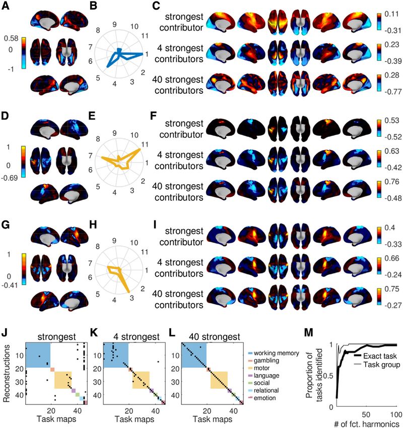
ll
Article                                                                                                                                     OPEN ACCESS

Figure 1. Workflow for the estimation of functional harmonics
(A) Brain activity measured with functional magnetic resonance imaging (fMRI) in resting state for 812 subjects provided by the Human Connectome Project (HCP;
900 subjects data release).
(B) Illustration of brain activity time series of three representative vertices on the cortex (x1, x2,., xn).
(C) The dense functional connectivity (FC) matrix computed from the temporal correlations between the time courses of each pair of vertices as shown in (B)
averaged across 812 subjects.
(D) Representation of the dense FC as a graph, where the edges indicate strong correlations between the corresponding vertices. The anatomical locations of the
vertices are color-coded (Glasser et al., 2016).
(E) Functional harmonics are estimated by the eigenvectors of the graph Laplacian computed on the graph representation of the FC. The first five functional
harmonics ordered from the lowest to higher spatial frequencies are illustrated on the FC graph representation (top), in the eigenvector format as 59,412 3 one-
dimensional vectors (middle), and on the cortical surface (bottom). Note that here we show the patterns on the left hemisphere for illustrative purposes, yet the
entire cortex was used in the analysis. Likewise, the graph representations in (D) and (E) are shown for a parcellated version of the FC matrix using the HCP
parcellation (Glasser et al., 2016), i.e., each node represents an HCP parcel, but the computation of the functional harmonics were performed on the dense FC
using 59,412 3 59,412 without any parcellation.

diagonal matrix that holds the degree (number of connections) of                  of the cortex with similar colors in the surface plots in Figure 2).
each vertex i in its diagonal entries dii.                                        Mathematically, as the eigenvalue increases, (spatial) graph fre-
    By definition, the first functional harmonic with eigenvalue 0 is             quency also increases (Shuman et al., 2013). This intrinsic link
constant over the whole cortex and is discarded. Figure 2 shows                   between the graph frequency and cortical scale implies that
the first 11 non-constant functional harmonics (referred to as j1,                functional harmonics yield not only a multi-dimensional, but
j2, /, j11), ordered starting from the lowest eigenvalue >0, and                  also a multiscale description of the cortex (Tian et al. 2020).
illustrating that each harmonic is a smoothly varying pattern on                  Note that the ordering by graph frequency is a property that
the cortex between a positive and a negative polarity (i.e., a                    emerges from the Laplace eigenfunctions and therefore is not
gradient). To give an intuitive interpretation of functional har-                 present in function bases proposed by other methods, such as
monics, we emphasize that the actual magnitudes of the gradi-                     principle component analysis (PCA) or independent component
ents plotted on the cortex are not per se meaningful, but the dif-                analysis (ICA), which, as a result, do not implicitly possess this
ference between the values assigned to two vertices reflects how                  multiscale property.
different they are in terms of their ‘‘functional connectivity profile’’
(i.e., their pattern of connectivity to the rest of the cortex).                  Functional harmonics capture sub-areal topographic
    With increasing eigenvalue, the functional harmonics become                   organization
increasingly more complex and segregate the cortex into an                        We first tested whether functional harmonics capture cortical or-
increasing number of nodal areas (Levy, 2006) (contiguous areas                   ganization on a sub-areal scale (i.e., within a cortical parcel).

                                                                                                            Cell Reports 36, 109554, August 24, 2021 3
ll
     OPEN ACCESS                                                                                                                  Article
                                                                                             Figure 2. Functional harmonics capture ex-
                                                                                             isting characterizations of functional anat-
                                                                                             omy
                                                                                             The first 11 non-constant functional harmonics
                                                                                             plotted on the cortical surface. It is clearly visible
                                                                                             that the first two functional harmonics (A and B)
                                                                                             constitute global gradients over the entire cortex,
                                                                                             whereas subsequent maps (C–K) include
                                                                                             increasingly more local details. In each functional
                                                                                             harmonic, known functional regions (e.g., C),
                                                                                             processing streams (e.g., E) or networks (e.g., B)
                                                                                             are discernible, and we have annotated the most
                                                                                             conspicuous ones. In order to illustrate that simi-
                                                                                             larly colored patches of cortex correspond to
                                                                                             known functional regions, borders of HCP parcels
                                                                                             have been added (white lines). V1–V4, visual areas
                                                                                             1 to 4; MT, middle temporal visual area; 24 dd, an
                                                                                             area that contains a higher order representation of
                                                                                             the hand; fusiform face complex, an area that re-
                                                                                             sponds specifically to images of human faces. The
                                                                                             functional harmonics were derived from the HCP’s
                                                                                             dense functional connectivity matrix, which is an
                                                                                             average of over 812 subjects.

                                                                                              are defined by the HCP (Glasser et al.,
                                                                                              2016) and form a topographic map of
                                                                                              the surface of the body on the cortex,
                                                                                              which is oriented orthogonally to the
                                                                                              anatomically defined areal boundaries.
                                                                                              These sub-areas correspond to repre-
                                                                                              sentations of the face, upper limbs (map-
                                                                                              ped by moving the hands), eyes, lower
                                                                                              limbs (mapped by moving the feet), and
                                                                                              trunk. We observed somatotopic map-
                                                                                              pings within functional harmonics 3 (c3),
                                                                                              7 (c7), and 11 (c11) (Figures 2C, 2G,
                                                                                              and 2K). Figure 3A illustrates the two-
                                                                                              dimensional subspace formed by func-
                                                                                              tional harmonics 3 (c3) and 11 (c11),
                                                                                              which accounts for the mapping of the
                                                                                              human body onto the somatotopic
                                                                                              regions of the cortex. This example
Specifically, we investigated whether functional harmonics cap-       also illustrates how specialized functional regions can be ex-
ture somatotopy (Penfield and Rasmussen, 1950) and retinotopy         plained by the interaction of functional harmonics across multi-
(Sereno et al., 2001), two major topographic mappings found in        ple dimensions within the functional harmonics framework
the brain. Topographic mappings represent sensory input on            instead of a single gradient (see Figures S1A–S1C for further
the cortical surface such that the relative positions of the recep-   examples).
tors that receive these inputs (e.g., the photo receptors in the         We derived a measure that we term ‘‘somatotopic silhouette
retina) are preserved. In a broader sense, it refers to any mapping   value,’’ which quantifies the degree to which each somatotopic
in which neurons that are close together on the cortical surface in   sub-area is delineated within these three functional harmonics
one functional area project to neurons that are close together in     (see STAR Methods for details). We compared our results to
another functional area. This organization leads to a functional      those obtained from spherical rotations (Alexander-Bloch
gradient within a specialized brain area (Haak et al., 2013).         et al., 2018) of functional harmonics 3 (c3), 7 (c7), and 11 (c11).
   In the HCP parcellation (Glasser et al., 2016), the sensori-       We found that for functional harmonic 3 (c3), the face and foot
motor cortex is, in each hemisphere, divided into five areas          areas were significantly separated from the rest of the cortex
(1, 2, 3a, 3b, and 4) that are defined cytoarchitechtonically         as well as other somatotopic areas; for functional harmonic 7
and using microstructural measures such as cortical thickness         (c7), we found the face and hand areas, and for functional
and myelination (see ‘‘Supplementary neuroanatomical results’’        harmonic 11 (c11), we found the hand areas to be significantly
in Glasser et al. [2016]). However, functional connectivity is not    separated (see Figure S2A; pcorr < 0.05 after Bonferroni correc-
homogeneous within these regions: five somatotopic sub-areas          tion, Monte Carlo tests with 300 rotated versions of the

4 Cell Reports 36, 109554, August 24, 2021
ll
Article                                                                                                                                   OPEN ACCESS

                                                                                                            Figure 3. Functional harmonics capture so-
                                                                                                            matotopy and retinotopy
                                                                                                         (A) Functional harmonics 3 (c3) and 11 (c11) in their
                                                                                                         own space. Vertices are color-coded according to
                                                                                                         their anatomical locations (see Figure 1D), and the
                                                                                                         location of 4 somatotopic areas in this space is
                                                                                                         annotated.
                                                                                                         (B and C) Retinotopies of functional harmonics 4
                                                                                                         (B, c4) and 8 (C, c8). Each panel shows, on the
                                                                                                         right, the colors of the respective functional
                                                                                                         harmonic in early visual areas V1–V4 on a polar
                                                                                                         plot of eccentricity (distance in degree from
                                                                                                         the fovea) and angle on the visual field (see
                                                                                                         legend at the bottom of the figure). On the left,
                                                                                                         the respective functional harmonic is shown on
a flat map of early visual cortex (left hemisphere). V1, V2, V3, and V4: visual areas 1, 2, 3, and 4. The shown figures are derived from the functional
harmonics obtained from the HCP’s dense functional connectivity matrix, which is an average over 812 subjects.

functional harmonics). This finding indicates that functional har-              areas occurs in a hierarchical fashion (i.e., although the sign
monics capture somatotopic organization in the cortex.                          changes in c1 and c2 follow the borders of the visual cortex as
   We next investigated the presence of retinotopic mapping of                  a whole, higher order functional harmonics respect various bor-
early visual regions (V1–V4), where cortical representations of                 ders occurring between V1–V4 within the visual cortex).
the visual field reflect the positions of the receptors in the retina
of the eye such that each vertex within the patterns of functional              Functional harmonics reveal specialized brain areas
harmonics is assigned an eccentricity (distance from the fovea)                 By visual inspection of Figure 2, one notices that patches of cor-
and a polar angle (position in the visual field, i.e., top, bottom,             tex that are colored in the same shade seem to correspond to a
left, right), according to the HCP retinotopy dataset (Benson                   certain degree to known specialized brain regions (parcels)
et al., 2018). It has previously been demonstrated that topo-                   delineated by the HCP parcellation (Glasser et al., 2016) or
graphic organization can be evaluated using functional connec-                  groups of such regions (all parcel borders are shown in Data
tivity gradients, either within the visual system (Glasser et al.,              S1, page 1). This suggests that the gradients described by func-
2016; Haak et al., 2018) or on a whole-brain level (Yeo et al.,                 tional harmonics are flat within many specialized cortical areas,
2011). Examples of polar plots of the retinotopic gradients are                 indicating a near-homogeneous functional connectivity profile.
shown in Figures 3B and 3C (all polar plots are shown in                        In order to quantify the homogeneity within cortical areas of
Figure S2B). To investigate the degree of agreement between                     the functional harmonics shown in Figure 2, we compared the
functional harmonics 1–11 (Figure 2) and the retinotopic map-                   within-area-variability to the average between-area-variability
pings, we measured the correlation between eccentricity as                      of each parcel. The resulting measure is similar to the silhouette
well as polar angle maps and functional harmonic patterns in                    value used in quantifying the quality of cluster solutions (de
V1–V4. We found significant correlations (pcorr < 0.05 after Bon-               Amorim and Hennig, 2015), but ranges between 0 and 1, where
ferroni correction) between the retinotopic eccentricity map and                a value close to 1 indicates that the functional harmonic is homo-
all functional harmonics 1–11 except functional harmonic 9 and                  geneous within that region. We refer to this measure as ‘‘modi-
also between the retinotopic angular map and functional har-                    fied silhouette value’’ (see STAR Methods for details).
monics 1–4, 7–9, and 11.                                                           We applied this analysis to four alternative function bases: (1)
   These results, obtained with whole-brain connectivity, demon-                eigenvectors of the dense FC matrix (Data S1, page 2), (2) eigen-
strate that retinotopic organization of the early visual areas is               vectors of the adjacency matrix (Data S1, page 3), (3) principal
implicitly present in the resting state brain activity (Yeo et al.,             components (PCA) (Data S1, page 4), and (4) independent com-
2011) and is revealed by the functional harmonic basis.                         ponents (ICA) (Data S1, page 55). The first two of these bases
We draw the reader’s attention to the fact that retinotopic                     were chosen in order to test the effect of the two major process-
organization has been shown in rich detail elsewhere, using                     ing steps that are necessary when transforming the dense FC
either retinotopic mapping techniques based on specific visual                  matrix into the Laplacian: (1) the adjacency matrix is obtained
stimuli (Benson et al., 2018; Sereno et al., 2001) or functional                from the dense FC matrix by binarizing (using k = 300 nearest
connectivity within the visual system (Glasser et al., 2016;                    neighbors), and (2) the graph Laplacian is obtained from the ad-
Haak et al., 2018). Thus, although functional harmonics capture                 jacency matrix by normalizing, i.e., taking into consideration the
some prominent aspects of retinotopic organization, there are                   degree of each vertex (LG = D  A). Furthermore, to relate the
also details that require more dedicated approaches.                            performance of functional harmonics to other well-known func-
   It is also important to note that the borders of visual areas V1–            tion bases, we also performed the analysis with the basis func-
V4, which were identified by the existence of orderly topographic               tions of (3) PCA and (4) ICA. As shown in Figure 4A, although
maps with clear visual field reversals (Sereno et al., 1995), corre-            all approaches exhibited moderate to high modified silhouette
spond to a sign reversal between the positive and negative polar-               values, indicating that they all reflected the functional organiza-
ity of the harmonic pattern in various functional harmonics (e.g.,              tion well, we found that functional harmonics had significantly
c5–c10). Yet in functional harmonics, the delineation of the visual             higher modified silhouette values than three of the four function

                                                                                                          Cell Reports 36, 109554, August 24, 2021 5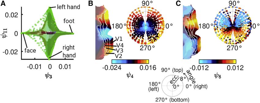
ll
     OPEN ACCESS                                                                                                                  Article
                                                                                             Figure 4. Functional harmonics capture
                                                                                             specialized brain areas
                                                                                             The modified silhouette values (y-axes in all panels)
                                                                                             quantifies the degree to which gradients described
                                                                                             by the functional harmonics as well as control ba-
                                                                                             sis sets are flat within the parcels defined by the
                                                                                             HCP parcellation. A modified silhouette value
                                                                                             close to 1 indicates homogeneous values within
                                                                                             HCP parcels.
                                                                                             (A) A comparison between the basis sets indicates
                                                                                             that functional harmonics and adjacency eigen-
                                                                                             vectors have significantly higher modified silhou-
                                                                                             ette values than the other three (Wilcoxon rank-sum
                                                                                             test, black bars indicate significant differences at
                                                                                             pcorr < 0.05 after Bonferroni correction). Each data
                                                                                             point is computed from a matrix that is an average
                                                                                             over 812 subjects.
                                                                                             (B–F) Modified silhouette values of the first 11 non-
                                                                                             constant components of each basis set (colored
                                                                                             circles; each data point is computed from a matrix
                                                                                             which is an average over 812 subjects) compared
                                                                                             to their rotations (gray crosses). The stars above
                                                                                             each column indicate significant silhouette values
                                                                                             (pcorr < 0.05 after Bonferroni correction, Monte
                                                                                             Carlo test with 220 spherical rotations per
                                                                                             component). Functional harmonics had the highest
                                                                                             number of significant modified silhouette values (10
                                                                                             out of 11).

bases; only eigenvectors of the adjacency matrix had equally         Although it should always be considered that harmonic modes
high modified silhouette values.                                     describe the structure of the underlying graph in a multi-dimen-
   We established significance of the modified silhouette values     sional fashion (as is illustrated for two dimensions in Figure 3A),
obtained from each functional harmonic by computing the              in the following, we provide some insight into the functional signif-
same measure also for n = 220 spherical rotations of the func-       icance of each of the functional harmonics shown in Figure 2.
tional harmonics (Alexander-Bloch et al., 2018) (stars above         Functional harmonics 1 (c1) and 2 (c2) correspond to previously
each column in Figures 4B–4F). By doing so, we ensured that ho-      identified large-scale gradients (Margulies et al., 2016) that delin-
mogeneous gradients within parcels are not due to the proper-        eate the separation between the major sensory and the uni- versus
ties of the harmonic decomposition alone, but are specific to        multimodal cortices in the brain, respectively (see Figure S1D). Fig-
their location on the cortical surface. As shown in Figure 4B,       ures 2A and 2B demonstrate the overlap between the visual and
we found that the modified silhouette values of 10 of the first      sensorimotor networks as defined in Yeo et al. (2011) and the gra-
11 functional harmonics were larger than those of the rotated        diental patterns of the first and second functional harmonics. We
harmonic basis (pcon < 0.05 after Bonferroni correction, Monte       observed that functional harmonic 3 (c3) reveals a finer subdivision
Carlo tests; see STAR Methods for details). The only exception       of the somatosensory/motor system (Zeharia et al., 2012, 2015;
to this finding was functional harmonic 4 (c4), which captures       Kuehn et al., 2017). The overlay of the borders of the five somato-
the retinotopic organization of early visual regions (Figure 3B;     topic areas defined by the HCP (Glasser et al., 2016; Barch et al.,
see above for a discussion of retinotopic organization of func-      2013) on the third functional harmonic are shown in Figure 2C.
tional harmonics), and which is relatively flat over the rest of     Similarly, in functional harmonic 4 (c4), we found a finer segregation
the cortex. When applied to the four alternative function bases,     of the visual system, following a retinotopic eccentricity gradient
the same analysis showed that for eigenvectors of the dense          (Benson et al., 2018). The overlay of the borders of early visual
FC matrix, only 7 out of 11 basis functions had a significantly      areas (V1–V4) on functional harmonic 4 (c4) are shown in Figure 2D
higher modified silhouette value than their spherical rotations;     (for further details on retinotopic and somatotopic mapping, see
for eigenvectors of the adjacency matrix, this number was 8          above).
out of 11, for PCA, only 3 PCs reached significance, and for            Qualitative evaluation of higher frequency functional har-
ICA, this number was 4 out of 11 (Figures 4C–4F). For qualitative    monics systematically revealed their link to more specialized
evaluation, the overlap between parcels and functional har-          complex brain function. The pattern observed in functional har-
monics as well as other bases are shown in Data S1, pages 1–5.       monic 5 (c5) (Figure 2E) is consistent with a functional network
                                                                     in which action and perception interact. In the negative polarity,
Functional harmonics yield graph frequency-specific                  we found primary visual, auditory, and somatosensory cortices,
brain networks                                                       whereas the regions in the positive polarity closely resemble the
In Margulies et al. (2016), the authors investigated the principal   sensory-motor pathway, which has been shown to mediate se-
cortical gradients captured by the first two functional harmonics.   lective interactions between resting state networks along the

6 Cell Reports 36, 109554, August 24, 2021
ll
Article OPEN ACCESS
visual hierarchy (Yeo et al., 2011). Parts of this pathway are functional harmonics (see STAR Methods for details). The 47
known to be modulated by visuospatial attention (Corbetta and maps consist of activation maps as well as contrasts derived
Shulman, 2002). The fact that we also found auditory and so- from 7 groups of tasks (working memory, motor, gambling, lan-
matosensory regions is in line with the idea that the interplay be- guage, social, emotional, and relational; see STAR Methods for
tween action and perception circuits—also known as active summaries). This reconstruction yields a coefficient (weight) for
inference (Friston et al., 2011; Adams et al., 2013)—is a multi- each functional harmonic, quantifying how much it contributes
modal process (Keysers et al., 2010; Hauk and Pulvermu €ller, to a certain task map. The set of all coefficients forms a spectrum
2004; Pulvermu €ller and Fadiga, 2010). In functional harmonic 6 equivalent to the power spectrum obtained from a Fourier trans-
(c6), auditory and visual areas were both localized in the positive form, in this case the power spectrum of the functional harmonic
polarity, forming a network related to audiovisual object basis. We quantified the goodness of fit by measuring the dis-
(including faces) recognition (Beauchamp et al., 2004; Levy tance between the original and the reconstructed task maps.
et al., 2001; Hasson et al., 2003) (i.e., recognition of the ‘‘outer We determined how well the first 11 non-constant functional
world’’). The negative polarity of functional harmonic 6 (c6) seg- harmonics shown in Figure 2 were able to approximate task
regates the somatotopic face area as well as parts of the default maps and compared their performance to the four alternative func-
mode network (DMN), a network of regions whose activity has tion bases mentioned above (eigenvectors of the FC, eigenvectors
been related to self-referential tasks (Gusnard et al., 2001). of the adjacency matrix, principal components, and independent
Thus, the negative polarity of functional harmonic 6 (c6) forms components), as well as spherical rotations of functional har-
a self-referential processing stream (Gusnard et al., 2001; Haxby monics (Alexander-Bloch et al., 2018). Note that the first 11 func-
et al., 2000; Leslie et al., 2004). Functional harmonic 7 (c7) pro- tional harmonics constitute 0.02% of the total functional har-
vides a finer somatotopic gradient, including a higher hand monic spectrum. Due to the intrinsic ordering of functional
area, 24 dd, in the medial cortex (Zeharia et al., 2015) (see Fig- harmonics by graph frequency, using only the first few functional
ure 2G and annotations in Figure 2C). Functional harmonics 8– harmonics omits more localized details of brain activity and thus,
10 (c8, c9, and c10) correspond to different subdivisions of higher the omitted information results in a reconstruction error.
order networks such as the frontoparietal network and DMN (see Figures 5A–5G show the average normalized reconstruction
Figures S3A–S3C). In particular, the DMN (Raichle et al., 2001) is errors for all groups of tasks and for all compared function bases.
delineated in the positive polarity of functional harmonic 9 (c9) First focusing on the functional harmonic basis (red line), the er-
(borders of the DMN as defined by Yeo et al. [2011] are overlaid ror drops from between 1.00 (for emotion) and 1.40 (for lan-
on functional harmonic 9 (c9) in Figure 2I). In functional harmonic guage) to between 0.65 (for emotion) and 0.78 (for language).
10 (c10), we found a significant correlation (r = 0.63, p = 4 $ This corresponds to a level of correlation between the original
1021) with the degree of auditory involvement of the functional task maps and the task maps reconstructed from the first 11
areas (Figure S3D). Functional harmonic 11 (c11), the first clearly non-constant functional harmonics of between 0.78 (for emotion)
asymmetric harmonic between the two hemispheres, yields and 0.69 (for language; see Figure S4A). Figure 5H illustrates the
the separation between the right and left somatotopic hand reconstruction procedure for one specific task (working memory:
areas (Pool et al., 2014). body) (see Data S1, page 6 for all tasks).
Overall, these results demonstrate that functional harmonics We next compared the reconstruction performance of the func-
provide a multitude of functionally relevant macroscopic net- tional harmonics to each alternative function basis, employing
works, each associated with a unique graph frequency. a Monte-Carlo analysis. Again, using only the first 11 non-
constant components, we found that reconstruction errors of
Relating rest and task with functional harmonics functional harmonics were significantly lower than those of their ro-
Functional harmonics, by definition, yield the extension of the tations for each of the task groups (all pcorr < 0.035, Monte-Carlo
Fourier basis to the functional connectivity of the human brain. tests with 1,000 permutations, Bonferroni corrected for multiple
Laplace eigenfunctions on a one-dimensional domain with cyclic comparisons), and significantly lower than those of the adjacency
boundary conditions (i.e., a circle) yield sine and cosine waves eigenvectors in six out of seven task groups (all pcorr < 0.035,
with different frequencies, which constitute the well-known Fourier Monte-Carlo tests with 1,000 permutation, Bonferroni corrected
basis. In a similar manner, we estimated the functional harmonics for multiple comparisons, except language, where p = 0.18 before
by computing the Laplace eigenfunctions on a graph encoding the correction for multiple comparisons, n.s.). In comparison to FC ei-
human brain’s functional connectivity (illustrated in Figure 1D). As genvectors, we found that functional harmonics performed signif-
such, the functional harmonics provide a frequency-specific func- icantly better in the reconstruction of motor tasks (pcorr < 0.035,
tion basis, in which any pattern of brain activity on the graph can be Monte-Carlo tests with 1,000 permutations, Bonferroni corrected
represented as a weighted combination of functional harmonics. for multiple comparisons), whereas there was no significant differ-
Given the experimental evidence showing that resting state func- ence in other task groups (all p > 0.01 before correction for multiple
tional connectivity reflects connectivity during task (Biswal et al., comparisons, n.s.). Compared to PCA and ICA, the reconstruction
1995; Deco et al., 2011; Buckner et al., 2013; Cole et al., 2016), errors of functional harmonics were significantly lower for motor
we tested how efficiently the functional harmonics derived from and working memory task groups (all pcorr < 0.035, Monte-Carlo
the resting state dense FC can represent task-induced cortical tests with 1,000 permutations, Bonferroni corrected for multiple
activity. comparisons), whereas for all other task groups, there were no sig-
To this end, we reconstructed 47 group-level task maps pro- nificant differences (all p > 0.01 before correction for multiple com-
vided by the HCP (Barch et al., 2013) from the superposition of parisons, n.s.). We also tested the performance of the alternative
Cell Reports 36, 109554, August 24, 2021 7ll
OPEN ACCESS Article
Figure 5. A small set of functional harmonics suffices to reconstruct diverse task activity maps
(A–G) Mean reconstruction errors for each of the 7 task groups and all 6 basis function sets when only the first 11 non-constant components are used (see Figure
S4 for results when using more components).
(H) One example for a reconstruction using a working memory task. The bottom panel is the original task activation map (working memory—body; see also Data
S1, page 6, panel l), and top panels use the number of harmonics indicated on the left to reconstruct it.
(I) Results of significance tests comparing normalized reconstruction errors of functional harmonics to other function bases (significant differences at pcorr < 0.05
after Bonferroni correction, Monte Carlo tests with 1,000 permutations). Top: When using only the first 11 components, functional harmonics outperformed each
of the other function bases. Bottom: When using the first 100 components (Figure S4), eigenvectors of the dense FC outperformed functional harmonics in 3 task
groups. All basis sets were derived from matrices which are averages over the same 812 subjects. The task activity maps (Cohen’s D activation contrast maps)
are based on 997 subjects.
8 Cell Reports 36, 109554, August 24, 2021ll
Article OPEN ACCESS
function bases when the first 100 components were used (Data S1, working memory task (Figure 6K), and 70% of all individual tasks.
page 6). We found several changes in contrast to the results with When the 40 functional harmonics with maximum contribution
only the first 11 components. First, FC eigenvectors outperformed were used, which corresponds to 0.1% of the complete spec-
functional harmonics in working memory, language, and social trum of functional harmonics, 44 out of 47 task maps were
tasks, whereas there was no significant difference in the other correctly identified from their reconstructions (Figure 6L).
task groups. Second, functional harmonics outperformed PCA in These results demonstrate that functional harmonics provide a
all seven task groups instead of only in motor and working memory. functionally relevant representation, where the brain activity
Third, for ICA, only 15 ICs were extracted, and when all of them accompanying different tasks can be unambiguously identified
were used, functional harmonics outperformed ICA in social from the activation profiles of a small range of functional
and emotional tasks in addition to working memory and motor harmonics.
tasks as before. The results remained the same for adjacency
eigenvectors. DISCUSSION
Figure 5I provides a summary of the results. The first 11 non-
constant functional harmonics performed at least as well as In this paper, we show that harmonic modes of the vertex-level
the tested alternative function bases in all task groups (i.e., functional connectivity of the human cortex, termed functional
none of the alternative function bases outperformed functional harmonics, link observations spanning several spatial scales,
harmonics in any of the task groups). When 100 components from sub-areal topographic mappings (somatotopy, retinotopy),
were used, FC eigenvectors outperformed functional harmonics to delineation of specialized functional brain areas, to the level of
in three task groups. large-scale networks and combinations thereof. In this view,
Next, we wanted to confirm that the decrease in reconstruc- functional integration and segregation on all these levels can
tion error did not merely reflect the fact that functional harmonics be accounted for by the same mathematical framework (i.e., har-
capture global features of brain activity maps common to all monic modes).
tasks. We designed an analysis that allowed us to test whether Although it is abundantly clear from the literature that modular
functional harmonics are able to capture brain activity that is and gradiental organization are complementary principles in the
specific to a task or group of tasks. Figures 6A, 6D, and 6G human brain, a mathematical framework that can account for
and Figures 6B, 6E, and 6H show three examples of task activa- both has thus far been missing. Within the functional harmonic
tion maps and the corresponding normalized power of the first framework, specialized regions as well as networks of function-
11 non-constant functional harmonics, respectively, revealing ally related regions correspond to patches of the cortical surface
how strongly each of the 11 functional harmonics shown in Fig- within which the gradiental patterns of functional harmonics are
ure 2 contributes to these particular task maps. In other words, relatively constant. Such constant regions are then increasingly
Figures 6B, 6E, and 6H show spectral representations of the segregated into finer regions through the interaction of functional
task maps in Figures 6A, 6D, and 6G. For qualitative evaluation, harmonics across multiple dimensions, as was recently shown to
we display the task activation maps reconstructed by superim- be the case for the subcortex by Tian et al. (2020).
posing functional harmonics in the order of their contribution We have interpreted the first 11 functional harmonics in rela-
strength for varying numbers of functional harmonics in Figures tion to existing characterizations of functional anatomy, like the
6C and 6F (see Data S1, page 6 for all tasks). Across all 47 concordance between functional harmonic 5 and the sensory-
task maps that were evaluated, the functional harmonic that motor pathway (Yeo et al., 2011). Thus, in this framework,
was the strongest contributor was always either the constant functional networks observed during rest and task emerge as a
functional harmonic or one of the first 11 non-constant har- graph frequency-specific function basis derived from the human
monics shown in Figure 2. resting state functional connectivity matrix.
In order to confirm that each spectral representation was spe- Harmonic modes are initially a dimensionality reduction tech-
cific to the task map from which it was obtained, we computed nique: they are estimated as the eigenvectors of the graph Lapla-
the distance between a given reconstructed map and all original cian. As such, they are ordered by graph frequency and orthogonal
task maps, resulting in a confusion matrix for each number of to one another. By definition, any pattern of cortical activity can be
harmonics with maximum contribution. If a spectral representa- expressed in this function basis as a superposition of functional
tion is indeed specific to a task map, the error should be minimal harmonics. In this new function basis, brain activation patterns
between a reconstruction and its corresponding task map measured during tasks are expressed in a convenient and
compared to the error of the reconstruction of the other 46 compact manner as task-specific spectra that quantify the contri-
task maps. The confusion matrices in Figures 6J–6L show the bution of each functional harmonic.
pairs of the original and reconstructed task activation maps We tested how well functional harmonics are able to capture
with the minimum distance when using 1, 4, and 40 functional task activity patterns (reconstruction errors, as shown in Fig-
harmonics with maximum contribution. Colored squares mark ure 5), as well as specialized brain regions (modified silhouette
the 7 task groups as in Figure 5. The proportion of unambigu- values, as shown in Figure 4), and compared them to other
ously identified tasks depending on the number of function bases on these two spatial scales. In both regards,
functional harmonics is shown in Figure 6M. We found that functional harmonics outperformed PCA and ICA, two very
sparse representations using the 4 functional harmonics with popular dimensionality reduction techniques, suggesting that
the largest power for each task are sufficient to unambiguously the non-linear nature of harmonic modes captures important
characterize the seven task groups with the exception of one properties of cortical organization. The remaining two function
Cell Reports 36, 109554, August 24, 2021 9ll
OPEN ACCESS Article
Figure 6. Functional harmonics provide a
characterization of task activity maps
(A) Original task map of the contrast between
working memory (face) and average working
memory from the HCP task dataset (Barch et al.,
2013).
(B) Spectral representation of the task map shown
in (A) (i.e., the normalized coefficients of the graph
Fourier transform quantify the contribution of the
first 11 non-constant functional harmonics to the
task map). The color indicates the task group (see
legend in L).
(C) Reconstruction of the task map in (A) when
using the functional harmonic with the strongest
contribution (highest coefficient) only, the four
functional harmonics with the strongest contribu-
tions, and the forty functional harmonics with the
strongest contributions.
(D–F) The same as (A)–(C) using the map of the
contrast between motor (right hand) and average
motor.
(G–I) The same as (A)–(C) using the map of the
contrast between motor (trunk) and average
motor.
(J–L) Confusion matrices. Black entries mark the
task map-reconstruction-pair that has the lowest
reconstruction error; colored squares indicate the
task group.
(M) Proportion of reconstructions, for each number
of harmonics, which have the minimum recon-
struction error with their exact original task map
(thick line) and a task map belonging to the same
group of tasks as the original map (thin line).
Functional harmonics were derived from the HCP’s
dense functional connectivity matrix, which is an
average of over 812 subjects. The task activity
maps (Cohen’s D activation contrast maps) are
based on 997 subjects.
bases were chosen because they constitute steps in our pro- functional networks that are activated in order to fulfill task de-
cessing pipeline. Eigenvectors of the dense FC performed mands. In addition, the continuous nature of functional har-
equally well or better than functional harmonics in task recon- monics enables them to combine brain regions into macroscopic
struction but performed significantly worse in capturing brain networks in a manner that allows the shape of the regions to vary
regions. This is interesting because it suggests that although depending on context (Salehi et al., 2020). Future research
details of the FC that are not preserved in the k = 300 nearest should evaluate whether the exact shapes of (combinations of)
neighbor graph shape task activity, the same does not seem to functional harmonics is in line with activation studies using
be true for parcel boundaries. In fact, with regard to the parcel- different tasks.
lation, eigenvectors of the adjacency matrix performed almost Beyond dimensionality reduction, the interpretability of func-
as well as functional harmonics, and significantly better than tional harmonics demonstrated in this paper, their multi-scale
FC eigenvectors, suggesting that keeping only the nearest properties, and their orthogonality make them a candidate orga-
neighbors is actually beneficial for delineating parcel borders. nizational principle that could explain how the brain flexibly
At the same time, eigenvectors of the adjacency matrix were switches between overlapping functional networks: functional
outperformed by functional harmonics in task reconstruction. harmonics are, by definition (Belkin and Niyogi, 2003), the pat-
It should be noted, however, that the differences between func- terns with the least overall variation on the cortex that respect
tional harmonics, FC eigenvectors, and adjacency eigenvectors the constraints posed by the functional relationships given by
were slight in reconstruction performance. We speculate that the FC. This implies that the average difference between neigh-
the reason for these contrasting results could be that the par- boring nodes in a graph representation is minimized. Intriguingly,
cellation captures average functional regions, whereas during theoretical work has shown that activation patterns on graphs in
task, parcel borders can shift and adapt (Salehi et al., 2020), which neighboring nodes co-activate lead to patterns with min-
consistent with the idea that transitions between parcels may imum free energy or entropy (Tomasi et al., 2013; Gu et al., 2018;
be gradiental (Bailey and Von Bonin, 1951; Bajada et al., 2020). Friston, 2010), and the transition between such patterns requires
The approach of functional harmonics allows interpreting minimal energy (Gu et al., 2015). This means that transitioning
cortical activity patterns as different combinations of the same between the functional networks instantiated by the functional
10 Cell Reports 36, 109554, August 24, 2021ll
Article OPEN ACCESS
harmonics in response to changing task demands is optimally fying the similarity of connectivity profiles in an intermediate
efficient. In addition, functional harmonics and the networks to step. The fact that the first two functional harmonics look virtually
which they correspond emerge in a wholly self-organizing identical to what was presented in Margulies et al. (2016) speaks
fashion from the functional connectivity. to the robustness of these principal cortical gradients toward the
Another important conceptual aspect of harmonic modes is specific technique (they also used a slightly different definition of
their grounding in physical theories linking them to standing the Laplacian, and k was around 6,000, i.e., 10% of the connec-
waves. This suggests that each spatial pattern on the graph tions were retained).
might be linked to a unique set of temporal frequencies. Such Considering that the principle of harmonic modes, when
a link would make it possible to interpret functional harmonic applied to the structural connectivity of the human brain—the
modes as ‘‘communication channels’’ along the lines of the ideas human connectome—has been shown to reveal functional
of multiplexing (Akam and Kullmann, 2014). In Haak et al. (2018), networks (Atasoy et al., 2016), our results point to the existence
the authors demonstrated that harmonic modes applied to the of the same fundamental principle in multiple aspects of
visual system were able to ‘‘tease apart’’ retinotopic gradients human brain function, including functional integration and
in separate harmonic modes. In parallel to this idea, we assert segregation. Beyond the results presented here, functional har-
that functional harmonics are able to ‘‘tease apart’’ overlapping monics suggest novel ways to understand the dynamics of the
functional networks subserved by specialized brain regions human brain in health and in pathology, as well as to explore
with multiple functions. For example, area MT, annotated in Fig- individual differences within this multi-dimensional harmonic
ures 2E and 2F, is part of the sensory-motor pathway (Figure 2E) representation.
as well as audiovisual processing (Figure 2F). The idea of
‘‘communication channels’’ suggests that depending on which STAR+METHODS
function area MT is fulfilling at any given moment, the network
that it is engaged in would synchronize using different temporal Detailed methods are provided in the online version of this paper
frequencies. and include the following:
Taken together, we conclude that although functional har-
monics perform very well in both capturing the HCP parcellation d KEY RESOURCES TABLE
and task map reconstruction, they offer specific benefits in terms d RESOURCE AVAILABILITY
of interpretability. This makes them an excellent choice not only B Lead contact
as a tool for dimensionality reduction, but for exploring cortical B Materials availability
organization. B Data and code availability
Despite the relative simplicity of the approach presented here d EXPERIMENTAL MODELS AND SUBJECT DETAILS
as compared to, e.g., ICA, there are several technical issues B General information
that need to be addressed in the future in order to ensure robust- B Dense functional connectivity matrix
ness of the eigenmaps discussed in this work. In particular, the B Retinotopy data
choice of how to set the weight in the adjacency matrix, the B Task maps
choice of Laplacian, and the choice of k (number of nearest neigh- d METHOD DETAILS
bors) are free parameters, the impact of which needs to B Software
be systematically explored in the future. Here, we obtained highly B Background: Functional Harmonics
interpretable cortical patterns using the technique of Laplacian B Control function bases
eigenmaps (Belkin and Niyogi, 2003) with a binary adjacency ma- B Modified silhouette values
trix with k = 300 nearest neighbors. Although this can be seen as a B Reconstructing task maps
standard approach, connection weights in the adjacency matrix B Somatotopic areas
could be set in a graph distance-dependent manner, or the orig- B Parcel borders for visualization
inal correlation values could be used (Belkin and Niyogi, 2003); B Third-party software
instead of the combinatorial Laplacian, one could use the sym- d QUANTIFICATION AND STATISTICAL ANALYSIS
metric normalized Laplacian. In this study, we did not find any sig- B Monte Carlo simulations
nificant difference when using the normalized Lapalcian. The B Auditory hierarchy
choice of k was previously discussed regarding a related tech-
SUPPLEMENTAL INFORMATION
nique, the Isomap algorithm (Tenenbaum et al., 2000; Balasubra-
manian et al., 2002): k should be chosen such that a Supplemental information can be found online at https://doi.org/10.1016/j.
balance between local and global features is achieved. The celrep.2021.109554.
amount of local and global connections has been suggested to
have a major impact on the performance of the harmonic decom- ACKNOWLEDGMENTS
position (Haak et al., 2018; Naze et al., 2021). At the same time, k
might vary in a subject- or state-specific manner, revealing K.G. and P.H. are supported by Swiss National Science Foundation (170873).
M.L.K. is supported by the Center for Music in the Brain funded by the Danish
further interesting properties of cortical organization.
National Research Foundation (DNRF117), and Centre for Eudaimonia and Hu-
It should be noted that our approach differs from that used in man Flourishing funded by the Pettit and Carlsberg Foundations. G.D. is sup-
Margulies et al. (2016) and Haak et al. (2018), in that it uses an ported Spanish National Research Project (PID2019-105772GB-I00 MCIU AEI)
adjacency matrix derived directly from the FC instead of quanti- funded by the Spanish Ministry of Science, Innovation and Universities (MCIU),
Cell Reports 36, 109554, August 24, 2021 11You can also read