Epithelial IL-33 appropriates exosome trafficking for secretion in chronic airway disease - JCI Insight
←
→
Page content transcription
If your browser does not render page correctly, please read the page content below
Epithelial IL-33 appropriates exosome trafficking for secretion in
chronic airway disease
Ella Katz-Kiriakos, … , Mark J. Miller, Jennifer Alexander-Brett
JCI Insight. 2021. https://doi.org/10.1172/jci.insight.136166.

 Research   In-Press Preview Immunology        Pulmonology

Graphical abstract

Find the latest version:
https://jci.me/136166/pdf
TITLE: Epithelial IL-33 appropriates exosome trafficking for secretion in chronic
airway disease

AUTHORS: Ella Katz-Kiriakos1, Deborah F. Steinberg1, Colin E. Kluender1, Omar A.

Osorio1, Catie Newsom-Stewart1, Arjun Baronia1, Derek E. Byers1, Michael J.

Holtzman1,2, Dawn Katafiasz3, Kristina L. Bailey3, Steven L. Brody1, Mark J. Miller4,

Jennifer Alexander-Brett1,5*

Affiliations:
1Department     of Medicine, Division of Pulmonary and Critical Care Medicine

Washington University School of Medicine, Saint Louis, MO, USA
2
    Department of Cell Biology and Physiology

Washington University School of Medicine, Saint Louis, MO, USA
3Department     of Medicine, Division of Pulmonary, Critical Care, Sleep and Allergy

University of Nebraska Medical Center, Omaha, Nebraska, USA
4Department     of Medicine, Division of Infectious Diseases

Washington University School of Medicine, Saint Louis, MO, USA
5Department     of Pathology and Immunology

Washington University School of Medicine, Saint Louis, MO, USA

*Corresponding    Author: Jennifer Alexander-Brett, Campus Box 8052 660 S. Euclid Ave.

Washington University St. Louis, MO 63110 phone: 314-273-1554 email:

jalexand@wustl.edu

                                                                                        2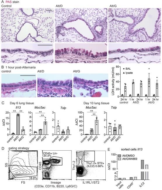
CONFLICT OF INTEREST STATEMENT: MJH serves as a member of the data and

safety monitoring board of clinical trials done by AstraZeneca and as president/founder

NuPeak Therapeutics. All other authors declare no conflict of interest.

                                                                                          3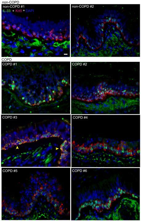
ABSTRACT

Interleukin-33 (IL-33) is a key mediator of chronic airway disease driven by type-2 immune

pathways, yet the non-classical secretory mechanism for this cytokine remains undefined.

We performed a comprehensive analysis in human airway epithelial cells, which revealed

that tonic IL-33 secretion is dependent on the ceramide biosynthetic enzyme neutral

sphingomyelinase 2 (nSMase2). IL-33 is co-secreted with exosomes by the nSMase2-

regulated multivesicular endosome (MVE) pathway as surface-bound cargo. In support

of these findings, human chronic obstructive pulmonary disease (COPD) specimens

exhibited increased epithelial expression of the abundantly secreted IL33Δ34 isoform and

augmented nSMase2 expression compared to non-COPD specimens. Using an

Alternaria-induced airway disease model, we found the nSMase2 inhibitor GW4869

abrogated both IL-33 and exosome secretion as well as downstream inflammatory

pathways. This work elucidates a novel aspect of IL-33 biology that may be targeted for

therapeutic benefit in chronic airway diseases driven by type-2 inflammation.

                                                                                        4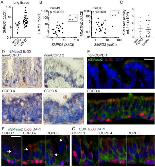
INTRODUCTION

       IL-33 is a cytokine abundantly expressed at mucosal barriers (1) that has been

shown to promote type 2-polarized immune programs in health and disease (2, 3). A role

for IL-33 in human type-2 driven chronic airway disease was highlighted by genome-wide

association studies linking IL33 and IL1RL1/ST2 with asthma (4-6), and increased IL-33

in serum, sputum or tissue from asthma (7, 8) and chronic obstructive pulmonary disease

(COPD) patients (9-11). Animal models have also supported a role for IL-33 in response

to virus (9) or allergen (12, 13) triggers of airway disease. The IL-33 system is also broadly

linked to pulmonary inflammation, arthritis, inflammatory bowel disease, hepatitis, heart

failure, central nervous system inflammation and cancer (reviewed in (2)). This wide-

ranging disease relevance emphasizes the necessity to understand how a nuclear-

targeted cytokine or ‘nucleokine’ lacking a classical secretion signal can be released from

intact cells to propagate inflammation.

       Human full-length IL-33 (IL-33full) is a 270-amino acid protein encoded by eight

exons that encompass the N-terminal ‘chromatin-interacting’ domain (NTD) and C-

terminal ‘cytokine’ domain (CTD). The NTD chromatin-interacting motif (14) appears to

be responsible for tightly sequestering IL-33 within the nucleus, presumably to regulate

the cytokine, as a specific nuclear function for full-length IL-33 protein has yet to be firmly

established (15). The CTD includes exons 5-8 and is sufficient to induce signaling through

the IL-33 receptor (IL-1RL1/ST2 and IL-1RAP) (16). Unlike the prototypic IL-1 family

member, IL-1β, IL-33 is not cleaved by inflammasome-associated caspases (17, 18).

Apoptotic and inflammatory proteases can process full-length IL-33 in vitro, which can

result in either enhanced function (19) or deactivation (20, 21). While these in vitro studies

                                                                                             5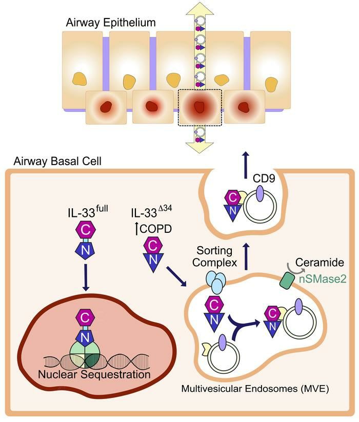
have shown processed cytokine retains activity, it remains unknown whether proteolysis

is required for activation or secretion of endogenous IL-33 in vivo.

       Interleukin-33 secretion has been investigated under multiple conditions and two

primary scenarios have unfolded in the literature (reviewed in (22)): passive ‘alarmin’

release from necrotic cells during tissue damage versus non-classical secretion from

intact cells. In the context of airway disease, passive IL-33 release could occur with acute

airway damage, but this transient activity is unlikely to account for maintenance of chronic

disease. A mechanism based on non-classical secretion appears to be the more likely

basis for persistent IL-33 activity. Support for this mechanism is provided by the recent

description of natural spliced IL33 isoforms lacking portions of the NTD (23, 24), which

may not be regulated by nuclear sequestration.

       To investigate the mechanism of epithelial IL-33 secretion in chronic airway

disease, we took advantage of the properties of these truncated IL33 isoforms. We

expressed IL-33 protein variants in primary human airway basal cells as dual-fluorescent

and peptide tagged fusion proteins, demonstrating secretion of intact protein through the

neutral sphingomyelinase-2 (nSMase2)-dependent multivesicular endosome pathway

(MVE). IL-33 can be co-secreted non-covalently bound to small extracellular vesicles

(~100-150 nm diameter) commonly referred to as exosomes (25). We also leveraged

chronic obstructive pulmonary disease (COPD) specimens to investigate this pathway

and found the IL-33 isoform lacking NTD exons 3 and 4 (IL33Δ34) and nSMase2 were

increased in COPD-derived specimens relative to non-COPD controls. Interleukin-33 was

isolated from COPD bronchial wash fluid and the primary species present was a

truncated, bioactive form with immunoreactivity consistent with the IL-33Δ34 variant. Using

                                                                                          6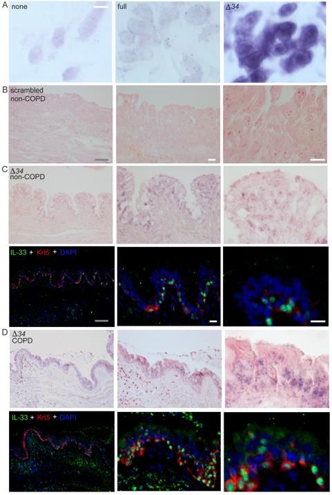
an Alternaria-induced model of airway disease, we demonstrated that blockade of

nSMase2 with GW4869 resulted in reduced IL-33 and exosome secretion in

bronchoalveolar lavage fluid and downstream type-2 inflammation. Together these data

reveal a mechanism for IL-33 secretion from intact airway cells and demonstrate a

potential avenue for development of novel therapeutics in type-2 endotypes of chronic

airway disease driven by this cytokine pathway.

                                                                                   7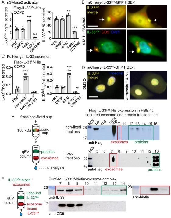
RESULTS

Tonic secretion of truncated IL-33 variants from intact airway cells

We leveraged a series of known N-terminal truncated IL33 isoforms (23, 24) lacking exons

3-5 (Figure 1A) to investigate the mechanism of non-classical secretion from intact airway

epithelial cells. We transduced airway basal cells with lentiviruses expressing dual-tagged

IL-33 fusion proteins, comprised of either N-terminal mCherry and C-terminal monomeric

eGFP (mCherry-IL-33-GFP) or N-terminal Flag and C-terminal 6-His tags (Flag-IL-33-His)

(Figure 1B). This strategy allowed simultaneous tracking of N- and C-terminal fragments,

should proteolysis occur in the process of secretion. We imaged live COPD basal cells

(Figure 1C) and stable HBE-1 cell lines (Figure S1A&B) expressing mCherry-IL-33-GFP

fusion proteins including full-length IL-33 (IL-33full), single exon-deletion variants (IL-33Δ3,

IL-33Δ4, IL-33Δ5), compound deletion variants (IL-33Δ34 and IL-33Δ345) and a non-natural

IL-33Δ2 variant (for comparison). Results demonstrated tight nuclear mCherry and GFP

signal for IL-33full and all single-exon deletion variants. In contrast, the compound deletion

variants exhibited mixed nuclear and cytoplasmic staining apparent by both

epifluorescence (Figure 1C) and confocal imaging (Figure S1B). The strict merged signal

for all variants indicated proteins were intact within respective cellular compartments.

       To determine whether altered cellular localization impacted IL-33 secretion

efficiency, we performed IL-33 ELISA on cell supernatants and lysates for epithelial cells

expressing IL-33 variants as both mCherry-GFP and Flag-His fusions (Figure 1D&E,

Figure S1C,D&E). In both COPD and non-COPD basal cells as well as the epithelial HBE-

1 cell line, we found IL-33Δ34 exhibited the most abundant protein expression and tonic

                                                                                              8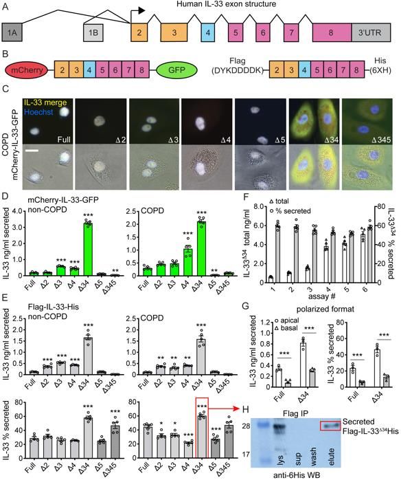
secretion among the isoforms tested. When normalized to account for differences in

protein expression levels (% secreted, Figure 1E, Figure S1C&D), IL-33Δ34 was still

secreted more efficiently than any other variant, which was stable over a 10-fold range of

total protein expression (Figure 1F). The IL-33Δ345 variant also exhibited increased

secretion efficiency, but total expression was well below other variants and near assay

detection limits.

       We also tested vectoral secretion of IL-33full and IL-33Δ34 from non-COPD basal

cells assayed in polarized format (Figure 1G). Flag-IL-33-His secretion was measured in

apical and basal compartments of confluent cultures grown on transwell supports, which

showed that both IL-33full and IL-33Δ34 were secreted in both directions, but more

abundantly from the apical surface. Similar to non-polarized format, the IL-33Δ34 variant

was secreted more efficiently (2-fold) over IL-33full. Results were not influenced by the pore

sizes in the transwell support (Figure 1G, Figure S1F).

       To investigate the role of proteolysis in tonic secretion, we Flag-immunoprecipitated

(Flag-IP) Flag-IL-33Δ34-His from cell supernatants and analyzed by anti-6His western blot

(Figure 1H). Flag-IP IL-33Δ34 protein from supernatant migrated at the same molecular

weight (MW) as protein from cell lysate and was detected by anti-6His western blot,

indicating that secreted protein was intact (not proteolytically processed). This is consistent

with imaging of mCherry-IL-33-GFP isoforms, which demonstrated merged signal in both

nuclear and cytoplasmic compartments.

       Together, these results demonstrate that the protein product of the natural IL33Δ34

isoform can be abundantly expressed in airway basal cells, exhibits cytoplasmic

accumulation due to lack of nuclear targeting, and is tonically-secreted at high levels

                                                                                             9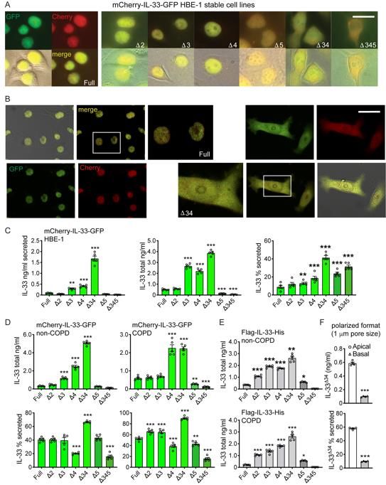
preferentially from the apical surface without proteolytic processing. These findings

support a model in which IL33 isoforms with altered cellular localization are released from

nuclear regulation to undergo tonic secretion from the base of the epithelium

predominantly toward the airway lumen.

Epithelial IL-33 and exosomes are secreted through the nSMase2-depedent

multivesicular endosome (MVE) pathway

The protein products of all IL33 isoforms lack a signal sequence to mediate secretion via

the ER-Golgi pathway, therefore the tonic secretion observed in our cellular assay must

occur via a non-classical mechanism. Multiple routes of non-classical protein secretion

have been described, including active transporters (26), cell-death inducing membrane

pores (27), budding from the plasma membrane (microvesicles) or multivesicular

endosomes (MVE) that fuse with the cell surface to release small intraluminal extracellular

vesicles or ‘exosomes’ containing protein and miRNA cargo (28).

      To address which non-classical secretion pathway was involved, we tested a panel

of chemical inhibitors in our IL-33 ELISA secretion assay. We performed inhibition

experiments in non-COPD and COPD basal cells and HBE-1 cells expressing Flag-IL-

33Δ34-His and mCherry-IL-33Δ34-GFP (Figure 2A, Figure S2A&B). The ER-Golgi inhibitors

brefeldin and monensin had no effect on secretion, however, we did observe a marked

inhibition of IL-33Δ34 secretion following treatment with GW4869, a non-competitive

antagonist of the ceramide synthetic enzyme neutral sphingomyelinase 2 (nSMase2) (29)

(Figure 2A). GW4869 was found to inhibit IL-33Δ34 secretion in both non-COPD and COPD

basal cells and HBE-1 cells, which was apparent as both the amount and % secreted

                                                                                        10
(Figure 2A, Figure S2A&B). Other putative nSMase2 inhibitors spiroepoxide (29),
glutathione (29) and cambinol (30) and the microautophagy inhibitor 3-methyladenine (3-
MA) (31) exhibited a modest effect on secretion, which was variably significant among
non-COPD, COPD and HBE-1 cells tested. When GW4869 was tested against the panel
of IL-33 variants in COPD cells, it was found to globally inhibit secretion for variants that
exhibit distinct cellular localization patterns and expression levels (Figure 2B, Figure S2C).
The ceramide biosynthetic enzyme neutral sphingomyelinase 2
(nSMase2/SMPD3) regulates the ESCRT-independent MVE pathway through maturation
and membrane fusion of multivesicular endosomes (29). Our observed inhibition of tonic
epithelial IL-33 secretion with the compound GW4869 suggested this that phenomenon
may be dependent on nSMase2 activity. We first examined SMPD3 expression in airway
basal cells, which revealed significantly increased expression in COPD relative to non-
COPD specimens (Figure 2C). NSMase2 enzyme activity was measured in a subset of
cultured cell lysates, and multiple COPD specimens exhibited increased activity compared
to non-COPD. Given these findings, we targeted the nSMase2-dependent MVE pathway
with shRNA knockdown and compared to mediators of the ESCRT-dependent MVE
(VPS4A) (32) and microautophagy (LAMP2) (33) pathways. Experiments using primary
cells also included the ROCK inhibitor Y27632, which functions to block microvesicle (MV)
shedding from the plasma membrane (34). In COPD cells, we performed serial rounds of
transduction with shRNA- and Flag-IL-33Δ34-His-expressing lentiviruses, followed by
selection and recovery (Figure 2D). Successful knockdown of targets was confirmed by
qPCR (Figure S2E). We were careful to fully recover cells prior to secretion assay, to
avoid ongoing cell death from selection complicating interpretation. Subsequent IL-33
11secretion assay demonstrated a 2-fold reduction in IL-33Δ34 secretion only with nSMase2
shRNA knockdown, based on both absolute and % secretion (Figure 2D, Figure S2D). As
a confirmatory approach we developed an HBE-1 SMPD3-/- cell line, which revealed
further (3-fold) reduction in IL-33Δ34 absolute and % secretion (Figure 2D, Figure S2G).
COPD cells expressing Flag-IL-33Δ34-His were also immunostained with anti-Flag and
corresponding antibodies for VPS4A, LAMP2, and nSMase2, demonstrating a vesicular
staining pattern for pathway intermediates and diffuse nucleocytoplasmic staining for IL-
33Δ34 (Figure 2E). Immunostaining was also performed in cells expressing nSMase2
shRNA and Flag-IL-33Δ34-His, which demonstrated a loss of nSMase2 staining and an
accumulation of cytoplasmic IL-33Δ34 (and loss of nuclear staining).
To investigate the impact of nSMase2 knockdown on extracellular vesicle (EV)
production, we analyzed EVs secreted from COPD cells (Figure 2F). Culture supernatants
were concentrated using a 100 kilodalton (kDa) cutoff centrifugal filter and the
concentrations of particlescontrol, suggesting that disruption of any pathway intermediate could have global effects
on vesicle biogenesis.
We also sought to determine whether this is a general phenomenon as IL-33
expression has been reported in multiple cell types, including some immune cell
populations (13, 35). We found that the human mast cell line HMC 1.2 exhibited 150-fold
lower SMPD3 expression compared to airway basal cells (Figure 2H) and therefore
transduced these cells with Flag-IL-33Δ34-His and performed secretion assay. Despite
adequate cellular protein expressed in cell lysate, no IL-33Δ34 protein could be detected
in cell supernatants, including under GW4869 treatment conditions (Figure 2I).
Together, these data reveal that tonic epithelial IL-33Δ34 secretion can be blocked
by nSmase2 inhibition using both pharmacologic and genetic approaches. This coupled
with reduction in secreted exosomes under these same conditions, strongly implicates the
nSMase2-dependent exosome biogenesis pathway in non-classical IL-33 secretion.
Parallel analysis in other cell types further suggests that IL-33 secretion could be a
specialized function of nSMase2-expressing cells.
Augmenting nSMase2 promotes IL-33 secretion
We used the nSMase2 activator 4-methylumbelliferone (4-MU, (36)) to determine whether
modulation of nSMase2 activity could promote IL-33Δ34 secretion. Treatment of Flag-IL-
33Δ34-His expressing COPD cells resulted in a 2-fold increase in secreted IL-33Δ34 protein
and a 10% increase in secretion efficiency, which could be reversed using the non-
competitive nSMase2 inhibitor GW4869 (Figure 3A). Live-cell imaging of the mCherry-IL-
33Δ34-GFP HBE-1 line treated with both the nSMase2 activator 4-MU and GW4869 in an
13effort to trap MVEs at the point of secretion demonstrated foci of merged IL-33 signal near
the plasma membrane (Figure 3B). Cells were then fixed under the same conditions and
immunostained for the exosome marker CD9, which demonstrated these IL-33-GFP foci
also contained CD9.
In contrast to IL-33Δ34, full-length IL-33 appears to be tightly sequestered in the
nucleus of airway cells. We next asked whether altered cytoplasmic trafficking of IL-33full
could drive secretion of this variant through the nSMase2-dependent MVE pathway. We
tested this under conditions of nuclear import inhibition with ivermectin (37) and with the
nSMase2 activator 4-MU. With ivermectin treatment, we observed a ~2-fold increase in
secreted IL-33full protein (Figure 3C) which could be reversed by GW4869. Likewise, 4-
MU induced a 3-fold enhancement of Flag-IL-33full-His secretion, also sensitive to GW486,
suggesting both nuclear import blockade and nSMase2 activation function to shunt
cytoplasmic IL-33full toward the MVE secretory pathway. To visualize altered trafficking of
IL-33full the mCherry-IL-33full-GFP HBE-1 line was treated with both of Ivermectin and 4-
MU and live-cell imaging was performed, which demonstrated accumulation of
cytoplasmic mCherry-IL-33full-GFP signal with sustained signal within the nucleus (Figure
3D). We interpret these results as augmented secretion due to altered trafficking of newly
synthesized (overexpressed) IL-33full protein rather than cytoplasmic translocation of
nuclear sequestered protein.
This data would suggest that recruitment of IL-33 protein to the nSMase2-
dependent MVE pathway could occur for any IL33 isoform expressed under cellular
conditions where the cytokine accumulates within the cytoplasm and that nSMase2
activation can enhance secretion efficiency accordingly.
14IL-33Δ34 is secreted with exosomes as surface-bound cargo
As the secretion of IL-33 and exosomes are both sensitive to nSMase2 activity, we next
investigated whether IL-33 was in fact associated with exosomes, potentially secreted as
cargo upon MVE fusion. We concentrated Flag-IL-33Δ34-His-expressing HBE-1
supernatant with a 100 kDa centrifugal filter and western blot analysis revealed IL-33Δ34
was retained above the filter, even though free protein (MW 25 kDa) would be expected
to flow-through (Figure 3E). We then resolved exosomes from free proteins by size
exclusion chromatography, and western blot on column fractions revealed IL-33Δ34 signal
was absent from exosome fraction 7 but present in free protein fractions 12-15 (Figure
3E). Recognizing that IL-33 could be oxidized in cell culture media (38), we repeated the
experiment with fixed, fresh culture supernatant. In fixed supernatant, IL-33Δ34 signal was
present in exosome fraction 7 but migrated at a higher molecular weight (likely as a result
of fixation). We therefore tested whether purified, recombinant IL-33Δ34 could bind to
separately isolated exosomes. We purified HBE-1 secreted exosomes by size exclusion
chromatography and incubated with biotinylated IL-33Δ34 protein (Figure 3F). Repeat size
exclusion chromatography on the mixture demonstrated clear elution of IL-33Δ34 within
exosome fractions 7-9, highlighted by CD9, and detected by both anti-IL-33 antibody and
streptavidin.
These findings demonstrate that fixation can trap secreted non-covalently bound
IL-33Δ34 in complex with exosomes in culture supernatant and that exogenously-applied
IL-33Δ34 can form a stable complex with purified exosomes. Collectively, these data
15illuminate a pathway for IL-33Δ34 secretion with exosomes as surface-bound cargo via the
nSMase2-dependent MVE pathway.
Expression of the IL33Δ34 isoform in human COPD
To provide context for our in vitro observations we examined human COPD tissue
specimens for expression of IL33 isoforms. We analyzed specimens from subjects with
very severe COPD undergoing lung transplantation compared to donor lungs unsuitable
for transplant (non-COPD) (Table S1). Using isoform-specific qPCR assays, we observed
that IL33full and IL33Δ34 isoforms were significantly increased in COPD tissue, unlike
IL33Δ3, IL33Δ4 and IL33Δ345 which were detected but not significantly upregulated (Figure
4A, Figure S4A&B). To define the expression pattern of IL33Δ34 in lung tissue, we
performed in situ hybridization using an isoform specific probe (Figure 4B, Figure S5A).
We found IL33Δ34 signal to be enriched in cells at the base of the epithelium in COPD
compared to non-COPD tissue (Figure 4B, Figure S5C&D), suggesting airway epithelial
basal cells were the primary source of increased IL33Δ34 transcript. When the same tissue
sections were immunostained for IL-33, protein staining could be observed in
corresponding regions with high IL33Δ34 probe staining (Figure S5D).
To further examine the protein product of IL33Δ34 in COPD tissue, we analyzed a
subset of non-COPD and COPD specimens for which matched tissue sections, protein
lysates and bronchial wash (BW) samples were available. We immunostained tissue
sections for IL-33 and the basal cell marker cytokeratin 5 (Krt5) in order to highlight the
cellular localization of IL-33 protein in COPD airways (Figure 4C, Figure S6).
Representative non-COPD tissues exhibited lower-intensity predominantly nuclear IL-33
16staining at the base of the airway epithelium, while COPD sections exhibited variable IL-
33 staining patterns, including intense nuclear, diffuse vesicular and in one specimen
strong signal co-localizing with the cytoplasmic basal cell marker Krt5 (Figure 4C, COPD
3). We next analyzed these specimens by western blot to characterize the MW of IL-33
protein products within tissue lysates and bronchial wash (BW) fluid. We used commercial
IL-33 antibodies raised against either NTD (exon 3-4) or CTD (exon 5), which were
validated against recombinant IL-33 variants (Figure 4D). Western blot in tissue using the
CTD antibody shows multiple variable-intensity bands in the MW ranges corresponding
to IL-33full (red) and IL-33Δ34 (blue) in both COPD and non-COPD specimens (Figure 4E).
NTD antibody staining from tissue lysates could not be interpreted due to high
background signal (not shown). Parallel analysis of BW samples demonstrated a ~28 kDa
band of variable intensity detected by the CTD antibody, but not the NTD antibody,
suggesting the absence of exon 3-4 epitope (Figure 4E). IL-33 protein was quantified in
equivalent tissue and BW samples (normalized to total protein) revealing significantly
elevated IL-33 levels in tissue and a trend toward increased levels in BW fluid. Soluble
IL-1RL1/ST2 was also quantified and found to be significantly reduced in COPD samples,
suggesting a deficiency in soluble receptor-mediated IL-33 neutralization in COPD BW
specimens. Among the matched samples in this analysis, COPD 3 was of particular
interest, as this specimen demonstrated strong cytoplasmic IL-33 signal in tissue, an
intense CTD-reactive 28 kDa band on western blot, and the highest IL-33 protein level
measured in BW by ELISA (highlighted in yellow, Figure 4C&E).
Analysis of a separate cohort of cultured airway basal cells also revealed increased
IL33full and IL33Δ34 expression (Figure 4F) compared to non-COPD controls. Similar to
17tissue specimens, IL33Δ3, IL33Δ4 and IL33Δ345 were detected but not significantly
upregulated (Figure S4B). Western blot analysis on a subset of these cells using NTD
and CTD antibodies as above, demonstrates again the presence of ~28 kDa band
reactive with CTD antibody but not NTD, which appears enriched in COPD basal cells
(Figure 4G). ELISA-quantified IL-33 in the airway cell cohort demonstrates increased total
protein in COPD specimens relative to non-COPD, similar to lung tissue specimens.
Together, these results provide support for enrichment of the spliced IL33Δ34
isoform in COPD airway epithelium, which we have found is capable of tonic secretion
from basal cells. One COPD specimen demonstrated particularly strong cytoplasmic IL-
33 signal in tissues with concomitant high levels of a truncated protein in BW that exhibits
an immunoreactivity profile consistent with the IL33Δ34 variant.
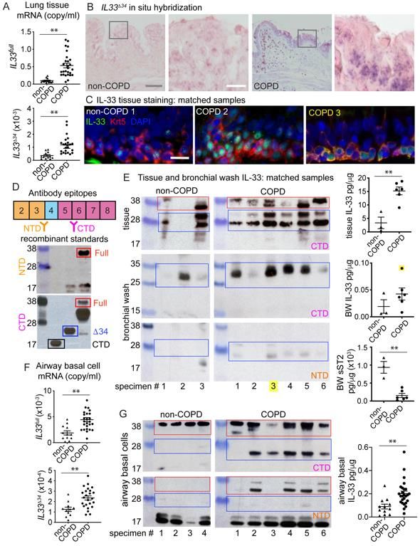
NSMase2 pathway in COPD specimens
NSMase2 (SMPD3) metabolizes sphingomyelin to generate ceramide and both lung
nSMase2 activity and ceramide metabolism have been shown to be altered in the setting
of cigarette smoking (39, 40) and COPD (41-43). We have shown that nSMase2 is
increased in COPD airway basal cells and regulates tonic IL-33 secretion, and therefore
extended the analysis to our cohort of COPD and non-COPD tissue specimens. We found
that SMPD3 exhibited highly variable expression in lung tissue and was increased in
multiple COPD specimens, in some by orders of magnitude (Figure 5A). SMPD3
expression correlated with the IL-33 receptor IL1RL1 and the major airway mucin
upregulated in COPD, MUC5AC and in some samples these three transcripts were
coincidentally increased by multiple orders of magnitude (boxed red data points, Figure
185B). Though SMPD3 expression trended higher in COPD tissue samples, this did not
translate to a difference in nSMase2 activity observed in a subset of specimens, likely in
part due to the 10-fold lower activity in tissue compared to airway cells (Figure 5C and
Figure 2C). Immunohistochemistry and immunofluorescence staining of nSMase2 and IL-
33 in tissue sections demonstrated a patchy basilar pattern in non-COPD and more
intense, diffuse staining pattern in COPD tissues (Figure 5D&E, Figure S7A&B).
Frequently nSMase2-enriched epithelium was coincident with cells exhibiting a vesicular
cytoplasmic IL-33 pattern that extended toward the airway lumen, examples for multiple
COPD specimens shown in Figure 5F. These regions were also stained for the exosome
marker CD9 (separately due to same antibody host species), which demonstrated a
striking linear-reticular pattern surrounding IL-33 positive basal cells extending to the
subepithelial and luminal surfaces, examples shown in Figure 5G.
Together these data reveal that SMPD3 expression and nSMase2 protein staining
are enriched in COPD specimens, and in some cases expression was strongly induced
concomitant with IL1RL1 and Muc5AC. NSMase2 and CD9 staining in proximity to cells
exhibiting cytoplasmic and/or vesicular IL-33 staining patterns suggests the appropriate
machinery is in place to facilitate secretion of IL-33-exosome complexes into the airway
lumen and interstitium.
Isolation of IL-33 and exosomes from COPD bronchial wash
In parallel with our in vitro analysis of IL-33 and exosomes secreted from cultured airway
basal cells, we sought to isolate and characterize endogenous components from
bronchial wash specimens. We performed this analysis with the COPD 3 sample exhibiting
19the highest IL-33 level, first by concentrating using a centrifugal 100 kDa filter, as in Figure
3E. Similar to culture supernatant, endogenous BW IL-33 was retained above the filter,
quantified in Figure 6A. Concentrated BW was fractionated by size exclusion
chromatography and ELISA-quantified IL-33 as well as total protein levels are shown in
Figure 6B. Peak exosome fraction 7 was analyzed by TRPS and DLS (Figure 6C, Figure
S3A&B), yielding vesicle size and distribution nearly identical to airway cell derived
exosomes and ultimately confirmed by transmission electron microscopy (Figure 6D).
Exosome properties were also consistent for multiple COPD specimens evaluated (Figure
S3B&D). Western blot was performed on BW exosome fraction 7 to verify CD9-positivity
and epithelial origin based on EpCAM staining (Figure 6E). Bronchial wash IL-33 largely
segregated from exosomes into free protein fractions as observed for non-fixed airway cell
supernatant. Western blot using CTD antibody confirmed the ~28 kDa band observed in
Figure 4E and IL-33 was again concentrated (10 kDa filter) and further resolved on a high-
resolution Superose 6 size-exclusion column. Endogenous IL-33 eluted at a volume
corresponding to MW 25 kDa based on standard curve (Figure 6G), consistent with the
calculated MW of IL-33Δ34. Purified endogenous BW IL-33 was then tested for bioactivity
in an HMC 1.2 activation assay (Figure 6H&I). Due to limited quantities of purified BW IL-
33, sample and controls were carefully matched for input concentration prior to assay
(Figure 6H). Endogenous BW-derived IL-33 was found to induce IL-8 secretion from HMC
1.2 cells with greater potency than commercial (CTD) protein at the same input
concentration (125 pg/ml), which was inhibited by anti-IL-1RL1 blocking antibody,
demonstrating specificity (Figure 3I).
20These results collectively reveal that endogenous IL-33 protein isolated from COPD
BW fluid exhibits biochemical properties consistent with IL-33Δ34 and retains bioactivity.
Furthermore, endogenous IL-33 was retained with higher-MW species during
concentration of BW fluid, similar to IL-33Δ34 secreted from airway cells. Exosomes derived
from COPD BW specimens also demonstrate a marker profile consistent with epithelial
origin, suggesting the bulk of exosomes secreted into COPD airway surface liquid are
epithelial derived.
NSMase2 inhibitor blocks IL-33 secretion and type-2 inflammation in vivo
We have uncovered a mechanism for tonic IL-33 secretion from human airway cells in
vitro and found support for this model in human COPD specimens. To test whether
blockade of nSMase2 activity could disrupt IL-33-mediated inflammation in vivo, we
employed an allergic airway disease model using the fungal allergen Alternaria alternata.
We selected the Alternaria model for this analysis because it is dependent on IL-33 for
induction of type-2 inflammation (44-46) and robustly induces IL-33 protein in
bronchoalveolar lavage (BAL) fluid (45). Regarding strategy for nSMase2 blockade, the
spontaneously derived mouse Smpd3 mutation characterized as fragilitas ossium (fro)
confers severe developmental abnormalities in mice, including osteogenesis imperfecta
and high perinatal mortality (47). We therefore chose to focus our efforts on the GW4869
compound that was effective in our in vitro studies and has been successfully applied to
other inflammatory mouse models (48, 49).
To induce type-2 driven airway disease in mice, we administered 5 doses of
Alternaria extract (or PBS) intranasally (i.n.) to mice on alternating days, as an extension
21of published protocols (44), and treated mice with either GW4869 (Alt/G) or vehicle control
(Alt/D) intraperitoneally (i.p.) beginning with the first dose of Alternaria, and daily thereafter
(Figure 7A). At 10 days post-Alternaria treatment, lung Il33 and Smpd3 mRNA were found
to be increased 3-fold in the Alt/D groups and not significantly reduced with GW4869
treatment (Figure 7B). Induction was limited to Il33, as other type-2 cytokines were not
impacted by Alternaria (Tslp unchanged, Figure S8C and Il25 not detected). Measurement
of IL-33 protein revealed a 3-fold induction of total IL-33 in lung tissue for both Alt/D and
Alt/G groups (Figure 7C). In contrast, GW4869 appeared to increase intracellular IL-33
protein levels 4-fold in cell suspension, suggesting retention of IL-33 in the setting of
nSMase2 blockade. Likewise, IL-33 protein was detected in bronchoalveolar lavage (BAL)
fluid in the Alt/D group at 1 hr. and 24 hr. following the 5th dose of Alternaria, which was
markedly decreased in the Alt/G group at both time points. Absolute BAL IL-33 protein was
approximately 6-fold higher in BAL at the 1 hr. versus 24 hr. time points (600 pg/ml versus
100 pg/ml, respectively), which is not reflected by normalized levels due to the high BAL
protein content immediately following Alternaria dose. We attribute induced BAL IL-33
protein to secretion rather than cellular necrosis, as the epithelium appears intact at 1 hr.
(Figure S8B) and 24 hr. (Figure S8A) post-Alternaria. Likewise, LDH activity in BAL fluid
is marginal compared to tissue lysate in samples at 1 and 24 hrs., with no observed
difference between Alternaria and control (Figure S8B).
Exosomes isolated from BAL fluid 24 hrs. after last Alternaria dose were analyzed
by TRPS and western blot. Results for pooled replicates show the Alt/D group exhibits
increased CD9 signal by western blot and particle number by TRPS, which is decreased
22to control level with GW4869 treatment. Exosome distribution and mean particle size for
Alt/D group are similar to human airway epithelial and BW derived exosomes (Figure 7D).
Interleukin-33 immunostaining of tissue sections demonstrate Alternaria-induced
expansion of IL-33-positive parenchymal cells with alveolar type-2 (AT2) morphology
(Figure 7E), similar to observations in other IL-33-dependent airway disease models (9).
This effect was observed in both Alt/D and Alt/G groups, consistent with GW4869
mediating blockade of IL-33 secretion rather than expression. In control tissue, IL-33
exhibited a predominant nuclear pattern, while in the Alt/D and Alt/G groups, many cells
exhibited a mixed nucleo-cytoplasmic IL-33 staining pattern (Figure 7E).
With respect to IL-33-induced type-2 inflammation, we analyzed the model at 6-
and 10-day time-points and found a 15-fold induction of lung Il13 mRNA at 6 days (3
doses) and 100-fold at 10 days (5 doses) (Figure 7F and Figure S8C). Subsequent
analyses were therefore performed at the 10-day time point. In addition to Il13, lung Il5
was also increased 5-fold, and both were substantially decreased with GW4869 treatment.
Likewise, lungs sorted for IL-1RL1+/Thy1.2+ innate lymphoid type-2 (ILC2) cells
demonstrated induction of ILC2s in the Alt/D group, which was partially blocked in the Alt/G
group (Figure 7G). Analysis of il13 expression in sorted cells from pooled replicates
demonstrates a 2-fold increase in Alt/D group that is reduced to control level in Alt/G group.
We observed a mild increase in Muc5ac 6 days post-Alternaria, which was further induced
at 10 days and was augmented with GW4869 treatment (Figure S8C). While qualitatively
PAS staining appeared diminished in Alt/G tissue sections (Figure S8A), it is possible that
other IL-33-indepdendent pathways contributing to mucus production are induced in this
model (50).
23Together these findings support our model developed in the human system
implicating the nSMase2-dependent exosome biogenesis pathway in lung epithelial IL-33
secretion. Disruption of type-2 inflammation in this model is not based on a substantial
reduction in Il33 expression, but rather inhibition of IL-33 secretion, implicating a novel
pathway in IL-33-induced chronic airway disease, which we have shown to be amenable
to therapeutic intervention.
24DISCUSSION
This study addresses multiple fundamental questions in the field of IL-33 biology
and advances our understanding of human chronic airway disease pathogenesis. We
show that the IL-33Δ34 isoform is increased in COPD and can undergo tonic secretion
from airway cells independent of proteolytic processing. We found that this mechanism
of secretion from intact airway cells is applicable to all IL-33 variants and occurs through
the nSMase2-dependent MVE pathway. Remarkably, IL-33 appears to be secreted as
surface-bound rather than an encapsulated exosome cargo. Identification of nSMase2 as
a key mediator of IL-33 secretion and demonstration of increased nSMase2 expression
in COPD specimens provides a connection between environmental triggers and non-
classical inflammatory cytokine secretion. We furthermore highlight a potential novel
therapeutic angle for disrupting the IL-33 system by demonstrating that nSMase2
inhibition can block IL-33 secretion and subsequent type-2 inflammation in a mouse
model of airway disease.
Our investigation revealed that tonic secretion of the IL-33Δ34 isoform is dependent
on the exosome biogenesis pathways regulated by nSMase2. In the context of prior work,
multiple stimuli for IL-33 release have been described based on the alarmin hypothesis,
including cryoshock (51), proteases (19), respiratory viral infection (52, 53) and allergens
including house dust mite (54). Comparatively, Alternaria extract has been reported to
induce alarmin release via cellular necrosis (55) or regulated secretion involving
purinergic receptors, intracellular calcium signaling and reactive oxygen species (56, 57).
We have also observed modest amounts of endogenous IL-33 secreted from intact airway
basal cells exposed to exogenous ATP (9). One common theme among these stimuli is
25that they all can induce extracellular vesicle flux as a cellular pro-survival signal (58).
Therefore, it is possible that a diverse array of cellular signals could converge upon a
fundamental cellular pathway that mediates non-classical protein and exosome secretion.
In this case, it appears such a pathway is co-opted by dysregulated IL33 isoforms
expressed in chronic airway disease. It is our expectation that IL-33 would not be the only
inflammatory mediator to utilize such a secretory mechanism, indeed this may be a
shared phenomenon among IL-1 family members (59, 60). Future studies of non-classical
cytokine secretion will be greatly informed by a more thorough understanding of functional
compensation between ESCRT-dependent and independent MVE pathways and their
intersection with chaperone-mediated microautophagy.
The results presented here provide key mechanistic insights into a potential role
for extracellular vesicle-mediated cellular communication in chronic airway disease
pathogenesis. Indeed, exosome biogenesis pathways have previously been linked to
chronic lung disease (61) and nSMase2/SMPD3 has been associated with eosinophilic
asthma (62) and found to be increased along with ceramide metabolites in smokers (43)
and COPD (42). We have found that nSMase2 is increased COPD specimens and
propose a model by which nSMase2 regulates secretion of IL-33 as surface-bound
exosome cargo. It will be important going forward to define the molecular interactions
between cytoplasmic IL-33 and chaperones that recruit this cytokine to the MVE pathway.
Likewise, it will be necessary to determine what role exosome-bound IL-33 plays in the
efficiency of IL-33 receptor signaling on effector cells. Future studies should also address
whether exosomes can enhance IL-33 stability, given the propensity of this cytokine to be
inactivated by protease digestion or oxidation in the extracellular milieu (38).
26With respect to the endogenous form of IL-33, our analysis of COPD BW IL-33
protein indicates a truncated bioactive form is present, with an antibody reactivity profile
consistent with the IL-33Δ34 isoform. Several prior studies have examined the issue of IL-
33 processing, mostly in the context of IL-33full protein. The flexible linker region encoded
by exon 4 is not a substrate of caspase-1, in contrast to IL-1β and IL-18 (17, 19, 20), but
is sensitive to multiple inflammatory proteases. Given that the secreted IL-33Δ34 isoform
lacks the protease-sensitive exon 4 linker, it is not surprising that we observe the protein
to be secreted in an intact form. Definitive characterization of endogenous IL-33 will
require mass spectrometry-based analysis of sufficient quantities of highly purified (non-
degraded) protein from relevant biological specimens, which remains a challenging
endeavor.
Regarding limitations of our approach, we understand that our in vitro observations
have been made in the context of protein overexpression, which has limitations and could
produce unexpected artifacts in any system. We have conducted our experiments
carefully to address the phenomenon with different tagged formats, diseased/non-
diseased primary cells and cell lines and under a range of expression levels and culture
formats to address any irregularities that may result from these experimental conditions.
With respect to our in vivo model, we recognize that COPD is a heterogeneous
disorder with multiple described inflammatory endotypes (63) among them type-2
predominant asthma-COPD overlap syndrome (64). Clear challenges remain in defining
and validating these endotypes in research and clinical care. In choosing a model system
for this study, we considered factors including variable severity of phenotype (smoking),
strain and/or pathogen-specific responses (virus) and distinct respiratory anatomy in mice
27and humans (IL-33 expression in type 2 pneumocytes in mice versus basal cells in
humans). Multiple studies to date have implicated IL-33 in Alternaria-induced respiratory
disease and have identified this fungal allergen as a potent stimulus for IL-33 secretion in
BAL fluid in vivo. With this in mind, we established the Alternaria model to test respiratory
IL-33 secretion and downstream inflammation with disruption of nSmase2 activity. We
recognize that this model does not encompass the full spectrum of COPD disease.
Clinical COPD care would indeed benefit from a better understanding of which
patients may respond more favorably to therapies targeting specific inflammatory
endotypes. Our COPD cohort includes sufficient material to explore disease mechanism,
but is comprised of severe COPD specimens, which limits our ability to correlate IL-33Δ34
or nSMase2 with COPD disease severity or expression-based metrics of type-2
endotypes (65). Future investigation and validation of pathways illuminated in this study
will require the addition of specimens across the spectrum of disease severity and
endotypes that incorporate protein- and exosome-focused sampling methods.
In summary, our analysis in human COPD illuminates a role for nSMase2 and
exosome pathways in the mechanism of IL-33 airway secretion, supported by
amelioration of type-2 inflammation with pharmacological blockade of nSMase2 in vivo.
This work reveals a novel aspect of IL-33 biology with potential to open a new area of
investigation in chronic airway disease and development of novel COPD therapeutics.
METHODS
(Additional methods described in Supplementary Materials)
Human Lung Samples and Study Design
28Clinical samples were obtained from consenting patients at the time of lung
transplantation from COPD recipients (n=27) with very severe disease (GOLD Stage IV)
during the period from 2011-2019 at Barnes-Jewish Hospital (BJH, St. Louis, MO).
Control samples were obtained from non-COPD donor lungs (n=13) that were not useable
for transplantation at BJH and University of Nebraska Medical Center (Omaha, NE).
There were no pre-determined inclusion or exclusion criteria beyond criteria for lung
transplant candidacy. To analyze tissue staining, gene expression and protein levels, lung
tissue samples were collected and processed for histopathology, RNA and protein
analysis from four different lung zones of each specimen. For this study, equivalent
quantities of the 4 lung areas were pooled for RNA and protein analysis to represent a
single sample per specimen. Tissue was homogenized in Trizol (Invitrogen) for all COPD
(n=27) and non-COPD (n=13) specimens for RNA extraction. COPD (n=14) and non-
COPD (n=6) specimens were minced and lysed in T-PER (Pierce) supplemented with
HALT protease inhibitor (Pierce), centrifuged at 10,000xg and supernatant collected for
protein analysis. COPD (n=14) and non-COPD (n=6) tissue specimens were fixed in 10%
neutral buffered formalin (ThermoFisher) prior to paraffin embedding and sectioning for
histopathology analysis. Airway basal cells were cultured from large airways (1st-3rd
generation) for COPD (n=26) and non-COPD (n=12) specimens and cells processed for
RNA and protein analysis. For a subset of COPD (n=6) and non-COPD (n=3) specimens,
bronchial wash (BW) was also performed at the time of specimen collection above.
Bronchial wash fluid was obtained from explanted lungs by gently injecting 100 ml of PBS
into maintstem bronchi and fluid recovered with passive return and gentle suctioning (to
preferentially return airway surface liquid and minimize alveolar lavage). Bronchial wash
29fluid was centrifuged at 100xg to pellet cells, HALT was added to supernatant prior to
storage for further analysis.
Exosome preparations and analysis
Extracellular vesicles were isolated from bronchial wash fluid and cell culture
supernatants in an analogous manner. Solutions were first spun at 2000xg to clarify,
followed by concentration using a centrifugal concentrator with 100 kilodalton (kDa)
molecular weight (MW) cutoff (Sartorius Vivaspin Turbo). For low abundance samples
(mouse BAL fluid, shRNA knockdown), samples of equivalent volumes were analyzed
following the concentration step. For samples of larger quantity (culture supernatants),
equivalent volumes of supernatant from confluent cultures were concentrated and
fractionated on a qEV 35 (Izon Science) size exclusion column. Fractions were collected
in PBS supplemented with HALT protease inhibitor and screened by dynamic light
scattering (DLS, Malvern NanoS) to verify vesicle size and homogeneity (polydispersity
index, PdI). Exosomes (100-150 nm) typically eluted in fraction 7-8 and free proteins in
fractions 12-17 when run according to manufacturer protocol. For experiments conducted
under fixed conditions, following clarification supernatant was fixed with 1%
paraformaldehyde for 5 minutes at room temperature and quenched with 1M Tris pH 8.0.
Supernatant was then concentrated similar to non-fixed sample and exosomes purified
using qEV 35 column; exosome integrity (size, homogeneity) was verified post-fixation.
For purified exosomes mixed with recombinant biotinylated IL-33∆34 protein,
approximately 1x10⁸ HBE-derived exosomes were incubated with 1 g IL-33∆34-biotin in
100 l PBS for 15 minutes at room temperature. Sample was then purified on a qEV 35
30column as above.
Exosome analysis was performed using a combination of dynamic light scattering (DLS),
transmission electron microscopy (TEM) and tunable resistive pulse sensing (TRPS).
Human and mouse exosomes were typically concentrated 10-fold and quantified by
TRPS (Izon Biosciences qNano device) using a 150 nm cutoff pore filter (NP150) and by
DLS (Malvern NanoS). Human COPD 3 BAL-derived exosomes were purified by qEV 35
column as above and prepared according to (66) for imaging on a JEOL JEM-1400 120
kV transmission electron microscope with an Advance Microscopy Technologies camera
system.
IL-33 secretion assays
Primary airway basal cells from non-COPD and COPD specimens and HBE-1 cell line
were cultured as described in Supplementary Methods on collagen coated tissue culture
plates (unless otherwise indicated). All secretion assays were performed at 37°C and 5%
CO2. Media was exchanged to fresh pre-warmed BEGM at beginning of assay and plates
were incubated for 2 hrs. Supernatant was clarified and cells were lysed in MPER (Pierce)
supplemented with HALT protease inhibitor (Pierce). For all secretion assays, IL-33
protein was quantified in supernatant and lysate using R&D commercial ELISA assays
(Supplementary Methods) with total assay protein (supernatant + lysate) and % secretion
(supernatant/(supernatant + lysate) x100) quantified based on standard curve. Some
experiments were performed in polarized format using Transwell® culture dishes (0.4 m
and 1.0 m pore size). Calculations were performed similarly for polarized format, with
31total assay protein (apical + basal + lysate) and % secretion (apical or basal/(apical +
basal + lysate) x100).
For chemical inhibition assays, all chemicals were solubilized in DMSO and filter
sterilized prior to use. As a control, DMSO was used at highest concentration required for
solubility in the assay. Inhibitors were pre-incubated with cells for 1 hr. prior to beginning
secretion assay and maintained in media during assay. Chemical concentrations used for
inhibition assay are as follows: PBS; DMSO vehicle control (2.5%); BD GolgiPlugTM
Brefeldin (1:1000), GolgiStopTM Monensin (1:1500), GW4869 (20 M), Cambinol (10 M),
Spiroepoxide (5 M), glutathione (5 M) and 3-methyladenine (3-MA, 5 M).
All secretion assays were performed with n=5 biological replicates (unless
otherwise indicated) and performed in triplicate.
Alternaria mouse model
Wild-type 5-week old male C57Bl/6 mice were purchased from Jackson Laboratories.
Alternaria alternata extract was purchased from Greer Laboratories and reconstituted and
adjusted to 1 mg/ml solution in sterile saline (based on BCA assay, Pierce). GW4869
(N,N’-Bis[4-(4,5-dihydro-1H-imidazol-2-yl)phenyl]-3,3’-p-phenylene-bis-acrylamide
dihydrochloride; molecular weight 577.5 g/mol; Sigma) was reconstituted in DMSO (20
mg/mL stock) and diluted into sterile saline prior to use. Experiments included n=11-14
mice per group and were repeated in triplicate. Five-week-old mice were either treated
with 25 µg intranasal Alternaria extract (in 25 µl) or PBS control (25 µl) under isoflurane
anesthesia every 48 hrs. for a total of 5 doses over 9 days. Mice in Alternaria groups were
also treated with 100 µl of 0.5 mg/mL GW4869 in 2.5% DMSO/saline (50 µg/mouse; 2-
322.5 µg/g body weight) or 100 µl of 2.5% DMSO/saline vehicle control every 24 hr. for 9
days, beginning on the same day as the first dose of Alternaria. Mice were analyzed 24
h after the final Alternaria dose except for 1 hr. post-Alternaria bronchoalveolar lavage
(BAL), for which mice were analyzed 1 hr. after the 5th dose. No behavioral problems were
observed during treatment. Mild body weight loss was observed in both Alternaria
treatment groups (5-10%), which was not different between groups and recovered by the
end of the experiment. RNA extraction, qPCR and ELISA and cell sorting are described
in Supplementary Materials; tissue and cell preparations were lysed in Trizol or
TPER/HALT for analysis. BAL fluid was obtained by intratracheal instillation of 0.7 ml PBS
with return volume ~0.3-0.4 mL and samples centrifuged to pellet cells. Supernatants
from n=3 mice were analyzed by ELISA (mouse IL-33 DuoSet (R&D Systems)
normalized to total protein by BCA assay) and an equivalent volume of each pooled and
concentrated with 100 kDa concentrator and analyzed by TRPS and western blot. Mouse
Alternaria model experiments were performed with accompanying analysis in triplicate.
Statistical analysis
For statistical analysis, Student’s t test was used for comparisons between two groups
and comparisons with 3 or more groups were analyzed using One-way ANOVA. For all
experiments, P value < 0.05 was considered statistically significant. Within individual
figure panels P-value information is indicated as follows: * = P < 0.05, ** = P < 0.01, *** =
P < 0.001 with symbols superimposed (and comparison groups indicated) on graphs
accordingly. Correlation analysis was performed based on Pearson’s coefficient. For all
33data in which 3 or more independent measurements are reported, data are displayed as
mean ± standard error of mean (SEM).
Study Approvals
All human studies were conducted with protocols approved by the Washington University
Institutional Review Board and written informed consent was obtained from study
participants. All experiments involving animals followed approved protocols by
Washington University’s Institutional Animal Care and Use Committee.
34ACKNOWLEDGEMENTS
Special thanks to Tom Brett, Gaya Amarasinghe and Jeff Haspel for critical reading of the
manuscript. We thank the Washington University Digestive Diseases Research Core
(NIDDK P30 DK052574) for providing qNano TRPS services. We thank Bill Eades and
the Siteman Center Flow Cytometry Core for FACS support. We thank the Washington
University Center for Cellular Imaging for Microscopy support (ORIP OD021629). We
thank the Washington University Genome Engineering and iPSC Center (GEiC) for
generating the HBE SMPD3-/- KO pool. We thank the Pulmonary Morphology Core for
tissue histology preparation. Support for this work was provided by NIH/NHLBI (K08
HL121168 and R01 HL52245, JAB), American Thoracic Society (Early Career
Investigator Award, JAB), Burroughs Wellcome Fund (Career Award for Medical Scientist,
JAB), and Doris Duke Foundation (Fund to Retain Clinical Scientists, JAB).
AUTHOR CONTRIBUTIONS
E.K., D.S., C.E.K., O.O., A.B., and J.A.B. designed and/or performed the experiments;
E.K., D.S., C.E.K., O.O., C.N.S. and J.A.B. prepared figures and wrote the manuscript;
S.L.B. and M.J.M. contributed to preparation and editing of manuscript; D.K., K.L.B. and
D.E.B. contributed to enrollment of human subjects and biobanking efforts; S.L.B., M.J.M.
and M.J.H. provided guidance with design/interpretation of experiments.
35References:
1. Pichery M, Mirey E, Mercier P, Lefrancais E, Dujardin A, Ortega N, et al.
Endogenous IL-33 is highly expressed in mouse epithelial barrier tissues, lymphoid
organs, brain, embryos, and inflamed tissues: in situ analysis using a novel Il-33-
LacZ gene trap reporter strain. J Immunol. 2012;188(7):3488-95.
2. Liew FY, Girard JP, and Turnquist HR. Interleukin-33 in health and disease. Nat
Rev Immunol. 2016;16(11):676-89.
3. Rostan O, Arshad MI, Piquet-Pellorce C, Robert-Gangneux F, Gangneux JP, and
Samson M. Crucial and diverse role of the interleukin-33/ST2 axis in infectious
diseases. Infect Immun. 2015;83(5):1738-48.
4. Moffatt MF, Gut IG, Demenais F, Strachan DP, Bouzigon E, Heath S, et al. A large-
scale, consortium-based genomewide association study of asthma. N Engl J Med.
2010;363(13):1211-21.
5. Bonnelykke K, Sleiman P, Nielsen K, Kreiner-Moller E, Mercader JM, Belgrave D,
et al. A genome-wide association study identifies CDHR3 as a susceptibility locus
for early childhood asthma with severe exacerbations. Nat Genet. 2014;46(1):51-
5.
6. Savenije OE, Mahachie John JM, Granell R, Kerkhof M, Dijk FN, de Jongste JC,
et al. Association of IL33-IL-1 receptor-like 1 (IL1RL1) pathway polymorphisms
with wheezing phenotypes and asthma in childhood. J Allergy Clin Immunol.
2014;134(1):170-7.
7. Prefontaine D, Lajoie-Kadoch S, Foley S, Audusseau S, Olivenstein R, Halayko
AJ, et al. Increased expression of IL-33 in severe asthma: evidence of expression
by airway smooth muscle cells. J Immunol. 2009;183(8):5094-103.
8. Wang Y, Wang L, and Hua S. Interleukin-33 in children with asthma: A systematic
review and meta-analysis. Allergol Immunopathol (Madr). 2017;45(4):387-92.
9. Byers DE, Alexander-Brett J, Patel AC, Agapov E, Dang-Vu G, Jin X, et al. Long-
term IL-33-producing epithelial progenitor cells in chronic obstructive lung disease.
J Clin Invest. 2013;123(9):3967-82.
10. Kim EY, Battaile JT, Patel AC, You Y, Agapov E, Grayson MH, et al. Persistent
activation of an innate immune response translates respiratory viral infection into
chronic inflammatory lung disease. Nat Med. 2008;14:633-40.
11. Xia J, Zhao J, Shang J, Li M, Zeng Z, Zhao J, et al. Increased IL-33 expression in
chronic obstructive pulmonary disease. Am J Physiol Lung Cell Mol Physiol.
2015;308(7):L619-27.
12. Bartemes KR, Iijima K, Kobayashi T, Kephart GM, McKenzie AN, and Kita H. IL-
33-responsive lineage- CD25+ CD44(hi) lymphoid cells mediate innate type 2
immunity and allergic inflammation in the lungs. J Immunol. 2012;188(3):1503-13.
13. Hardman CS, Panova V, and McKenzie AN. IL-33 citrine reporter mice reveal the
temporal and spatial expression of IL-33 during allergic lung inflammation. Eur J
Immunol. 2013;43(2):488-98.
14. Roussel L, Erard M, Cayrol C, and Girard JP. Molecular mimicry between IL-33
and KSHV for attachment to chromatin through the H2A-H2B acidic pocket. EMBO
Rep. 2008;9(10):1006-12.
36You can also read