Huntingtin and Its Role in Mechanisms of RNA-Mediated Toxicity
←
→
Page content transcription
If your browser does not render page correctly, please read the page content below
toxins
Review
Huntingtin and Its Role in Mechanisms of
RNA-Mediated Toxicity
Annika Heinz              , Deepti Kailash Nabariya and Sybille Krauss *

                                           Institute of Biology, University of Siegen, 57076 Siegen, North Rhine-Westphalia, Germany;
                                           Annika.Heinz@uni-siegen.de (A.H.); Deepti.Nabariya@uni-siegen.de (D.K.N.)
                                           * Correspondence: sybille.krauss@uni-siegen.de

                                           Abstract: Huntington’s disease (HD) is caused by a CAG-repeat expansion mutation in the Hunt-
                                           ingtin (HTT) gene. It is characterized by progressive psychiatric and neurological symptoms in
                                           combination with a progressive movement disorder. Despite the ubiquitous expression of HTT,
                                           pathological changes occur quite selectively in the central nervous system. Since the discovery of HD
                                           more than 150 years ago, a lot of research on molecular mechanisms contributing to neurotoxicity
                                           has remained the focal point. While traditionally, the protein encoded by the HTT gene remained
                                           the cynosure for researchers and was extensively reviewed elsewhere, several studies in the last few
                                           years clearly indicated the contribution of the mutant RNA transcript to cellular dysfunction as well.
                                           In this review, we outline recent studies on RNA-mediated molecular mechanisms that are linked to
                                           cellular dysfunction in HD models. These mechanisms include mis-splicing, aberrant translation,
                                           deregulation of the miRNA machinery, deregulated RNA transport and abnormal regulation of
                                           mitochondrial RNA. Furthermore, we summarize recent therapeutical approaches targeting the
                                           mutant HTT transcript. While currently available treatments are of a palliative nature only and do
                                           not halt the disease progression, recent clinical studies provide hope that these novel RNA-targeting
                                           strategies will lead to better therapeutic approaches.


                                    Keywords: huntingtin; RNA toxicity; CAG repeat; neurodegeneration; RNA hairpin; RNA binding
                                           protein (RBP); mis-splicing; translation; RNA-targeting compound
Citation: Heinz, A.; Nabariya, D.K.;
Krauss, S. Huntingtin and Its Role in
                                           Key Contribution: Structural changes in the RNA transcripts, for example, the folding of expanded
Mechanisms of RNA-Mediated
Toxicity. Toxins 2021, 13, 487. https://
                                           CAG repeats into a hairpin, can lead to cellular dysfunction that is linked to the development of neu-
doi.org/10.3390/toxins13070487             rodegenerative diseases. This RNA-mediated toxicity is due to aberrant interaction of RNA binding
                                           proteins or other transcripts that attach to this hairpin and, upon binding, execute aberrant activities,
Received: 26 May 2021                      including deregulated splicing, aberrant translation and aberrant function of the miRNA machinery.
Accepted: 11 July 2021
Published: 14 July 2021

Publisher’s Note: MDPI stays neutral       1. Introduction
with regard to jurisdictional claims in
                                                 Short tandem repeats, such as trinucleotide-repeats, are a frequent motif in the human
published maps and institutional affil-
                                           genome, with the trinucleotide CAG being one of the most common repeat motifs [1].
iations.
                                           These repeats are polymorphic and variable in length. One proposed mechanism that is
                                           responsible for the variability is the so-called strand-slippage during DNA replication.
                                           This slippage is a consequence of a detachment of the DNA polymerase, followed by
                                           misalignment on the template strand with the repeated DNA fragment being looped out.
Copyright: © 2021 by the authors.          If this mispairing between the template strand and the nascent strand occurs in such a way
Licensee MDPI, Basel, Switzerland.         that the loop structure is built on the nascent strand, then the repeat number is elevated
This article is an open access article
                                           (Figure 1) [2].
distributed under the terms and
                                                 Upon repeat expansion beyond a certain threshold, CAG-repeat expansion is asso-
conditions of the Creative Commons
                                           ciated with the development of neurodegenerative diseases [3,4]. CAG repeats, like all
Attribution (CC BY) license (https://
                                           CXG repeats (with X being any nucleotide), are overrepresented in exons and occur most
creativecommons.org/licenses/by/
                                           frequently within the open reading frame [5,6]. When translated into protein, CAG repeats
4.0/).

Toxins 2021, 13, 487. https://doi.org/10.3390/toxins13070487                                                    https://www.mdpi.com/journal/toxins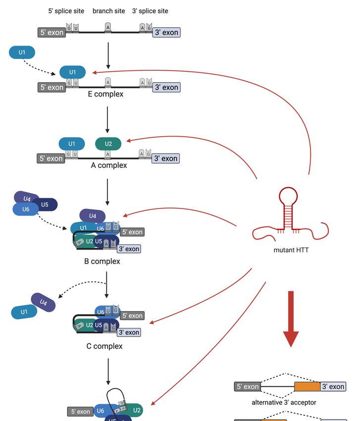
Toxins 2021, 13, 487 2 of 25
Toxins 2021, 13, x FOR PEER REVIEW code for polyglutamine tracts. Thus, CAG-repeat expansion disorders in which the CAG
2 of 26
repeat is located in the coding region are also called polyglutamine diseases, with the most
common one being Huntington’s disease (HD).
Figure 1. Schematic illustration of a CAG-repeat expansion during DNA replication, its transcription
into RNA
Figure and three-dimensional
1. Schematic illustration ofRNA folding dependent
a CAG-repeat expansionon the CAG-repeat
during length. Upper
DNA replication, itspart of
transcrip-
the picture: During DNA replication, the polymerase uses the template
tion into RNA and three-dimensional RNA folding dependent on the CAG-repeat length. Upper strand (blue) to synthesize a
nascent strand (red). CAG-repeat expansion during replication can be explained
part of the picture: During DNA replication, the polymerase uses the template strand (blue) to syn- by strand-slippage.
The polymerase
thesize detaches
a nascent strand from
(red). the template
CAG-repeat strand and
expansion reattaches
during such that
replication can abepart of the repeat
explained by strand-
sequenceThe
slippage. is looped out. Duedetaches
polymerase to this misalignment, the nascent
from the template strand
strand and has an increased
reattaches number
such that aofpart
CAG of the
repeats.
repeat Lower panel:
sequence the CAG
is looped out.repeat
Due tocontaining mRNA foldsthe
this misalignment, into a three-dimensional
nascent strand has anstructure
increasedthatnum-
depends
ber of CAG onrepeats.
the repeat length.
Lower mRNA
panel: thewith
CAG either normal
repeat (left) or mutant
containing mRNA(right) CAGarepeat
folds into numbers
three-dimensional
structure
are shown. thatThedepends
mutant on CAGtherepeat
repeatfolds
length.
into mRNA with either
a characteristic normal
hairpin (left)that
structure or mutant (right)
sticks out to theCAG
repeat numbers
side. The mutantare shown. The
CAG-repeat mutant
hairpin CAG repeat
is highlighted in folds
red andintothea corresponding
characteristic hairpin structure that
short CAG-repeat
sticks out to
sequence in the
the side.
normal Thetranscript
mutant CAG-repeat
is highlightedhairpin
in green.is highlighted
Created with in BioRender.com
red and the corresponding
and the
short CAG-repeat sequence in the normal transcript is highlighted
RNA Folding Form V2.3 (http://www.unafold.org/mfold/applications/rna-folding-form-v2.php, in green. Created with BioRen-
der.com
accessedandon 22 theMarch
RNA 2021).
Folding Form V2.3 (http://www.unafold.org/mfold/applications/rna-folding-
form-v2.php, accessed on 22 March 2021).
Upon repeat expansion beyond a certain threshold, CAG-repeat expansion is associ-
ated with the development of neurodegenerative diseases [3,4]. CAG repeats, like all CXG
repeats (with X being any nucleotide), are overrepresented in exons and occur most fre-
quently within the open reading frame [5,6]. When translated into protein, CAG repeats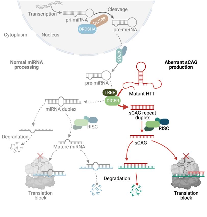
Toxins 2021, 13, 487                                                                                          3 of 25

                             Clinical features of HD include three main groups: behavioral symptoms, cogni-
                       tive difficulties and involuntary movements. The disease progresses over approximately
                       15–20 years and is ultimately lethal. Most patients (up to 50%) show some behavioral
                       changes, such as depression, even before HD is diagnosed [7]. A high depression-associated
                       suicide rate is seen in HD patients [8]. The rate of progression of early-onset psychiatric
                       symptoms is variable and does not correlate with the rate of development of other symp-
                       toms, such as chorea and cognitive difficulties [9]. The cognitive difficulties often succeed
                       initial behavioral changes, but at the time of diagnosis, most HD patients show significant
                       cognitive impairment. This progresses slowly over many years, finally culminating in
                       dementia. The third clinical feature in HD is chorea (from Greek χoρεία, a circle dance). It
                       describes a progressive movement disorder that is characterized by the loss of voluntary
                       movements and the development of involuntary movements. The movement disorder
                       starts with short suppressible unintended movements, for example, of the hands or the
                       face. Later the involuntary movements affect more and more muscle groups as the disease
                       progresses [10]. Other movement symptoms include bradykinesia and marked postural
                       abnormalities [11].
                             In HD, the mutant CAG repeat is located within exon1 of the Huntingtin (HTT) gene
                       on chromosome 4p16.3. The HTT gene product is a large protein of approximately 348 kDa.
                       Upon expansion of the polyglutamine tract, the HTT protein, just like all polyglutamine pro-
                       teins, tends to misfold and aggregate. HTT-polyglutamine protein aggregation in the central
                       nervous system is a pathological hallmark of the disease and pathogenesis was consistently
                       associated with the abnormal function of mutant HTT protein [12–14]. A well-established
                       example of abnormal function of the mutant HTT protein is the down-regulation of the
                       brain-derived neurotrophic factor (BDNF) via transcriptional alteration [15,16]. BDNF
                       is involved in several processes in the nervous system, such as neuronal differentiation,
                       synaptic plasticity, dendritic complexity and neuronal survival [17]; low levels of BNDF
                       cause degeneration of striatal projection neurons in HD [16,18,19]. Because proteolytic
                       cleavage of mutant HTT protein is an established rate-limiting step in the aggregation
                       process, a lot of research has focused on the N-terminal fragment of the mutant HTT
                       protein that contains the polyglutamine tract. Interestingly, the second cleavage product
                       of mutant HTT, the non-polyglutamine C-terminal fragment, was also reported to cause
                       cellular toxicity [20]. However, the role of aggregates and aggregate-prone polyglutamine
                       protein in pathogenesis was reviewed extensively elsewhere [21–25] and we will focus on
                       RNA-mediated molecular mechanisms that are linked to cellular dysfunction in HD in
                       this review.
                             In the last couple of years, there has been emerging evidence indicating the contri-
                       bution of RNA-mediated abnormal functions to pathogenesis. Several lines of evidence
                       indicate that mutant CAG repeats can induce RNA-mediated toxicity in in vivo models
                       expressing untranslated repeats [26–30]. In addition, there are also examples of human dis-
                       eases, in which an expanded CAG repeat is located in the untranslated region, suggesting
                       that CAG-repeat RNA can cause disease development, even in the absence of a polyg-
                       lutamine protein (these diseases include SCA12 (spinocerebellar Ataxia Type 12, OMIM
                       604326), SCA8 (spinocerebellar ataxia type 8, OMIM 608768) and Huntington disease-like 2
                       (OMIM 606438)).

                       2. RNA Structure and Misfolding
                            One way of explaining a toxic gain-of-function of an RNA molecule is by aberrant
                       attachment to proteins or other RNAs, where the three-dimensional structure is the key for
                       intermolecular binding. RNA is most often a single-stranded molecule that folds onto itself
                       to minimize its free energy. It can form fully paired and non-canonically paired regions,
                       such as hairpins, internal loops, bulges, multi-branch loops and pseudoknots (Figure 2).
                       These structural motifs are important recognition sites for RNA–protein and RNA–RNA
                       interactions. While the three-dimensional structure of an RNA molecule is crucial to
                       its physiological function, its misfolding can lead to the deregulation of various cellular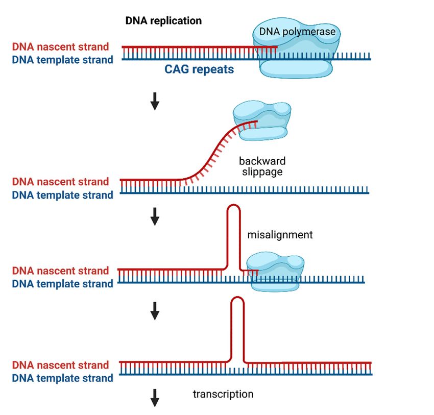
RNA interactions. While the three-dimensional structure of an RNA molecule is crucial to
Toxins 2021, 13, 487 its physiological function, its misfolding can lead to the deregulation of various 4 of 25cellular
processes, resulting in a toxic gain-of-function under disease conditions [31]. With respect
to CAG-repeat RNA, a toxic gain-of-function is triggered by an aberrantly folded RNA
hairpin motif
processes, in the repeat
resulting in a toxiclocation that is notunder
gain-of-function present under
disease healthy[31].
conditions conditions. Hydrogen
With respect
bonding
to CAG-repeat RNA, a toxic gain-of-function is triggered by an aberrantly folded RNA CAG-
between nucleobases plays a crucial role in RNA folding. In the expanded
repeat
hairpinregions,
motif in the RNA location
the repeat folds onto thatitself
is not with
presenttheunder
opposing
healthystrands of the
conditions. CAG repeat,
Hydrogen
forming
bondingclassic
between Watson–Crick
nucleobases plays contacts between
a crucial role inthe
RNA G folding.
and C positions and wobble
In the expanded CAG- pairs
repeat regions, the RNA folds onto itself with the opposing strands
at the A–A mismatches (Figure 2). The stability and the length of the hairpin are depend- of the CAG repeat,
forming
ent on theclassic
numberWatson–Crick contactsWhile
of CAG repeats. between the G and C positions
in non-mutant and wobble
HTT transcripts, thepairs
CAG at repeat
the A–A mismatches (Figure 2). The stability and the length of the hairpin are dependent
pairs with an adjacent CCG repeat, expansion of the CAG repeat leads to the folding of a
on the number of CAG repeats. While in non-mutant HTT transcripts, the CAG repeat
double-stranded hairpin structure of pure CAG repeats. The hairpin can be divided into
pairs with an adjacent CCG repeat, expansion of the CAG repeat leads to the folding of a
three parts: the base
double-stranded of the
hairpin hairpin,
structure ofthe
pure stem
CAGregion that
repeats. Theis hairpin
built upcan of be
thedivided
double-stranded
into
CAG
threeregion andbase
parts: the a terminal loop of
of the hairpin, theeither
stem four
regionorthat
seven nucleotides.
is built The size of the termi-
up of the double-stranded
nal
CAG loop depends
region on the repeat
and a terminal loop ofnumber,
either fourwith evennucleotides.
or seven numbers mainlyThe sizeresulting in loops of
of the terminal
four
loopnucleotides
depends on the andrepeat
odd numbers
number, with mainly
even resulting
numbers mainlyin loops of seven
resulting nucleotides.
in loops of four Not
nucleotides
only and odd numbers
do the CAG-repeat lengths mainly resulting
influence in loops
hairpin of seven
stability butnucleotides.
also specific Not only
repeat-flank-
do the CAG-repeat lengths influence hairpin stability but also specific
ing regions at the stem can stabilize the hairpin structure by serving as a G–C clamp [32]. repeat-flanking
regions
These at the stemformed
aberrantly can stabilize
mutant theCAG-repeat
hairpin structure by serving
hairpin motifs asinterfere
a G–C clampwith[32].
theThese
normal cel-
aberrantly formed mutant CAG-repeat hairpin motifs interfere with the normal cellular
lular functions by aberrantly recruiting RNA-binding proteins [32,33]. The sequestration
functions by aberrantly recruiting RNA-binding proteins [32,33]. The sequestration of these
ofproteins
these proteins to the
to the hairpin hairpin
hinders thehinders
functionthe function
of several of several
pathways they pathways
are a part of,they are a part
leading
of,toleading to the breakdown
the breakdown of the cellular ofsystem.
the cellular system.
Schematic illustration
Figure2.2.Schematic
Figure illustrationofofthree-dimensional
three-dimensional RNARNAfolding structures.
folding RNA can
structures. RNA form
canfully
form fully
paired and non-canonically paired regions, such as hairpins, internal loops, bulges,
paired and non-canonically paired regions, such as hairpins, internal loops, bulges, multi-branch multi-branch
loopsand
loops andpseudoknots
pseudoknots (upper
(upper panel).
panel). CAG-repeat
CAG-repeatregions
regionscancanfold
foldinto hairpins
into hairpinsin which
in whichthe the op-
opposing strands of the CAG-repeat form Watson–Crick contacts between the G
posing strands of the CAG-repeat form Watson–Crick contacts between the G and C positions (in- and C positions
(indicated
dicated as red
as red lines)
lines) andand wobble
wobble pairs
pairs at at
thethe A–Amismatches
A–A mismatches(lower
(lower panel). In this
panel). In this picture,
picture,aa termi-
terminal
nal loop ofloop of seven
seven nucleotides
nucleotides is shown.
is shown. Created
Created with
with BioRender.com.
BioRender.com.
3. RNA Localization and RNA Granules
An essential part of spatial protein translation control is the regulation of cytoplasmic
mRNA localization. RNA is transcribed in the nucleus and, after maturation into mRNA,
binds to specific proteins that trigger mRNA export to the cytoplasm, where the mRNA
can be translated into protein. The discovery that mutant CAG repeats promote nuclear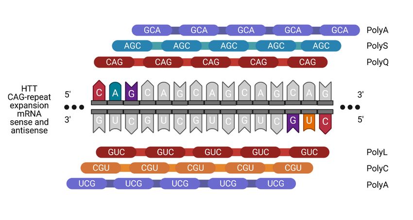
Toxins 2021, 13, 487                                                                                            5 of 25

                       3. RNA Localization and RNA Granules
                             An essential part of spatial protein translation control is the regulation of cytoplasmic
                       mRNA localization. RNA is transcribed in the nucleus and, after maturation into mRNA,
                       binds to specific proteins that trigger mRNA export to the cytoplasm, where the mRNA
                       can be translated into protein. The discovery that mutant CAG repeats promote nuclear
                       retention and formation of RNA foci brought a new vision in the field of neurological
                       disorders. Apparently, the mutant CAG-repeat RNA gets retained inside the nucleus
                       due to aberrant RNA–protein interactions. Such interactions may pave the way to strong
                       sequestration of other proteins, such as muscleblind 1 (MBNL1), which is an important
                       protein in foci formation [34]. RNAs with CAG-repeat expansions were found within foci,
                       along with MBNL1 in HD cells [35]. RNA foci occur in various cell models of HD, such
                       as lymphoblasts, fibroblasts and neuronal progenitors [36]. The presence of RNA foci
                       leads to alterations in cellular pathways, apoptosis initiation and abnormal alternative
                       splicing [37]. However, whether the nuclear accumulation of CAG-repeat transcripts
                       represents a stable fraction that remains in the nucleus or whether CAG-repeat transcripts
                       travel to the cytoplasm after temporary participation in the formation of foci remains
                       unclear [35].
                             One important phenomenon that leads to RNA toxicity upon CAG-repeat expansion
                       and that is linked to aberrant RNA localization is RNA granulation. The maintenance
                       of neuronal circuits and synaptic strength is largely dependent on the local translation
                       of mRNAs [38]. Neuronal RNA granules are a transport mechanism of mRNAs and
                       other proteins to the synapses. Pertaining to their complex morphology, neurons rely on
                       various internal factors to achieve precise compartmentalization of external signals. This is
                       partially achieved by decentralizing the control of gene expression and by the dynamic local
                       translation of mRNA that is transported to axon terminals and dendrites. Both the transport
                       and the translation of these mRNAs are tightly regulated. During transport, mRNA
                       translation is repressed inside the transport cargo. The release of translational repression
                       is temporally and spatially controlled and triggered either by signaling molecules or by
                       synaptic activity. Although the exact mechanistic process remains an integral part of several
                       ongoing studies, it is clear that neuronal RNA binding proteins are required for the targeting
                       of mRNA and translational regulation. They recognize the regulatory sequences, recruit
                       translational repressors and enable the formation of multi-molecular ribonucleoprotein
                       complexes (RNPs). These RNPs assemble to form neuronal RNP granules. In addition
                       to these RNP granules, neuronal cells also contain other cytoplasmic RNP granules that
                       include processing bodies (PBs) and stress granules (SGs). However, these different classes
                       of granules sometimes contain common proteins in their cargo and use similar mechanisms
                       to control mRNA translation or decay [39,40]. Neuronal granules often contain large
                       amounts of ribosomal and other RNA-binding proteins. Proteins such as FMRP (fragile X
                       mental retardation protein), Staufen and UPF1 are key regulators of translational silencing
                       and activation in neurons [40]. Often, these proteins are trapped by the mutant HTT mRNA,
                       leading to the breakdown of the transport machinery. Studies by Savas et al. suggest that
                       the HTT protein also plays an important role in RNA transport and translation within
                       neuronal granules [41]. The mutant RNA obstructs the axonal transport and compromises
                       the synaptic excitability of the neuron. This failure of receptor delivery may lead to
                       excitotoxic damage of the neuron [42].
                             PBs are formed via phase separation within the cytoplasm and play a role in mRNA
                       degradation [43]. They also serve as sites for miRNA-mediated translational silenc-
                       ing [44,45]. Ago2 is an integral protein that facilitates neuronal HTT-mediated gene
                       silencing and copurifies with the HTT protein. HTT and Ago2 associate and localize
                       themselves in PBs in order to contribute to post-translational gene silencing [41]. En-
                       dogenous HTT protein also associates with Ago2 in neuronal granules and supports the
                       transport of mRNA to dendrites. The knockdown of HTT results in improper Ago2 distri-
                       bution. Previous studies also reported that the integrity of PBs is compromised in striatal
                       cells and primary neurons that express endogenous mutant HTT [41].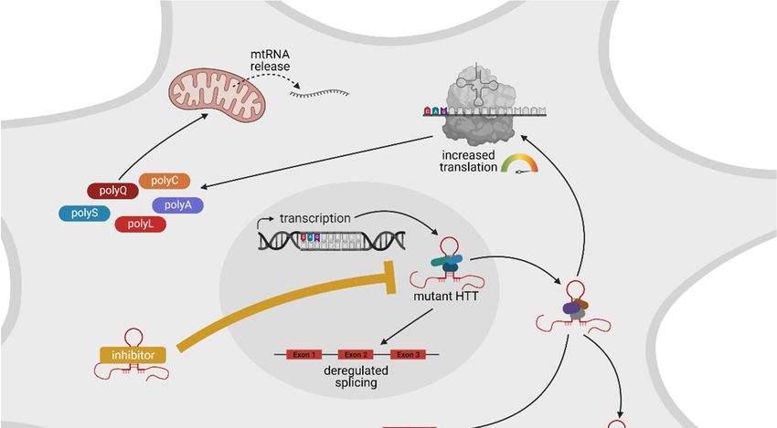
Toxins 2021, 13, 487                                                                                            6 of 25

                             SGs are dense attachments of RNAs and RNA-binding proteins that form in response
                       to cell stress. They can be seen as a storage granule for untranslated mRNAs. The stored
                       mRNAs can go down one of three paths to promote cell survival: further storage, re-
                       initiation of translation or decay [46]. To facilitate degradation, SGs transfer transcripts to
                       the adjacent PBs [47]. The pathophysiology of most neurodegenerative disorders includes
                       the presence of oxidative stress. In response to oxidative stress, eukaryotes either activate
                       defense mechanisms that help cell survival or initiate apoptotic pathways [48]. SGs are
                       nucleated through the binding of core mRNA-binding proteins (RBPs) to mRNA. These
                       core RBPs often contain polyglycine-rich domains and can recruit proteins that are linked
                       to several neurodegenerative disorders [49]. RBPs include several molecules, such as
                       optineurin, angiogenin, ataxin-2, hnRNPA1, SMN-1, TIA-1, TDP-43, TTP, FUS and B2,
                       which are known to associate with SGs. Mutations and malfunctions in these RBPs could
                       cause direct neurodegeneration [50,51].

                       4. Deregulated Splicing
                            One approach to identifying mechanisms of RNA-mediated toxicity of mutant CAG-
                       repeat transcripts is to analyze the proteome that aberrantly binds to expanded CAG
                       repeats. Schilling et al. purified proteins that are captured by HTT RNA in a repeat-
                       length-dependent manner and identified them using mass spectrometry [52]. Of the 36
                       proteins binding HTT RNA with a higher affinity for RNAs harboring mutant CAG repeats,
                       32 proteins are functionally connected to splicing. This finding indicates that one major
                       mechanism of RNA-mediated toxicity is deregulated splicing. Indeed, several mis-splicing
                       events were detected in different models of HD.
                            Aberrant splicing events that occur upon the expression of mutant CAG-repeat RNA
                       can be generally grouped into two groups: in splicing events affecting the CAG-repeat
                       transcript itself and in splicing events affecting other transcripts. Both groups of mis-
                       splicing events were shown in different CAG disease models (Figure 3).
                            The first group, namely, mis-splicing of the mutant CAG-repeat transcript itself, occurs
                       in diverse HD models and disease tissues. Here, the partial splicing of mutant HTT from
                       exon 1 to exon 2 leads to the production of a small polyadenylated transcript encoding a
                       neurotoxic mutant HTT protein fragment, which contains the expanded polyglutamine
                       stretch [53,54]. This incomplete splicing of mutant HTT from exon 1 to exon 2 can be
                       detected in vivo in diverse HD mouse models [54,55]. Importantly, the level of mis-spliced
                       transcript correlates with the onset of behavioral phenotypes and the striatal disposition
                       of polyglutamine–protein aggregates [55]. Furthermore, in an HD patient’s tissue, this
                       mis-spliced HTT transcript can be detected. Tissues that express this mis-spliced transcript
                       include fibroblasts, cortex, hippocampus and cerebellum [54,56]. Mechanistically, the
                       production of the mis-spliced HTT transcript may be caused by one or more splicing
                       factors that aberrantly bind to the mutant transcript. For example, the splicing factor
                       SRSF6 binds to the mutant transcript [52,54] and manipulation of the expression level of
                       SRSF6 modulates this aberrant splicing event [57]. However, in mouse models, incomplete
                       splicing remained unaffected after the reduction of SRSF6 to 50% of wild-type levels [58].
                       Thus, it is likely that more than one splicing factor is involved in the mis-splicing of HTT
                       transcripts with expanded CAG repeats.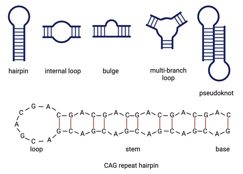
Toxins 2021, 13,
Toxins 2021, 13, 487
                 x FOR PEER REVIEW                                                                                            77 of
                                                                                                                                 of 26
                                                                                                                                    25

      Figure 3. Proteins that aberrantly bind to HTT RNA with expanded CAG repeats regulate splicing. A schematic of the
     Figure
      splicing3.process
                 Proteinsis that aberrantly
                             shown.         bind toisHTT
                                    RNA splicing          RNA that
                                                      a process withremoves
                                                                     expanded CAG repeats
                                                                            non-coding    regulate
                                                                                       sequences   splicing.
                                                                                                 (introns)   A schematic
                                                                                                           from pre-mRNA of and
                                                                                                                            the
     splicing process is shown. RNA splicing is a process that removes non-coding sequences (introns) from pre-mRNA and
     joins the protein-coding sequences (exons) together, which is carried out by the spliceosome. The spliceosome consists of
     several proteins and small nuclear RNAs. The major spliceosome includes the small nuclear ribonucleoproteins (snRNPs)
     U1 (blue), U2 (green), U4 (violet), U5 (dark blue) and U6 (blue). These snRNPs sequentially attach to the pre-mRNA and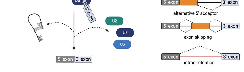
Toxins 2021, 13, 487 8 of 25
joins the protein-coding sequences (exons) together, which is carried out by the spliceosome. The spliceosome consists of
several proteins and small nuclear RNAs. The major spliceosome includes the small nuclear ribonucleoproteins (snRNPs)
U1 (blue), U2 (green), U4 (violet), U5 (dark blue) and U6 (blue). These snRNPs sequentially attach to the pre-mRNA and
carry out splicing, starting with U1, which binds at the 50 splice site of the exon. U2 assembles at the branch site and then
the U5 and U4–U6 complexes attach, resulting in the for-mation of the precatalytic spliceosome. Afterward, U4 and U1
are released from the pre-spliceosomal complex, the 50 splice site gets cleaved and a looped lariat structure containing
the intronic sequence is removed. Finally, the two exons are linked. HTT RNA with an expanded CAG repeat (hairpin
depicted in red) aberrantly sticks to the snRNPs U1, U2, U4, U5 and U6. This results in aberrant splicing events, including
intron retention, exon skipping or the use of alternative 30 or 50 acceptor sites. Created with BioRender.com (accessed on 22
March 2021).
The second group of aberrant splicing events that occur upon the expression of mutant
HTT affects other transcripts; several examples of this group were found in different stud-
ies. For example, the CREB1 transcript is mis-spliced upon the expression of mutant HTT.
Mechanistically, CREB1 mis-splicing involves recruitment of the splicing factor PRPF8 to
mutant HTT RNA. Importantly, CREB1 mis-splicing is also seen in HD brains [52]. Other
examples of transcripts that are mis-spliced upon the expression of CAG-repeat constructs
include INSR, SERCA1, CLCN1 and LDB3. Here, the overexpression of the splicing regula-
tor MBNL1 partially reverses splicing abnormalities, suggesting that MBNL1 is one of the
splice factors that are mechanistically involved here [59]. In a more general approach to
identify mis-spliced transcripts in the motor cortex from seven human HD brains via deep
RNA-seq analysis, 593 differential alternative splicing events between HD and control
brains were detected [60]. In the same study, Lin et al. also evaluated the expression levels
of splicing factors in HD patients’ brains. They reported significantly altered expression
levels and found that the splicing factor PTBP1 impacts disease-associated splicing [60].
Additionally, in a recent study, Elorza et al. performed intersect-RNA-seq analyses of
human postmortem striatal tissue and of an early symptomatic mouse model in which
neuronal loss and gliosis were not yet present. This, in combination with a human–mouse
parallel motif scan analysis, allowed for the identification of a shared mis-splicing sig-
nature that was triggered by the CAG-repeat mutation in both species and gives rise
to a total of 949 one-to-one orthologs that are differentially spliced in both human and
mouse [61]. In addition, via human–mouse parallel complementary motif searches on
common mis-spliced events, the authors concluded a network of candidate upstream
splicing factors with reduced protein levels in both species that may be mechanistically
involved in mis-splicing [61]. Interestingly, some of these splicing factors, for exam-
ple, U2AF2 and HNRNPC, were previously found to aberrantly bind to CAG-repeat
RNA [52].
All these data indicate that the sequestration of splicing factors to mutant CAG-repeat
RNA affects splicing of both the CAG-repeat RNA itself and other transcripts and, thus,
one major mechanism of RNA-mediated toxicity is deregulated splicing.
5. Aberrant Translation
Upon splicing and maturation of the mRNA, protein translation can be initiated,
leading to protein synthesis from the mature mRNA transcript. The translation of
expanded CAG-repeat mRNA results in a neurotoxic polyglutamine protein. This event
can be seen as a pathogenic function, in connection with the neurotoxicity of the mutant
CAG-repeat RNA.
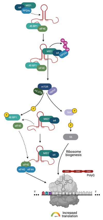
In addition to splicing factors, translation factors also bind length-dependently to CAG-
repeat mRNA such that mutant, i.e., expanded repeats, recruit significantly more protein.
One example of protein complexes that aberrantly attach to the mutant CAG-repeat hairpin
is a protein complex consisting of MID1 (midline 1), the 40S ribosomal S6 kinase (S6K) and
the catalytic subunit of protein phosphatase 2A (PP2Ac) [62]. PP2A is one of the central
serine/threonine phosphatases and is therefore responsible for the dephosphorylation of a
large amount of the serine/threonine phosphorylated molecules in cells. This phosphatase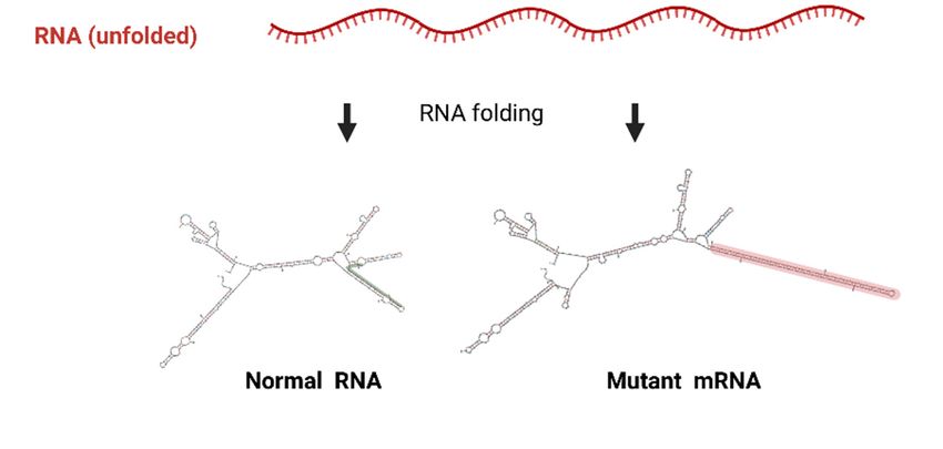
Toxins 2021, 13, 487                                                                                           9 of 25

                       consists of various subunits. The core is the already mentioned C-subunit (PP2Ac), which
                       dephosphorylates a large number of substrates in vitro. In cells, PP2Ac is bound to either
                       one or two of its regulatory subunits that modify its enzymatic activity and substrate
                       specificity. In this context, MID1 binds to the alpha4 protein, which is one of the regulatory
                       subunits of PP2A. Upon binding, MID1 catalyzes the polyubiquitination of PP2Ac, which
                       marks PP2Ac for degradation in the proteasome. Thus, MID1 acts as a negative regulator
                       of PP2A [63,64].
                             Additionally, MID1 also influences the activity of the kinase mTOR (mammalian
                       target of rapamycin) in the mTORC1 protein complex, which consists of the proteins
                       mTOR, mLST8 (mammalian lethal with SEC13 protein 8) and raptor (regulatory-associated
                       protein of mTOR). Here, MID1 regulates the composition of the mTOR/raptor complex.
                       MID1-deficient cells have an increased level of PP2Ac, which, in the complex with its
                       regulatory B-alpha subunit, inhibits the mTOR/raptor binding and thus the activity of the
                       mTOR/raptor complex. Both proteins, PP2A and mTOR, play an important role in trans-
                       lational regulation [65]. Among other functions, mTOR phosphorylates and activates the
                       protein S6K, which plays an important role in the induction of translation [66]. Activated
                       S6K phosphorylates and activates its target protein S6, a ribosomal subunit. At the same
                       time, the negative translation regulator 4E-BP1 (eukaryotic translation initiation factor
                       4E (eIF4E)-binding protein 1) is inactivated by phosphorylation via mTOR. As a result,
                       4E-BP1 is released from the 50 end of the RNA and loses its attachment to its interaction
                       partner elF4E (eukaryotic translation initiation factor 4E). elF4E can then bind to elF4G
                       and elF4A and the resulting complex binds to cofactors and to the 5’UTR. Consequently,
                       structural changes affect the conformation of the RNA in conjunction with the translation
                       initiation factor complex such that the ribosomal proteins are recruited, the ribosome is
                       assembled and translation is enabled. mTOR and PP2A, which are regulated by MID1, can
                       direct the translation of MID1-bound mRNAs by changing the phosphorylation status of
                       S6K and 4E-BP1. Thus, the repeat-length-dependent binding of the MID1 protein complex
                       leads to an increased translation rate of the mutant CAG-repeat mRNA. Therefore, the
                       expanded CAG-repeat mRNA can recruit translation factors and stimulate its own trans-
                       lation rate, which can be seen as a toxic gain of function of the mutant CAG transcript
                       (Figure 4). Interestingly, MID1 binds to multiple CAG-repeat mRNAs regardless of the
                       repeat-flanking sequences so that translation induction by MID1 occurs in cell models
                       of multiple CAG-repeat diseases, such as HD and spinocerebellar ataxia types 2 (SCA2),
                       3 (SCA3) and 7 (SCA7) [67]. Consequently, inhibiting the binding of the MID1 complex
                       could be an encouraging mechanism for suppressing the increased translation of expanded
                       CAG-repeat mRNA in several CAG-repeat diseases, including HD.
                             Generally, the translation of mRNA is initiated at the start codon (AUG). This also
                       applies to the open reading frame of the RNAs with expanded CAG repeats, which are
                       translated into polyglutamine protein. Until 2011, it was assumed that, in the frame with
                       the start codon, only the polyglutamine protein is produced from the mutant CAG repeats.
                       However, in 2011, Zu et al. showed an AUG-independent possibility of translation since
                       mutation of the only AUG codon in ATXN8 transcripts with CAG-repeat expansion did
                       not prevent protein translation [68]. The repeat-associated non-AUG (RAN) translation
                       occurs in GC-rich RNA sections. Such GC-rich regions are not only typical for CAG-repeat
                       diseases, such as HD, but also occur in diseases caused by expansion of other repeats (such
                       as CXG or GGGGCC).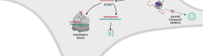
Toxins
Toxins       13, x13,
       2021,2021,  FOR487PEER REVIEW                                                                                               10 of10
                                                                                                                                         26of 25

          Figure 4. The MID1 protein   complex
                                  Figure  4. The induces translation
                                                  MID1 protein         of HTT
                                                                  complex       mRNA
                                                                           induces      with expanded
                                                                                     translation of HTTCAG
                                                                                                         mRNArepeats.
                                                                                                                 with MID1   (depicted
                                                                                                                       expanded    CAG in
                                                                                                                                        re-
          teal) attaches to HTT mRNA
                                  peats.with
                                         MID1expanded  CAG
                                                (depicted  inrepeats  (hairpintodepicted
                                                              teal) attaches     HTT mRNAin red)with
                                                                                                and mediates
                                                                                                     expandedthe binding
                                                                                                               CAG        of translational
                                                                                                                      repeats  (hairpin de-
                                  picted in red) and mediates the binding of translational regulators, including PP2A (depicted in
                                  blue). PP2A and its opposing kinase mTOR (depicted in dark blue) control the phospho-dependent
                                  activity of S6K (depicted in lilac) and 4E-BP1 (depicted in light green). 4E-BP1 is a negative regulator
target S6, which is a subunit of the ribosome. These two mTOR-dependent phosphorylation events
promote ribosome assembly on the RNA and thus promote translation. Besides recruiting PP2A and
S6K to the RNA hairpin, MID1 induces mTOR activity and simultaneously inhibits the activity of
PP2A by inducing its proteasomal degradation. Thus, MID1 indirectly stimulates translation. Cre-
ated with BioRender.com.
Toxins 2021, 13, 487 11 of 25
Generally, the translation of mRNA is initiated at the start codon (AUG). This also
applies to the open reading frame of the RNAs with expanded CAG repeats, which are
translated
regulators, including into(depicted
PP2A polyglutamine
in blue).protein.
PP2A andUntil
its 2011, it was
opposing assumed
kinase mTORthat, in thein
(depicted frame
dark with
blue) control
the start codon, only the polyglutamine protein is produced from the
the phospho-dependent activity of S6K (depicted in lilac) and 4E-BP1 (depicted in light green). 4E-BP1 mutant CAGisre-a negative
peats. However,
regulator of translation in 2011,translation
that suppresses Zu et al. showed an AUG-independent
when bound possibility
to the 50 end of an RNA. of translation
Its phosphorylation by mTOR
since mutation
leads to the detachment from ofRNAtheand
only AUG
the codon
release in ATXN8
of this transcripts
translational block. Atwith
the CAG-repeat
same time, S6K expansion
gets activated by
didvia
phosphorylation not prevent
mTOR. protein translation
Phospho-activated [68]. The repeat-associated
S6K phosphorylates its target S6, whichnon-AUG (RAN)
is a subunit of thetransla-
ribosome. These
tion occurs in GC-rich RNA sections. Such GC-rich regions are not
two mTOR-dependent phosphorylation events promote ribosome assembly on the RNA and thus promote only typical for CAG-translation.
repeat diseases, such as HD, but also occur in diseases caused by expansion
Besides recruiting PP2A and S6K to the RNA hairpin, MID1 induces mTOR activity and simultaneously inhibits of other re- the
peats
activity of PP2A by (such as CXG
inducing or GGGGCC).
its proteasomal degradation. Thus, MID1 indirectly stimulates translation. Created with
BioRender.com. Since both the sense and the antisense strands of the DNA can be transcribed into
RNA, bidirectional GC-rich mRNAs can be formed from the same gene locus. Since no
Since both the sense and the antisense strands of the DNA can be transcribed into
AUG start codon is required for the RAN translation, these two complementary mRNAs
RNA, bidirectional GC-rich mRNAs can be formed from the same gene locus. Since no
can theoretically be translated. Thus, two expanded repeat mRNAs (sense and antisense)
AUG start codon is required for the RAN translation, these two complementary mRNAs
can be translated in all six reading frames into six different potentially toxic proteins (Fig-
can theoretically be translated. Thus, two expanded repeat mRNAs (sense and antisense)
ure 5).
can be translated in all six reading frames into six different potentially toxic proteins
(Figure 5).
Figure 5. Schematic overview of an RAN translation. Mutant HTT is transcribed from both sense and antisense strands and
both resulting transcripts (CAG and CUG repeats) fold into hairpin structures that aberrantly recruit RNA-binding proteins
Figure
(RBPs). This leads 5. Schematic
to an overview
RAN translation of an RAN translation.
(repeat-associated non-AUGMutant HTT iswhich
translation), transcribed fromform
is a special bothofsense
translation that
andan
does not require antisense
AUG startstrands
codonand
butboth resulting
can start transcripts
at any base in all(CAG and CUG
3 reading repeats)
frames. fold into
This leads hairpin of aberrant
to translation
protein speciesstructures that aberrantly
in all 6 reading recruit RNA-binding
frames: polyalanine proteins (RBPs).
(polyA), polyserine (polyS), This leads to an(polyQ),
polyglutamine RAN translation
polyleucine (polyL)
(repeat-associated non-AUG translation), which is a special form of translation that does not re-
and polycysteine (polyC) proteins. Created with BioRender.com.
quire an AUG start codon but can start at any base in all 3 reading frames. This leads to translation
of aberrant protein Fourspecies in all 6HTT-RAN
mutant reading frames: polyalanine
translation (polyA),
proteins polyserine polyserine,
(polyalanine, (polyS), poly-
polyleucine and
glutamine (polyQ), polyleucine (polyL) and polycysteine (polyC) proteins. Created with BioRen-
polycysteine) from the sense and antisense transcripts were found in 2015 by analyzing
der.com.
human HD autopsy brain samples [69]. This finding was followed by several research
projects that led to inconsistent results. For example, one study was conducted on C.
Four mutant HTT-RAN translation proteins (polyalanine, polyserine, polyleucine
elegans expressing HD-RAN homopolymers. These contained codon-varied tags and
and polycysteine) from the sense and antisense transcripts were found in 2015 by analyz-
different CAG-repeat lengths. This study showed that only the polyL protein induced
ing human HD autopsy brain samples [69]. This finding was followed by several research
significant toxicity depending on the CAG-repeat length, although all RAN translation
proteins aggregated [70]. In contrast, different results were observed in the brains of two
different mouse models: (1) a model that enabled RAN translation but did not express
HTT with the polyglutamine tract translated from the start codon, and (2) a model that
expressed N-terminal polyglutamine HTT. Interestingly, no RAN translation products
could be identified and only those mice that expressed N-terminal polyglutamine HTT
showed an HD phenotype [71]. It should be noted that these studies are difficult to compare
due to their different model systems and experimental set-ups. Further experiments are
needed to conclude the relationship between RAN translation and neurotoxicity in HD
and other neurodegenerative CAG-repeat disorders.Toxins 2021, 13, 487 12 of 25
Taken together, all the above-mentioned experiments suggest that mutant CAG-repeat
mRNAs can aberrantly recruit translation factors and promote their own translation, not
only in the open reading frame defined by the AUG start codon but also in different reading
frames starting at the repeat sequence.
Besides this aberrant translation triggered by the CAG-repeat mRNA, translation in
HD is also affected by two further pathogenic mechanisms that are linked to the mutant
HTT protein. On one hand, mutant HTT protein interacts with ribosomal proteins and can
lead to ribosomal stalling, thereby disrupting translation [72]. On the other hand, mutant
HTT protein represses the expression of the mitochondrial metabolic regulator peroxisome
proliferator-activated receptor gamma co-activator 1α (PGC-1α) [73,74]. In addition to its
metabolic functions, PGC-1α is involved in ribosome biogenesis. Consequently, in HD,
ribosome biogenesis and thus protein synthesis is reduced, as shown, for example, in
the muscle biopsies of HD patients [75]. In summary, the general translation machinery
is impacted by both mutant HTT mRNA and mutant HTT protein, with the mutant
HTT mRNA pushing towards its own translation and the mutant HTT protein impacting
ribosomes. Thus, the general protein synthesis is disturbed by two hits in HD.
6. Deregulation of the microRNA Machinery
Another group of RNA-binding proteins that get trapped by mutant CAG-repeat
RNA, resulting in aberrant function, includes RNA processing enzymes that are involved
in the generation of small non-coding RNAs. Interestingly, more than 90% of the human
genome is transcribed but does not translate into protein [76]. These transcripts are
called non-coding RNAs and, beyond the fact that it is still not clear whether all of these
transcripts are functional, there are multiple examples of molecular functions of non-coding
RNAs [77]. For example, microRNAs (miRNAs) are endogenously expressed short non-
coding RNAs that downregulate gene expression of protein-coding genes in several cellular
processes [78–80]. miRNAs bind to complementary sequences in their target mRNAs of
protein-coding genes. miRNAs are transcribed as full-length transcripts called the primary
RNA (pri-miRNA) that go through a sequence of cleavage steps to produce small double-
stranded RNAs of 18–23 nucleotides. The first intranuclear cleavage step involves a protein
complex containing the Drosha and DGCR8 (DiGeorge syndrome critical region in gene
8) and results in an RNA fragment of 60–70 bp, which is referred to as precursor miRNA
(pre-miRNA) [81–83]. The pre-miRNA is then transported to the cytoplasm, passing the
nuclear pore with the help of exportin 5 [84,85]. The second cleavage step is carried
out by the enzyme DICER in combination with TRBP (trans-activation response RNA-
binding protein) and results in an 18–23 bp RNA duplex of the mature miRNA and its
antisense strand. While the antisense strand is released and degraded [86–89], the mature
miRNA then associates with and guides the RNA-induced silencing complex (RISC) to its
complementary target mRNA. The miRNA–RISC complex can either induce degradation
of the target mRNA or suppress its translation [90–94] (Figure 6).
Interestingly, some of the enzymes involved in miRNA cleavage aberrantly bind to
mutant HTT RNA at its CAG-repeat region, resulting in a DICER-dependent production of
small CAG-repeated RNAs (sCAGs) withToxins 2021, 13, 487 13 of 25
sCAGs are also found in HD patient brain tissue [97]. The detrimental effect of sCAGs can
be explained in two ways: either by the silencing of CTG-containing genes or by aberrant
recruitment of the miRNA-cleavage machinery and, thus, deregulation of miRNA synthesis.
Indeed, the expression of several miRNAs is deregulated in HD [100], as well as other
CAG-repeat-expansion diseases [101–103]. However, the exact molecular contribution of
the miRNA machinery and sCAGs in pathogenesis requires further study, especially since
21, 13, x FOR PEER REVIEW 14 of 26
another layer of complexity is observed in HD, where mutant polyglutamine protein also
interacts with a member of the RISC complex, namely, Argonaute (Ago2) [104].
Figure 6. Deregulation of the miRNA machinery and production of sCAGs. miRNAs bind to complementary sequences in
their target mRNAs of protein-coding genes. miRNAs are transcribed as primary RNA (pri-miRNA) that undergo serial
cleavage steps
Figurewithin the nucleus due
6. Deregulation to miRNA
of the enzymesmachinery
including DROSHA
and productionand DGCR8, as well
of sCAGs. as outside
miRNAs bindthe
to nucleus
com- due
to enzymes including DICER and TRBP. This cleavage results in small double-stranded RNAs
plementary sequences in their target mRNAs of protein-coding genes. miRNAs are transcribed as with 18–23 nucleotides,
of which the mature
primary miRNA
RNA associatesthat
(pri-miRNA) withundergo
the RISCserial
complex, while
cleavage the antisense
steps within thestrand
nucleusis degraded. The miRNA
due to enzymes
then guides the RISCDROSHA
including to its complementary
and DGCR8,target mRNA,
as well wherethe
as outside it either
nucleusinduces
due todegradation of the target
enzymes including mRNA or
DICER
suppressesand
its translation.
TRBP. ThisThe mutant results
cleavage HTT RNA hairpin
in small aberrantly recruits
double-stranded DICER,
RNAs withwhich
18–23 leads to the cleavage
nucleotides, of the long
of which
double-stranded CAG repeats
the mature miRNAinto fragments
associates with
with the22RISC
nt, termed sCAGs.
complex, whileThese
the sCAGs associate
antisense strandwith the RISC and
is degraded. The recruit
the RISC tomiRNA
mRNAsthen withguides the RISC tosequences.
complementary its complementary target
This, in turn, mRNA,
results where
in the it either
aberrant induces inhibition
translational degrada- of the
mRNAs with tionCUG-motifs.
of the target mRNA
Created or BioRender.com.
with suppresses its translation. The mutant HTT RNA hairpin aberrantly
recruits DICER, which leads to the cleavage of the long double-stranded CAG repeats into frag-
ments with 22 nt, termed sCAGs. These sCAGs associate with the RISC and recruit the RISC to
mRNAs with complementary sequences. This, in turn, results in the aberrant translational inhibition
of the mRNAs with CUG-motifs. Created with BioRender.com.
Another layer at which the miRNA machinery can affect cellular disease-mechanisms
is by changing the protein expression of RNA-binding proteins that aberrantly bind to
mutant HTT RNA and exhibit abnormal function in association with the CAG-repeat
RNA. For example, miRNAs that regulate the expression of the MID1 complex [105] couldToxins 2021, 13, 487 14 of 25
Another layer at which the miRNA machinery can affect cellular disease-mechanisms
is by changing the protein expression of RNA-binding proteins that aberrantly bind to
mutant HTT RNA and exhibit abnormal function in association with the CAG-repeat
RNA. For example, miRNAs that regulate the expression of the MID1 complex [105]
could change the translation rate of mutant HTT by repressing MID1 and, thus, could
counteract the increased translation of polyglutamine protein. Another example involves
miRNAs targeting important modifiers in HD pathogenesis, such as BDNF [106]. Indeed,
differential expression of miRNAs was detected in HD tissue [107]. Future studies
should aim at identifying the exact mechanisms of miRNA-dependent processes in
disease development.
7. Mitochondrial RNA
While in all the above-mentioned studies, the aberrant interaction of mutant HTT
mRNA was described, abnormal RNA-function in HD also occurs at a different level,
namely, in the mitochondrial RNA (mtRNA). Generally, mitochondria are important for
cell viability and several lines of evidence have connected mutant HTT protein with
mitochondrial dysregulation [108,109]. For example, striatal cells expressing mutant HTT
protein exhibit a substantial increase in the mitochondria-released reactive oxygen species
(ROS). As mtDNA is a major target of oxidative stress in HD, a dramatic decrease in
the mtDNA in mutant HTT cells is seen [110]. Additionally, defects in mitochondrial
Ca2+ handling contribute to HD pathogenesis [111]. Interestingly, the suppression of p53
leads to a substantial reversal of mutant HTT-induced mitochondrial depolarization and
cytotoxicity [112], indicating an integral role of p53 in mitochondrial dysfunction caused
by mutant HTT protein. While these effects on mitochondrial function are linked to the
aberrant function of the mutant HTT protein, in this review, we aimed to focus on RNA-
mediated aberrant mechanisms. In this respect, there is evidence that mitochondrial RNA
(mtRNA) plays an important role in imparting mitochondrial dysfunction. In an interesting
experiment conducted by Lee et al. cell-type-specific transcriptomic analyses were done in
order to study the gene expression changes in human HD and mouse models of HD by
single nuclear RNA sequencing (snRNA-seq) and translating ribosome affinity purification
(TRAP). They conducted striatal cell-type-specific transcriptomic studies in both human
and mouse models. In HD, they observed the cytosolic release of mtRNA, a potent innate
immunogen in striatal spiny projection neurons that led to the upregulation of innate
signaling responses. They also showed that the released mtRNAs bind to innate immune
sensor protein kinase R (PKR), which, when inhibited, could lead to the suppression
of innate immune pathway signaling [113]. Recently, a group of scientists developed a
new human neuronal cell model by using neural stem cells (ReNcell VM NSCs). These
cells were transduced to express HTT exon 1 with variable lengths of CAG repeats.
Investigations showed repeat-length-dependent formation of intranuclear inclusions
during neurogenesis, as well as marked mitochondrial dysfunction [114]. Taken together,
these observations suggest that not only the mutant CAG-repeat mRNA transcript plays
a role in RNA-mediated toxicity but mtRNA is also deregulated and pushes pathogenic
mechanisms. However, further studies that focus on elucidating the mechanisms of
mtRNA-mediated neurotoxicity are required to understand the mtRNA’s toxic gain of
function in HD.
8. Compounds
Given the fact that in HD, at least two neurotoxic gene products (the mutant transcript
and the mutant protein that is cleaved into two neurotoxic N- and C-terminal fragments)
contribute to neurodegeneration, an ideal therapeutic approach would target both the
mutant RNA and protein. A two-photon Ca2+ imaging study in Hdh150 knock-in mice
by Arnoux et al. showed neuronal hyperactivity in the visual cortex well before disease
onset (VFDO), suggesting that therapeutic intervention may be required at an early stage.
Small bioavailable molecules that are capable of crossing the blood–brain barrier would beToxins 2021, 13, 487 15 of 25
ideal therapeutics to inhibit such pathogenic cellular processes, for example, by reducing
aberrant recruitment of RBPs to mutant HTT RNA. One compound that was studied in the
context of VFDO is dimethylbiguanide metformin, which is an FDA-approved, low-cost
type II diabetes drug. Metformin suppresses mutant HTT translation via interfering with
the MID1 complex. This occurs through the metformin-mediated disassembly of the MID1
complex that leads to the activation of PP2A. This, in turn, decreases protein translation
and, thus, the protein level of mutant HTT both in cell cultures and in vivo. In addition,
metformin treatment reverses early-onset network dysregulation in HD mice [115]. In
2017, metformin was also shown to improve central phenotypic traits by normalizing ERK
signaling in a mouse model for a CGG-repeat disorder, namely, fragile X syndrome [116].
Thus, metformin seems to be promising for the therapy of CXG repeat expansion diseases,
including HD. However, to assess metformin‘s effect in HD patients, clinical studies are
required and, thus, further research is needed to show the consequences of its long-term
administration in HD patients.
Another molecule that was studied in HD models is furamidine. In silico identification
of CAG binding ligands led to the identification of furamidine, which binds to HTT exon1
RNA in vitro. The RNA binding of one known mutant HTT interactor, namely, MID1,
was blocked by furamidine, as shown, for example, in in vitro RNA protein pulldown
experiments. As a consequence of blocking MID1 binding to mutant HTT RNA, the
compound also reduces HTT translation and protein levels in an HD cell-line model.
However, furamidine binding is not specific for CAG-repeat RNA. In addition to CAG
repeats, it also binds AU-RNAs, CUG-RNAs and the DNA minor groove [117]. Thus,
undesirable side effects are expected, which provide an argument against HD treatment
with furamidine.
Another interesting compound for HD therapy is one of the flavonoids. Flavonoids
are known for their health benefits and can act as antioxidants, anticancer agents, neuronal
protection and neurodegenerative disease prevention. One flavonoid, namely, myricetin,
was shown to interact with the CAG motif and, thus, prevent both the translation of mutant
HTT protein and the sequestration of MBNL1. This happens through the interaction of
myricetin with RNA via base stacking at the A–A mismatch of the hairpin structure from
expanded CAG repeats. Additionally, in a 3-nitropropionic acid (3-NP)-induced HD rat
model, the oral supplementation of myricetin prevented mitochondrial dysfunction and
oxidative stress, along with motor deficits [118]. Further research will show whether the
therapeutic results on myricetin can be translated from rats to humans.
In 2019, Khan et al. designed and synthesized several pyridocoumarin derivatives to
attach to CAG motifs. Two of these derivatives turned out to be promising: both molecules
showed higher affinity and selectivity for expanded CAG-repeat RNA when compared
to regular duplex AU-paired RNA. Interestingly, both molecules are cell-permeable and
exhibit low toxicity to healthy fibroblasts. Additionally, they are also capable of reducing
the level of polyglutamine aggregation in cell cultures. Further research will show whether
these compounds have the same beneficial effects in vivo.
9. Antisense Oligonucleotides
Antisense oligonucleotides (AONs) are short (8–50 bases), synthetically produced
nucleic acids that bind complimentarily through Watson–Crick base pairing to a specific
sequence within their target RNA. Depending on their chemical design and target sequence,
AONs act through different mechanisms, including mechanisms that promote degradation
of the target mRNA, mechanisms that lead to translational inhibition or mechanisms
affecting splicing. For example, if a DNA AON binds its target mRNA, thereby forming
an RNA–DNA heteroduplex, this duplex is detected by endogenous enzymes, such as
ribonuclease H (RNase H), which cleaves the RNA strand. If an AON binds to its target pre-
mRNA at intron/exon junctions, it can mask splicing enhancers and repressor sequences,
thereby modulating splicing events. If an AON binds to its target mRNA, it can also
sterically block the 40S and 60S ribosomal subunits from attaching, thereby blockingToxins 2021, 13, 487 16 of 25
translation (reviewed in [119]). AONs have been in clinical use for many years. For example,
in 1998, formivirsen obtained approval for use in the treatment of cytomegalovirus retinitis
in patients with immunodeficiency [120].
Although a lot of research has been conducted and several attempts have been made
to develop therapeutic approaches, no disease-modifying treatment options are routinely
available to date, and current medical management is restricted to supportive care. This
may change with the development of HTT-lowering AONs. Several reports performed in
mouse models showed that AONs can be beneficial.
For example, Kordasiewicz et al. showed that the injection of an AON into the
cerebrospinal fluid of a symptomatic HD mouse model delayed disease progression [121].
The AON in this study not only reduced the expression of mutant HTT but also that of
the normal HTT allele. In a different approach, Ostergaard et al. developed an AONs
treatment approach that is specific for the mutant HTT allele by targeting single nucleotide
polymorphisms (SNPs) that are associated with the repeat expansion [122]. Another
way to specifically target the disease allele is to target the CAG-repeat region. Indeed,
using a (CUG)7 AON showed beneficial effects in HD models, along with models of
other CAG-repeat-expansion diseases, such as spinocerebellar ataxia type 1 (SCA1) and
SCA3 [123–125]. Based on these encouraging results, AONs represent an interesting HTT-
lowering strategy for HD. If AONs are used to target the CAG repeat, it could not only
prove to be a promising therapeutic tool to diminish the mutant HTT in HD but also help
in perturbing the effects of other polyglutamine diseases.
10. Clinical Trials and Novel Therapies
To this point, there is no therapy that can cure CAG-repeat diseases, such as HD.
The currently available treatments impart only symptomatic relief but do not stop disease
progression. Through physiotherapy or speech therapy, the patient learns to manage
involuntary movements or speech difficulties. Neuroleptics or tetrabenazine (indirectly
acting as antidopaminergic) are used as medication. These substances help to inhibit
uncontrollable muscle movements [10]. Psychotherapies are used to treat depression. In
addition, genetic counseling can be of importance for the families of the patients.
Various therapeutic approaches for curing HD are being explored in current research
projects and clinical studies. A primary goal is to develop therapies that will take effect
before symptoms appear, as it is not certain that the symptom-free phenotype can be
restored later in HD. During this period, the post-transcriptional level offers opportunities
for promising therapeutic approaches since the targeting of the mutant transcript and,
consequently, the lack of the mutant protein would deprive most of the known pathomech-
anisms.
One option for targeting the mutant transcript involves AONs. There are two different
approaches using AONs that are currently used in clinical trials for HD treatment. In the
first approach, AONs targeting the HTT transcripts from both alleles (normal and mutant)
are used. An example is an AON called tominersen. In preclinical studies on model
organisms, the use of tominersen led to significant changes. After 14 days of treatment of
BACHD mice (which express human mutant HTT) with the AON tominersen, the HTT
mRNA level was reduced by up to 80%. This reduction lasted for 16 weeks. In addition,
symptomatic improvement was observed in three different HD mouse models. In this
context, the motor deficits in young animals were almost completely restored. Next, this
AON was tested in clinical trials. In a phase 1b/2a clinical study conducted in 2015, the
tominersen doses were administered intrathecally. Notably, this Ionis-Pharmaceuticals-
funded study showed that tominersen decreased levels of the mutant HTT protein in
cerebrospinal fluid (CSF) in patients with early stages of HD. Thus, this attempt awakens
new hope for a curative treatment. However, it is unclear whether lowering wild-type
HTT in an organism has long-term consequences. The phase 3 study on tominersen
(GENERATION-HD; NCT03761849; sponsored by Hoffmann-La Roche), which aimed to
clarify these consequences, began in January 2019. This trial was planned to end in 2022You can also read