Molecular basis of mRNA transport by a kinesin-1-atypical tropomyosin complex - Genes Dev
←
→
Page content transcription
If your browser does not render page correctly, please read the page content below
Downloaded from genesdev.cshlp.org on August 13, 2021 - Published by Cold Spring Harbor Laboratory Press

Molecular basis of mRNA transport by a
kinesin-1–atypical tropomyosin complex
Lyudmila Dimitrova-Paternoga,1,2,3 Pravin Kumar Ankush Jagtap,2 Anna Cyrklaff,1 Vaishali,1,4
Karine Lapouge,5 Peter Sehr,6 Kathryn Perez,5 Simone Heber,1 Christian Löw,3 Janosch Hennig,2
and Anne Ephrussi1
1
 Developmental Biology Unit, European Molecular Biology Laboratory (EMBL) Heidelberg, 69117 Heidelberg, Germany;
2
 Structural and Computational Biology Unit, EMBL Heidelberg, 69117 Heidelberg, Germany; 3Centre for Structural Systems
Biology (CSSB), Deutsches Elektronen-Synchrotron (DESY), EMBL Hamburg, 22607 Hamburg, Germany; 4Faculty of Biosciences,
Heidelberg University, 69120 Heidelberg, Germany; 5Protein Expression and Purification Core Facility, EMBL Heidelberg, 69117
Heidelberg, Germany; 6Chemical Biology Core Facility, EMBL Heidelberg, 69117 Heidelberg, Germany

Kinesin-1 carries cargos including proteins, RNAs, vesicles, and pathogens over long distances within cells. The
mechanochemical cycle of kinesins is well described, but how they establish cargo specificity is not fully under-
stood. Transport of oskar mRNA to the posterior pole of the Drosophila oocyte is mediated by Drosophila kinesin-1,
also called kinesin heavy chain (Khc), and a putative cargo adaptor, the atypical tropomyosin, aTm1. How the
proteins cooperate in mRNA transport is unknown. Here, we present the high-resolution crystal structure of a
Khc–aTm1 complex. The proteins form a tripartite coiled coil comprising two in-register Khc chains and one aTm1
chain, in antiparallel orientation. We show that aTm1 binds to an evolutionarily conserved cargo binding site on
Khc, and mutational analysis confirms the importance of this interaction for mRNA transport in vivo. Furthermore,
we demonstrate that Khc binds RNA directly and that it does so via its alternative cargo binding domain, which
forms a positively charged joint surface with aTm1, as well as through its adjacent auxiliary microtubule binding
domain. Finally, we show that aTm1 plays a stabilizing role in the interaction of Khc with RNA, which distinguishes
aTm1 from classical motor adaptors.
[Keywords: kinesin; kinesin adaptor; kinesin–atypical tropomyosin complex; mRNA transport; oskar mRNA]
Supplemental material is available for this article.
Received March 5, 2021; revised version accepted May 14, 2021.

Intracellular transport is an essential cellular function                   dendrites, cell migration, and embryonic axis formation
that is ensured by a vast cytoskeletal network and three                    (Hirokawa et al. 2009).
major classes of motor proteins, namely kinesins, dyne-                        Kinesin-1 harbors an N-terminal motor domain, fol-
ins, and myosins (Hirokawa et al. 2009; de Lanerolle                        lowed by a long coiled-coil stalk that mediates homodi-
2012; Olenick and Holzbaur 2019). Whereas myosins are                       merization, and a flexible tail with regulatory function
responsible for short-range transport along randomly ori-                   (Fig. 1A; Seeger and Rice 2013). The two motor domains
ented actin filaments, kinesins and dyneins provide                         are powered by ATP hydrolysis, which elicits conforma-
long-range directional transport on a polarized microtu-                    tional changes that allow the kinesin molecule to move
bule network (Titus 2018). Kinesin-1, a microtubule plus                    processively along microtubules (Qin et al. 2020). The
end-directed motor, was the first kinesin identified and                    kinesin-1 stalk associates with cargos either directly or
is well characterized (Vale et al. 1985; Hirokawa et al.                    via adaptor proteins that bind to the C-terminal region
2009). It transports diverse cargos including proteins, or-                 of the domain (Cross and Dodding 2019). Although a large
ganelles, mRNA nucleoprotein complexes (mRNPs), and                         portion of kinesin-1 transport is mediated by the adaptor
viruses (Chudinova and Nadezhdina 2018; Garcin and                          kinesin light chain (Klc), the transport of mRNA cargos
Straube 2019; Banerjee et al. 2020) and is essential for nu-                such as mRNP granules in dendrites or oskar in the Dro-
merous processes such as endoplasmic reticulum (ER)-                        sophila oocyte appears to be independent of Klc (Diefen-
Golgi transport, mitochondrial distribution in axons and                    bach et al. 1998; Kanai et al. 2004; Loiseau et al. 2010;
                                                                            Cross and Dodding 2019).

Corresponding authors: anne.ephrussi@embl.org,
janosch.hennig@embl.de
Article published online ahead of print. Article and publication date are   © 2021 Dimitrova-Paternoga et al. This article, published in Genes & De-
online at http://www.genesdev.org/cgi/doi/10.1101/gad.348443.121. Free-     velopment, is available under a Creative Commons License (Attribution-
ly available online through the Genes & Development Open Access             NonCommercial 4.0 International), as described at http://creativecom-
option.                                                                     mons.org/licenses/by-nc/4.0/.

                  GENES & DEVELOPMENT 35:1–16 Published by Cold Spring Harbor Laboratory Press; ISSN 0890-9369/21; www.genesdev.org               1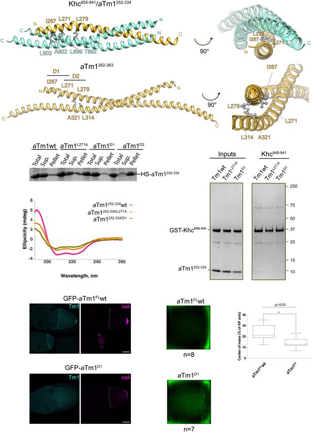
Downloaded from genesdev.cshlp.org on August 13, 2021 - Published by Cold Spring Harbor Laboratory Press

Dimitrova-Paternoga et al.

A                                                                                       Figure 1. aTm1 interacts with the alterna-
                                                                                        tive cargo binding domain of Khc. (A) Sche-
                                                                                        matic representation of aTm1 and kinesin
                                                                                        heavy chain (Khc). aTm1 has a central
                                                                                        coiled-coil domain, flanked by mostly un-
                                                                                        structured N- and C-terminal regions. Khc
                                                                                        comprises an N-terminal motor-domain
                                                                                        (amino acids 1–345), followed by a long
B                                          C                                            coiled-coil stalk (amino acids 345–902) and
                                                                                        an unstructured tail (amino acids 902–975).
                                                                                        The stalk includes the kinesin light chain
                                                                                        (Klc) binding region (amino acids 792–839)
                                                                                        and an alternative cargo binding region (ami-
                                                                                        no acids 848–902). The tail comprises an
                                                                                        ATP-independent       microtubule      binding
                                                                                        domain (amino acids 910–938) and an IAK
                                                                                        (isoleucine-alanine-lysine) motif (amino acids
                                                                                        938–950), responsible for the autoinhibition
                                                                                        of Khc (Williams et al. 2014). Although in
                                                                                        many studies the alternative cargo binding
                                                                                        region has been considered part of the tail,
D
                                                                                        based on its structural and functional fea-
                                                                                        tures, we categorize it as part of the coiled-
                                                                                        coil stalk. (B) Yeast two-hybrid analysis
                                                                                        shows that aTm1 and Khc interact. The
                                                                                        Khc variants were tagged with the GAL4
                                                                                        binding domain, and the aTm1 variants
                                                                                        were fused to the GAL4 activation domain.
                                                                                        Yeast cells were cotransformed with different
                                                                                        combinations of the above-mentioned con-
                                                                                        structs and spotted in two 10-fold dilutions
                                                                                        on SDC−Leu−Trp and SDC−Leu−Trp−His
                                                                                        plates. (C ) A GST (glutathione S-transferase)
                                                                                        pull-down assay reveals direct binding of
                                                                                        aTm1 to kinesin: GST-tagged Khc1–264 and
                                                                                        Khc265–975 were immobilized on glutathione
                                                                                        beads and mixed with E.coli lysates contain-
                                                                                        ing recombinantly expressed His6-SUMO-
                                                                                        aTm1-FL and His6-SUMO-GFP (Supplemen-
                                                                                        tal Fig S1A). The eluates were analyzed by
                                                                                        SDS-PAGE. The asterisk indicates the inter-
                                                                                        action of aTm1FL with Khc265–975. (D)
                                                                                        aTm1247–335 interacts with the alternative
                                                                                        cargo binding domain of Khc848–941. In vitro
                                                                                        binding of GST-tagged Khc truncations (sche-
matic top left) to different aTm1 truncations (schematic top right), premixed with E.coli whole-cell lysate. The asterisk indicates the
interaction of aTm1247–334 with Khc848–941.

   In Drosophila, localization of oskar mRNA to the poste-            ports oskar to the posterior pole where the mRNA is local-
rior pole of the developing oocyte drives abdominal pat-              ized (for review, see Trcek and Lehmann 2019). oskar
terning and germline formation in the embryo (Ephrussi                mRNA transport depends on a region of Khc downstream
and Lehmann 1992). The directional transport of RNA is                from the Klc binding site that includes an alternative car-
mediated by both the dynein and kinesin transport ma-                 go binding domain and an ATP-independent microtubule
chineries (Clark et al. 2007; Zimyanin et al. 2008). In the           binding site (Williams et al. 2014).
germline syncytium, the oocyte is transcriptionally quies-               Moreover, kinesin-mediated oskar transport requires a
cent and relies on mRNAs provided by 15 interconnected                number of cis and trans factors. One of these factors is a
nurse cells for its growth and development (Roth and                  unique isoform of the actin binding protein tropomyosin
Lynch 2009). Upon transcription in the nurse cells, oskar             1, known as Tm1-I/C, which we refer to here as atypical
mRNA is transported into the oocyte on the microtubule                or aTm1 for simplicity (Loiseau et al. 2010; Veeranan-Kar-
cytoskeleton by the minus end-directed dynein motor                   megam et al. 2016; Gáspár et al. 2017a). In the absence of
complex (Clark et al. 2007). Within the oocyte, the micro-            aTm1, oskar mRNA fails to localize to the posterior pole
tubule plus end-directed motor kinesin-1 or kinesin heavy             of the oocyte, as kinesin recruitment to oskar mRNA re-
chain (Khc) in Drosophila (Lawrence et al. 2004) trans-               quires aTm1 function (Erdélyi et al. 1995; Gáspár et al.

2      GENES & DEVELOPMENT
Downloaded from genesdev.cshlp.org on August 13, 2021 - Published by Cold Spring Harbor Laboratory Press
mRNA transport by kinesin–atypical tropomyosin 1
2017a). Furthermore, the fact that tethering of the Khc fragments of the proteins (Fig. 1D; Supplemental Fig.
motor domain to oskar restores posterior localization of S1B,C). This revealed that aTm1247–335 binds Khc848–941
the mRNA in a Tm1 protein-null background suggested (Fig. 1D, panel 4), which comprises the alternative cargo
that aTm1 is an essential adaptor for Khc-mediated oskar binding region and the ATP-independent microtubule
transport (Gáspár et al. 2017a). binding region of Khc (Fig. 1D; Seiler et al. 2000; Williams
Here, we show that aTm1 homodimers adopt an unusu- et al. 2014). Veeranan-Karmegam et al. (2016) have previ-
al antiparallel coiled-coil conformation. Moreover, aTm1 ously suggested that aTm1 interacts with residues 914–
binds to an alternative conserved cargo binding region in 936 of Khc. However, the fact that Khc902–941 did not
Khc, forming a tripartite coiled-coil complex with a posi- interact with aTm1 in our assays (Fig. 1D, panel 5) indi-
tively charged surface. Finally, we show in vivo that the cates that aTm1247–335 binds to the conserved alternative
function of aTm1 is ensured by its Khc interacting domain cargo binding site of Khc (residues 848–902) (Supplemen-
and that this atypical Tm1 functions in stabilizing the in- tal Fig. S1D).
teraction of Khc with RNA rather than as a classical
adaptor.
The alternative cargo binding region of Khc is essential
for oskar mRNA localization
Results
The alternative cargo binding region in Khc was originally
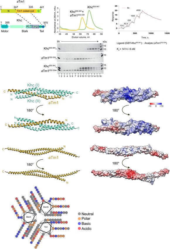
aTm1 binds to the alternative cargo binding site of Khc
identified in Neurospora crassa kinesin (Nkin), where
It was previously reported that aTm1 is necessary for the deletion of this site abolished the ability of the protein
recruitment of Khc to oskar mRNA and that the two pro- to rescue the Nkin null mutant phenotype (Seiler et al.
teins physically interact (Veeranan-Karmegam et al. 2016; 2000). In Drosophila, deletion of the entire C terminus
Gáspár et al. 2017a). To elucidate the molecular basis of of Khc, which includes the alternative cargo binding re-
the aTm1-Khc interaction, we first performed a yeast gion and the regulatory tail, also led to an oskar mRNA
two-hybrid analysis, which confirmed that aTm1 (Tm1- localization defect among others (Williams et al. 2014).
FL) binds both full-length Khc (Khc-FL) and Khc265–975 To investigate the relevance of this cargo binding region
(Fig. 1B). Because the interaction of Khc and aTm1 was for oskar mRNA transport, we expressed RNAi-resistant
formerly tested by coimmunoprecipitation (IP) experi- Khc-FL-mKate2 and Khc855–911Δ-mKate2 (wherein the
ments from S2 cells or Drosophila ovarian lysates, neither cargo binding domain is deleted) fusion proteins in a
of which is performed in a nucleic acid-free environment khc-RNAi background (Fig. 2A; Supplemental Fig. S2).
(Veeranan-Karmegam et al. 2016; Gáspár et al. 2017a), we Khc855–911Δ localized to the posterior pole of the oocyte
also probed the interaction by pull-down of recombinant (Fig. 2B, upper right panel), indicating that Khc motor
Khc and aTm1 proteins in the presence of DNase and RN- function and polarity of the microtubule cytoskeleton
ase (Fig. 1C; inputs in Supplemental Fig. S1A). Binding of were preserved. However, oskar mRNA failed to localize
aTm1 to Khc was preserved under these conditions (Fig. at the posterior pole (Fig. 2B, bottom right panel) and the
1C, right panel, lanes 5,6), indicating the specific and di- eggs produced failed to hatch (Fig. 2C), demonstrating
rect interaction of aTm1 with Khc. that the alternative cargo binding region of Drosophila
To further narrow down the aTm1 and Khc interacting Khc, and likely its interaction with aTm1, play a major
regions, we performed GST pull-down assays on shorter role in oskar mRNA transport.
A Figure 2. oskar mRNA localization depends on
the alternative cargo binding domain of Khc. (A)
Schematic of Khc-FL and KhcΔ855–911 transgenic
constructs resistant to khc-RNAi used in this study.
The pink stars indicate silent point mutations in
the region targeted by khc-RNAi; KhcΔ855–911 lacks
B C the alternative cargo binding domain. (B) Deletion
of the alternative cargo binding region of Khc leads
to oskar mRNA mislocalization. (Top middle and
right panels) Khc-FL and KhcΔ855–911 variants tagged
with mKate2 were expressed in a khc-RNAi back-
ground. Khc variants and khc-RNAi were expressed
in the germline, which includes nurse cells and the
oocyte (see schematic top left). (Bottom panels)
oskar mRNA was visualized by single-molecule
(sm)FISH. Scale bar, 25 µm. (C ) Hatching rates of
eggs from flies expressing Khc-FL or KhcΔ855–911
transgenes in Khc knockdown background.
GENES & DEVELOPMENT 3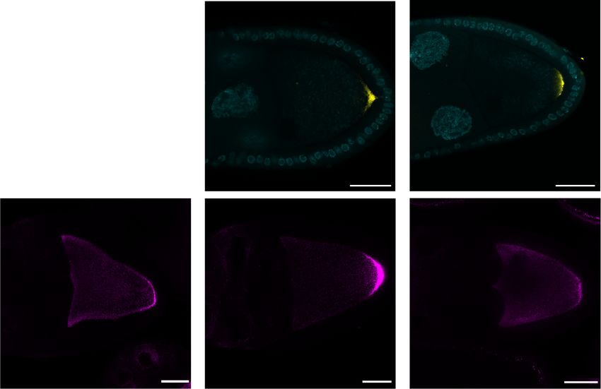
Downloaded from genesdev.cshlp.org on August 13, 2021 - Published by Cold Spring Harbor Laboratory Press

Dimitrova-Paternoga et al.

The Khc interaction domain of aTm1 is essential for oskar               Fig. S3C). These data show that the core domain of aTm1
localization                                                            (residues 247–334) identified as the Khc binding domain is
                                                                        essential for oskar mRNA localization.
The region of aTm1 (residues 247–335) that interacts with                  Interestingly, despite supporting oskar mRNA localiza-
Khc consists of a coiled coil that is also present in the clas-         tion, GFP-aTm1247–334, which lacks a major portion of the
sical Tm1-A isoform (residues 282–335), preceded by a                   unique N-terminal region of aTm1, does not accumulate
unique N-terminal sequence predicted to be at least par-                at the posterior pole of the oocyte, in contrast to aTm1FL
tially helical (Fig. 1D). To understand the functional sig-             or aTm11–334. This suggests that the aTm1 N-terminal
nificance of the aTm1–Khc interaction, we generated                     domain interacts with components of the pole plasm or
GFP-tagged aTm1 truncations in which the Khc interac-                   with oskar mRNA itself, resulting in aTm1 accumulation
tion site was either present or absent and tested them in               at the oocyte posterior.
vivo. We expressed the constructs in Tm1 eg9 mutant flies,
in which aTm1 is not present and oskar mRNA fails to
localize at the posterior of the oocyte (Supplemental Fig.              aTm1 coiled-coil dimers adopt an antiparallel
S3A–C, first panel; Erdélyi et al. 1995; Gáspár et al.                  configuration
2017a).                                                                 To gain insight into the molecular basis of aTm1–Khc in-
   Expression of transgenic full-length aTm1 (aTm1FL) and               teraction, we used crystallography to determine the struc-
aTm11–334 restored oskar localization to the posterior pole             ture of aTm1 alone and in complex with Khc. The classical
of Tm1 eg9 oocytes (Fig. 3A–C), suggesting that the aTm1                actin-binding tropomyosins (Tm1-PG and Tm1-PA in Dro-
C-terminal region is nonessential for this function. Expres-            sophila) are highly conserved in eukaryotes (Fig. 4A),
sion of the minimal Khc interaction region of aTm1                      where they form long parallel coiled coils that polymerize
(aTm1247–334) also promoted oskar localization in                       in a head-to-tail fashion along the actin filaments (Parry
Tm1 eg9 oocytes. Although the RNA was not as concentrat-                and Squire 1973). Residues 81–165 of Tm1-PG are present
ed at the posterior pole as upon aTm1FL or aTm11–334 ex-                in aTm1 whose coiled-coil region is further extended by 20
pression (Fig. 3B,C; analysis was performed as in Gaspar                unique residues at the N terminus (aTm1262–366) (Fig. 4A),
et al. 2014), the female progeny developed ovaries and                  which we included in our constructs.
were fertile. Neither aTm11–247 nor aTm1335–441 could re-                 We performed crystallization trials with different
store oskar localization in Tm1 eg9 oocytes (Supplemental               aTm1 truncations (Fig. 4A) and obtained crystals for

            A

            B                                                                          C

Figure 3. aTm1247–334 rescues oskar mRNA localization in Tm1 eg9/Tm1 eg9 mutant. (A) Confocal images of Tm1 eg9/Tm1 eg9 egg cham-
bers expressing GFP-tagged aTm1FL, aTm11–334, and aTm1247–334 variants. oskar mRNA was visualized by smFISH. Scale bar, 25 µm. (B)
Mean oskar distribution (green) in stage 9 oocytes from the flies in A. n indicates the number of oocytes analyzed. (C ) Position of oskar
mRNA center of mass relative to the geometric center of the oocyte represented in box plots with minimum and maximum whiskers. The
analysis was performed as in Gaspar et al. (2014).

4      GENES & DEVELOPMENT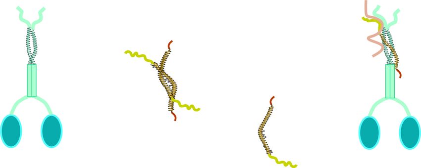
Downloaded from genesdev.cshlp.org on August 13, 2021 - Published by Cold Spring Harbor Laboratory Press

                                                                                     mRNA transport by kinesin–atypical tropomyosin 1

A                                                                                       Figure 4. aTm1 forms an antiparallel
                                                                                        coiled coil. (A) Multiple sequence align-
                                                                                        ment of three Tm1 isoforms from Droso-
                                                                                        phila melanogaster. The shared coiled-coil
                                                                                        region of aTm1 extends N-terminally into
                                                                                        a unique predicted α helix (the predicted
                                                                                        secondary structure is indicated under the
                                                                                        alignment where regions with thick gray
                                                                                        bars indicate α helices). Tm1-PG and
                                                                                        Tm1-PA are two classical actin-binding
                                                                                        nonmuscle isoforms that lack this N-termi-
                                                                                        nal extension. The boundaries of the crys-
                                                                                        tallized constructs of aTm1 are indicated
                                                                                        above the alignment. The alignment was
                                                                                        done by ESPript 3 (Robert and Gouet
                                                                                        2014). (B) aTm1 coiled-coil region crystal-
                                                                                        lized in an antiparallel conformation. Crys-
B                                                         C                             tal structures of aTm1262–363 (top panel)
                                                                                        and aTm1270–334 (middle panel) and their
                                                                                        superimposition (bottom panel). Residues
                                                                                        261–358 are resolved in the crystal struc-
                                                                                        ture of the aTm1262–363 construct. Residues
                                                                                        277–319 in the first coil and residues 273–
                                                                                        330 in the second coil are resolved in
                                                                                        aTm1270–334. (C) The coiled coil of aTm1
                                                                                        is stabilized primarily by hydrophobic in-
                                                                                        teractions. Helical wheel diagram of
                                                                                        aTm1262–363 as generated by DrawCoil 1.0
                                                                                        (https://grigoryanlab.org/drawcoil).    The
                                                                                        dashed lines connecting Arg292 and
                                                                                        Asp299 indicate salt bridges between these
                                                                                        residues.

aTm1262–363 and aTm1270–334 constructs that diffracted to          aTm1–Khc interaction domains form a triple coiled coil
a resolution of 2.45 Å and 2.3 Å, respectively (Fig. 4B). The      of two kinesin chains and one aTm1 chain
structures were solved by ab initio molecular replacement
with Ample and were refined to an Rwork/Rfree of 0.25/0.29         We next reconstituted, biophysically characterized, and
and of 0.23/0.27 for Tm1262–363 and Tm1270–334, respec-            crystallized the minimal aTm1–Khc complex. To do so,
tively (see Supplemental Table S1 for structural statistics;       the initially identified protein boundaries (Fig. 1D) were
Bibby et al. 2012). Residues 261–358 were fitted into the          further refined to improve protein solubility and yields,
density of the aTm1262–363 crystal structure, whereas res-         after which individually purified aTm1252–334 and
idues 277–319 in the first coil and residues 273–330 in the        Khc855–941 were mixed and analyzed by gel filtration
second coil were resolved in aTm1270–334.                          (Fig. 5A). Upon addition of aTm1, the Khc peak shifted
   Both truncations crystallized as antiparallel coiled coils      to a higher apparent molecular weight, indicating com-
with an average backbone RMSD of 1 Å between residues              plex formation. Interestingly, the aTm1 peak fractions
278 and 328 (Fig. 4B). A helical wheel diagram of                  shifted to a later retention time (larger elution volume) in-
aTm1262–363 in Figure 4C demonstrates that the two α he-           dicative of compaction of the protein in the complex upon
lices are held together primarily by hydrophobic residues          binding (Fig. 5A, middle gel [lanes 7–9] and lower gel [lanes
at the a and d positions of the heptad repeats, which is typ-      8–10]). Also, although we mixed the proteins in a 1:1 ratio
ical for coiled-coil structures. The interaction is addition-      before injection onto the column (Fig. 5A, right panel,
ally stabilized by two symmetric salt bridges on the               lowest gel, lane 1), the ratio of the bands in the complex
surface from Arg292 and Asp299 of chain A to Asp299                peak (Fig. 5A, right panel, lowest gel, lanes 8–12), as well
and Arg292 of chain B, respectively.                               as the presence of an additional aTm1 peak (Fig. 5A, right
   The unusual antiparallel coiled-coil conformation of            panel, lowest gel, lanes 3–7) indicate that Khc is in excess
aTm1 distinguishes it from the canonical Tm1 isoforms,             in the complex. To determine the affinity of the
which form parallel coiled coils, and might be an addition-        aTm1–Khc interaction, we performed surface plasmon
al factor attributing to it specific functions.                    resonance (SPR) experiments using an anti-GST antibody

                                                                                                    GENES & DEVELOPMENT            5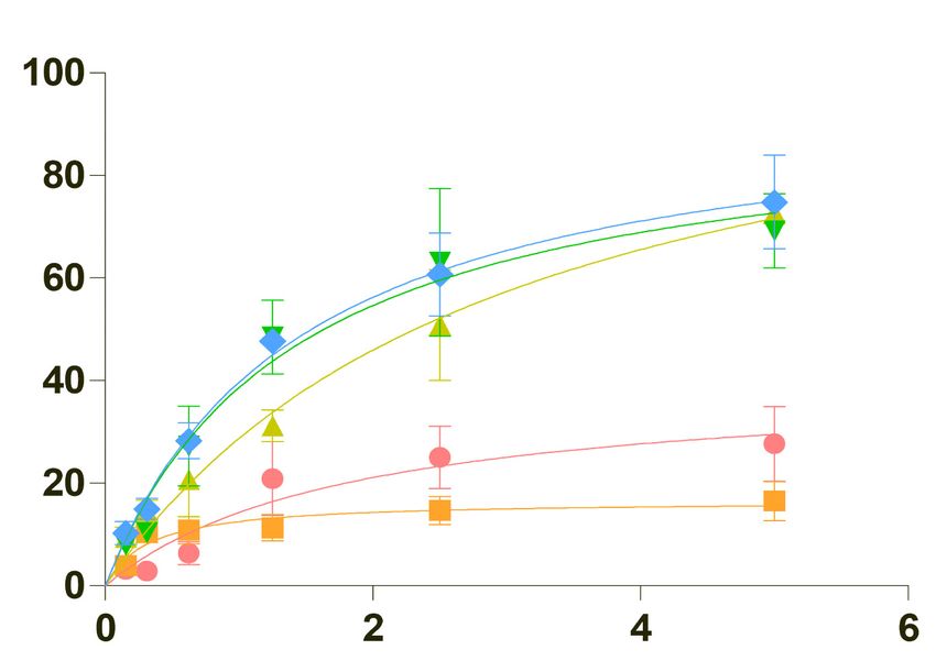
Downloaded from genesdev.cshlp.org on August 13, 2021 - Published by Cold Spring Harbor Laboratory Press

                A                                                                     B

                C

                D

Figure 5. The aTm1–Khc complex forms a tripartite coiled coil. (A) Reconstitution of an aTm1–Khc complex, consisting of the minimal
interaction regions of the two proteins (left schematic). Size exclusion chromatography (S200 resin) of Khc855–941, aTm1252–334, and a 1:1
mixture of both proteins. The UV trace of aTm1 alone could not be recorded because of the very low extinction coefficient of this trun-
cation. Peak fractions were analyzed by gel electrophoresis in the bottom panel. (B) Binding of aTm1252–334 to Khc848–941 analyzed by sur-
face plasmon resonance (SPR). GST-tagged Khc848–941 was used as a ligand, and increasing concentrations (indicated above the sensogram
in nanomoles) of aTm1 were added as analyte. Ligand to analyte was fitted using a standard 1:1 kinetic model with a Kd = 141 ± 6 nM. (C)
Crystal structure of the aTm1252–334/Khc855–941 complex. Two different helical views (left panels) and the corresponding electrostatic
views (right panels) are presented. The two Khc chains (aquamarine) are parallel and in register, whereas the single aTm1 chain (gold)
is in an antiparallel conformation. The crystal structures consist of aTm1 (residues 259–324), KhcI (residues 855–923), and KhcII (residues
855–916). The bottom two left and right panels represent ribbon and electrostatic views of aTm1262–363 for comparison. (D) The triple
coiled coil of the aTm1–Khc complex is stabilized primarily by hydrophobic interactions; helical wheel diagram generated by DrawCoil
1.0 (https://grigoryanlab.org/drawcoil).

6      GENES & DEVELOPMENT
Downloaded from genesdev.cshlp.org on August 13, 2021 - Published by Cold Spring Harbor Laboratory Press

                                                                                     mRNA transport by kinesin–atypical tropomyosin 1

coated chip allowing immobilization of GST-Khc848–941.             aTm1I267A/L271A/L279A (aTm1T1) at the unique N-terminal
aTm1252–334 was added stepwise (fivefold increasing con-           extension of aTm1, and aTm1L296A/L300A/L307A (aTm1T2)
centration at each step), and the resulting sensorgram             in the middle of the aTm1 coiled coil—and a combination
could be fitted to a standard 1:1 kinetic model with a dis-        of the two (aTm1Hexa) (Fig. 6A; Supplemental Fig. S5C).
sociation constant (Kd) of 141 ± 6 nM (Fig. 5B).                   Both the aTm1T1 and aTm1Hexa mutations fully abolished
   The aTm1252–334–Khc855–942 complex crystallized in a            interaction with Khc, as determined by in vitro binding
P21 space group. A data set was collected to a resolution          assays (Supplemental Fig. S5D). However, although we
of 2.3 Å and a model obtained by ab initio molecular re-           changed the target residues to alanines in order to preserve
placement with Arcimboldo (Rodríguez et al. 2009). The             the helical propensity of aTm1, circular dichroism (CD)
structure was refined to a final Rwork/Rfree of 0.24/0.28, in-     spectroscopy showed that the mutations affected substan-
corporating aTm1 (residues 259–324), KhcI (residues 855–           tially the structure of the aTm1 homodimers (Supplemen-
923), and KhcII (residues 855–916) in 1:2 (aTm1:Khc) stoi-         tal Fig. S5E).
chiometry (Fig. 5C). The trimeric structure comprises two             As the triple mutant T2 showed residual binding to
parallel, in-register Khc chains and a single, antiparallel        Khc, we focused on T1 and designed two double mu-
Tm1 chain, with the Khc C-terminal residues in close               tants, aTm1I267A/L271A (aTm1D1) and aTm1 L271A/L279A
proximity to the aTm1 N-terminal region. This arrange-             (aTm1D2) (Fig. 6A). The mutations introduced in aTm1D2
ment suggests that the coiled coil of aTm1 opens to ac-            substantially reduced the solubility of aTm1252–334 since
commodate the coiled-coil stalk of Khc. A possible                 most of the recombinantly expressed protein precipitated
explanation for the mechanism of aTm1–Khc complex                  after centrifugation of the E.coli lysate (Fig. 6B). This
formation would be a lower dimerization propensity of              might be due to the fact that L279 lies at the junction of
the aTm1 homotypic coiled coil. To estimate the Kd of              the two aTm1 chains in close proximity to L314 and
the aTm1 homodimer, we performed an isothermal titra-              A321 of the second chain and is therefore essential for
tion calorimetry (ITC) experiment whereby we fitted the            the stability of the aTm1 coiled coil. This again demon-
heat changes upon dilution of aTm1252–334 (Supplemental            strates how the fact that aTm1 uses the same surface to
Fig. S4A), and obtained a Kd of 10 µM. Thus, aTm1 has a            form a homodimer and to interact with Khc renders it dif-
higher affinity to Khc (Kd in the nanomolar range) (Fig.           ficult to generate an aTm1 mutant that has no effect on
5B) than to another aTm1, which explains why aTm1                  the structure of aTm1 yet disrupts its interaction with
forms a complex with the Khc homodimer.                            Khc. Thus, even the two mutations in aTm1D1 appear to
   Superimposition of the aTm1 homodimer on the Khc–               destabilize the coiled coil to some extent, as a SEC-
aTm1 complex structure revealed no major changes of                MALS (size exclusion chromatography multiangle light
the aTm1 helix except for a kink caused by the looping             scattering) measurement indicates that aTm1D1 behaves
out of T277 (Supplemental Fig. S4B). As crystals of Khc            as a mixture of dimers and monomers (Supplemental
truncations were only obtained in the presence of aTm1,            Fig. S5F). Nonetheless, the aTm1D1 mutations did not af-
we could not evaluate the effect of aTm1 binding on                fect solubility of the protein and had only a moderate ef-
Khc conformation.                                                  fect on its secondary structure as assessed by CD
   The triple coiled coil is held together by van der Waals        spectroscopy (Fig. 6B,C). More importantly, aTm1D1 in-
forces between mostly hydrophobic residues at “a” and              teraction with Khc was notably reduced in our GST
“d” positions of each helix (Fig. 5D). Additionally, the sur-      pull-down assays and as evaluated by SPR (Fig. 6D; Sup-
face of the complex is characterized by a positively               plemental Fig. S5G).
charged patch, not present in the aTm1262–363 homo-                   To assess aTm1D1 function in vivo, we generated a GFP-
dimer, formed by the polar side chains of K884, K887,              tagged aTm1D1 transgene and tested its ability to rescue
and R888 of Khc (Fig. 5C, right panels), important for             oskar mRNA localization in tm1 eg1 oocytes. smFISH re-
RNA binding (see below).                                           vealed that the bulk of oskar mRNA was unlocalized,
                                                                   with only trace amounts detected at the posterior pole
                                                                   (Fig. 6E; Supplemental Fig. S5H), further confirming the
Mutational analysis of aTm1–Khc interaction contacts
                                                                   importance of the aTm1–Khc interaction for oskar
To verify the interaction surface of aTm1 and Khc, we              mRNA transport.
generated single point mutants that would be expected
to disrupt complex formation. We first mutated two ami-
                                                                   aTm1 stabilizes the interaction of Khc with RNA
no acid residues in the unique region of the aTm1 coiled
coil, L271 at the hydrophobic core of the complex, and             Previous findings suggest a direct interaction of aTm1
Q278, whose polar side chain is positioned near the hydro-         with the oskar 3′ UTR, supporting the notion that
phobic core due to a kink in aTm1 (Supplemental Figs.              aTm1 might function as a Khc adaptor for RNA (Gáspár
S4B, S5A). Mutating aTm1 L271 to the smaller nonpolar              et al. 2017a). We therefore tested whether aTm1 interacts
residue alanine or the bulkier, charged arginine only mild-        with poly (U) RNA in vitro. Considering the rescue of the
ly affected the interaction with Khc, whereas the Q278A            Tm1eg9/eg9 loss-of-function mutant by aTm11–334 (Fig. 3),
mutation had no effect (Supplemental Fig. S5B, left and            we focused our analysis on this portion of the protein.
middle panels). This might be due to the extensive hydro-          Since recombinantly expressed aTm11–335 was unstable
phobic core of the aTm1–Khc complex. To achieve disrup-            and gave rise to two proteolytic products (Supplemental
tion of the interaction, we designed two triple mutants—           Fig. S6A), we generated further truncations of aTm1 to

                                                                                                    GENES & DEVELOPMENT            7
Downloaded from genesdev.cshlp.org on August 13, 2021 - Published by Cold Spring Harbor Laboratory Press
Dimitrova-Paternoga et al.
A
B D
C
E F G
Figure 6. Mutational analysis of the aTm1–Khc interaction. (A) Designing double point mutations at the hydrophobic core of aTm1–Khc
coiled coil. Ribbon views of aTm1–Khc complex (top panels) or aTm1 homodimer (bottom panels) in two different orientations. Three
residues—I267, L271, and L279—at the hydrophobic core of the trimeric complex are highlighted. In aTm1D1, residues I267 and L271
are mutated to alanine. In aTm1D2, residues L271 and L279 are mutated to alanine. (B) aTm1D2 has significantly reduced solubility. Total,
supernatant, and pellet fractions of E. coli lysate with recombinantly expressed His-SUMO tagged aTm1252–334 wild-type and mutant var-
iants were analyzed on SDS-PAGE and stained with Coomassie. (C) aTm1D1 has the characteristic coiled-coil fold. (Magenta) CD analysis
of aTm1wt, (orange) aTm1L271A , (green) aTm1D1. (D) aTm1D1 mutation causes strong reduction of the interaction with Khc. Pull-down
assays of GST-tagged Khc848–942 with different variants of aTm1252–334. (Left panel) Purified aTm1 variants were mixed in excess with
Khc848–942, immobilized on GSH beads. (Right panel) After incubation and extensive washing, the eluates were analyzed on SDS-
PAGE and stained with Coomassie. (E) Tm1D1 causes oskar mRNA localization defects in vivo. (Left panels) Egg chambers (stage 9)
from flies expressing GFP-aTm1-FL and GFP-aTm1D1 in the Tm1 eg9 /Tm1/eg9 background. (Right panels) oskar mRNA was detected
by smFISH. Scale bar, 25 µm. (F ) Mean oskar distribution (green) in stage 9 oocytes from the flies in E. n corresponds to the number of
analyzed oocytes. (G) Position of oskar mRNA center of mass relative to the geometric center of the oocyte, represented in box plot
with minimum and maximum whiskers. GFP-aTm1FL wt panels and quantification in E–G are the same as in Figure 3.
8 GENES & DEVELOPMENTDownloaded from genesdev.cshlp.org on August 13, 2021 - Published by Cold Spring Harbor Laboratory Press
mRNA transport by kinesin–atypical tropomyosin 1
probe its interaction with RNA. Previously we showed by teraction surface. These data are consistent with the find-
NMR, which allows detection of stable as well as tran- ing of Williams et al. (2014) that Khc1–910, which lacks the
sient interactions, that Tm11–246, which encompasses auxiliary microtubule binding domain, does not support
most of the low complexity N-terminal region of aTm1, oskar mRNA localization.
interacts directly with U25 RNA (Vaishali et al. 2021). Taken together, our findings demonstrate that Khc can
Moreover, electrophoretic mobility shift assays (EMSAs) bind RNA directly and that aTm1 acts in stabilizing rath-
showed that the C-terminal region of aTm11–334, er than in mediating the Khc–RNA interaction, distin-
Tm1252–334, binds oligo-(U25) only weakly, with a guishing aTm1 from a classical kinesin adaptor.
Kd >10 µM, as compared with Tm154–335, which demon-
strated significantly higher affinity for RNA (Fig. 7A).
These findings suggest that a major RNA binding activity Discussion
of aTm1 lies within the N-domain (residues 1–246).
As deletion of the N domain of aTm1 had only a mild In recent decades, mechanisms of mRNA transport and
effect on oskar localization (Fig. 3; Supplemental Fig. the composition of mRNP transport complexes have
S3), we suspected the presence of an additional RNA bind- been extensively studied, yet only a few adaptor proteins
ing moiety within the Khc–aTm1 complex. Indeed, as linking RNAs to the transport machineries have been
mentioned above, the structure of the aTm1–Khc com- identified. These include She2p/She3p for myosin-based
plex displays a joint positively charged surface (Fig. 5C). RNA transport in S. cerevisiae (Takizawa and Vale 2000),
We therefore performed EMSAs with Khc855–941 alone or and Egalitarian/BicD proteins for dynein-based transport
in complex with aTm1. Khc on its own bound RNA in Drosophila (Dienstbier and Li 2009; Dienstbier et al.
with high affinity (high nanomolar range) and, at higher 2009). The atypical tropomyosin isoform aTm1 was re-
concentrations, appeared to form oligomers with RNA cently proposed to function as an adaptor for Khc-mediat-
that failed to enter the gel (Fig. 7B). Upon addition of ed transport of oskar mRNA in Drosophila (Veeranan-
Tm1252–334 or Tm154–335 to Khc in a 1:2 ratio, the affinity Karmegam et al. 2016; Gáspár et al. 2017a). Classical
increased and the protein–RNA complexes could be re- Tm1 isoforms form parallel coiled coils that polymerize
solved (Fig. 7B). The affinity increase for Tm154–335, as ex- in head-to-tail fashion along actin filaments and are
pected, has a stronger effect, as the N domain contributes known to regulate different functions of the actin cyto-
to RNA binding. As an independent method to assess the skeleton (Parry and Squire 1973; Gunning et al. 2015).
interaction of aTm1–Khc with RNA, we performed filter aTm1, which is present in Drosophilidae, has a shortened
binding assays, which overall confirmed the EMSA results coiled-coil region flanked on both sides by unique, intrin-
showing that the interaction of Khc with RNA in solution sically disordered N-terminal and C-terminal sequences.
is enhanced almost to the same extent by Tm1252–334 and Crystal structures of the aTm1 coiled coil presented in
Tm154–334 (Fig. 7C). Together, these data show that aTm1 this study reveal an antiparallel conformation with a
prevents formation of Khc–RNA aggregates and that the coiled-coil region extended by 20 amino acids beyond the
aTm1 and Khc synergize in RNA binding. conserved region (Fig. 4). Coiled coils play vital roles in
To ascertain that the interaction of Khc with RNA is not nearly all biological processes and are among the most
due to stickiness of the Khc855–941 truncation, we also per- studied structural motifs (Truebestein and Leonard
formed EMSAs with the full-length Khc, alone or in the 2016). A few point mutations in the hydrophobic core are
presence of Tm1252–334 or Tm154–335. This confirmed sufficient to cause a switch in the orientation of the coiled
that Khc-FL binds to RNA and that its affinity for RNA in- coil or the number of coils involved in the interaction (Ma-
creases when aTm1 is present (Supplemental Fig. S6B). son and Arndt 2004; Malashkevich et al. 2015). Therefore,
The Khc855–941 fragment used in our RNA binding and it is likely that the unique extension of the aTm1 coiled-
crystallization experiments contains both the aTm1 bind- coil region causes the antiparallel topology of the two
ing site (residues 855–909) and the ATP-independent mi- chains. Importantly, two point mutations in this region
crotubule binding site (AMB, residues 910–938). To have a dramatic effect on aTm1’s interaction with Khc,
determine which of the two regions mediates RNA bind- which suggests that the unique sequence contributes to
ing, we tested two further truncations from which the defining the specificity of aTm1 for Khc, both at the sec-
AMB was either partially or fully deleted (Khc855–920 and ondary and tertiary structure level (Fig. 6). Additionally,
Khc855–909). Both proteins, and especially Khc855–909, the switched orientation of the aTm1 coiled coil prevents
showed reduced binding to RNA (Supplemental Fig. interaction of the protein with actin and thus prevent its
S6C). To test for involvement of the positively charged interference with the function of the classical actin-bind-
patch on the surface of aTm1–Khc in RNA binding, we ing tropomyosin isoforms (Cho et al. 2016).
mutated Lys884, Lys887, and Arg888 of Khc to alanines Although we could not determine a structure of the Khc
(Khc855–941/TA) (Supplemental Fig. S6D). This reduced dimer on its own, we assume that its conformation does
the binding of Khc to RNA but had no effect on protein not change significantly when bound to aTm1. This is
folding as measured by CD spectroscopy or on binding corroborated by rotary shadowing negative stain EM im-
to aTm1 as assessed by a GST pull-down assay (Supple- ages of Khc, which show that the motor domains are sym-
mental Fig. S6E–G). These experiments show that both metrically attached to a long rod, a conformation that can
the aTm1 binding site and the AMB contribute to RNA be achieved only when Khc is a parallel in-register coiled
binding, most likely by formation of an extended RNA in- coil (Hirokawa et al. 1989). The aTm1 homodimeric
GENES & DEVELOPMENT 9Downloaded from genesdev.cshlp.org on August 13, 2021 - Published by Cold Spring Harbor Laboratory Press
Khc
A B
Khc 855 cargo Mt 941
252 334 54 335
Tm1 cc N Tm1 cc 855 cargo Mt 941 Tm1 cc
334 252
aTm1252-334 aTm154-335 Khc855-941 Khc855-941+aTm1252-334
10 10μM 10 10 μM
0.15 0.15 0.15 0.15
Bound
Bound probe
probe
Bound
probe
Free
probe
Free
probe
U25 U25
C Khc
Khc 855 cargo Mt 941
855 cargo Mt 941 Tm1 cc N
Binding, %
335 54
855-941 855-941 54-335
Khc Khc +aTm1
10 10 μM
0.15 0.15
Bound
probe
Concentration, μM
855-941
Khc
aTm1252-334
Free
aTm154-335 probe
Khc855-941+aTm1252-334
Khc855-941+aTm154-335 U25
D
oskar mRNA
AM
AM
B
B
weak interaction strong interaction
?
aTm1
Khc
Figure 7. aTm1 as a kinesin modulator. (A) aTm1-N domain binds to RNA. Electrophoretic mobility shift assay (EMSA) of aTm1252–334
and aTm154–335 variants. 32P-labeled oligo-(U25) was incubated with twofold increasing concentrations (0.15, 0.3 ….10 µM) of recombi-
nantly purified proteins in 15-µL reactions as indicated. The samples were then loaded on 6% native gel and visualized by autoradiogra-
phy. Free probe was run in the first lane. (B) Khc binds to RNA alone and better in the presence of aTm1. Electrophoretic mobility shift
assay (EMSA) of Khc855–942 alone and in the presence of aTm1252–334 (left gel) or of aTm154–335 (right gel). For the complex, aTm1 was
added to Khc in 1:2 molar ratio prior to performing serial dilutions. (C ) Nitrocellulose filter binding assay of aTm1 and Khc variants alone
and in complex. 32P-labeled oligo-(U25) probe was incubated with increasing concentrations of proteins and protein complexes. The mix-
ture was then passed through a nitrocellulose filter, and the radioactive probe left on the filter was measured as a fraction of the total probe
that was added to the reaction. (D) Hypothetical model of aTm1 as a modulator of Khc interaction with RNA. aTm1 and Khc each bind
RNA weakly on their own. To accommodate the Khc coiled coil, the aTm1 homodimer opens, allowing formation of a tripartite coiled coil
composed of one aTm1 and two Khc chains. The second aTm1 might be rearranged and promote formation of higher order structures.
Binding of aTm1 to Khc might elicit conformational changes in Khc or, alternatively, stabilize the Khc RNA binding surface and thus
allow the Khc-aTm1 complex to interact with higher affinity and/or specificity with RNA. Since the region with which Khc interacts
with RNA incorporates both the alternative cargo binding domain and the adjacent auxiliary microtubule binding domain, the binding
of aTm1 to Khc might regulate the kinetic activity of Khc.
10 GENES & DEVELOPMENTDownloaded from genesdev.cshlp.org on August 13, 2021 - Published by Cold Spring Harbor Laboratory Press
mRNA transport by kinesin–atypical tropomyosin 1
coiled coil, on the other hand, must open to allow binding region of Khc (Figs. 1, 7). Therefore, it is appealing to imag-
to the kinesin homodimer, thus posing the question of ine aTm1 as an adaptor for Khc as well. However, deletion
what happens to the second aTm1 chain (Fig. 7D). It might of the N domain in vivo resulted in only minor oskar
form an individual complex with another Khc homodimer mRNA mislocalization, and sole expression of the Khc
or, alternatively, rearrange such that it remains bound to binding domain almost restored fully the accumulation of
the portion of the aTm1 coiled coil not in complex with oskar at the posterior pole of the oocyte (Fig. 3). As demon-
Khc. This arrangement can further provide a platform strated, Khc binds to RNA also on its own, and its RNA af-
for the formation of higher-order structures. finity increases in the presence of aTm1 (Fig. 7). Moreover,
The interaction of Tm1 with Khc involves the alterna- the Khc-RNA interaction appears to be mediated by both
tive cargo binding region of Khc, which lies immediately the aTm1 binding surface and the adjacent ATP-indepen-
downstream from the Klc binding domain (Fig. 1). This re- dent microtubule binding region (Supplemental Fig. S6).
gion was initially identified as cargo binding in the fungus This explains the strong effect of deletion of the ATP-inde-
N. crassa and was subsequently shown to be the major pendent microtubule binding region on oskar mRNA local-
binding site for other adaptors and their respective cargos ization (Williams et al. 2014). Therefore, aTm1 appears to
(Diefenbach et al. 2002, 2004; Setou et al. 2002; Kanai act as a stabilizer of the Khc interaction with RNA rather
et al. 2004; Glater et al. 2006). We have shown that dele- than as a classical adaptor (Fig. 7).
tion of this region in Drosophila Khc results in oskar mis- It is possible that aTm1, upon binding, induces a confor-
localization and female sterility (Fig. 2), a phenotype more mation change of Khc, leading to increased RNA binding.
severe than that of complete loss of aTm1, in which case This is supported by the structure reported here, as part of
the eggs still hatch and develop into adult sterile flies. the AMB site of Khc appears to acquire a helical conforma-
This suggests that the alternative cargo binding region tion (resides 909–920), although predicted to be disor-
of Khc might have functions beyond oskar mRNA locali- dered. Despite the lack of an isolated Khc structure for
zation also in Drosophila. For example, it has been shown comparison, it can be speculated that the binding of
that the mitochondrial adaptor Milton binds also to the al- aTm1 stabilizes the helical conformation of the alterna-
ternative cargo binding domain, suggesting that in tive cargo binding domain that extends into the AMB
KhcΔ855–911 mitochondrial transport to and within the oo- site and thus enables a higher-affinity interaction of Khc
cyte might also be affected (Cox and Spradling 2006). with RNA. Interestingly, downstream from AMB (resi-
Moreover, Milton along with SNAP25, which is an adap- dues 938–950) (Williams et al. 2014) is the Khc IAK auto-
tor for vesicular transport, and the ribosome receptor p180 inhibitory motif, which interacts with the motor domain
appear to bind Khc also via coiled-coil interactions, sug- of Khc and keeps the protein inactive in the absence of car-
gesting a binding mechanism similar to the aTm1–Khc in- go (Kaan et al. 2011). Because of the antiparallel topology
teraction described here (Diefenbach et al. 2002, 2004; of the aTm1 chain within the aTm1–Khc complex, the N
Glater et al. 2006; Randall et al. 2013). domain of aTm1 is in proximity to the IAK motif of Khc.
Functionally, coiled coils allow the formation of long Thus, besides enabling the binding of cargo to Khc, aTm1
rigid structures, especially when they involve more than might also play a role in release of the autoinhibited con-
two chains, and they may play the role of “molecular rul- formation of Khc. This might explain the observation
ers,” spacing the different functional regions of a protein that, upon deletion of the N domain of aTm1, most but
(Truebestein and Leonard 2016). Thus, with the Klc bind- not all oskar localizes to the oocyte posterior pole (Fig.
ing site and the alterative cargo binding region adjacent 3; Supplemental Fig. S3).
to one another and potentially prone to steric hindrance, Our high-resolution structure of the atypical Tm1
the coiled-coil structure might provide space for the simul- (aTm1) suggests that the unique antiparallel coiled-coil
taneous binding of cargos to these two regions. It will be in- conformation of its dimers might underlie the specific
teresting in the future to determine whether different functions of this isoform. Together with our high-resolu-
cargos can indeed be cotransported by a single Khc tion structure of the complex of aTm1 with Khc, our find-
molecule. ings show that aTm1 stabilizes the interaction of Khc
Retrograde transport by different kinesins along microtu- with RNA. Whether aTm1 binding increases Khc’s specif-
bules comprises a major fraction of the intracellular trans- icity for oskar mRNPs alone or through additional pro-
port of mRNP granules. Surprisingly however, there are to tein-protein or protein-RNA interactions in vivo
date scarce mechanistic details of how the cargo might be remains to be investigated. Future identification of the
recognized and how it can be attached to Khc. Only recent- aTm1 interactome and of the oskar mRNA sequence or
ly, the transport of β-actin and β2Β-tubulin mRNAs was re- structure to which the aTm1–Khc complex binds will be
constituted in vitro, whereby APC (adenomatous polyposis essential to elucidate the full mechanism whereby this
coli) links the mRNAs to kinesin-2 through the cargo adap- RNA transport complex recognizes its different cargos.
tor KAP3 (Baumann et al. 2020). Another study suggests
that the transport of some axonal mRNPs depends on
KLC1, which also implies a more classical adaptor-based Materials and methods
transport mechanism (Fukuda et al. 2021). Here, we have
shown that the N domain (residues 1–246) of aTm1 binds Plasmid construction
RNA, whereas the region immediately adjacent to it (resi- Cloning techniques including digestion, ligation, and DNA elec-
dues 247–334) interacts with the alternative cargo binding trophoresis were performed essentially as described in Sambrook
GENES & DEVELOPMENT 11Downloaded from genesdev.cshlp.org on August 13, 2021 - Published by Cold Spring Harbor Laboratory Press
Dimitrova-Paternoga et al.
et al. (1989) unless indicated otherwise. Mutagenesis was per- His6-SUMO tagged proteins were affinity-purified by a His
formed according to Liu and Naismith (2008). All GST-tagged TRAP Ni column (GE Healthcare) and eluted over an imidazole
Khc constructs (Khc1–264, Khc265–975, Khc1–345, Khc345–848, gradient (40–600 mM). After cleavage of the tag by Senp2 prote-
Khc848–941, Khc941–975, Khc902–941, Khc559–941, Khc623–941, and ase, the proteins were passed one more time over a His TRAP
Khc786–941) were cloned between BamHI and EcoRI sites of the Ni column to remove the tag and protease. Finally, the eluates
pGEX6p-1 vector (Amersham). All His6-SUMO tagged Tm1 were further separated on a Superdex 200 16/600 column in a
(Tm1-FL, Tm11–335, Tm1247–378, Tm11–246, Tm1247–334, and 20 mM Tris-HCl (pH 7.5), 150 mM NaCl, and 0.5 mM TCEP buff-
Tm1335–441) and Khc (Khc855–941, Khc855–909, and Khc855–920) trun- er for crystallization and binding assays and 20 mM HEPES-Na
cations were cloned between BamHI and SacI sites of the (pH 7.5), 150 mM NaCl, and 0.5 mM TCEP for surface plasmon
pETM11-His6-SUMO plasmid (derived from pBR322, H. Besir, resonance (SPR) and CD spectroscopy.
Protein Expression and Purification Core Facility, EMBL Heidel- His6-SUMO-SNAP-tagged Khc-FL was expressed in 1 L of SF-21
berg). The simple modular architecture research tool (SMART) insect cells at 1 × 10⁶ cells/mL shaking culture infected with
was used for designing the truncation boundaries (Letunic and 10 mL of P1 virus stock. After expression for 3 d at 27.5°C, cells
Bork 2018). were harvested by centrifugation at 1000g for 20 min at 4°C.
For yeast two-hybrid assays, Khc-FL, Khc265–975, and Khc265–975 The pellet was lysed in a dounce tissue grinder in lysis buffer
were cloned between NdeI and BamHI sites of the pG4BDN22 (20 mM Tris/HCl at pH 7.5, 500 mM NaCl, 1 mM MgCl2, 0.1
vector, whereas Tm1-FL, Tm11–247, and Tm1247–441 were cloned mM ATP, 2 mM DTT, 5% glycerol, 0.2% Tween-20). The lysate
between NdeI and BamHI sites of the pG4ADHAN111 vector was cleared by centrifugation and the soluble protein fraction was
(Thoms et al. 2015). affinity-purified on a HisTrap Excel column (GE Healthcare). Af-
The Tm1 integrase constructs were based on pUASp2-emGFP- ter elution in a 0–300 mM imidazole gradient, the HisSUMO-
Tm1RI (Gáspár et al. 2017a). Essentially, the aTm1 open reading SNAP-fusion tag was cleaved by 3C protease digest upon dialysis
frame (ORF) and 3′ UTR were cloned between BamHI and BglII in 25 mM HEPES/KOH (pH 7.3), 150 mM KCl, 1 mM MgCl2,
sites of the pUASp vector (Rørth 1998). emGFP was inserted 0.1 mM ATP, and 2 mM DTT for 16 h before further purification
downstream from the pUAS promoter in-frame with the Tm1 by anion exchange chromatography on a HiTrap Q HP column
coding sequence. To generate pUASp-attB-ΔK10-Tm1 constructs, (GE Healthcare), followed by size exclusion chromatography
the region of pUASp2-emGFP-Tm1 containing the pUAS promot- (SEC) on a Superdex 200 10/300 Increase column (GE Healthcare).
er, emGFP, Tm1-FL, or truncations’ ORFs, and Tm1 3′ UTR re- For expression of seleno-methionine (SeMet)-labeled
gion were PCR-amplified and inserted between NotI and XbaI Tm1252–334, we used the B834(DE3) methionine auxotrophic
sites of pUASp-attB-ΔK10 plasmid using In-Fusion cloning ac- E. coli strain. After transformation, the cells were grown in min-
cording to the manufacturer’s instructions (Takara). imal M9 medium containing 50 µg/mL methionine overnight at
To generate the pKhcp-attB-ΔK10-Khc-FL construct, we ampli- 37°C. The bacteria were then scaled up to the volume of the ex-
fied the 2R:16266960–16273471 region from fly genomic DNA pression culture and grown to OD600 = 1 in minimal medium sup-
and inserted it between Eco147I and BamHI sites of pUASp-attB- plemented with methionine. The cells were then harvested,
ΔK10 vector, removing the pUAS promoter. We then performed resuspended in minimal medium without methionine, and
site-directed mutagenesis using the following oligos: 5′ -GTCT starved for 4–8 h before the addition of 50 µg/mL SeMet to the cul-
ACCTGTTTGATAAAGTCTTCAAACCGAATGCATC-3′ (fwd) ture. After 30 min, protein expression was induced with 200 mM
and 5′ -TTTATCAAACAGGTAGACCTTGCCCTGCAGACGA IPTG for 16 h at 18°C (for a detailed protocol see https://www
TG-3′ (rev) to change the region targeted by Khc-RNAi (Blooming- .embl.de/pepcore/pepcore_services/protein_expression/ecoli/
ton Stock 35409). The mKate2 coding sequence was added in-frame seleno).
at the C terminus of the Khc ORF using the ligation-free cloning ap-
proach. To generate the pUASp-attB-ΔK10-KhcΔ855–911 truncation,
we performed inverse PCR with oligos starting at the boundaries of Yeast two-hybrid assay
the truncation, followed by ligation of the product and transforma-
The yeast two-hybrid analysis was performed according to James
tion in E.coli.
et al. (1996). The yeast two-hybrid strain PJ69-4a was transformed
Khc-FL was cloned with a HisSUMO-SNAP-tag between
with the indicated aTm1 or Khc fragments cloned in pAS or
BamHI and HindIII sites of the pFastBacDual vector. All plasmids
pG4ADHAN vectors (Clontech Laboratories, Inc.), and plated
used in this study are summarized in Supplemental Table S2.
on SDC−TRP−LEU or SDC−TRP−LEU−HIS plates. A positive
interaction was monitored by growth on SDC−TRP−LEU−HIS
Protein expression and purification plates. pTD1-1 (Clontech Laboratories, Inc.), which contains
pGAL4-AD SV40 large T-antigen, and pVA3-1 (Clontech Labora-
GST- and His6-SUMO-tagged proteins were expressed in BL21-
tories, Inc.), which contains pGAL4-BD-tagged murine p53, were
CodonPlus(DE3)-RIL cells (Stratagene) by isopropyl β-D-1-thioga-
used as positive controls.
lactopyranoside (IPTG) induction for 16 h at 18°C. Cells were
grown in Luria-Bertani (LB) medium (0.5% [w/v] yeast extract
[MP], 1% [w/v] tryptone [MP], 0.5% [w/v] NaCl at pH 7.2) supple-
GST pull-down assays
mented with antibiotics (100 µg/mL ampicillin or 10 µg/mL kana-
mycin and 34 µg/mL chloramphenicol). After harvesting, the Proteins for pull-down assays were purified in 20 mM Tris-HCl
pellets were lysed by a microfluidizer processor (Microfluidics) (pH 7.5), 150 mM NaCl, 5% glycerol, and 0.01% NP-40 binding
in 500 mM NaCl, 20 mM Tris-HCl (pH 7.5), 5% glycerol, 0.01% buffer. Baits were purified on gluthatione sepharose beads (see
NP-40, and 40 mM imidazole (only in the case of His6-tagged pro- above). Lysates from E. coli expressing the protein of interest,
teins) buffer supplemented with protease inhibitor cocktail E. coli lysates mixed with prepurified bait proteins or pure pro-
(Roche) and 5 mM β-mercaptoethanol. The lysates were subse- teins were added with bait protein five times in excess of the
quently cleared by centrifugation at 18,000 rpm for 20 min. prey protein, immobilized on GSH beads. After incubation on a
For GST-tagged proteins, the lysates were incubated with glu- turning wheel at 4°C and extensive washing, the proteins were
thatione sepharose 4B (GE Healthcare) for 1 h at 4°C. After wash- eluted in sample buffer (200 mM Tris-HCl at pH 6.8, 8% [w/v]
ing, the beads were directly used for binding assays (see below). SDS, 40% glycerol, 0.4% bromophenol blue, 100 mM DTT) for
12 GENES & DEVELOPMENTDownloaded from genesdev.cshlp.org on August 13, 2021 - Published by Cold Spring Harbor Laboratory Press
mRNA transport by kinesin–atypical tropomyosin 1
30 sec at 90°C. The eluates were analyzed by SDS-PAGE and (Cytiva) in 10 mM HEPES (pH 7.5), 150 mM NaCl, and 5 mM
stained with Coomassie. TCEP buffer at room temperature. The column was coupled to
a MALS system (MiniDAWN and Optilab, Wyatt Technology).
Data were analyzed using the Astra 7 software (Wyatt Technolo-
Crystallization, data collection, and structure determination
gy). Measurements were performed in triplicate.
Tm1262–363 was concentrated to 36 mg/mL and mixed in 1:1 ratio
(100 nL:100 nL) with 0.1 M Tris-HCl (pH 8) and 40% MPD reser-
Isothermal titration calorimetry (ITC)
voir solution at 20°C. Rod-shaped crystals were obtained by sit-
ting drop vapor diffusion method between day 21 and 35. Experiments were performed using a Microcal PEAQ-ITC (Mal-
Crystals were harvested in mother liquor without additional vern Instrument GmbH). The protein was dialyzed overnight at
cryoprotectant. Tm1270–334 produced needle-like crystals 1 d after 4°C against the ITC buffer (20 mM HEPES at pH 7.5, 150 mM
setting the hanging drops at 20 mg/mL in 1:1 ratio with 0.1 M NaCl, 5 mM TCEP). The Tm1 WT protein in the syringe, at a con-
NH4Ac (pH 4.6) and 15% (w/v) PEG 4000 reservoir solution. centration of 1 mM, was injected into the cell containing the buff-
The crystals were harvested in mother liquor supplemented er. Titrations were performed at 20°C or 25°C and consisted of 13
with 25% glycerol. or 25 injections. The data were fitted using the dissociation fitting
Tm1252–334/Khc855–942 complex crystals were obtained after model of the MicroCal PEAQ-ITC analysis software (Malvern).
mixing the SeMet-labeled Tm1252–334 and unlabeled Khc855–942
proteins in 1:1 ratio followed by further purification by size exclu-
Fly stocks and husbandry
sion chromatography. The peak fractions corresponding to the
complex were concentrated to 4.5 mg/mL and mixed in 1:1 ratio Tm1 and Khc transgenic flies were generated by site-specific inte-
(100 nL:100 nL) with 0.2 M NaAc, 20% PEG 3350. Thin plate- gration of the respective pUASp-attB-ΔK10 or pKhcp-attB-ΔK10
shaped crystals were obtained after 1 d, and crystals were harvest- constructs (see above) with Φc31 integrase in VK18 {vas-phi-
ed in mother liquor supplemented with 25% glycerol. ZH2A, PBac(y[+]-attP-9A)VK00018, Bellen laboratory} fly line.
Diffraction data sets were recorded at ID-29 beamline at the Eu- emGFP-Tm1-FL, truncations, and point mutations were driven
ropean Synchrotron Radiation Facility (ESRF), Grenoble, France. by one copy of oskGal4 ((Telley et al. 2012); FBtp0083699) in
Data were processed with XDS (Kabsch 2010), and the structures the Tm1 eg9/Tm1 eg9 background (FBal0049223) (Erdélyi et al.
were solved by ab initio molecular replacement with Ample 1995). Khc-FL-mKate2 and Khc855–911Δ-mKate2 were constitu-
(Bibby et al. 2012) or Archimboldo (Rodríguez et al. 2009). Model tively expressed by the Khc endogenous promoter in the UASp-
building and several cycles of refinement were performed with Khc RNAi Trip Line GL00330 (Staller et al. 2013) background
Coot (Emsley et al. 2010) and with the Phenix suite (Liebschner where the expression of Khc-RNAi was driven by oskGal4.
et al. 2019), respectively. All fly stocks were grown at 21°C–25°C in vials on standard
The crystal structures are deposited at the Protein Data Bank cornmeal agar. Prior to dissection, freshly hatched flies were fed
under the following accession codes: Tm1262–363 (7BJG), with dried yeast for 1–2 d.
Tm1270–334 (7BJN), and Tm1252–334/Khc855–942 complex (7BJS).
Single-molecule fluorescent in situ hybridization (smFISH)
Circular dichroism spectroscopy
Forty-two probes against the oskar mRNA coding region and 3′
The proteins were purified in 20 mM HEPES/NaOH (pH 7.5), 150 UTR were labeled with Atto633 according to Gáspár et al.
mM NaCl, and 0.5 mM TCEP for circular dichroism (CD) spectro- (2017b). smFISH was performed essentially as in Gáspár et al.
scopy. Prior to measurement, the salt was diluted to 50 mM (2017a). In short, two to three pairs of Drosophila ovaries were
NaCl. The measurements were performed at a 20 μM concentra- fixed with 2% (v/v) PFA and 0.05% (v/v) Triton X-100 in PBS
tion in a 0.2-mm cuvette at 20°C using a Jasco J-815 CD spectrom- (pH 7.4) for 20 min on an orbital shaker, followed by two washes
eter. The monitored wavelength range was from 240 to 190 nm in with PBT (PBS + 0.1% [v/v] Triton X-100 at pH 7.4) for 10 min
0.1-nm steps with an average of five points per wavelength. The each. Ovaries were then prehybridized in 100 µL of hybridization
data was plotted using GraphPad Prism 5. buffer (300 mM NaCl, 30 mM sodium citrate at pH 7.0, 15% [v/v]
ethylene carbonate, 1 mM EDTA, 50 µg/mL heparin, 100 µg/mL
salmon sperm DNA, 1% [v/v] Triton X-100) for 30 min at 42°C, to
Surface plasmon resonance (SPR)
which 100 µL of probe mixture (25 nM per individual oligonucle-
Surface plasmon resonance (SPR) measurements were conducted otide in hybridization buffer) was added for an additional 2–3 h at
using a BIAcore T200 instrument (GE Healthcare) in 20 mM 42°C. After hybridization, the following washes were carried out
HEPES-Na, pH 7.5, 150 mM NaCl, 0.5 mM TCEP, 0.005% to remove excess probes: hybridization buffer, hybridization buff-
Tween20, and 0.05% BSA at 25°C. Anti-GST antibody from the er to PBT in a 1:1 mixture, PBT (10 min at 42°C each), and PBT at
GST Capture kit was covalently immobilized to a CM5 sensor room temperature. The samples were then mounted in 80% (v/v)
chip according to the supplier‘s instructions (GE Healthcare), 2,2-thiodiethanol (TDE) in PBS and viewed using a Leica TCS SP8
and GST-tagged Khc848–941 or GST were site-directed affinity- confocal microscope with a 63× 1.4 NA objective. To detect GFP-
captured to the sample and reference flow cell, respectively. tagged Tm1 variants, the native fluorescence of GFP was visual-
The binding of Tm1252–334 at 15.625, 62.5, 250, 1000, and 4000 ized. Because the mKate2 tag was not visible after smFISH, we
nM was measured in a single cycle kinetic with 90 sec injections imaged the ovaries directly after fixation in 2% PFA/PBT for 20
and a final dissociation for 600 sec at a flow rate of 30 µL/min. The min. The images were analyzed in ImageJ/Fiji. Analysis of the
interaction was analyzed either as steady state affinity or by a ki- oskar mRNA center of mass distribution was performed accord-
netic fit of the sensorgrams using a 1:1 binding model. ing to Gáspár et al. (2014) and plotted with GraphPad Prism 5.
Size exclusion chromatography multiangle light scattering (SEC-MALS) Immunological techniques
One-hundred microliters of Tm1 WT or Tm1 D1 (2.0 mg/mL) was For Western blots, dissected ovaries were lysed in 2× SDS sample
injected onto a Superdex 200 10/300 GL gel filtration column buffer (100 mM Tris-HCl at pH 6.8, 4% [w/v] SDS, 20% glycerol,
GENES & DEVELOPMENT 13You can also read