The Mouse Heart Mitochondria N Terminome Provides Insights into ClpXP-Mediated Proteolysis
←
→
Page content transcription
If your browser does not render page correctly, please read the page content below
RESEARCH

The Mouse Heart Mitochondria N Terminome
Provides Insights into ClpXP-Mediated Proteolysis
Authors
Eduard Hofsetz, Fatih Demir, Karolina Szczepanowska, Alexandra Kukat,
Jayachandran N. Kizhakkedathu, Aleksandra Trifunovic, and Pitter F. Huesgen

Correspondence                      Graphical Abstract
aleksandra.trifunovic@uk-
koeln.de;
p.huesgen@fz-juelich.de

In Brief

                                                                                                   Downloaded from https://www.mcponline.org by guest on January 14, 2021
We combined quantitative pro-
teomics with N terminome profil-
ing and substrate trapping
AP-MS to identify new ClpXP
substrates. Mitochondrial protein
N termini isolated from mouse
hearts revealed frequent amino-
peptidase processing after MTS
cleavage and many internal pro-
tease-generated neo-N termini.
CLPP-deficient mice showed
perturbed processing patterns
with overall accumulation of pro-
tease-generated neo-N termini.
New high-confidence candidate
ClpXP substrates were identified
by strong changes in N termini
abundance or pull-downs with
inactive CLPP and validated us-
ing immunoblotting and cyclo-
heximide-chase experiments.

Highlights

• Mitochondrial heart N terminome shows aminopeptidase processing after MTS cleavage.

• CLPP-deficiency alters protein processing patterns in mouse heart mitochondria.

• Candidate substrates identified by N termini accumulation and interaction with inactive ClpXP.

• UQCRC1, HSPA9 and OAT validated biochemically as high confidence ClpXP substrates.

Hofsetz et al., 2020, Mol Cell Proteomics 19(8), 1330 –1345
August 2020 © 2020 Hofsetz et al. Published under exclusive license by The American Society for
Biochemistry and Molecular Biology, Inc.
https://doi.org/10.1074/mcp.RA120.002082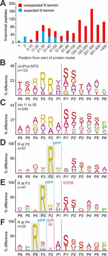
RESEARCH                                                                                                                               los

The Mouse Heart Mitochondria N Terminome
Provides Insights into ClpXP-Mediated
Proteolysis
Eduard Hofsetz1,2, Fatih Demir3x , Karolina Szczepanowska1,2, Alexandra Kukat1,2,
Jayachandran N. Kizhakkedathu4, Aleksandra Trifunovic1,2,*, and Pitter F. Huesgen2,3,5,*x

The mammalian mitochondrial proteome consists of more                                   Mitochondria are essential organelles for almost all eukary-
than 1100 annotated proteins and their proteostasis is                               otic cells; thus, sustaining the integrity of their proteins is
regulated by only a few ATP-dependent protease com-                                  crucial for cell viability. Given that the proteome is of dual
plexes. Technical advances in protein mass spectrometry                              origin, mitochondria face the difficulty of coordinating protein
allowed for detailed description of the mitoproteome from                            synthesis, import, and maintenance. Because of the high

                                                                                                                                                              Downloaded from https://www.mcponline.org by guest on January 14, 2021
different species and tissues and their changes under                                mitochondrial compartmentalization, soluble proteins in the
specific conditions. However, protease-substrate rela-                               matrix or the intermembrane space, as well as hydrophobic
tions within mitochondria are still poorly understood.                               proteins in the inner or outer membrane, must be processed
Here, we combined Terminal Amine Isotope Labeling of
                                                                                     independently. Mitochondria have several levels of protein
Substrates (TAILS) N termini profiling of heart mitochon-
                                                                                     quality control operating on the molecular or cellular level. As
dria proteomes isolated from wild type and Clppⴚ/ⴚ mice
                                                                                     a result of the alphaproteobacterial origin of mitochondria,
with a classical substrate-trapping screen using FLAG-
tagged proteolytically active and inactive CLPP variants                             most of the proteolytic quality control system shares strong
to identify new ClpXP substrates in mammalian mito-                                  similarity between mitochondria and today’s proteobacteria,
chondria. Using TAILS, we identified N termini of more                               such as Escherichia coli. Proteolytic control within mitochon-
than 200 mitochondrial proteins. Expected N termini                                  dria is predominantly performed by ATP-dependent pro-
confirmed sequence determinants for mitochondrial tar-                               teases, which include the soluble ClpXP and LONP1 pro-
geting signal (MTS) cleavage and subsequent N-termi-                                 teases in the mitochondrial matrix and membrane-bound
nal processing after import, but the majority were pro-                              i-AAA and m-AAA proteases that face the intermembrane
tease-generated neo-N termini mapping to positions                                   space and matrix, respectively (1). Whereas LONP1 is mainly
within the proteins. Quantitative comparison revealed                                attributed with quality control of misfolded (2) and oxidatively
widespread changes in protein processing patterns, in-                               damaged proteins (3), or mtDNA maintenance via the proteo-
cluding both strong increases or decreases in the abun-                              lytic control of mitochondrial transcription factor A (TFAM) (4),
dance of specific neo-N termini, as well as an overall                               relatively little is known about substrates and physiological
increase in the abundance of protease-generated neo-N                                relevance of the caseinolytic peptidase ClpXP (1). In E. coli,
termini in CLPP-deficient mitochondria that indicated
                                                                                     up to 60% of all protein degradation seems to be ATP-de-
altered mitochondrial proteostasis. Based on the com-
                                                                                     pendent proteolysis executed by the Lon and ClpXP/ClpAP
bination of altered processing patterns, protein accu-
                                                                                     proteases (5). Both proteases have specific regulatory sub-
mulation and stabilization in CLPP-deficient mice and
interaction with CLPP, we identified OAT, HSPA9 and                                  strates as well as housekeeping functions, and it seems that
POLDIP2 and as novel bona fide ClpXP substrates. Fi-                                 also in bacteria, most unfolded or damaged proteins are
nally, we propose that ClpXP participates in the coop-                               degraded by the Lon protease (5). Remarkably, tightly folded
erative degradation of UQCRC1. Together, our data pro-                               substrate domains can result in stalling of processive degra-
vide the first landscape of the heart mitochondria N                                 dation by ClpP in bacteria, resulting in the release of pro-
terminome and give further insights into regulatory and                              cessed proteoforms. A prominent example is the partial N-
assisted proteolysis mediated by ClpXP.                                              terminal proteolysis of DnaX by Caulobacter crescentus

From the 1Institute for Mitochondrial Diseases and Aging at CECAD Research Centre, and Center for Molecular Medicine Cologne (CMMC),
   Medical Faculty, University of Cologne, Cologne, Germany; 2Cologne Excellence Cluster on Cellular Stress Responses in Aging Associated
   Diseases (CECAD), Cologne, Germany, Medical Faculty and University Hospital, University of Cologne, Cologne, Germany; 3Central Institute
   for Engineering, Electronics and Analytics, ZEA-3, Forschungszentrum Jülich, Germany; 4Centre for Blood Research, School of Biomedical
   Engineering, Department of Pathology & Laboratory Medicine, Department of Chemistry, University of British Columbia, Vancouver, British
   Columbia, Canada; 5Institute for Biochemistry, Faculty of Mathematics and Natural Sciences, University of Cologne, Cologne, Germany
This article contains supplemental data.
* For correspondence: Aleksandra Trifunovic, aleksandra.trifunovic@uk-koeln.de; Pitter F. Huesgen, p.huesgen@fz-juelich.de.

1330 Mol Cell Proteomics (2020) 19(8) 1330 –1345
© 2020 Hofsetz et al. Published under exclusive license by The American Society for Biochemistry and Molecular Biology, Inc.   DOI 10.1074/mcp.RA120.002082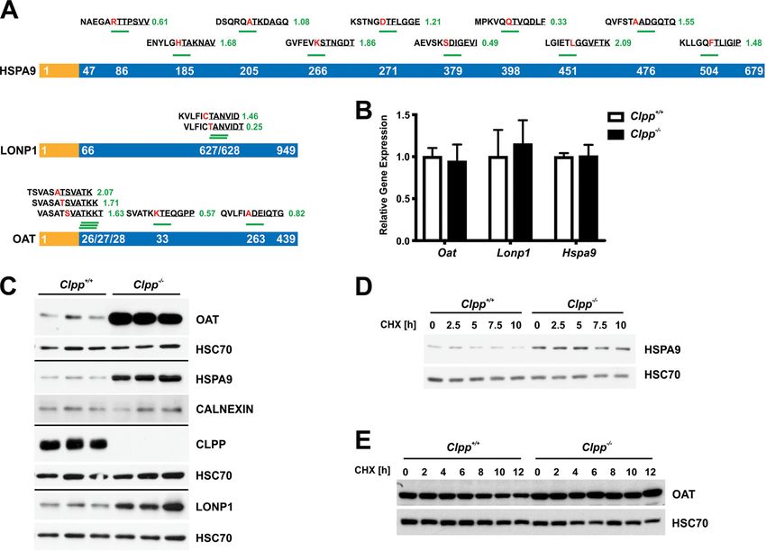
Mouse CLPP Degradomics

ClpXP, which generated distinct DnaX proteoforms that are           essing by the aminopeptidases ICP55 and OCT1 (21–23) and
important to support normal growth (6).                             identified the proapoptotic protein Smac/DIABLO as a sub-
   The functional ClpXP protease consists of the protease           strate of the mitochondrial rhomboid protease PARL (24). N
subunit CLPP (Caseinolytic Peptidase ATP-dependent, Pro-            termini profiling in mitochondria isolated from human cells has
teolytic Subunit), and the ATP-dependent chaperone CLPX             further been applied to characterize protein processing early
(7). In the presence of CLPX and ATP, the two CLPP hepta-           after activation of the intrinsic apoptosis pathway (25) and in
meric rings form the catalytic tetradecamer chamber with 14         response to zinc and rapamycin treatment (26).
serine/histidine/aspartate triads that catalyze peptide cleav-         Here, we combined two mass spectrometry-based ap-
age (8). The chaperone CLPX forms hexameric rings that              proaches to investigate the impact of CLPP on the mitopro-
stack on one or both sides of the CLPP chamber to allow             teome and systematically identify putative ClpXP substrates.
ATP-dependent protein unfolding and translocation through           Profiling of protein N termini in control and CLPP-deficient
the axial channel into the catalytic core (9, 10). In the absence   heart mitochondria by Terminal Amine Isotope Labeling of
of CLPX unfoldase, which also ensures the identification of         Substrates (TAILS) (27) revealed altered cleavages in accu-
specific substrates, CLPP is still able to cleave small, unspe-     mulating candidate ClpXP substrates, including OAT and
cific peptides (10). The 3-dimensional structure and biochem-       HSPA9, as well as moderate but widespread alterations in
ical properties of ClpXP have been well described in bacteria       mitochondrial protein processing. In addition, we identified
and are largely conserved in eukaryotes (7, 8). However, sub-       putative substrates by expressing FLAG-tagged, proteolyti-

                                                                                                                                              Downloaded from https://www.mcponline.org by guest on January 14, 2021
strates, specific adaptors, and the function of mitochondrial       cally active and inactive versions of CLPP in Clpp⫺/⫺ mouse
ClpXP remain mostly enigmatic.                                      embryonic fibroblasts (MEFs), followed by affinity purification
   We have recently shown that the whole-body CLPP knock-           and mass spectrometric quantification (9). Using this ap-
out mice (Clpp⫺/⫺) are sterile with shorter stature, as ob-         proach, we identified a small subset of specifically trapped
served in Perrault syndrome patients (11, 12). Remarkably,          mitochondrial matrix proteins including UQCRC1, POLDIP2,
they live a normal life-span with improved glucose metabo-          and NDUFV2 as candidate substrates. Cycloheximide-chase
lism that renders them resistant to diet-induced obesity (11).      demonstrated stabilization of HSPA9, OAT, and UQCRC1 in
Particularly in the heart, mammalian CLPP is an important           Clpp⫺/⫺ MEFs, validating these proteins as novel bona fide
regulator of mitochondrial protein synthesis through the re-        ClpXP substrates. Taken together, our results provide a first
moval of ERAL1 from the small ribosomal subunits to allow           large-scale experimental characterization of protein matura-
assembly of the functional mitoribosome (13). Contrary to           tion and processing in heart mitochondria and support the
nematodes, CLPP appears not to be needed for the activation         hypothesis that mammalian ClpXP primarily functions as a
or maintenance of the mammalian mitochondrial unfolded              proteolytic regulator of specific substrates, but also directly or
protein response (UPRmt) (14). Unexpectedly, loss of CLPP           indirectly contributes to mitochondrial protein quality control.
alleviates the strong mitochondrial cardiomyopathy and par-
tially restores the diminished respiration in animals with a                           EXPERIMENTAL PROCEDURES
strong dysregulation of mitochondrial protein synthesis (14).          Mouse Strains and Ethics—Clpp⫺/⫺ mice were generated, main-
Together, these results question our current understanding of       tained and genotyped as previously described (13). 17-weeks-old
ClpXP-mediated proteolysis in mammals, while introducing            animals were used for the TAILS analysis. All experiments were
                                                                    approved and permitted by the Animal Ethics Committee of North-
CLPP as a possible novel target for therapeutic intervention in     Rhein-Westphalia (Landesamt für Natur, Umwelt und Verbrauch-
mitochondrial diseases (14).                                        erschutz Nordrhein-Westfalen; LANUV) following German and Euro-
   Mass spectrometry-based proteomics has become the                pean Union regulations. All animal work was performed in accordance
predominant tool for unbiased characterization of protease          with recommendations and guidelines of the Federation of European
substrates and function (15). Quantitative proteomics can re-       Laboratory Animal Science Associations (FELASA).
                                                                       Mitochondrial Preparations from Hearts—Mitochondria were iso-
veal candidate substrates of degradative enzymes, including         lated from wild type and Clpp⫺/⫺ hearts at the age of 17 weeks as
ClpXP, based on the change in abundance between different           previously described (13). In short, freshly isolated hearts were ho-
genotypes and treatments (13), whereas the combination with         mogenized with a Teflon homogenizer in mitochondria isolation buffer
affinity enrichment of native or epitope-tagged active and          (100 mM sucrose, 50 mM KCl, 1 mM EDTA, 20 mM TES, pH 7.2) with
inactive versions of a protease can identify interaction part-      the addition of subtilisin A (1 mg/g heart) and 0.2% BSA (fatty acid
                                                                    free). Crude mitochondrial fractions were obtained through differential
ners, regulatory proteins and substrates (13, 15). In addition,     centrifugation at 8500 ⫻ g, 800 ⫻ g, and again at 8500 ⫻ g to remove
a variety of protocols for selective enrichment, identification     fat, heavy fractions, and to pellet mitochondria, respectively. The
and quantification of protein amino (N)- or carboxyl (C)-termi-     mitochondrial pellet was washed with mitochondria isolation buffer
nal peptides (16, 17) enable unbiased identification of proc-       without subtilisin A/BSA and either stored at ⫺80 °C for Western blot
essing patterns and direct determination of protease sub-           analysis in the same buffer or resuspended in 6 M GuHCl for TAILS
                                                                    experiments.
strates with their precise cleavage sites (18, 19). Global             Cell Lines—Wild type and Clpp⫺/⫺ MEFs were isolated, immortal-
profiling of mitochondrial protein N termini was used to define     ized and cultured in high glucose DMEM at 37 °C and 5% CO2 as
the MTS cleavage sites (20, 21), revealed subsequent proc-          previously described (28).

                                                                               Mol Cell Proteomics (2020) 19(8) 1330 –1345 1331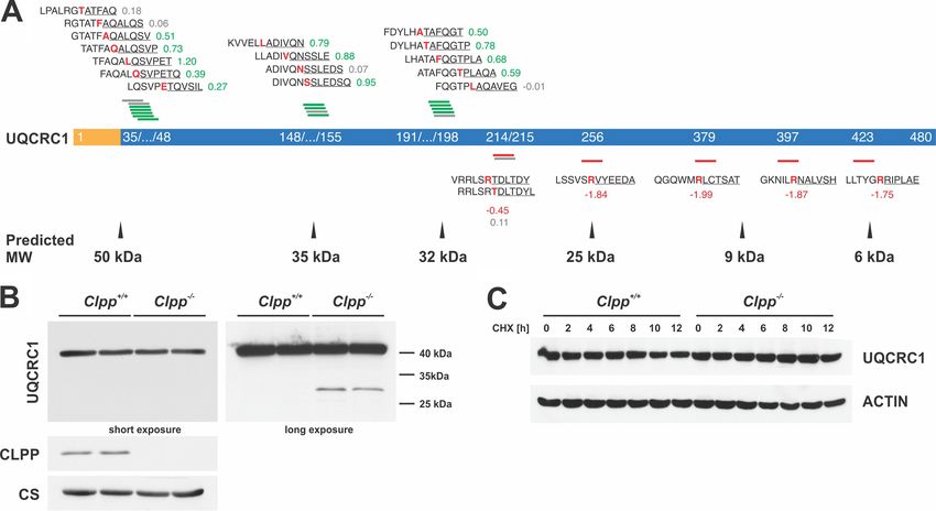
Mouse CLPP Degradomics
TABLE I
Antibodies, manufacturer and used conditions
Antibody Manufacturer Cat. Nr. Conditions
CLPP Sigma HPA040262 1:1000 (5% Milk PBST)
CLPX Sigma HPA040262 1:1000 (5% Milk PBST)
C1QBP/P32 Millipore AB2991 1:1000 (5% Milk PBST)
UQCRC1 Molecular Probes 459140 1:2000 (5% Milk PBST)
HSPA9 Abcam 82591 1:1000 (5% Milk PBST)
LONP1 Abcam ab82591 1:1000 (5% Milk PBST)
OAT Abcam ab137679 1:1000 (5% Milk PBST)
CALNEXIN Calbiochem 208880 1:1000 (5% Milk PBST)
-ACTIN Sigma A5441 1:5000 (5% Milk PBST)
HSC70 SantaCruz sc-7298 1:5000 (5% Milk PBST)
POLDIP2 CUSABIO CSB-PA896496LA01HU 1:2000 (5% Milk PBST)
TOMM20 SantaCruz sc-17764 1:2000 (5% Milk PBST)
FLAG M2 Sigma F1804 1:1000 (5% Milk PBST)
TABLE II modification and labeling of primary amines as described (29). The
Primers used for qPCR following labeling scheme was used: control, light label (12CH2O);
Clpp⫺/⫺ mutant, heavy label (13CD2O). Labeled samples were di-
Downloaded from https://www.mcponline.org by guest on January 14, 2021
Gene FWD 5⬘-3⬘ REV 5⬘-3⬘
gested with trypsin (Serva, Electrophoresis, Heidelberg, Germany)
Hspa9 ATGGCTGGAATGGCCTTAGC ACCCAAATCAATACCAACCACTG overnight, then a small aliquot of ⬃10 g proteome was withdrawn to
Lonp1 ATGACCGTCCCGGATGTGT CCTCCACGATCTTGATAAAGCG control for digestion efficiency by SDS-page analysis and labeling
Oat GGAGTCCACACCTCAGTCG CCACATCCCACATATAAATGCCT efficiency by shotgun proteome analysis (designated “preTAILS”
Hprt GCCCCAAAATGGTTAAGGTT TTGCGCTCATCTTAGGCTTT samples). For enrichment of N-terminal peptides, free trypsin-gener-
ated primary amines were subsequently coupled to HMW-ALD poly-
mer and internal peptides bound to the polymer removed by ultrafil-
CLPP Substrate Trapping in MEFs—Three independent Clpp⫺/⫺
tration, leaving enriched N-terminal peptides as “TAILS” samples in
MEF lines were transfected with each empty vector (NEG), CLPP-WT-
the flow-through. All peptides were desalted using in-house packed
FLAG (WT) and a catalytically inactive version with Ser149 mutated to
C18 StageTips (28) and additionally crudely fractionated (15–20-30 –
Ala, CLPP-TRAP-FLAG (TRAP) (13) using the Nucleofector (Lonza,
50% ACN) at neutral pH in 20 mM ammonium bicarbonate buffer
Basel, Switzerland) electroporation kit according to the manufacturer
(AmBiC) using SepPak C18 cartridges (Waters, Eschborn, Germany).
instruction. 72h after transfection, 80% confluent cells were har-
Samples were analyzed using an Ultimate 3000 RSLCnano HPLC
vested with trypsin, washed twice with PBS and lysed for 45 min in
(Thermo, Germering, Germany) operated in a two-column setup (Ac-
300 l IP buffer (Thermo Fisher Scientific, Schwerte). Samples were
incubated with 30 l ␣-FLAG magnetic beads (Sigma) over night at claim PepMap 100 C18, particle size 3 m, ID 75 m for trap and ID
4 °C on a rotating wheel. On the following day, beads were washed 4 50 m for analytical column; trap column length 2 cm, analytical
times with IP buffer and bound proteins were eluted in 70 l Elution column length 25 cm, Thermo) at a flow rate of 350 ml/min at 60 °C.
buffer (Thermo Fisher Scientific). Samples were neutralized with 10 l The nano LC system was on-line coupled to an Impact II high reso-
1 M Tris/HCl pH 7.5, snap-frozen and stored at ⫺80 °C. 1% of each lution Q-TOF (Bruker, Bremen, Germany) via a CaptiveSpray nano ESI
Lysate (L), unbound proteins (F), first washing solution (W) and 10% of source (Bruker) with a NanoBooster (Bruker) engaged to saturate the
the Elution fractions (E) were used for SDS-PAGE controls. Samples nitrogen gas stream with acetonitrile essentially as described (30).
were prepared for proteomic analysis as previously described (13). Mass Spectrometry Data Analysis—For analysis of preTAILS and
Cycloheximide (CHX) Chase Experiments—CHX chase experi- TAILS samples, spectra were matched to peptide sequences at a
ments were performed as previously described (13) and Western FDR of 0.01 using MaxQuant (31), v.1.6.0.16 with the UniProt mouse
blotting membranes were incubated with antibodies against newly proteome (canonical ⫹ isoforms, release 2017_10, 60717 entries) as
identified candidate substrates. For Fig.7C, the published loading a database for searches with standard Bruker QTOF instrument set-
control (ACTIN) for UQCRC1 was reused as the same membrane was tings, which included 0.07 Da MS precursor tolerance in the first
probed with anti-UQCRC1 (13). New CHX chase experiments were search, 0.006 Da precursor mass tolerance in the second search after
performed for OAT and HSPA9. All antibodies used in this study are recalibration, and MS/MS spectra matching tolerance of 40 ppm.
listed in Table I. Mass spectra acquired from preTAILS samples were searched twice:
RNA Isolation and qPCR—Isolated heart RNA was treated with To control efficiency of the different labeling steps, searches consid-
DNase (DNA-free Kit, Thermo Fisher Scientific) and subsequently ered trypsin as digestion enzyme allowing for up to four missed
reverse transcribed with the High-Capacity cDNA Reverse Transcrip- cleavages, Cys carbamidomethylation was set as fixed modification,
tion Kit (Thermo Fisher Scientific). Gene expression levels were de- and isotopically light (⫹28.031300) and heavy (⫹36.075670) dimethy-
termined with qPCR technique using Brilliant III Ultra-Fast SYBR lation of Lys residues and Met oxidation as variable modifications.
Green qPCR Master Mix (Agilent Technologies, Waldbronn, Ger- This search confirmed that ⬎98% of the identified Lys residues were
many). Samples were adjusted for total RNA content by Hypoxan- dimethyl labeled and ⬍ 6% of the identified peptides were cleaved
thine-guanine phosphoribosyltransferase (Hprt). Primers used are after dimethylated Lys residues, in agreement with inefficient trypsin
listed in Table II. activity toward dimethyl modified Lys (27, 32). Therefore, for analysis
N Terminome Profiling—N-terminal peptides were enriched from of protein abundance changes, a second analysis was performed
isolated heart mitochondria using TAILS (Terminal Amine Isotope considering ArgC as digestion enzyme, duplex isotope labeling by
Labeling of Substrates) with differential formaldehyde isotopes light (⫹28.031300) and/or heavy (⫹36.075670) dimethylation of Lys
(12CH2O and 13CD2O) and sodium cyanoborohydride (NaBH3CN) for residues, Cys carbamidomethylation as fixed and Met oxidation as
1332 Mol Cell Proteomics (2020) 19(8) 1330 –1345
Mouse CLPP Degradomics

variable modification (33). Search parameters for N termini identifica-     tochondrial protein processing in CLPP-deficient mice, we
tion and quantification were set to semi-specific (free N terminus)         first isolated mitochondria from 17 weeks-old wild type and
ArgC as digestion enzyme, isotope labeling by light (⫹28.031300)
and/or heavy (⫹36.075670) dimethylation of Lys residues and peptide
                                                                            Clpp⫺/⫺ hearts, extracted proteomes under denaturing con-
N termini, Cys carbamidomethylation as fixed and Met oxidation,             ditions and enriched N-terminal peptides by Terminal Amine
N-terminal acetylation (⫹42.010565) or N-terminal pyroGlu formation         Isotope Labeling of Substrates (27). In brief, primary amines at
from Glu (-18.010565) or Gln (-17.026549) as variable modifications.        protein N termini and Lys side chains in wild type and Clpp⫺/⫺
The “requantify” and “match between runs” functions were enabled.           mitoproteomes were differentially labeled with stable isotopes
False discovery rates, estimated using an appended reverse-decoy
protein database, were set to 0.01 at PSM and protein levels. For
                                                                            (light and heavy formaldehyde, respectively), before trypsin
preTAILS samples, MaxQuant protein ratios were recalculated con-            digestion (Fig. 1A). A small “preTAILS” sample was withdrawn
sidering only matching Lys-containing quantified peptides. For TAILS        after digestion and before N termini enrichment to control
data, identified N-terminal peptides were annotated based on the            dimethyl labeling efficiency and determine changes in overall
modificationSpecificPeptides.txt file of the MaxQuant output folder         protein abundance. Peptides with a trypsin-generated ␣-
with information from Uniprot.org and sequence windows using an
in-house script (MaxQuant Advanced N Termini Interpreter, MANTI.pl
                                                                            amine were further captured by reaction with an aldehyde-
version 3.7, https://sourceforge.net/projects/manti/). Pyro-Glu modi-       functionalized high molecular weight polymer and removed by
fied peptides may arise during sample preparation after tryptic digest      filtration, whereas in vitro dimethylated or endogenously N-
and were therefore excluded from further analysis, as were peptide          terminally modified proteins were enriched in the flow-through
identifications that did not contain a C-terminal Arg or reported with a    (27). Mass spectrometric analysis of the preTAILS showed
nonsense dimethyl modification at N-terminal Pro. Dimethylated or

                                                                                                                                                Downloaded from https://www.mcponline.org by guest on January 14, 2021
acetylated peptides matching to positions 1 or 2 of a protein model or
                                                                            efficient dimethyl labeling (⬎98%) and identified 371 proteins
termini starting within 5 amino acid window deviation from UniProt-         (Fig. 1B), including 271 proteins with annotated mitochondrial
annotated MTS cleavage sites were defined as “expected” termini,            location (Fig. 1C, supplemental Table S1) as retrieved from the
whereas all peptides matching to positions within the protein model         UniProt database (36). A total of 1558 N-terminal peptides
were considered “unexpected” protein N termini, which include               from 322 proteins were identified after enrichment by TAILS
mostly protease-generated neo-N termini but also unannotated pro-
teoforms arising from alternative splicing or use of alternative trans-
                                                                            (Fig. 1A, supplemental Table S2). 1058 N-terminal peptides
lation initiation sites (19). Further data analysis was performed with      were derived from 214 proteins with UniProt-annotated
Perseus version 1.6.6 (34).                                                 known or predicted mitochondrial localization (Fig. 1B, sup-
   For the CLPP-TRAP experiment, spectra were matched to peptide            plemental Table S3). The biological replicates were highly
sequences using MaxQuant v1.5.3 (31). using the UniProt Mouse               reproducible, with 775 N-terminal peptides identified in all
protein database (release 2015_01, 44654 entries) and standard set-
tings for the QExactive instrument, Thermo Scientific, Bremen, Ger-
                                                                            three experiments and further 334 N-terminal peptides in 2 of
many, including 20 ppm MS precursor tolerance in the first search,          the 3 experiments (supplemental Fig. S1A). Overall, a large
4.5 ppm precursor mass tolerance in the second search after recali-         overlap with our previous quantitative heart proteome analysis
bration, and MS/MS spectra matching tolerance of 20 ppm. Enzyme             was observed (Fig. 1B, 1C) (13). Next, we retrieved position-
specificity set to trypsin with two missed cleavages allowed, cys           specific annotations for each identified N-terminal peptide,
alkylation with iodoacetamide was set as fixed and Met oxidation as
variable modifications. Label free quantification and match between
                                                                            including information on protein domain structure and func-
runs was enabled with preset standard settings. False discovery             tion for the corresponding protein models from UniProt. 262
rates, estimated using an appended reverse-decoy protein database,          N-terminal peptides mapped to ”expected” positions, which
were set to 0.01 at PSM and protein levels. Further data analysis was       we here define as N termini starting at positions 1 or 2 of the
performed with Perseus version 1.6.6 (34). Proportional Venn dia-           protein models or within 5 amino acids of known or pre-
grams were drawn with the BioVenn web application (35).
   Experimental Design and Statistical Rationale—All experiments
                                                                            dicted signal peptide (SP), mitochondrial targeting signal
were performed in biological triplicates for each experimental condi-       (MTS) or propeptide cleavage sites (Fig. 1D). A majority of
tion. In the trapping experiment, proteins were tested for differential     1296 N-terminal peptides from 248 proteins mapped to
abundance between CLPP-FLAG, CLPP-TRAP-FLAG and control                     unexpected sites within the predicted protein sequences,
conditions by multi-sample ANOVA as implemented in Perseus,                 representing N termini of unknown origin that likely are
v.1.6.6 (34). Differences associated with Benjamini-Hochberg cor-
rected ANOVA q-value ⬍0.05 were considered significant. Quantita-
                                                                            mostly protease-generated neo-N termini (Fig. 1D). These
tive analysis of the N-terminal peptides considered only peptides           include peptides from short-lived degradation intermediates
quantified in at least 2 out of the 3 biological replicates. The signifi-   but may also indicate processed proteoforms with unknown
cance of the difference in the median abundance between expected            physiological function, unannotated MTS cleavage sites or
and unexpected protein N termini were tested by Mann-Whitney U              proteoforms arising from alternative splicing or use of alter-
test. Additionally, a cut-off for strong changes in N termini abundance
was defined based on the boxplot analysis of 120 N termini mapping
                                                                            native translation initiation sites (19, 37). Most of the protein
to proteins that showed no significant change in abundance in a             N termini mapping to positions 1 and 2 or annotated as
previous label-free LC-MS proteome analysis (13).                           secreted peptide (SP) cleavage sites were derived from
                                                                            contaminating non-mitochondrial proteins and were ex-
                               RESULTS
                                                                            cluded (Fig. 1D).
  Profiling of Mitochondrial Protein N Termini in Wild Type                     Characteristics of the Mitochondrial Mouse Heart N Termi-
and CLPP-deficient Mouse Hearts—To gain insights into mi-                   nome—We then limited further analysis to the 1058 N-termi-

                                                                                      Mol Cell Proteomics (2020) 19(8) 1330 –1345 1333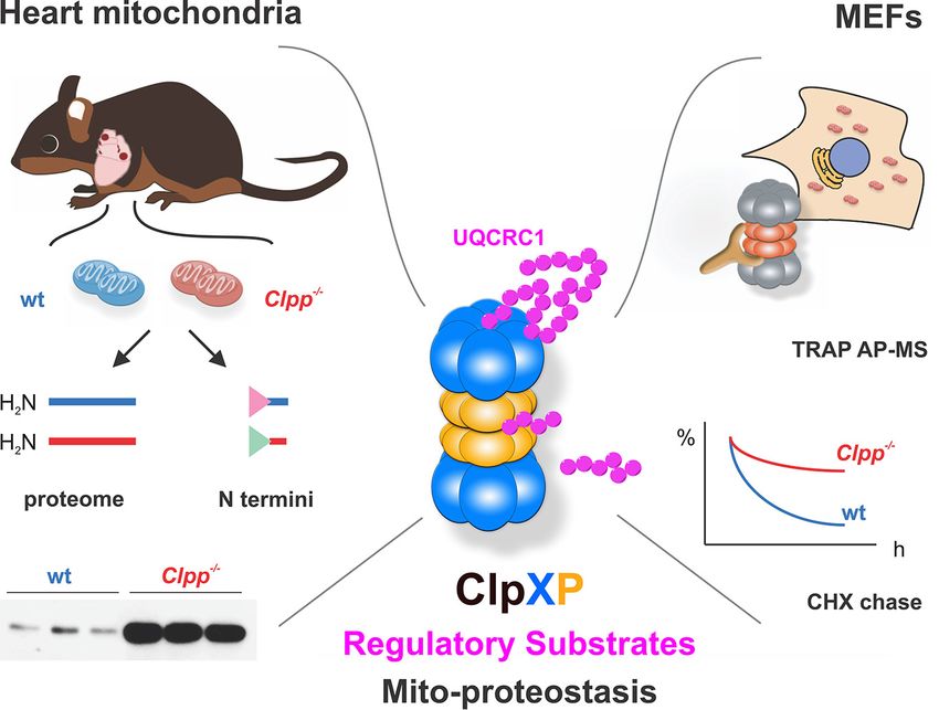
Mouse CLPP Degradomics
Downloaded from https://www.mcponline.org by guest on January 14, 2021
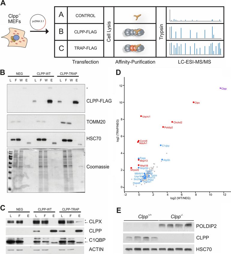
FIG. 1. Mouse heart N terminome profiling. A, Scheme of the TAILS workflow. Protein N termini and Lys side chains from wild type (wt)
and Clpp⫺/⫺ mitochondrial proteins were labeled with light and heavy formaldehyde, respectively, pooled and digested with trypsin. A
“preTAILS” aliquot was withdrawn for labeling control and determination of protein abundance changes. In the next reaction, peptides with
unlabeled N termini resulting from tryptic digestion were covalently captured with a high-molecular weight aldehyde-functionalized ALD-HPG
polymer. Removal of the polymer with bound peptides by ultrafiltration left labeled N-terminal peptides highly enriched in the flow-through for
LC-MS/MS analysis. B, Overlap of all proteins identified by the preTAILS and TAILS in this analysis and our previous LFQ proteome analysis
(13). C, Overlap of proteins with UniProt-annotated mitochondrial localization between preTAILS, TAILS and LFQ data sets. D, Positional
annotation classifying protein N-terminal peptides identified after TAILS enrichment into 5 categories, those matching position 1 or 2 of the
protein model, matching within 5 amino acids of annotated or predicted signal peptide (SP), mitochondrial targeting signal (MTS) cleavage
sites, propeptide maturation sites (PP) or those matching to “unexpected” positions within the protein model. Red bars indicate proteins with
mitochondrial location as annotated by UniProt.
nal peptides matching 963 unique N termini in 214 proteins P2 (Fig. 2B), reminiscent of the well-described “twin Arg mo-
with UniProt-annotated mitochondrial location (supplemental tif” arising from the preferred MTS cleavage with Arg at P2
Table S3). Notably, 172 and 93 of these N termini were pre- and subsequent processing by aminopeptidase ICP55-medi-
viously found by Calvo et al. in mitochondria isolated from ated cleavage (20, 25, 41, 42). However, the additional over-
mouse liver and kidney, respectively (20). Most nuclear en- representation of Arg at P4 indicated further aminopeptidase
coded proteins targeted to the mitochondria carry an MTS activity. A similar pattern was observed in an iceLogo analysis
that is cleaved off by the mitochondrial processing peptidase of the 246 non-redundant cleavage sites derived from all
(MPP) after import (38). Hence it was not surprising that 123 N-terminal peptides in bins 11 to 50 (Fig. 2C), including MTS
non-redundant N termini were observed within 5 amino acid cleavage sites not yet annotated in UniProt, as for example
(aa) distance from UniProt-annotated MTS cleavage sites MRPS22 (supplemental Fig. S2B). Further iceLogo analysis of
(supplemental Fig. S2A). Many proteins showed N-terminal the 57 N termini with Arg at P2 showed the typical character-
ragging, i.e. sequential truncation by 1 or more amino acids, istics of MTS cleavage resulting in stable N-terminal peptides
indicating further aminopeptidase-mediated processing (sup- frequently starting with Ser (Fig. 2D), whereas the 47 N termini
plemental Fig. S2A, supplemental Table S3). This is frequently with Arg at P3 additionally showed an overrepresentation of
observed in many different compartments and biological sys- aromatic residues Phe and Tyr at P1 (Fig. 2A) in agreement
tems (17, 30, 39). However, in many cases the most prevalent with the well-described cleavage site preference of ICP55 for
N terminus can be determined by spectral counts (20), as for Phe, Tyr and Leu (20, 23, 41– 43). In many cases, protein N
example the N terminus starting at Thr26 for ornithine amino- termini were frequently further truncated by an unidentified
transferase (OAT) (Supplemental Fig. S2B). We further ana- aminopeptidase after MMP and/or ICP55-cleavage. However,
lyzed the position of the N-terminal peptides in relation to the neither iceLogo analysis of the 29 cleavage sites with Arg at
protein model (Fig. 2A). As expected, most N termini mapping P4 (Fig. 2F) nor inspection of individual examples for well-
within 5 amino acids of MTS cleavage sites were observed in supported ragged protein N termini (supplemental Fig. S2B)
the bins between 10 to 50 amino acids from the start (20, 40). revealed stringent sequence features for this aminopeptidase
Visualization of the cleavage site sequence derived from the activity.
123 N termini mapping close to UniProt-annotated MTS Overall, the most N-terminal positions of expected N termini
cleavage site revealed an overrepresentation of Arg at P3 and within 5 residues of the known or predicted UniProt-anno-
1334 Mol Cell Proteomics (2020) 19(8) 1330 –1345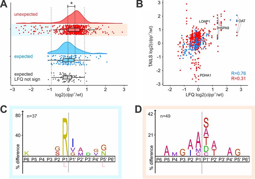
Mouse CLPP Degradomics

                                                                              tated MTS were almost exclusively occupied by Ala (24.6%),
                                                                              Ser (23.0%), Gly (12%), Thr (10.4%) and Met (8.7%) (supple-
                                                                              mental Fig. S3), which are all amino acids classified as stabi-
                                                                              lizing according to the bacterial N-end rule (44). In contrast,
                                                                              protease-generated neo-N termini mapping to positions
                                                                              within the proteins’ sequence featured branched aliphatic
                                                                              residues Ile (4.2%), Val (5.7%) and the secondary destabiliz-
                                                                              ing Asp (10.3%) and Glu (5.7%) that were rarely present at this
                                                                              position in expected N termini (supplemental Fig. S3). This
                                                                              supports the hypothesis that a significant proportion of the
                                                                              identified unexpected N-terminal peptides may represent
                                                                              degradation intermediates, which might be recognized by a
                                                                              yet unidentified N-recognin.
                                                                                 Neo-N termini Reveal Changes in Protein Processing in
                                                                              CLPP-deficient Heart Mitochondria—Next we wished to de-
                                                                              termine differences in N termini abundance between CLPP-
                                                                              deficient and wt heart mitochondria. To account for variations

                                                                                                                                                         Downloaded from https://www.mcponline.org by guest on January 14, 2021
                                                                              in the amount of co-purified cytosolic proteins in the mito-
                                                                              chondrial preparations, we first normalized the ratios in each
                                                                              experiment to the median fold change of the 200 N-terminal
                                                                              peptides matching to expected positions in mitochondrial
                                                                              proteins (Fig. 1D, P1/2 or MTS of mitochondrial proteins). We
                                                                              then further restricted the analysis to the 777 N-terminal pep-
                                                                              tides quantified in at least 2 of the 3 biological replicates
                                                                              (supplemental Table S3), which showed very good reproduc-
                                                                              ibility for paired single peptide data (supplemental Fig. S4A).
                                                                              The median ratio of the 154 expected N-terminal peptides
                                                                              showed the expected symmetric distribution centered around
                                                                              the expected 1:1 ratio (Fig. 3A; log2(Clpp⫺/⫺/wt) ⫽ 0). In
                                                                              contrast, the distribution of 623 unexpected N-terminal pep-
                                                                              tides was moderately, but significantly shifted (Mann-Whitney
                                                                              U test p-val ⬍0.001) to about 25% higher abundance in
                                                                              CLPP-deficient hearts (Fig. 3A; ⌬median log2(Clpp⫺/⫺/wt) un-
                                                                              expected - expected ⫽ 0.32). To further investigate this strik-
                                                                              ing difference, we next compared the N termini abundance
                                                                              with the corresponding protein abundance determined in
                                                                              our previous LFQ proteome (13), as this data set provided
                                                                              quantitative information for more proteins identified by
                                                                              TAILS than our limited preTAILS data set (Fig. 1C). The
                                                                              abundance ratio of the 198 proteins quantified in both pre-
                                                                              TAILS and LFQ data sets correlated well (Pearson correla-
                                                                              tion 0.73, -lg(p-val)⬎15.3), demonstrating that the two in-
                                                                              dependent experiments with mitochondria isolated form
                                                                              different mouse cohorts were indeed comparable (supple-
                                                                              mental Fig. S4B). In total, 636 of the 777 quantified TAILS-

                                                                              overrepresented in (B) 123 unique cleavage sites matching within 5 aa
                                                                              of annotated MTS cleavage sites and (C) 246 unique cleavage sites
   FIG. 2. Analysis of the mitochondrial mouse heart N terminome.             derived from peptides mapping to positions ⬎10 and ⬍51. Further
A, Start position of the 1058 identified N-terminal peptides in relation to   iceLogos visualize amino acids overrepresented among (D) 57 cleav-
the corresponding protein model. Blue indicates expected termini map-         ages sites with Arg at P2, (E) 47 cleavage sites with Arg at P3, and (F)
ping to positions 1 or 2 or within 5 residues from a UniProt-annotated        29 cleavage sites with Arg at P4. The dashed black line indicates start
mitochondrial targeting signal (MTS) cleavage site, not annotated un-         of experimentally determined N termini, dashed blue line indicates pu-
expected N termini are shown in red. IceLogos show amino acids                tative MPP cleavage site, dashed red line putative ICP55 cleavage site.

                                                                                          Mol Cell Proteomics (2020) 19(8) 1330 –1345 1335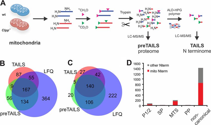
Mouse CLPP Degradomics

                                                                                                                                                  Downloaded from https://www.mcponline.org by guest on January 14, 2021
   FIG. 3. Quantitative analysis of N termini abundance in Clppⴚ/ⴚ and wt mouse heart mitochondria. A, Abundance in Clpp⫺/⫺ and wt
for 154 expected N-terminal peptides matching within 5 amino acids distance from UniProt annotated translation start or maturation sites and
for 623 N-terminal peptides matching to unexpected positions with the corresponding protein model. Asterisk indicates significant difference
between the mean values of the two distributions (Mann-Whitney U test p-val ⬍0.001), emphasized by dashed line. Red, unexpected N termini;
blue, expected N termini; gray, subset of 93 unexpected N termini matching to proteins that showed no significant change in abundance in
the LFQ data set used to define cut-off values of significant accumulation (light orange) or depletion (light blue) of unexpected N termini. B,
Abundance of N-terminal peptides compared with the corresponding protein abundance determined by label-free quantification. Red,
unexpected N termini; blue, expected N termini. Pearson correlation for each group is indicated. C, iceLogo of 37 unexpected N termini with
reduced abundance in CLPP-deficient mitochondria (log2(Clpp⫺/⫺/wt)⬍0.9). D, iceLogo of 49 unexpected N termini accumulating in CLPP-
deficient mitochondria (log2(Clpp⫺/⫺/wt⬎0.9).

enriched N-terminal peptides matched to 163 proteins                     logically highly relevant even if only a fraction of the precur-
quantified in the LFQ data set, of which 165 N-terminal                  sor protein is processed (37).
peptides came from 46 proteins with significantly different                 Differentially Accumulating N Termini Reveal Candidate
abundance (Student⬘s t test p-val ⬍0.05). The 120 expected               ClpXP Substrates—There are many potential direct and indi-
mitochondrial N-terminal peptides correlated well with the               rect causes for differential accumulation of both expected and
overall protein abundance (Fig. 3B, Pearson correlation ⫽                protease-generated neo-N termini in the absence of CLPP.
0.76, -lg(p-val)⬎323). This suggested that the known pro-                For example, expected N termini of direct ClpXP substrates
tein N termini largely reflect overall abundance of the cor-             would be expected to accumulate with the stabilized protein
responding proteins, which further suggests that these                   in the absence of CLPP, whereas the N-terminal peptides
constitute the major proteoforms. In contrast, the 389 un-               derived from CLPP cleavage would be drastically reduced in
expected protein neo-N termini showed divergent accumu-                  abundance. However, a plethora of indirect effects are likely
lation and reduction and correlated only poorly with                     to alter the behavior of protein abundance and processing
the corresponding protein abundance (Fig. 3B, Pearson                    patterns in vivo, for example by changes in transcription and
correlation 0.31, -lg(p-val) ⫽ 12.3), indicating widespread              translation or differential activation of proteolytic activities that
changes in mitochondrial protein processing in the absence               cut the same substrates at the same or different sites. Nev-
of CLPP. Furthermore, most neo-N termini poorly correlat-                ertheless, we reasoned that at least some direct ClpXP sub-
ing with protein abundance came from proteins that were                  strates, such as stabilized proteoforms with N-terminal ClpXP
not significantly affected by CLPP-deficiency, indicating the            degrons and N termini generated by ClpXP cleavages, would
cleavages affected only a minor fraction of the correspond-              show strong accumulation in the knockout or wild type, re-
ing precursor proteins, consistent with short-lived degrada-             spectively, compared with the 93 expected N-terminal pep-
tion intermediates. Nevertheless, proteolytically generated              tides from proteins showing no significant change in abun-
proteoforms that acquired a new function can be physio-                  dance in our previous analysis (13). Based on a boxplot

1336 Mol Cell Proteomics (2020) 19(8) 1330 –1345
Mouse CLPP Degradomics
Downloaded from https://www.mcponline.org by guest on January 14, 2021
FIG. 4. Validation of candidate ClpXP substrates identified by TAILS. A, Putative ClpXP substrates with increased N termini abundance.
MTS (yellow) and mature proteins (blue) are shown with the starting positions of the accumulating N termini indicated by white numbers. Above
the proteins, cleavage windows are stated with the difference in abundance (log2(Clpp⫺/⫺/wt)). Amino acids at the P1 position preceding the
cleavage site are highlighted in red, the detected peptide sequence is underlined. B, Western blots of steady state protein levels in heart
lysates. HSC70 and CALNEXIN were used as loading controls for the respective blots. C, Relative gene expression with qPCR of Oat, Lonp1
and Hspa9. D, CHX chase experiment of HSPA9 in MEFs. HSC70 was used as loading control. E, CHX chase experiment of OAT in MEFs.
HSC70 was used as loading control.
analysis of these 93 unchanged termini (Fig. 3A), we chose trast, the 37 neo-N termini that were more prevalent in wt, the
log-transformed ratios ⫾ 0.9, or more than ⫾ 1.5 times the best candidates for direct CLPP-mediated cleavage events,
interquartile range, as cut-off values for determining N-termi- showed a clear overrepresentation of Arg at P1 preceding the
nal peptides with significantly altered abundance (irrespective cleavage site (Fig. 3D).
of protein abundance). This revealed 10 expected and 49 One of the proteins with strong differential accumulation of
unexpected N termini that were strongly accumulating in N-terminal peptides was OAT (Ornithine Aminotransferase),
CLPP-deficient heart mitochondria, and 6 expected and 37 where three ragged expected N termini mapping to the
unexpected N termini strongly reduced in abundance. We known MTS cleavage site accumulated ⬃4-fold, in agreement
speculated that the unexpected N-terminal peptides accumu- with the protein accumulation observed previously (LFQ
lating in the absence of CLPP might reveal an N-terminal log2(Clpp⫺/⫺/wt) ⫽ 2.71) (13). Notably, two additional pro-
degron for substrate recognition by ClpXP. However, se- tease-generated N termini were much less affected and there-
quence logo analysis of these N termini showed an overrep- fore likely represent intermediates from CLPP-independent
resentation of the stabilizing residues Ala, Ser and Thr and the degradation (Fig. 3B, Fig. 4A). We also detected increased
potentially destabilizing Asp and Cys as N-terminal residues abundance of N termini from other proteins that showed
(Fig. 3C), reminiscent of the amino acid abundance observed significantly elevated protein abundance in our previous study
for expected protein termini (supplemental Fig. S3). In con- of the Clpp⫺/⫺ heart mitoproteome, including HSPA9 (LFQ
Mol Cell Proteomics (2020) 19(8) 1330 –1345 1337Mouse CLPP Degradomics
log2(Clpp⫺/⫺/wt) ⫽ 1.34) and LONP1 (LFQ log2(Clpp⫺/⫺/wt) ⫽ were included in our previous substrate trapping screen (13).
0.57) (Fig. 3B, Fig. 4A) (13). Neo-N termini of these proteins Although this demonstrated that protease substrates can be
were distributed across the protein sequences and the iden- co-purified with an active protease, “trapped” substrates are
tified cleavage sites differed strongly from those accumulating expected to accumulate to higher abundance when purified
in the wild type, in line with the overall motifs (Fig. 3C, 3D). with an inactive protease that mitigates direct degradation in
Thus, the detected accumulating neo-N termini are likely the vivo (49). In order to obtain such quantitative data, we re-
result of alternative quality control proteolysis that is activated peated the CLPP substrate trapping experiment in triplicate
in the absence of ClpXP-mediated substrate regulation. Im- with some modifications to the protocol, including the use of
munoblot analysis confirmed that these proteins (OAT, whole cell lysates instead of isolated mitochondria (Fig. 5A).
HSPA9 and LONP1) accumulated in CLPP-deficient heart This enabled faster and thus more reproducible isolation
lysates (Fig. 4B), although transcript levels were unchanged of CLPP-associated proteins for the quantitative approach,
(Fig. 4C). We further tested if they are indeed bona fide ClpXP albeit at the cost of a lower coverage of low abundant mito-
substrates by cycloheximide (CHX) chase experiments in cell chondrial proteins. Both CLPP-FLAG constructs were ex-
culture, where cytoplasmic protein synthesis is blocked by pressed to similar extend in CLPP-deficient MEFs and immu-
CHX treatment and degradation of selected proteins followed noprecipitation with anti-FLAG magnetic beads yielded
over time. As expected, the amount of both HSPA9 (Fig. 4D) virtually complete recovery of CLPP-(TRAP)-FLAG protein
and OAT (Fig. 4E) decreased over time in wild type cells after and minimal contamination with abundant non-interactors
Downloaded from https://www.mcponline.org by guest on January 14, 2021
CHX treatment. In contrast, both proteins were stabilized in such as HSC70 and TOMM20 (Fig. 5B) or ACTIN (Fig. 5C).
CLPP-deficient cells (Fig. 4D and 4E), although this was less Mass spectrometric analysis of whole lysate pull-downs iden-
clear for HSPA9 because of the increased overall abundance tified 60 mitochondrial proteins, of which 23 and 12 mitochon-
(Fig. 4D). We conclude that OAT and likely also HSPA9 are drial proteins were significantly more abundant after immuno-
substrates of ClpXP in murine mitochondria, which is further precipitation with the CLPP-TRAP mutant and CLPP wild
supported by their interaction with ClpXP in Podospora an- type, respectively, compared with the negative control (Multi-
serina (45). In contrast, LONP1 was not consistently stabilized sample ANOVA with Benjamini-Hochberg FDR ⬍0.05, fol-
in CLPP-deficient cells in independent CHX chase experi- lowed by Tukey⬘s PostHoc test with p-val ⬍0.05 to discrim-
ments (data not shown). At present, we can therefore not rule inate between the different groups, Fig. 5D; supplemental
out that other factors determine LONP1 lifetime, particularly Table S4). As expected and previously observed, the interact-
because LONP1 itself is a stress-induced protease activated ing ATPase CLPX (8) was highly enriched in both wild type
by the impaired mitochondrial proteostasis (46, 47). and inactive IPs, whereas below detection limit in the negative
Although we did not observe many examples of N-terminal control. Furthermore, C1QBP/P32, a previously suggested
protein processing that were absent in Clpp⫺/⫺ heart mito- ClpXP interactor (10), was also purified with both CLPP vari-
chondria, a strongly diminished terminus (log2(Clpp⫺/⫺/wt) ⫽ ants (Fig. 5D, Table III). Interestingly, CLPX resulted in sub-
⫺2.41) 15 amino acids after the mitochondrial targeting signal strate-like co-purification, as it was significantly more en-
(MTS) cleavage site of PDHA1 (Fig. 3B, supplemental Fig. riched with inactive CLPP-TRAP than with wild type CLPP
S5A) represented a possible proteolytic modification that is (Fig. 5D, Table III). An increased interaction of CLPX and
virtually indistinguishable from the full-length protein when inactive CLPP might arise from trapped substrates anchoring
analyzed on standard SDS-PAGE (supplemental Fig. S5B) or the ATPase to the proteolytic chamber, thus stabilizing the
in the LFQ proteome data set. The pyruvate dehydrogenase is interaction of all three components. Indeed, the stabilization
a large complex within the mitochondrial matrix and regulates of CLPX with the catalytically inactive CLPP variant also lead
the oxidative decarboxylation of pyruvate to generate Acetyl- to a small but not significant enrichment of the CLPX interac-
CoA, the entry metabolite of the TCA cycle. Its short-term tor C1QBP in the CLPP-TRAP fraction (log2(TRAP/NEG) ⫽
regulation is mediated through the phosphorylation of the 4.94 versus log2(WT/NEG) ⫽ 3.10). However, because of in-
alpha subunit encoded by the Pdha1 gene (48). Although creased steady-state CLPX levels in Clpp⫺/⫺ mice (13), we
pyruvate dehydrogenase (PDH) activity in the heart was not cannot rule out that CLPX is also a ClpXP substrate through
affected by CLPP-depletion (supplemental Fig. S5C), it is self-regulating proteolysis. Among the proteins highly en-
possible that the truncated PDHA1 protein has altered phos- riched with CLPP-TRAP, polymerase delta-interacting protein
phorylation properties and its processing provides an addi- 2 (POLDIP2) and coiled-coil-helix-coiled-coil-helix domain
tional layer of regulation. containing 2 (CHCHD2) were already detected in our initial
Identification of Putative ClpXP Substrates by Substrate substrate screen (13). Interestingly, POLDIP2 was recently
Trapping—Previously, we reported several ClpXP substrates reported to interact with CLPX to regulate the degradation of
that were initially detected in a substrate screen based on the ACSM1 (50) and co-purified with human CLPP after cross-
affinity purification of wild type and catalytically inactive CLPP linking in HepG2 cells (51). However, we observed an in-
variants from mitochondria purified from MEFs. We noted that creased amount of POLDIP2 protein in Clpp⫺/⫺ hearts both in
many proteins with altered proteolytic processing patterns our previous proteome analysis (13), and by immunoblotting
1338 Mol Cell Proteomics (2020) 19(8) 1330 –1345Mouse CLPP Degradomics
Downloaded from https://www.mcponline.org by guest on January 14, 2021
FIG. 5. Identification of candidate ClpXP substrates by trapping. A, Trapping workflow: Control, CLPP-WT and CLPP-TRAP containing
plasmids were transfected into Clpp⫺/⫺ MEFs. Cells were lysed and FLAG-tagged CLPP was affinity purified with magnetic beads. CLPP and
bound proteins were eluted from the beads, subjected to trypsin digestion and quantified with LC-MS. B, Western blotting and Coomassie-stained
gel of total lysate (L), flow-through (F), washing (W) and elution (E) fractions. TOMM20 and HSC70 were used as controls for unspecific mitochondrial
and cytosolic contaminations, respectively. C, Western blotting of total lysate (L), flow-through (F) and elution (E) fractions. CLPX and C1QBP/P32
were used as positive controls for proteins known to interact with Clp(X)P. * represents unspecific antibody binding. D, Scatter plot of proteins
co-enrichment in immunoprecipitates of FLAG-CLPP-TRAP and FLAG-CLPP-WT compared with control. Red dots indicate significantly enriched
in CLPP-TRAP over CLPP-WT, blue dots proteins significantly enriched with CLPP-TRAP over negative control. The tight binding ATPase subunit
CLPX is highlighted in violet. E, Western blotting of POLDIP2 steady state levels in heart lysates, HSC70 was used as control.
Mol Cell Proteomics (2020) 19(8) 1330 –1345 1339Mouse CLPP Degradomics
TABLE III
High confidence ClpXP substrates significantly enriched in CLPP-TRAP over CLPP-WT
Gene names TAILS LFQ log2 (Clpp⫺/⫺/WT) ANOVA q-value log2 (TRAP/WT) Peptides Function
Clpx No 6.52 0.00 2.27 30 Known Clp(X)P binding partners
Chchd2; Zbed5 No 1.18 0.00 2.98 5
Poldip2 No 2.53 0.00 3.66 18
Mrpl13 No n.d. 0.00 3.20 6 Mitochondrial Translation
Mrpl18 Yes n.d. 0.01 3.65 2
Mrps22 Yes 1.18 0.02 3.35 6
Ndufv1 Yes ⫺0.84 0.00 5.45 10 Respiratory Chain
Uqcrc1 Yes ⫺0.13 0.00 8.04 16
Ccrn4l No n.d. 0.01 5.27 9 Metabolism
TABLE IV
Putative ClpXP substrates and interactors significantly enriched in CLPP-TRAP over NEG
Gene names TAILS ANOVA q-value log2 (TRAP/NEG) log2 (WT/NEG) Peptides Function
Clpp No 0.00 11.65 11.52 11 Known Clp(X)P binding partners
Downloaded from https://www.mcponline.org by guest on January 14, 2021
C1qbp No 0.00 4.95 3.10 5
Mrpl12 No 0.01 1.23 0.94 3 Mitochondrial
Mrpl39 Yes 0.02 2.31 0 4 Translation
Atp5a1 Yes 0.02 1.06 0.93 14 Respiratory Chain
Atp5b Yes 0.02 1.44 0.89 12
Atp5h;Gm10250 Yes 0.00 3.27 3.30 4
Tyms No 0.01 1.36 1.43 13 Metabolism
Mthfd1l No 0.00 1.23 0.97 46
Fpgs No 0.03 3.38 0 3
Gstp1 No 0.03 1.63 0.80 3
Slc25a3 Yes 0.04 1.04 0.76 3 Other
Usp15 No 0.01 1.26 0.60 8
Phb2 No 0.00 1.18 0.88 11
(Fig. 5E), which may indicate a protease-substrate relation- plex I (CI) subunit NDUFV1 and the complex III (CIII) subunit
ship. CHCHD2 recently gained attention as mutations in this UQCRC1 (Fig. 5D, Table III), did not show the intuitively ex-
gene have been associated with neurodegenerative diseases, pected increased abundance in Clpp⫺/⫺ heart mitochondria
including Parkinson’s Diseases (52). It was proposed that in our previous LFQ data set (13).
CHCHD2 competes with OPA1, a dynamin-like GTPase that Confirmation of Complex I N-module Subunits as ClpXP
regulates mitochondrial fusion and cristae structure, for the Substrates—We have recently provided strong evidence that
binding to C1QBP/P32, a previously identified CLPX interac- NDUFV1, together with the other CI core N-module subunit
tor (53, 54). NDUFV2, is under direct proteolytic control by ClpXP (55).
Besides these candidates, further 6 proteins with annotated Counterintuitively, NDUFV1, the trapped subunit of CI (Fig.
mitochondrial localization bound significantly stronger to 5D) exhibited decreased abundance of the expected N ter-
CLPP-TRAP than to active CLPP and were therefore consid- minus at Ser21 after MTS processing (Fig. 6A), in agreement
ered as high confidence ClpXP substrates (Table III), whereas with the reduced protein abundance observed in our previ-
additional 12 proteins were, like C1QBP, significantly en- ous LFQ proteome analysis (13). NDUFV2 was detected in 2
riched with inactive CLPP-TRAP over the negative control, out of 3 CLPP-TRAP replicates and not in any CLPP-WT or
suggesting putative ClpXP substrates or interactors (Table IV). control immunoprecipitations but did not meet our criteria
Most candidates displayed a similar trend in the initial screen for significance after imputation of missing values for
(13), and mainly comprise proteins involved in mitochondrial ANOVA analysis (supplemental Table S4). N-terminal pep-
translation, the respiratory chain and metabolism. Multiple tides of NDUFV2 indicated reduced abundance of the ex-
candidates obtained from the substrate screen were also pected MTS-processed N terminus as well as neo-N termini
detected in the N terminome data, which largely recapitulated indicating altered degradation (Fig. 6B). Immunoblotting
increased steady state protein/degradation intermediate lev- confirmed reduced NDUFV2 steady-state levels in CLPP-
els in Clpp⫺/⫺ heart mitochondria (supplemental Fig. S6). deficient heart mitochondria and differential accumulation
However, two of the proteins with the strongest accumula- of degradation intermediates (Fig. 6C). Together, this data
tion in CLPP-TRAP over wild type, the respiratory chain com- further supports NDUFV1 and NDUFV2 as direct ClpXP
1340 Mol Cell Proteomics (2020) 19(8) 1330 –1345Mouse CLPP Degradomics
Downloaded from https://www.mcponline.org by guest on January 14, 2021
FIG. 6. Altered proteolytic processing of NDUFV1 and NDUFV2 in CLPP-deficient mice. A, Scheme of NDUFV1 with identified N termini
indicated. MTS (yellow) and mature protein (blue) are shown with starting position of detected N termini. Above and below the protein, identified
N-terminal peptides (underlined) are shown with the associated log2(Clpp⫺/⫺/wt) and the preceding sequence. Amino acids at the P1 position
are highlighted in red. N termini with increased abundance are depicted in green, with decreased abundance in red and unchanged abundance
in gray. B, Schemes of NDUFV2 with identified N termini using the same color scheme and predicted masses of the corresponding
proteoforms. C, Immunoblot analysis of NDUFV2 with quantification of the corresponding bands. Differences between wild type and Clpp⫺/⫺
were tested using a two-tailed Student’s t test. *** indicates p-val ⬍0.001.
substrates, which serves to maintain CI fully functional by immunoblotting (Fig. 7B) and an increased UQCRC1 half-life
enabling selective exchange of N-module components in in cycloheximide-chase experiments (Fig. 7C). Together these
the pre-existing CI (55). results suggest that UQCRC1 stability and abundance is reg-
Validation of UQCRC1 As a New ClpXP Substrate—To un- ulated by a proteolytic cascade that involves final degradation
derstand the underlying protease-substrate relation, we fur- by the ClpXP machinery.
ther investigated the pattern of UQCRC1-derived N termini
identified by the TAILS experiment. Like other abundant pro- DISCUSSION
teins, including subunits of the respiratory chain complexes, In bacteria, substrate recognition, selectivity and timing of
the CIII subunit UQCRC1 was detected with several distinct N degradation by the caseinolytic protease ClpP are regulated
termini (Fig. 7A, supplemental Table S3). Whereas the abun- by its interactions with different AAA⫹ ATPase subunits and
dance of the expected N terminus after MTS cleavage was not adapter molecules (56). For example, arginine phosphoryla-
affected in Clpp⫺/⫺, three groups of neo-N termini in close tion marks substrates for degradation by ClpAP in Bacillus
proximity around the 40 aa, the 150 aa, and the 190 aa subtilis (57), whereas ClpXP recognizes and degrades E. coli
position showed increased levels in Clpp⫺/⫺ mitochondria. proteins marked with specific N- and C-terminal sequences
On the other hand, 5 distinct neo-N termini further toward the (9). In contrast, it is not clear how specific proteins are marked
C terminus of the protein had decreased levels in the knock- for selective degradation by ClpXP in mitochondria.
out (Fig. 7A). This pattern suggests that UQCRC1 is initially Our N-degradomic analysis of mouse heart mitochondria
processed by an unknown protease; a process unaffected by identified 110 expected N termini mapping at or close to the
the loss of CLPP and consistent with the unaltered steady- predicted MTS cleavage sites, which showed good agree-
state levels in the label-free proteome analysis of Clpp⫺/⫺ ment with previously reported cleavage site characteristics
heart mitochondria (13). The released truncated proteoforms and N-terminal amino acid prevalence (20, 25, 42). Mapping
of UQCRC1 (30 –35 kDa) appear to contain degrons that are of the 1058 N-terminal peptides from mitochondrial proteins
recognized by ClpXP, as the loss of CLPP increased the to positions within the respective protein models suggested
abundance of N termini of the truncated protein and de- that a few more N termini may be derived from MTS process-
creased those produced in the presence of ClpXP. In agree- ing and subsequent maturation, but the vast majority indi-
ment with this interpretation, we observed the accumulation cated proteolytic cleavage often at multiple sites within the
of a truncated form of UQCRC1 in Clpp⫺/⫺ mitochondria by target proteins. These protease-generated neo-N termini
Mol Cell Proteomics (2020) 19(8) 1330 –1345 1341You can also read