The Effects of Selective Inhibition of Histone Deacetylase 1 and 3 in Huntington's Disease Mice
←
→
Page content transcription
If your browser does not render page correctly, please read the page content below
ORIGINAL RESEARCH
                                                                                                                                             published: 17 February 2021
                                                                                                                                        doi: 10.3389/fnmol.2021.616886

                                            The Effects of Selective Inhibition of
                                            Histone Deacetylase 1 and 3 in
                                            Huntington’s Disease Mice
                                            Katharina Hecklau 1,2 , Susanne Mueller 2,3,5 , Stefan Paul Koch 3,5 ,
                                            Mustafa Hussain Mehkary 1,2 , Busra Kilic 1,2 , Christoph Harms 2,3,4 ,
                                            Philipp Boehm-Sturm 2,3,5 and Ferah Yildirim 1,2,4*
                                            1
                                              Department of Neuropsychiatry, Department of Psychiatry and Psychotherapy, Charité – Universitätsmedizin Berlin, Berlin,
                                            Germany, 2 NeuroCure Cluster of Excellence, Charité – Universitätsmedizin Berlin, Berlin, Germany, 3 Department
                                            of Experimental Neurology, Center for Stroke Research Berlin (CSB), Charité – Universitätsmedizin Berlin, Berlin, Germany,
                                            4
                                              Einstein Center for Neurosciences Berlin, Charité – Universitätsmedizin Berlin, Berlin, Germany, 5 Charité Core Facility 7T
                                            Experimental MRIs, Charité – Universitätsmedizin Berlin, Berlin, Germany

                                            Huntington’s disease (HD) is an autosomal dominant neurodegenerative disease
                                            characterized by a late clinical onset of psychiatric, cognitive, and motor symptoms.
                                            Transcriptional dysregulation is an early and central disease mechanism which is
                                            accompanied by epigenetic alterations in HD. Previous studies demonstrated that
                                            targeting transcriptional changes by inhibition of histone deacetylases (HDACs),
                                            especially the class I HDACs, provides therapeutic effects. Yet, their exact mechanisms
                                            of action and the features of HD pathology, on which these inhibitors act remain to
                                            be elucidated. Here, using transcriptional profiling, we found that selective inhibition of
                                            HDAC1 and HDAC3 by RGFP109 alleviated transcriptional dysregulation of a number
                           Edited by:
                                            of genes, including the transcription factor genes Neurod2 and Nr4a2, and gene sets
                    Carmen Villmann,
           Julius Maximilian University     and programs, especially those that are associated to insulin-like growth factor pathway,
                of Würzburg, Germany        in the striatum of R6/1 mice. RGFP109 treatment led to a modest improvement of the
                       Reviewed by:         motor skill learning and coordination deficit on the RotaRod test, while it did not alter the
                    Sandrine Betuing,
       Sorbonne Universités, France
                                            locomotor and anxiety-like phenotypes in R6/1 animals. We also found, by volumetric
                    Anthony Hannan,         MRI, a widespread brain atrophy in the R6/1 mice at the symptomatic disease stage,
The University of Melbourne, Australia
                                            on which RGFP109 showed no significant effects. Collectively, our combined work
                 *Correspondence:
                                            suggests that specific HDAC1 and HDAC3 inhibition may offer benefits for alleviating
                          Ferah Yildirim
             ferah.yildirim@charite.de      the motor phenotypic deficits and transcriptional dysregulation in HD.
                                            Keywords: HDAC inhibition, RGFP109, transcriptional dysregulation, Huntington’s disease, R6/1 mouse model,
        Received: 13 October 2020
                                            volumetric MRI
        Accepted: 27 January 2021
       Published: 17 February 2021
                           Citation:       INTRODUCTION
     Hecklau K, Mueller S, Koch SP,
     Mehkary MH, Kilic B, Harms C,         Huntington’s disease (HD), an autosomal dominant neurodegenerative disease caused by CAG
Boehm-Sturm P and Yildirim F (2021)
                                           repeat expansions in the exon I of the Huntingtin (HTT) gene (The Huntington’s Disease
   The Effects of Selective Inhibition
     of Histone Deacetylase 1 and 3
                                           Collaborative Research Group, 1993), is characterized by progressive psychiatric, motor, and
       in Huntington’s Disease Mice.       cognitive symptoms and is fatal. In HTT exon 1, normal individuals have 7–34 CAG repeats,
   Front. Mol. Neurosci. 14:616886.        while HD patients display more than 40 and in juvenile cases even more than 100 CAG repeats
   doi: 10.3389/fnmol.2021.616886          (Duyao et al., 1993). Currently, no disease-modifying treatment is available. Mutant HTT gene

Frontiers in Molecular Neuroscience | www.frontiersin.org                           1                                         February 2021 | Volume 14 | Article 616886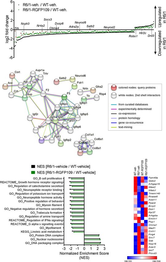
Hecklau et al.                                                                                                       Histone Deacetylase Inhibition in HD

causes brain region-specific neuronal dysfunction and                         provide significant support for the disease-modifying therapeutic
degeneration that is most prominent in the striatum during                    potential of epigenome-targeting strategies such as inhibition of
early disease stages and spreads to other brain regions when the              histone deacetylation.
disease progresses (Tabrizi et al., 2012).                                        Initially, HDAC inhibitors have been studied and applied
    Transcriptional dysregulation is an early and central                     in cancer research identifying non-selective HDAC inhibitors
pathogenic mechanism in Huntington’s disease which has been                   such as Trichostatin A (TSA) and Vorinostat as anti-cancer
demonstrated in cell and animal models of HD as well as in                    drugs. In recent years, however, selective HDAC inhibitors have
human HD brain (Cha, 2000; Hodges et al., 2006; Seredenina                    been developed (Wagner et al., 2015) and there is growing
and Luthi-Carter, 2012). Transcriptional repression of many                   evidence that especially HDAC class I inhibitors are effective in
genes coding for neurotransmitters, neurotrophins, and their                  suppressing pathogenic mechanisms in mouse models of HD
receptors is a hallmark of HD, while genes that are part of                   (Thomas et al., 2008; Chiu et al., 2011; Lim et al., 2011; Jia et al.,
stress-response pathways were shown to be upregulated in HD.                  2012, 2016; Chen et al., 2013; Suelves et al., 2017). RGFP109
Some of the key neuronal genes that are consistently reported                 is a HDAC class I inhibitor selective for HDAC1 and HDAC3.
to be repressed across HD patients and animal models include                  RGFP109 confirmed safety in a recent Phase I clinical trial
brain-derived neurotrophic factor (Bdnf ), preproenkephalin                   in patients of Friedreich Ataxia Syndrome and enhanced the
(Penk), dopamine receptor 2 (Drd2), and dopamine receptor 1a                  mRNA levels of the key disease gene frataxin (FXN) in patient
(Drd1). Accordingly, it has been shown that overexpression of                 blood mononuclear cells indicating its potentials for disease
Bdnf and Penk is neuroprotective and improves disease outcome                 amelioration (Soragni et al., 2014). In another study, RGFP109
in HD (Zuccato et al., 2005; Zuccato et al., 2011; Bissonnette                treatment led to significantly decreased levels of L-dopa induced
et al., 2013). Furthermore, dopamine agonist and antagonists                  dyskinesia in a Parkinson’s disease marmosets model (Johnston
play an important role in symptomatic treatment of HD patients                et al., 2013). A short-term treatment of HD R6/2 mice with
(Cepeda et al., 2014; Rangel-Barajas et al., 2015; Coppen and                 RGFP109 modified the expression levels of 4 out of 13 measured
Roos, 2017). However, the mechanisms driving transcriptional                  disease-associated genes in the striatum (Jia et al., 2012). So
dysregulation in HD are not fully understood. It was suggested                far, a comprehensive investigation of the effects of RGFP109 on
that mutant HTT directly interacts with transcription factors                 different aspects of HD, such as behavioral disease phenotypes or
and DNA (Benn et al., 2008). On the other hand there is strong                brain atrophy, has not been carried out.
evidence for epigenetic mechanisms such as histone acetylation,                   In the present study, we tested the therapeutic effects of
histone ubiquitination, histone trimethylation (H3K9me3,                      the selective HDAC inhibitor RGFP109 in the R6/1 mouse
H3K4me3), and DNA methylation contributing to selective                       model of HD. The outcome of HDAC1 and HDAC3 inhibition
changes in gene expression in HD pathogenesis (Ferrante et al.,               was assessed by a set of behavioral tests and genome-wide
2004; Ryu et al., 2006; Seo et al., 2008; Bett et al., 2009; Vashishtha       transcriptional analysis of the striatum. Moreover, we performed
et al., 2013; Hervas-Corpion et al., 2018).                                   volumetric magnetic resonance imaging (MRI) of the brain
    Histone hyperacetylation by histone acetyltransferases (HATs)             in living animals. Our results demonstrate that inhibition of
is generally associated with gene expression and histone                      HDAC1 and HDAC3 modestly alleviates the short-term motor
hypoacetylation by histone deacetylases (HDACs) with gene                     skill learning deficits, accompanied by a partial repair effect on
repression (Kurdistani et al., 2004). Global as well as gene specific         global gene expression changes in the striatum of R6/1 mice. MR
histone hypoacetylation at promoters of down-regulated genes                  imaging showed, on the other hand, that RGFP109 treatment
were shown in various HD models (Sadri-Vakili et al., 2007;                   exerted only very small, non-significant changes on the atrophy
Yildirim et al., 2019). Mutant HTT leads to hypoacetylation by                of specific brain regions in the R6/1 mice. Collectively, these
directly binding HATs such as CBP, reducing their activity and                findings present evidence for beneficial effects of specific HDAC1
resulting in gene repression (Steffan et al., 2000; NuciforaJr.,              and HDAC3 inhibition on transcriptional dysregulation and
Sasaki et al., 2001; Cong et al., 2005; Jiang et al., 2006).                  motor skill learning and coordination deficits, two key aspects of
Overexpressing CBP reverses these effects and leads to decreased              HD pathology, in HD mice.
mutant HTT toxicity (Steffan et al., 2000; Jiang et al., 2006).
Similar neuroprotective effects are achieved by inhibiting
the opponent, HDACs. HDAC inhibition has been shown                           RESULTS
to ameliorate transcriptional changes in HD and improve
behavioral deficits across different experimental models (Steffan             HDAC1 and HDAC3 Inhibition by
et al., 2001; Ferrante et al., 2003, 2004; Hockly et al., 2003;
Butler and Bates, 2006; Ryu et al., 2006; Kazantsev and
                                                                              RGFP109 Provides a Modest Positive
Thompson, 2008; Pallos et al., 2008). Beyond HD, such chromatin               Effect on Motor Learning Deficits Typical
targeting strategies exert therapeutic effects in other neurological          of HD in R6/1 Mice
conditions such as ischemic injury, Alzheimer disease, and                    R6/1 transgenic mice faithfully recapitulate many of the disease
Amyotrophic lateral sclerosis as previously reported by us                    features of human HD, such as transcriptional dysregulation,
and others (Ryu et al., 2005; Faraco et al., 2006; Meisel                     progressive impairments of both motor and cognitive functions
et al., 2006; Yildirim et al., 2008; Harrison and Dexter, 2013;               (Naver et al., 2003; Hodges et al., 2008; Brooks et al.,
Schweizer et al., 2015; Yang et al., 2017). These findings                    2012; Yildirim et al., 2019), brain atrophy and mutant HTT

Frontiers in Molecular Neuroscience | www.frontiersin.org                 2                                   February 2021 | Volume 14 | Article 616886
Hecklau et al.                                                                                                         Histone Deacetylase Inhibition in HD

accumulation (Bayram-Weston et al., 2012). In our study, to                     were found between vehicle- and RGFP109-treated R6/1 mice
evaluate if the specific HDAC inhibitor RGFP109 could alleviate                 for any of these parameters. Next, we performed the elevated
the transcriptional dysregulation in HD in vivo and improve                     plus maze test, which has been developed for measuring anxiety
the characteristic neuroanatomical HD features and behavioral                   in rodents (Walf and Frye, 2007). As previously described
deficits, we treated R6/1 mice (11–14 weeks of age) with                        (Naver et al., 2003), R6/1 mice showed trends to visit the open
30 mg/kg (i.p.) RGFP109 five times a week for 3 weeks (total of                 arms more frequently (7.6 ± 4.2 for WT-vehicle, 12.8 ± 6.0
23 days) (Figure 1A). R6/1 mice display a range of characteristic               for R6/1-vehicle; p = 0.137), spend more time (13.6 ± 11.2 s
behavioral deficits such as motor abnormalities, learning and                   for WT-vehicle, 26.3 ± 13.6 s for R6/1-vehicle; p = 0.163),
memory impairments, and reduced level of anxiety (Naver et al.,                 and travel longer distances (38.8 ± 29.2 cm for WT-vehicle,
2003; Hodges et al., 2008; Brooks et al., 2012). Thus, we examined              78.0 ± 45.5 cm for R6/1-vehicle; p = 0.198) in the open arms
the R6/1 mice using different behavioral tests including RotaRod,               regardless of treatment. Taking the reduced locomotor activity
open field, and elevated plus maze during the course of the                     of R6/1s into account, vehicle-treated R6/1 mice displayed a
study (Figure 1A).                                                              significantly less anxious phenotype than vehicle-treated wild
    For evaluating the effect of HDAC1 and HDAC3 inhibition on                  type mice (visits, time, and path in open arm normalized to
motor coordination, balance, and motor skill learning in R6/1                   total path). Treatment with RGFP109 did not exert a significant
mice, we performed the accelerating RotaRod test. Figure 1B                     effect on this phenotype neither in R6/1 nor in wild type mice
(left) depicts the latency to fall as mean of four consecutive trials           (Figure 1D and Supplementary Image S1C).
for each animal. Comparison of the vehicle-treated R6/1 and                         Lastly, abnormal limb clasping upon tail suspension can be
wild type animals revealed an HD-characteristic phenotype with                  used to demonstrate the presence of a neurological phenotype
significantly shorter time to stay on the rod in R6/1s. The latency             in HD mice. At the end of our study, before the sacrifice
to fall was significantly reduced by the main genotype effect [F (1 ,           of mice (at 14–17 weeks of age), several R6/1 animals
40) = 23.6 with p < 0.0001] while treatment had only a minor                    exhibited first degree forelimb clasping (score 0.5). However,
effect on motor coordination [F (1 , 40) = 1.09 with p = 0.304].                RGFP109 administration did not affect the clasping behavior
However, mice with treatment stayed 26 s longer on the rod,                     (Supplementary Image S1D). During the course of the study,
albeit not being significant (mean of all trial: 101.8 ± 44.2 s for             body weight of the animals was not influenced by HDAC
R6/1-vehicle and 127.8 ± 50.5 s for R6/1-RGFP109; p = 0.651)                    inhibitor administration indicating that the treatment was in
(Figure 1B, left). This was also observed for the speed at                      general well tolerated. As expected, a reduction in the weight of
fall (mean of all trials: 18.1 ± 5.7 rpm for R6/1-vehicle and                   the R6/1 mice was observed with disease progression, on which
21.4 ± 7.4 rpm for R6/1-RGFP109; p = 0.672) (Supplementary                      HDACi treatment showed no effect (Mangiarini et al., 1996;
Image S1A). In contrast, inhibition of HDAC1 and HDAC3 in                       Naver et al., 2003; Supplementary Image S1E).
R6/1 mice led to a small but significant improvement in RotaRod                     Altogether, these results demonstrate that RGFP109 treatment
performance over the course of the four consecutive trials,                     exerts a modest positive effect on the motor skill learning deficits
indicating enhanced motor learning skills in HDACi-treated R6/1                 while not affecting the general locomotor activity or anxiety-
animals (q = 0.017 for trial 4 vs. trial 1 for latency to fall; q = 0.026       related phenotypes in HD mice.
for trial 4 vs. trial 1 for speed at fall) (Figure 1B, middle and
right). Notably, RGFP109 treatment in R6/1 animals resulted in
a significant increase in time to stay on the rod compared to
                                                                                RGFP109 Treatment Alleviates, in Part,
vehicle-treated R6/1s in the fourth trial (103.1 ± 40.0 s for R6/1-             the Global Gene Expression Changes in
vehicle and 153.3 ± 64.6 s for R6/1-RGFP109 and a difference                    the Striatum of R6/1 Mice
of 50.2 s; q = 0.048) (Figure 1B, middle). Albeit not statistically             Aberrant transcriptional regulation in brain, especially in
significant, a similar trend was observed for the speed at fall                 striatum and cortex, is an early and central feature of HD
(mean difference = 7.08; q = 0.052) (Figure 1B, right). Vehicle-                pathogenesis in models and patients. In the R6/1 model,
treated R6/1 mice as well as vehicle- and RGFP109-treated                       consistent transcriptional dysregulation in the brain was shown
wild type animals did not exhibit significant changes in motor                  starting with the earliest microarray reports (Kuhn et al., 2007;
learning abilities during the four trials. These findings indicate              Benn et al., 2010) and more recently by RNA-sequencing
that HDAC1 and HDAC3 inhibition by RGFP109 modestly                             studies (Gallardo-Orihuela et al., 2019; Yildirim et al., 2019).
ameliorates short-term motor learning deficits in R6/1 mice.                    Among the genes with aberrant expression in HD striatum are
    We next analyzed if the general locomotor activity in the                   transcriptional regulators (e.g., Fos, Egr1, Npas4, and Polr2a)
open field was also affected by RGFP109 treatment in R6/1 mice.                 as well as key neuronal genes important for neurotransmitter
Locomotor activity is known to be impaired in the R6/1 line as                  signaling (e.g., Drd1, Drd2, Grin3a, and Ppp1r1b) and synaptic
well as in other HD models (Naver et al., 2003; Hodges et al.,                  plasticity (e.g., Arc and Syp) (Seredenina and Luthi-Carter, 2012;
2008). Accordingly, we observed a highly significant reduction                  Vashishtha et al., 2013; Yildirim et al., 2019). To examine the
in the total distance traveled as well as in the average velocity               impact of RGFP109 treatment on transcriptional dysregulation,
and vertical activity (supported rearing) in vehicle-treated                    we conducted transcriptome analysis of the striatum by RNA-
R6/1 animals compared to vehicle-treated wild type littermates                  sequencing (RNA-seq) (n = 5 for WT-vehicle, n = 6 for R6/1-
indicating a hypoactive phenotype in HD animals (Figure 1C and                  vehicle, n = 6 for WT-RGFP109, n = 7 for R6/1-RGFP109).
Supplementary Image S1B). However, no significant differences                   All animals were sacrificed 18 h after the final injection and

Frontiers in Molecular Neuroscience | www.frontiersin.org                   3                                   February 2021 | Volume 14 | Article 616886
Hecklau et al. Histone Deacetylase Inhibition in HD FIGURE 1 | RGFP109 treatment improves motor learning in R6/1 mice. (A) Schematic of treatment and experimental procedure. (B) Motor performance and learning on a RotaRod: Latency to fall as mean of all trials, latency to fall and speed at fall as learning curve over the course of four subsequent trials. (C) Open field exploration evaluating general locomotor activity measured as total distance traveled. (D) Anxiety-related behavior determined by elevated plus maze test showing visits to open arm and path traveled in open arm normalized to the total path. WT, wild type. Data presented as mean ± SEM; n = 10 [WT-vehicle], n = 12 (13, Open Field) [R6/1-vehicle], n = 10 [WT-RGFP109], n = 12 [R6/1-RGF109]; *p < 0.05, **p < 0.01, ****p < 0.0001, ns, not significant by two-way ANOVA with Tukey’s multiple comparisons test (B-left graph, C,D); # q < 0.05, ## q < 0.01, ns, not significant by mixed-effects analysis with two-stage linear step-up procedure of Benjamini, Krieger and Yekutieli as post-tests by controlling the False Discovery Rate (individually comparing each group to each other group) (B-learning curves: comparison of trial 4 between groups—indicated vertically, comparison of trial 4 vs. trial 1 for R6/1-RGFP109—indicated horizontally). the striatum was used for RNA isolation and subsequent age of sacrifice compared to wild type mice in the vehicle- analyses (Figure 1A). treated group (FDR, q < 0.1; log2 FC > | 0.5|) with 1,171 genes Analysis of the RNA-seq data revealed that 1,461 genes were (80%) being downregulated in R6/1 animals (Supplementary significantly differentially expressed in the R6/1 striatum at the Data Sheet S1). As expected, among these, key HD genes such Frontiers in Molecular Neuroscience | www.frontiersin.org 4 February 2021 | Volume 14 | Article 616886
Hecklau et al. Histone Deacetylase Inhibition in HD
as Drd1, Drd2, Ppp1r1b, Penk, and Adora2a were downregulated. analysis. Expanding the network analysis with 20 proteins
Analysis of the RNA-seq data from the RGFP109-treated R6/1 associated with the query proteins revealed that 15 out of
striata showed that the HDACi treatment significantly changed the 43 genes belong to predicted protein-protein interactions
the expression of 43 genes in R6/1 animals (FDR, q < 0.1; (Figure 2B). Notably, the largest cluster in the network included
log2 FC > | 0.5|), of which 36 genes were also differentially many members of the Insulin-like growth factor (IGF) pathway,
expressed in R6/1s compared to wild types (Supplementary suggesting activation of this pathway by RGFP109. In line,
Data Sheet S1). Fourteen out of these 36 genes were changed evaluation of the functional enrichment of all connected and
toward wild type expression levels upon RGFP109 treatment in disconnected nodes in the network for GO biological process
R6/1 animals (Figure 2A, highlighted in red). Among these are terms revealed the enrichment of regulation of insulin-like
transcription factor genes such as Neurod2, Neurod6, and Nr4a2, growth factor receptor signaling pathway as well as several other
the transcriptional regulator Satb2, the thyrotropin-releasing terms that are relevant for HD pathology, such as negative
hormone Trh, the neuropeptide-like molecule Nxph3, and the regulation of transmission of nerve impulse and learning or
nicotinic acetylcholine receptor Chrna4 genes. We validated the memory (Supplementary Data Sheet S2).
RNA-seq findings by quantitative RT-PCR for several typical Further, using log2 fold changes for ranking genes, we
HD genes (Drd1, Drd2, Ppp1r1b, Adora2a, Arc, Egr1, Polr2a, performed gene set enrichment analysis (GSEA) to find the
and Grin3a), for the effect of HDAC1 and HDAC3 inhibition gene sets associated to the functional transcriptional changes
on the transcriptional changes in R6/1s (Nr4a2, Satb2, Folr1, in HD and those induced by HDAC1 and HDAC3 inhibition.
and Otx2) (although not significant in qRT-PCR data), as well This analysis showed that normalized enrichment scores of
as for several genes that were previously connected to motor 43% (24 out of 55) of the gene sets that were negatively
skill learning behavior in rodents (Dpysl2, Wars, and Cpne5) enriched in disease (R6/1-vehicle/WT-vehicle comparison) were
(D’Amours et al., 2011) in the striatum of vehicle- and RGFP109- changed by at least 5% toward wild type levels in RGFP109-
treated wild type and R6/1 mice (Supplementary Image S2A). treated R6/1 mice (enrichment scores shifting toward 0
The complete lists of significantly differentially expressed genes comparing drug-treated R6/1s vs. vehicle-treated wild type mice).
for the R6/1 and wild type comparison as well as for the Among these were gene sets associated with neuropeptide
treatment effect in R6/1 mice in RNA-seq data are presented in receptor binding, negative regulation of hormone secretion,
Supplementary Data Sheet S1. and regulation of IFNα signaling, that were increased toward
Further analysis of the datasets, without imposing cutoff wild type levels. In contrast, among the positively enriched
criteria on the data for statistical significance, revealed a gene sets in R6/1s, only protein DNA complex and DNA
trend for a larger potential effect of RGFP109 treatment on packaging complex were lowered toward wild type levels
expression of the dysregulated genes in R6/1 striatum, as upon treatment (by 2.4 and 2.3%, respectively). Of note, the
illustrated in Figure 2A. Of the 1,171 downregulated genes in neuroactive ligand receptor interaction gene set showed further
the vehicle-treated R6/1s, expression of 21% (244) were enhanced enhancement of the disease-associated enrichment pattern
by >20% by the HDACi treatment of R6/1s and among these upon HDAC1 and HDAC3 inhibition in R6/1 animals (by
were genes such as adrenoceptor alpha 2a (Adra2a) and 1b 6.4%), while the remaining gene sets enriched in disease (31)
(Adra1b), suppressor of cytokine signaling 3 (Socs3), and dual were not changed or changed by less than 5% by RGFP109
specificity phosphatase 6 (Dusp6), whose expression showed a treatment and these include actin filament based movement
shift toward wild type levels. Similarly, expression of 13% (39) and potassium channel complex gene sets (Figure 2C and
of the genes upregulated in the vehicle-treated R6/1 striatum Supplementary Data Sheet S2).
were reduced by >20% by the RGFP109 treatment and some We next compared our results with a previous report which
of these genes were 5-hydroxytryptamine (serotonin) receptor examined the effects of a similar HDACi, HDACi 4b, on genome-
2c (Htr2c), roundabout guidance receptor 1 (Robo1), and wide gene expression profiles in the striatum of R6/2 mice using
dopamine receptor d3 (Drd3) (Figure 2A and Supplementary microarray (Thomas et al., 2008). Of 56 genes, which were shown
Image S2B). To get a better functional view of genes that to be restored in the striatum by HDACi 4b treatment of R6/2
were alleviated by HDACi treatment in R6/1 animals, we mice, 42 were detected by RNA-seq in our study. Of these 42
performed gene ontology (GO) enrichment analysis of these genes, only 6 were significantly dysregulated in the striatum
genes that were changed toward wild type expression levels in of R6/1 mice at the age of 14–17 weeks, which corresponds
RGFP109-treated R6/1 animals by at least 20%. These genes to a less progressed disease stage compared to the R6/2s, and
were enriched for several GO terms that are relevant for 2 of these genes, Arhgap15 and Gsto2, were restored by more
brain physiological processes, such as neuropeptide hormone than 20% by RGFP109 treatment of the R6/1s in our study.
activity, regulation of secretion, and second-messenger-mediated To explore a more inclusive list of potentially therapeutically
signaling (Supplementary Image S2C). Supplementary Data relevant genes, we examined all expression changes, regardless of
Sheet S2 shows the complete list of enriched GO terms their statistical significance, which revealed that of the 42 genes
(FDR, q < 0.1). previously reported to be restored by HDACi 4b, 25 showed a
Next, in order to functionally decipher the effect of RGFP109 change toward the wild type levels after RGFP109 administration
treatment on gene programs in the striatum of R6/1s, we in R6/1 mice in our study (Figure 2D and Supplementary Data
performed protein-protein interaction network analysis of the Sheet S1), providing a list of genes whose expression levels are
proteins assigned from the 43 differentially expressed genes using consistently ameliorated by specific HDAC1/3 inhibitors across
STRING (Szklarczyk et al., 2019) followed by gene ontology different HD models.
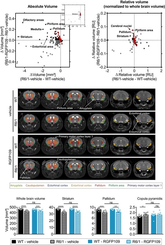
Frontiers in Molecular Neuroscience | www.frontiersin.org 5 February 2021 | Volume 14 | Article 616886Hecklau et al. Histone Deacetylase Inhibition in HD FIGURE 2 | Inhibition of HDAC1 and HDAC3 by RGFP109 has a partial repair effect on global gene expression changes in the striatum of R6/1 mice. (A) 283 genes, significantly differentially expressed between WT and R6/1 mice (black) (Cuffdiff 2; FDR, q < 0.1; log2 FC > | 0.5|), are changed toward wild type expression levels in RGFP109-treated R6/1 animals by at least 20% using no cutoff criteria on the data for statistical significance (green). Significantly differentially expressed genes upon RGFP109 treatment in R6/1 mice (R6/1-RGFP109/R6/1-vehicle; FDR, q < 0.1; log2 FC > | 0.5|) are framed in red. Selected genes are labeled. (B) STRING protein-protein interaction network analysis. Forty three genes differentially expressed between R6/1-vehicle and R6/1-RGFP109 (FDR, q < 0.1; log2 FC > | 0.5|) were used as input and assigned to proteins (query proteins). The maximum number of 2nd shell interactors was set to 20. Network nodes represent proteins. Edges represent protein-protein associations. Disconnected nodes in the network are hidden. (C) Gene set enrichment analysis (GSEA—Broad Institute) for gene ontology, KEGG pathway, and REACTOME pathway gene sets. Genes were ranked based on log2 fold changes. The bar graph shows gene sets with significant different normalized enrichment scores (NES) between WT-vehicle and R6/1-vehicle mice (FDR, q < 0.1). NESs of RGFP109-treated R6/1 compared to WT-vehicle mice are plotted for the same gene sets. Gene sets with positive NES and the top 15 gene sets with highest difference between both comparisons for negative NES are plotted. (D) Heatmap for gene expression levels of 25 genes, that show a change toward wild type levels by RGFP109 treatment in R6/1 mice, from a list of genes whose expression were restored by HDACi treatment in a previous study (Thomas et al., 2008). Mean FPKM value/group is shown. Each row is a gene and each column is a group. n = 5 [WT-vehicle], n = 6 [R6/1-vehicle], n = 7 [R6/1-RGFP109]. Frontiers in Molecular Neuroscience | www.frontiersin.org 6 February 2021 | Volume 14 | Article 616886
Hecklau et al.                                                                                                  Histone Deacetylase Inhibition in HD

   Collectively, RGFP109 treatment, at least to a certain extent,         animals (Figure 3B and Supplementary Image S3B). 144 brain
repairs the transcriptional effects of mutant Huntingtin gene             regions with significant volume change were shared between
expression, causing a significant change in the expression of             the two analysis strategies (Supplementary Image S3C). Similar
43 genes, in the striatum of R6/1 mice and induced collective             to the absolute volume analysis, RGFP109 treatment did not
changes in the expression of a number of gene sets and                    show any significant effects on volumes of brain regions in
pathways, some of which were associated to biological functions           R6/1 mice after correcting for the whole brain atrophy. Non-
and processes that are relevant for neuronal physiology and               significant trends toward an improved volume change of at least
HD pathogenesis.                                                          20% upon drug treatment in R6/1 mice was detectable for 25%
                                                                          (55/220) of the regions showing a significant volume difference
Effects of RGFP109 Treatment on the                                       in R6/1 mice (Figure 3B, framed red). Seventeen brain regions
                                                                          and sub-regions that show a trend toward positive effect by
HD-Associated Atrophy of Specific Brain                                   drug treatment by more than 20% in R6/1s were shared in
Areas in the R6/1 Mice Measured by                                        both analysis strategies. These include piriform area (molecular
Volumetric MRI                                                            layer), ectorhinal area, intermediodorsal nucleus of the thalamus,
One hallmark of HD is neuronal degeneration which manifests               and prelimbic area (layer 5) (Supplementary Images S3D,E
predominantly in the striatum, particularly the caudoputamen              and Supplementary Data Sheet S3). Figure 3C depicts selected
and dorsal striatum in HD patients. In addition, brain atrophy            regions that were previously implicated in motor skill learning
of variable severity can be observed in several other brain               (amygdala, caudoputamen, ectorhinal cortex, entorhinal cortex,
regions, such as the cerebral cortex, total white matter, amygdala,       pallidum, piriform area, primary motor cortex) in representative
hippocampus, and brainstem (Rosas et al., 2003; Tabrizi et al.,           examples of vehicle- or HDACi-treated wild type and R6/1 mice
2012). The R6/1 mouse model closely recapitulates the neuronal            (Tamakoshi et al., 2014; Scholz et al., 2015; Badea et al., 2019).
degeneration seen in HD patient brain. These mice exhibit                     In summary, matching the Allen brain atlas to MR images
progressive brain atrophy in the striatum (especially the posterior       allowed us to investigate the HD mouse neuroanatomy and
striatum) and cortex (especially in the retrosplenial areas)              the impact of RGFP109 treatment on it in great detail. While
as well as a subtle expansion of posterior ventricular spaces             we detected a large number of brain regions and sub-regions
(Rattray et al., 2013).                                                   with significant volume change in R6/1 animals, HDAC1 and
    By registering the Allen brain atlas to T2w MR images                 HDAC3 inhibition exhibited only trends toward slowing these
(Koch et al., 2019) we used an unbiased approach to detect                volumetric differences in the brains of R6/1 mice at this
regional and sub-regional changes in R6/1 mice upon RGFP109               progressed disease stage.
treatment (Figure 1A). This analysis revealed 419 brain regions
and sub-regions (55% of all analyzed regions) that showed
a significant volume difference between vehicle-treated R6/1
                                                                          RGFP109 Treatment Does Not Affect
and wild type mice at the age of 13–16 weeks of age (FDR,                 Aggregate Formation or Bulk Histone
q < 0.1) (Figure 3A and Supplementary Image S3A). Of                      H3K27acetylation Levels in R6/1 Mice
these, the majority (403 regions, 96%) exhibited a significant            HD is characterized by mutant HTT protein aggregate formation,
volume decrease in R6/1 mice including striatum, pallidum,                which displays a histopathological basis of transcriptional
hippocampal region, and piriform area. In contrast, 16 regions            dysregulation, neuronal degeneration, and behavioral deficits.
(4%) showed a significant volume increase in HD mice, e.g.,               Given the effect of RGFP109 on short-term motor skill learning,
copula pyramidis, orbital area (medial part, layer 1 and 2), and          we examined whether RGFP109 treatment has an impact on
supratrigeminal nucleus (Figure 3D and Supplementary Data                 this neuropathological hallmark of HD. For this purpose, we
Sheet S3). RGFP109 treatment did not induce any significant               analyzed EM48 immunoreactivity in striatal sections visualizing
changes in the volumes of the afore-mentioned 419 brain                   aggregated mutant HTT. We analyzed 8 separate images per
substructures in R6/1 mice at this age. However, 61 regions               section, each covering a 34,192 µm2 area with 123 DAPI + cells
(15%) with significant decrease or increase in R6/1 animals               in average in the dorsal stratum directly lateral to the lateral
showed non-significant trends toward a reversal of the volume             ventricle. By using ImageJ software, we defined particles larger
change upon drug treatment by at least 20% (Figure 3A,                    than 15 µm2 as nuclei. Blinded manual counting of the
framed red). Some of these regions include central amygdalar              number of aggregates showed that RGFP109 did not affect
nucleus (medial part), pallidum (ventral region), and prelimbic           the number of mutant HTT protein aggregates in R6/1 mice
area. Supplementary Data Sheet S3 contains the complete                   (Supplementary Image S4A).
list of brain regions with significant volume difference in                   To examine the effects of RGFP109 treatment on global
R6/1 animals and those that show a trend toward change                    histone acetylation, we determined bulk acetylation patterns
by the treatment.                                                         at lysine 27 of histone H3 (H3K27ac) in protein extracts of
    As the overall brain volumes in R6/1 mice were reduced                striatal tissue via immunoblotting. Increased H3K27ac levels
(Supplementary Image S3E, first graph), we utilized a second              can be found at active promoters and enhancers indicating
approach that corrects the regional and sub-regional volumes              high transcriptional activity (Wang et al., 2008). Our Western
by the whole brain volume. This resulted in 220 regions (29%              blotting analysis showed no differences in acetylated H3K27
of all analyzed regions) showing a significant change in R6/1             levels between R6/1 and wild type mice, regardless of treatment.

Frontiers in Molecular Neuroscience | www.frontiersin.org             7                                  February 2021 | Volume 14 | Article 616886
Hecklau et al. Histone Deacetylase Inhibition in HD FIGURE 3 | Effect of RGFP109 treatment on HD-associated atrophic brain substructures in R6/1 mice. Mouse brain MR images were registered to the Allen brain atlas. Volumes of substructures (absolute or normalized to whole brain volumes) were analyzed by two-sample t-test (FDR, q < 0.1). (A) Mean absolute volume differences of brain regions with significant volume change in R6/1 compared to WT mice (419). (B) Mean relative volume differences of brain regions with significant volume change in R6/1 compared to WT mice (220). (A,B) Black: substructures affected positively by drug (not significant; red frame: >20% of R6/1-WT volume difference); Gray: substructures affected negatively by drug (not significant). (C) MR images, exemplarity shown for three individuals; selected regions are color-coded. Anatomical labels are based on the Allen brain atlas. (D) Absolute volumes of selected brain regions. Data presented as mean ± SEM; n = 10 [WT-vehicle], n = 13 [R6/1-vehicle], n = 10 [WT-RGFP109], n = 12 [R6/1-RGFP109]; *q < 0.05, **q < 0.01, ***q < 0.001, ****q < 0.0001, ns, not significant. As control for equal histone amounts, we determined total H3 While we did not detect increases in global H3K27ac patterns levels from the same samples. Similarly, we did not observe in striatum by the HDACi treatment, temporally dynamic differences between the groups (Supplementary Image S4B). and site-specific changes in acetylation levels not detected Frontiers in Molecular Neuroscience | www.frontiersin.org 8 February 2021 | Volume 14 | Article 616886
Hecklau et al.                                                                                                  Histone Deacetylase Inhibition in HD

by immunoblotting of total tissue could be associated to the             other behavioral tests to better distinguish motor skill learning
transcriptional changes observed in HD.                                  from motor coordination and balance. In line with our findings,
                                                                         Suelves et al. demonstrated that selective HDAC3 inhibition
                                                                         by RGFP966 in the HdhQ7/Q111 knock-in mouse model of
DISCUSSION                                                               HD (50 mg/kg of RGFP966 three times per week from 3 to
                                                                         6.5 months) prevents corticostriatal-dependent motor learning
This study suggests that treating HD mice with a specific                deficits from trial four on testing animals for four times per day
HDAC1 and HDAC3 inhibitor has benefits on multiple aspects               for 3 consecutive days (Suelves et al., 2017). Similarly, treatment
of HD. Inhibiting histone modifying enzymes, in particular               of N171-82Q HD mice with RGFP966 over a period of 10 weeks
class I HDACs, has been previously shown to improve disease              with 10 or 25 mg/kg per week showed improved motor function,
features, such as the phenotypic and neuropathological deficits          accompanied by neuroprotective effects on striatal volume, and
and dysregulation of selected genes, in various models of HD             significant alterations of the expression of 3 immune pathway
(Thomas et al., 2008; Chiu et al., 2011; Lim et al., 2011; Jia           genes (chemokine (C-C motif) ligand 17 (Ccl17), macrophage
et al., 2012, 2016; Chen et al., 2013; Suelves et al., 2017). The        migration inhibitory factor (Mif ), interleukin 13 (Il13); measured
goal of our study was to evaluate the effects of the specific            by PCR) (Jia et al., 2016). Notably, in the latter study, the
HDAC1 and 3 inhibitor RGFP109 in an unbiased approach                    beneficial effects of RGFP966 on the RotaRod task was observed
on the genome-wide transcriptional dysregulation and on the              in female HD mice only, suggesting potential differences in
phenotypic deficits in HD mice using the R6/1 model. Further,            HDACi’s effects depending on sex. While a sex comparison was
by performing MRI measurements on living mice, we conducted              not within the scope of our study, it would be important to test
a detailed analysis of the brain volumetric changes in the R6/1          RGFP109, which showed positive effects in male R6/1 mice in
mice. Our findings demonstrate that the specific inhibition of           this current study, also on female R6/1s in future. In a mouse
HDAC1 and HDAC3 by RGFP109 modestly improves the motor                   model of Friedreich ataxia prolonged RGFP109 treatment led to
learning skills and alleviates, at least to a certain extent, the        modest improvements in motor coordination performance and
dysregulation of a number of genes and gene sets in the R6/1             locomotor activity (Sandi et al., 2011). It has been shown that
mice. By volumetric MRI, we detected extensive atrophic changes          improvement on the RotaRod mainly requires a change in the
in the brains of the R6/1 mice, on which RGFP109 exerted                 motor strategy to master the task rather than an enhancement in
no significant effects. Given our results and data from others,          general locomotor activity (Buitrago et al., 2004). Accordingly, we
early targeting of transcriptional dysregulation by specific HDAC        did not observe improvement in general locomotor activity in the
inhibition may alleviate key transcriptional and phenotypic              open field test upon RGFP109 treatment in R6/1 animals.
aspects of HD pathology.                                                    To investigate the potential molecular correlates of our
    In a previous study, a short-term treatment regime with              behavioral findings, we carried out a genome-wide analysis of
RGFP109 (subcutaneous injections of 150 mg/kg/day for 3 days)            the transcriptional changes, which was not done before in the
partially reversed four selected disease-associated genes in the         afore-mentioned studies, in the striata of vehicle- and drug-
striatum in HD R6/2 mice as measured by PCR (Jia et al., 2012).          treated R6/1 and wild type mice. Our RNA-seq data showed the
However, the effects of RGFP109 treatment on the behavioral              altered expression of a large number of genes in the striatum
deficits and on gene expression throughout the genome as well as         of R6/1 mouse, which included also those that are well-known
on the brain atrophy have not been evaluated in HD. Here, using          to be dysregulated in HD, e.g., Drd1, Drd2, Penk, and Adora2a
the R6/1 mice, which is less progressive than the R6/2 model,            (Seredenina and Luthi-Carter, 2012). RGFP109 administration
we chose to use a lower therapeutic dose of 30 mg/kg based on            showed, in part, repair effects on the global aberrant gene
a previous Parkinson’s disease study (Johnston et al., 2013) and         expression changes in the striatum of R6/1 mice, affecting 43
applied a longer treatment regime (3 weeks with five injections          individual genes significantly. Some of these genes were the
per week), aiming to minimize potential toxic side effects by            thyrotropin-releasing hormone gene Trh, the neuropeptide-like
reducing high drug exposure.                                             molecule gene Nxph3, the nicotinic acetylcholine receptor gene
    Among the most characteristic behavioral deficits in HD mice         Chrna4, and transcriptional regulator genes such as Neurod2,
are motor impairment and deficit in motor skill learning, which          Neurod6, Nr4a2, and Satb2, whose change in expression may
are caused by the dysfunction of corticostriatal circuits (Backman       likely result in alterations in the expression of their target genes.
et al., 1997; Lawrence et al., 2000; Cybulska-Klosowicz et al.,          Further, Arhgap15 (Rho GTPase activating protein 15) and Gsto2
2004; Mazarakis et al., 2005). The RotaRod task is a useful              (Glutathione S-Transferase Omega 2), genes whose expression
marker for detecting early HD phenotypes in R6/1 animals                 were completely rescued in a previous report by the HDAC1 and
from as early as 8 weeks of age (Brooks et al., 2012). Our               HDAC3 selective inhibitor HDACi 4b in R6/2s (Thomas et al.,
study demonstrates that 3-week-long treatment with 30 mg/kg              2008), were also restored in our study upon RGFP109 treatment
RGFP109 modestly ameliorates motor skill learning deficits in            in R6/1s, supporting a similar mode of action of these selective
this mouse model of HD. While we obtained this finding by                HDAC inhibitors across different models of HD. Of note, loss
testing the animals on the RotaRod for up to four trials on the          of Arhgap15 gene, a member of the Rac signaling pathway, was
same day, in future studies, it would be important to analyze            shown to cause decreased synaptic density and cognitive deficits
the animals across several days to assess learning over longer           in mouse and its mutations were identified in association with
time frames as well as to use modified RotaRod protocols or              neurological and cognitive deficits in patients with Intellectual

Frontiers in Molecular Neuroscience | www.frontiersin.org            9                                   February 2021 | Volume 14 | Article 616886
Hecklau et al. Histone Deacetylase Inhibition in HD
Disability (Zamboni et al., 2018), revealing the rescue of its progressive whole brain volume loss is evident (Tabrizi et al.,
expression by HDACi treatment as potentially therapeutically 2012). Furthermore, in addition to the striatum, other brain
relevant in HD. Beyond these single gene expression changes, regions known to be affected in HD patients, such as cerebral
we found that a number of gene sets and pathways associated cortex, amygdala, hippocampus, and brainstem (Rosas et al.,
with neurophysiological functions relevant for HD pathology 2003), are also changed in R6/1 mice. Similar to the modest
were changed upon treatment and these changes could be linked transcriptomic changes we observed upon RGFP109 treatment,
to the observed phenotypic changes. Notably, all three analyses volumes of specific brain regions and sub-regions showed only
tools we utilized, Gene ontology, GSEA and String network, non-significant trends toward an alleviation upon HDAC1 and
commonly revealed enrichment of hormone activity related HDAC3 inhibition in R6/1 mice at this disease stage. Of note,
pathways, indicating in particular the activation of Insulin-like these regions include some of the brain areas that were previously
growth factor pathway by RGFP109 treatment. Both IGF-1 and implicated in motor skill learning behavior in rodents, such
IGF-2 were previously reported to exert robust protective effects as the amygdala, ectorhinal cortex, entorhinal cortex, pallidum,
in HD (Lopes et al., 2014; Garcia-Huerta et al., 2020), suggesting piriform area, and primary motor cortex (Tamakoshi et al., 2014;
that induction of IGF pathway by RGFP109 may be one of Scholz et al., 2015; Badea et al., 2019).
the key mechanisms contributing to RGFP109’s beneficial effects In summary, our study suggests that treatment with the
observed in our study. HDAC inhibitor RGFP109 provides benefits on transcriptional
In an animal study of Friedreich Ataxia it was shown that dysregulation and motor skill learning and coordination deficits,
long-term RGFP109 treatment (100 mg/kg over a period of while not affecting the locomotor and anxiety-like deficits and
5 months with five injections per week) increased especially the progressive brain atrophy in the R6/1 mice. Further studies
local H3K9ac and H4K5ac levels directly at the frataxin gene should include also relevant immunohistochemical assessments
accompanied by higher frataxin gene expression levels in mouse of the brain for elucidating potential synaptic plasticity changes
brain, whereas global H3 and H4 acetylation patterns did not that may contribute to the observed phenotypic effects of HDAC
significantly increase by the drug (Sandi et al., 2011). Next inhibition in HD mice. Although the R6/1 line recapitulates
to the spatial changes of specific histone acetylation patterns, several key features of HD, such as motor and cognitive
temporal differences of global and local histone acetylation deficits, transcriptional dysregulation, accumulation of mutant
have been observed in another Friedreich Ataxia mouse study Htt aggregates and brain atrophy, establishing direct mechanistic
using a single RGFP109 administration (with 150 mg/kg) (Rai links between molecular pathology and specific behavioral
et al., 2010). In this study, global H3 acetylation in the brain deficits has been a challenge so far (Rattray et al., 2013). Further
increased to a maximum level at 4 h after injection and totally work using an HD mouse model with slower progression,
disappeared at 24 h, whereas H4K5ac and H3K14ac at the intervention earlier in the time course of pathology, a longer
frataxin gene were shown to increase between 12 and 24 h (Rai treatment regime with various doses or a combination of these
et al., 2010). In contrast to the aforementioned studies in mouse factors in a well-powered sex-balanced cohort may enhance
models of Friedreich Ataxia, we did not observe increased global the leveraging of the therapeutic potentials of this selective
H3K27 acetylation by RGFP109 treatment measured by Western HDAC inhibitor in HD. Nevertheless, we have demonstrated
blotting of total striatal extracts. Supporting our findings of no that HDAC1/3 inhibitor RGFP109 modestly improved motor
change in global histone acetylation after RGFP109 treatment, skill learning deficits and alleviated transcriptional dysregulation,
a previous report showed histone acetylation changes only at characteristic disease features, in HD mice. Epigenome-targeting
specific promoters using a similar HDAC inhibitor, HDACi 4b, strategies may be viable approaches for targeting transcriptional
in the R6/2 mice (Thomas et al., 2008). dysregulation in HD and also in other neuropsychiatric diseases
Aiming at examining different key features of HD, on which without significant genetic causation.
RGFP109 treatment may have an effect, we performed MRI
measurements to study the structural brain changes in HD mice.
By registering the Allen brain atlas to MR images, we provided MATERIALS AND METHODS
a complete list of regional and sub-regional volumetric changes
of the brains of R6/1 animals, which by far expands the set of Animals
regions so far shown to be changed in this HD model. Using Hemizygous R6/1 mice, expressing exon 1 of the human HTT
longitudinal in vivo MRI, a previous study detected reduction in gene with an estimated repeat expansion range of 115–150
both global brain volume as well as brain sub-regional volumes CAGs, were purchased from the Jackson Laboratory. R6/1
when corrected for global volume change in R6/1 mice over mice were maintained on a C57BL/6J background crossing
time, showing ubiquitous shrinkage of the striatum and the male R6/1 with female C57BL/6J. Genotypes were determined
somatosensory cortices (Rattray et al., 2013), comparable with by PCR analyses (R6/1-fwd: 50 -CCGCTCAGGTTCTGCTTTTA-
what we detected with our approach here. In contrast, increases 30 ; R6/1-rev: 50 -GGCTGAGGAAGCTGAGGAG-30 ). The repeat
in regional and sub-regional volumes when corrected for whole length was 143 ± 4 CAGs, as detected by sequencing of the
brain volume should be viewed cautiously, as they indicate a less genotyping PCR products of selected animals (including some of
pronounced shrinkage of that brain area compared to the volume the mice used in this study). Littermates were randomly divided
reduction of the whole brain, rather than an actual regional into four groups: WT-vehicle (n = 10), R6/1-vehicle (n = 13),
size increase. In pre-symptomatic and symptomatic HD patients WT-RGFP109 (n = 10), R6/1-RGFP109 (n = 12). All mice used
Frontiers in Molecular Neuroscience | www.frontiersin.org 10 February 2021 | Volume 14 | Article 616886Hecklau et al. Histone Deacetylase Inhibition in HD
in the present study were housed together in groups of maximal Accelerating RotaRod
four animals; if possible, genotype and treatment were mixed in One day before the actual test, mice were trained on the RotaRod
individual cages. Weight of the mice were monitored throughout (TSE Systems) three times at constant speed (4 rpm for 60 s)
the study. No animal was excluded due to excess weight loss and two times with accelerating speed (4–40 rpm over a period
(exclusion criterium: ≥25% weight loss compared to the weight of 5 min) with 30 min interval between the sessions. During the
at the start of the experiment). One animal (R6/1-vehicle) died training mice were placed back on the rod if they have fallen
during the course of the experiment (after completion of the MRI down. On the following day data were recorded for four test trials
measurement and open field test, before performing Rotarod and using the TSE RotaRod software, which detects the latency to fall
elevated plus maze tests) due to unknown reasons. Animals were in seconds and the rod rotational speed at fall in rpm. The test
housed under pathogen free conditions with ad libitum access trials were done by accelerating the rod from 4 to 40 rpm over a
to food and water on a 12 h light/12 h dark cycle at constant period of 5 min with at least 30 min interval between the sessions.
temperature (22 ± 2◦ C) and humidity (55 ± 10%). All animal The rod was cleaned between animal trials to remove any odors.
experiments were approved by the local animal care committee
of Charité-Universitätsmedizin Berlin and by the Landesamt für Open Field
Gesundheit und Soziales Berlin (license number G0314/16) and The open field test was performed in a gray open-top 50 × 50 cm
conducted according to the institutional guidelines. All efforts arena (height 40 cm) located in a sound-attenuated observation
were made to minimize all unnecessary suffering of animals. chamber. Mice were individually placed near the wall of the
In line with our license for animal experiments and the 3Rs box and locomotor activity was measured as total distance
principles, for reducing the data variation and thereby keeping traveled and average velocity over a period of 10 min. VideoMot2
the required number of mice for the experiments to a minimum, Software, TSE Systems was used to track and record all animals
only male mice were used in this study. (standard measuring mode based on center of gravity). Vertical
activity (supported rearing) was manually assessed analyzing the
RGFP109 Treatment of Mice recorded videos (experimenter was blinded for treatment and
The HDAC1 and HDAC3 inhibitor RGFP109 (RG2833; CAS genotype). The field was thoroughly cleaned between the animals
No. 1215493-56-3) was purchased from Selleckchem. RGFP109 to avoid any odor.
was dissolved in dimethyl sulfoxide and diluted in 0.9% NaCl
(1:2) directly before use (final concentration: 15 mg/ml). Mice Elevated Plus Maze
were administered RGFP109 (30 mg/kg body weight) or an The elevated plus maze test was performed in a sound-attenuated
equal volume of vehicle solution by intraperitoneal injections observation chamber. The maze configuration consisted of five
five times a week for 3 weeks (total of 23 days) starting at the main regions: two open arms (29.5 × 5 cm), two closed arms
age of 11–14 weeks. All injections and behavioral tests were (29.5 × 5 cm) and a middle area connecting the arms. The
done around the same time of the day to avoid any biochemical plus maze was raised 68 cm above the floor. Animals were
and physiological changes over the experiments. Mice were placed in the center of the maze toward one of the closed arms.
deeply anesthetized using isoflurane and sacrificed by cervical Movements were tracked (standard measuring mode based on
dislocation 18 h after the final injection. Brains were removed center of gravity) and recorded for 5 min using VideoMot2
and separated into right and left hemispheres. One hemisphere Software (TSE Systems). Number, duration, and path of visits
was flash frozen at −80◦ C in methyl butane (Sigma-Aldrich) to the open arms were calculated. The maze was rigorously
for immunohistochemical analysis, one was further dissected for cleaned between the animals to remove any scent clues left by the
RNA extraction and immunoblot analyses of striatum. previous subject mouse.
Behavioral Assessment MRI Measurements
Motor coordination, balance as well as motor skill learning MRI measurements were performed under 1–2% isoflurane
were assessed by accelerating RotaRod performance test. The anesthesia in a 70:30 nitrous oxide:oxygen mixture. Temperature
open field test was used to examine general locomotor activity. was maintained through a circulating warm water system.
Mouse emotional state, fearfulness, arousal, and anxiety were Respiration rate was monitored during the measurements (Small
evaluated by the elevated plus maze test assessing exploration Animal Instruments, Inc., Stony Brook, NY). T2-weighted
and motor activity in a new open environment. On the day of images were acquired on a 7T MR scanner (PharmaScan
the individual tests, animals were moved to the test room 30 min 70/16 US; Bruker, Ettlingen, Germany) using a 20 mm
prior to the start of the test to allow sufficient time to habituate. quadrature volume resonator (Rapid Biomed). To cover the
All data recorded, regardless of individual behavior, were used whole brain, a 2D T2-weighted RARE pulse sequence was
for the analyses. used with 32 contiguous axial slices with 0.5 mm slice
At the end of the treatment course, mice were tested for thickness and in-plane field of view of 25.6 × 25.6 mm. The
forelimb and hindlimb clasping behavior by suspending each imaging parameters were: matrix size 256 × 256, echo time
mouse by the tail, 20 cm above their home cage for up to spacing 1TE = 12 ms, repetition time TR/effective echo time
30 s (lack of any clasping behavior scored 0; rapid movement TE = 4,200/36 ms, bandwidth = 46,875 Hz, RARE factor 8, 4
of forelimbs scored 0.5; forelimb clasping behavior toward the averages, acquisition time 6:43 min.
abdomen scored 1; forelimb and hindlimb clasping behavior The Allen brain atlas (Lein et al., 2007) was registered to
toward the abdomen scored 2). individual MR images using the MATLAB toolbox ANTX (Koch
Frontiers in Molecular Neuroscience | www.frontiersin.org 11 February 2021 | Volume 14 | Article 616886Hecklau et al. Histone Deacetylase Inhibition in HD
et al., 2019) and the volume of each brain region was measured in All expressed genes, with FPKM > 0.1 in at least one sample
mm3 . For statistical comparison of brain region volume between (18,844) were used as background.
groups a t-test (FDR, q < 0.1) was applied. Only regions with a
size of >0.1mm3 (corresponding to 20 voxels) in at least one of Gene Set Enrichment Analysis
the analyzed groups were included in the analysis. Both, absolute For gene enrichment analysis only genes with FPKM >0.1 in
brain region volumes as well as brain region volumes normalized at least one of the groups analyzed were used. Pseudocounts
to whole brain volume were evaluated. of 0.05 FPKM were added to every gene to circumvent inflated
fold changes at low expressed genes. Mouse gene symbols
Gene Expression Analysis by RNA-Seq, were converted into human gene symbols using gene IDs
from BioMart - Ensembl. Enriched genes sets were analyzed
RNA-Seq Data Analysis, and qRT-PCR
using the software from Broad Institute [GSEA (Mootha
RNA Isolation
et al., 2003; Subramanian et al., 2005) with MgSigDB gene
Flash-frozen tissues were homogenized in QIAzol Lysis Reagent
set collections for gene ontology, KEGG, and REACTOME
(Qiagen) followed by RNA extraction using the miRNeasy Kit
pathways]. One thousand gene-set-wise permutations were
(Qiagen). Libraries for RNA-seq analysis were prepared using the
performed to generate the null distributions. Genes were ranked
TruSeq RNA kit from Illumina. Two pools with twelve libraries
in descending order based on log2 fold changes. Enrichment
each [pool 1: WT-vehicle (n = 3), R6/1-vehicle (n = 3), WT-
scores were calculated using the classic statistic (unweighted).
RGFP109 (n = 3), R6/1-RGF109 (n = 3); pool 2: WT-vehicle
Gene sets with FDR, q < 0.1 were considered significant.
(n = 2), R6/1-vehicle (n = 3), WT-RGFP109 (n = 3), R6/1-
RGF109 (n = 4)] were sequenced on a Illumina HiSeq4000 Quantitative PCR
instrument (1 × 51 bp). cDNA synthesis was performed with M-MLV reverse
transcriptase (Promega) and random hexanucleotides. mRNA
RNA-Seq Mapping and Analysis
expression levels were assessed by quantitative real-time PCR
Raw single-end reads of cDNA fragments were aligned to
using SYBR Green dye-based PCR amplification (Thermo Fisher
the mouse transcriptome (RefSeq, mm10) using the “RNA-
Scientific) and the QuantStudio 3 detection system (Applied
Seq Alignment Workflow” from BaseSpace, Illumina (version
biosystems). Primer sequences are listed in Supplementary
1.1.0) with STAR aligner (version STAR_2.5.0b) for mapping
Table S1. mRNA expression levels were calculated relative
and Cufflinks (version 2.2.1) for fragments per kilobase of exon
to housekeeping gene Actb according to following equation:
per million fragments mapped (FPKM) estimation of reference
2[Ct(Actb)−Ct(targetgene)] .
genes. Differential gene expression analysis was performed
using “Cufflinks Assembly & DE Workflow” from BaseSpace,
Illumina (version 2.1.0) with Cuffdiff 2 (Cufflinks, version
Immunoblot Analysis
2.2.1). Differential expressed genes with FDR, q < 0.1 were Flash-frozen tissues were homogenized in RIPA buffer [50 mM
considered significant. Tris pH7.4, 150 mM NaCl, 1% Triton X-100, 0.1% SDS, 1%
sodium deoxycholate and protease inhibitor cocktail (Thermo
STRING Analysis Scientific)], incubated for 2 h at 4◦ C, sonicated and clarified
Protein-protein interaction network analysis was performed (12,000 × g). Equal amounts of protein (20 µg) were
using STRING v11 (Szklarczyk et al., 2019). 43 genes differentially subjected to SDS-PAGE and subsequently transferred to a
expressed between R6/1-vehicle and R6/1-RGFP109 (FDR, nitrocellulose membrane. Membranes were blocked and then
q < 0.1; log2 FC > | 0.5|) were used as input and automatically incubated with primary antibodies (anti-H3K27ac, abcam; anti-
assigned to proteins by the program (Gm11549 could not H3, Cell Signaling Technology; anti-β Actin, Cell Signaling
be assigned). The minimum required interaction score was Technology) at 4◦ C overnight. Staining with secondary HRP-
set to 0.7 (high confidence). The maximum number of 2nd conjugated antibody was performed at room temperature for 1 h.
shell interactors was set to 20. The full network is shown, Detection of the membrane was carried out using SuperSignalTM
meaning the edges indicate both functional and physical West Dura Extended Duration Substrate (Thermo Fisher
protein associations. All interaction sources were used (text- Scientific). Histone intensities were compared and normalized
mining, experiments, databases, co-expression, neighborhood, to beta-Actin intensities from the same blot using ImageJ
gene fusion, co-occurrence). Functional enrichment of the (ImageJ software, NIH).
network for gene sub-ontology “biological process” was evaluated
from all 1st shell (query proteins) and 2nd shell (max 20) proteins. Immunohistochemistry
The hemispheres were cut in coronal sections of 30 µm using
GOrilla Analysis a Leica CM1950 cryostat. Sections were dried on glass slides at
Functional enrichment for gene ontology terms (biological room temperature before fixating in an Aceton:Methanol (1:1)
process, molecular function, and cellular component) were solution for 10 min at −20◦ C. After a short drying period,
calculated using the two unranked list approach (target and sections were rehydrated in PBS and blocked for 1 h in 10% goat-
background lists) from GOrilla (Eden et al., 2009). As target gene serum in PBS with 0.1% Triton-X 100. Huntingtin aggregates
list all genes significantly different in R6/1-vehicle/WT-vehicle were stained at 4◦ C over night (EM48, Millipore; 1:500, diluted
comparison (FDR, q < 0.1; log2FC > | 0.5|) and affected by in 10% goat-serum in PBS). Slides were washed in PBS and
RGFP109-treatment in R6/1 mice by at least 20% (283) were used. incubated with a secondary antibody (anti-mouse, Rhodamine
Frontiers in Molecular Neuroscience | www.frontiersin.org 12 February 2021 | Volume 14 | Article 616886You can also read