Impact of Adaptive Thermogenesis in Mice on the Treatment of Obesity - MDPI
←
→
Page content transcription
If your browser does not render page correctly, please read the page content below
cells
Review
Impact of Adaptive Thermogenesis in Mice on the
Treatment of Obesity
Marianela Bastías-Pérez 1 , Sebastián Zagmutt 1 , M Carmen Soler-Vázquez 1 , Dolors Serra 1,2, * ,
Paula Mera 1,2, * and Laura Herrero 1,2, *
 1    Department of Biochemistry and Physiology, School of Pharmacy and Food Sciences, Institut de Biomedicina
      de la Universitat de Barcelona (IBUB), Universitat de Barcelona, E-08028 Barcelona, Spain
 2    Centro de Investigación Biomédica en Red de Fisiopatología de la Obesidad y la Nutrición (CIBEROBN),
      Instituto de Salud Carlos III, E-28029 Madrid, Spain
 *    Correspondence: dserra@ub.edu (D.S.); pmera@ub.edu (P.M.); lherrero@ub.edu (L.H.)

 Received: 16 December 2019; Accepted: 27 January 2020; Published: 28 January 2020

 Abstract: Obesity and associated metabolic diseases have become a priority area of study due to
 the exponential increase in their prevalence and the corresponding health and economic impact.
 In the last decade, brown adipose tissue has become an attractive target to treat obesity. However,
 environmental variables such as temperature and the dynamics of energy expenditure could influence
 brown adipose tissue activity. Currently, most metabolic studies are carried out at a room temperature
 of 21 ◦ C, which is considered a thermoneutral zone for adult humans. However, in mice this chronic
 cold temperature triggers an increase in their adaptive thermogenesis. In this review, we aim to
 cover important aspects related to the adaptation of animals to room temperature, the influence of
 housing and temperature on the development of metabolic phenotypes in experimental mice and their
 translation to human physiology. Mice studies performed in chronic cold or thermoneutral conditions
 allow us to better understand underlying physiological mechanisms for successful, reproducible
 translation into humans in the fight against obesity and metabolic diseases.

 Keywords: obesity; adaptive thermogenesis; brown adipose tissue; basal metabolic rate;
 thermoneutrality; chronic cold; ambient temperature and body temperature

1. Introduction
     Endotherms, such as mammals, are organisms that use the heat released during cell metabolism
to maintain a stable internal temperature [1]. This constant central temperature maintenance favors
metabolic conditions so that enzymatic reactions can be carried out optimally, allowing endothermic
organisms to be active and adapt to various environments through internal thermoregulation [2,3].
     Since a stable core temperature is essential for the survival of endotherms, endothermic animals
do everything possible to defend their core temperature in colder environments (Figure 1). However,
when central temperature defense is not possible, such as during food shortages or seasonal cold
periods, many endotherms, including mice, leave homeothermy and engage in seasonal drowsiness or
hibernation to conserve energy [4,5].
     The experimental mouse, Mus musculus, is one of the most commonly used model organisms
for studies of metabolism, immunity and cardiovascular physiology, and for modelling human
diseases [6–8]. The reason is the conservation of genes between mice and humans, along with the
growing repertoire of genetic tools that allow the manipulation of mouse genes to decipher mechanisms
underlying physiological and pathophysiological processes. Therefore, we assume that research in
mice will provide valuable information on human biology. Although this is true in most studies,

Cells 2020, 9, 316; doi:10.3390/cells9020316                                        www.mdpi.com/journal/cells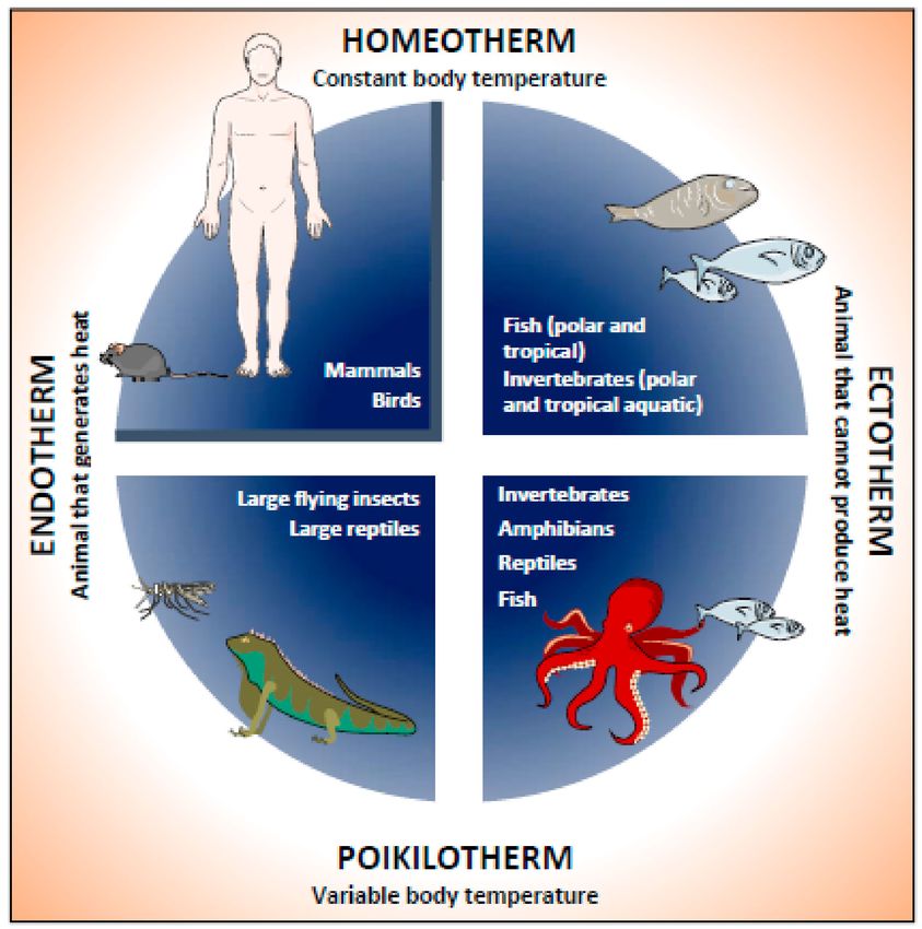
Cells 2020, 9, 316                                                                                       2 of 20

there is a considerable difference between the physiology of mice and humans that could directly bias
the preclinical findings [9].

      Figure 1. Classification of animals according to their body temperature and adaptation to room
      temperature. Animals are classified according to their way of acquiring body heat and their ability
      to adapt to room temperature. Endothermic animals can produce heat endogenously. Ectothermal
      animals cannot produce their own heat, so they rely on ambient temperature. In addition, animals can
      be classified into homeotherms that can keep their body temperature constant, or poikilotherms that
      have a variable body temperature. Humans and mice are mammals that are classified as homeothermal
      endotherms. However, when subjected to an ambient temperature well below their thermoneutrality,
      they experience physiological responses that trigger adaptive thermogenesis.

     Like other small mammals, the mouse has a large surface area and a small body mass. This makes
mice vulnerable to fluctuations in the ambient temperature (Ta ), especially when it falls below their
thermoneutral temperature (29–31 ◦ C) [9–13]. Mammals try to maintain their core temperature through
the adaptive capacity of thermoregulation. Thus, the mouse uses various adaptations to keep thermal
homeostasis in colder environments. For instance, the function of brown adipose tissue (BAT) is to
maintain body temperature through a process called thermogenesis or heat production. Currently,
most metabolic studies involving rodents are carried out at 21 ◦ C, which is a thermoneutral zone in
adult humans but is below the thermoneutral zone in mice. As a consequence, research studies in
mice that are housed at 21 ◦ C may not directly apply to humans, who live mainly in their comfort
zone or neutrality [6,7,10]. For this reason, it is necessary to understand how Ta affects metabolic and
cardiovascular phenotypes in mice, and the importance of this variable in the modelling of human
diseases in rodents.
     As animal models and measurement techniques become increasingly accurate and sophisticated,
environmental variables become critical for research development. A stable, defined environment
is essential to generate consistent experimental results that support both replication and valid
interpretations of the data. Previous studies have shown how mice adaptation to Ta alters their
disease phenotype [14–16]. Consequently, Ta might be a variable to consider in metabolic studies to
guarantee valid interpretation of experimental results, consistent conclusions and greater certainty in
the translation of preclinical experiments to clinical studies.
     The prevention and treatment of obesity has become a health priority. There is an alarming
increase in the prevalence of obesity and associated metabolic diseases, including type 2 diabetes
mellitus (T2D) and cardiovascular disease [17]. The health and economic impact of monitoring and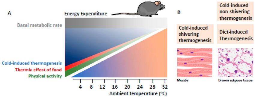
Cells 2020, 9, 316                                                                                  3 of 20

managing obesity and associated complications is also remarkable. Lifestyle changes, such as dietary
interventions and/or increased physical activity, have been widely recommended to prevent and treat
obesity. However, it is essential to determine why, in general, many obese individuals are exceptionally
resistant to treatment and voluntary weight loss is so difficult to achieve and sustain over time. Thus,
a better understanding of energy homeostasis is essential.
     In this review, we aim to cover important aspects related to the adaptation of animals to Ta ,
the influence of Ta on the development of metabolic phenotypes in experimental mice, and their
translation into human physiology.

2. Classification of Animals According to Body Temperature and Their Adaptation to
Ambient Temperature
      All living beings are sensitive to a minimum, optimum and maximum temperature. Due to
environmental adaptations, organisms are conditioned to their habitat in different climatic zones.
Accordingly, they are classified into eurytherms (tolerant to a wide variation of external temperatures)
and stenotherms (tolerant to a narrow range of ambient temperatures) [18] (Figure 1).
      The temperature of an animal is the amount of heat per unit of tissue mass and is a balance
between heat production and exchange, a key determinant in reproduction and development [19].
Body temperature (Tb ) is defined as the reflection of the thermal energy that is retained in the body’s
molecules. Based on the stability of Tb , animal species can be classified as either poikilotherms or
homeotherms [19] (Figure 1). Poikilotherms are animals with a variable Tb , i.e., their temperature
changes in response to environmental conditions. In contrast, homeotherms are animals that maintain
a relatively stable Tb . Most homeotherms manage to maintain a constant Tb through physiological
processes that regulate production rates and heat loss. The difference between poikilotherms and
homeotherms depends on the animal’s physiology and the nature of the environment. An animal can
maintain a constant Tb if it inhabits an environment with a constant Ta . Thermoregulation mechanisms
are understood as the physiological strategy that an animal uses to control temperature within the
desired range [20]. According to these thermoregulation mechanisms, animals are also described as
ectotherms and endotherms (Figure 1).
      In addition, animals can control their Tb through their behavior. Behavioral thermoregulation can
be used to control the body temperature of a poikilotherm or to reduce the cost of thermoregulation in
a homeotherm [20]. In ectotherms, the environment and behavioral thermoregulation determine the
Tb . In contrast, endotherms are vertebrates that generate internal heat to maintain a given Tb .
      Most mammals and birds (as illustrated in Figure 1) are classified as homeotherms because Tb is
stable, and endotherms since they thermoregulate Tb through metabolic heat and the thermal insulation
capacity of the animal.

3. The Use of Experimental Mice as a Model in Human Research

3.1. Relationship Between Body Size and Physiological Temperature
      Since the 1990s, genetic mouse models have been used to study obesity and energy balance.
The cloning and characterization of mutant genes associated with obesity led to the discovery of proteins
such as leptin [21], its receptor [22] and melanocyte-stimulating hormone [23], among others [24–26],
that cause monogenic obesity in mice and humans [27]. Together, these studies validated the use of the
mouse in the modelling of biological diseases related to energy homeostasis. Nonetheless, in energy
homeostasis studies, the thermal physiology of the experimental model of choice must be considered [8].
Mice and humans are both endothermic mammals with the ability to thermoregulate to maintain
a constant Tb . Yet the process of thermoregulation has important physiological differences between the
two species that we should bear in mind as researchers. For instance, the size of an animal influences
its thermal biology through its surface/volume ratio. The larger the individual is, the smaller the ratio.
Body surface area is proportional to the power of 2/3 to mass and it is an important determinant in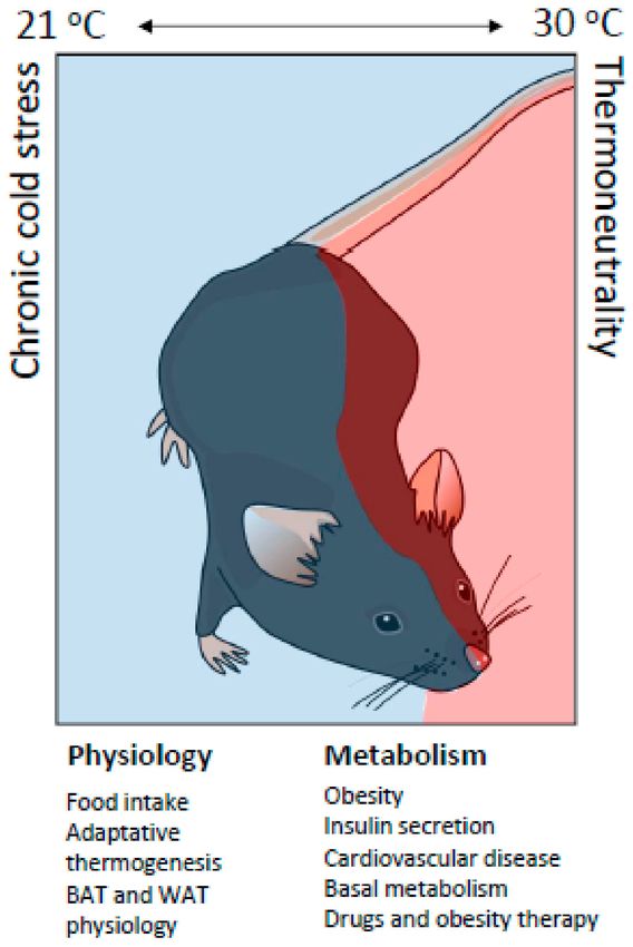
Cells 2020, 9, 316                                                                                          4 of 20

heat loss [28,29]. Adult humans are approximately 3000 times heavier than mice (75 kg vs. 25 g).
As thermal biology depends on body size, it is important to consider this significant difference in
inter-species dimensions. Homeothermic endotherms, such as mice and humans, must dissipate the
excess heat produced by their metabolism across the body surface. Humans have a larger body size
with a lower relative surface area, which leads to less heat loss. In contrast, mice have a smaller body
size for a greater relative surface area, and thus greater heat loss.
      Mice and humans have a similar internal Tb average of 37.0 ◦ C in humans and 36.6 ◦ C in
mice [30], which is within the characteristic range in mammals. Humans generate heat primarily as
a by-product of metabolism, without as much need for additional heat generation mechanisms. In fact,
human physiology is mostly aimed at heat dissipation. In contrast, the small size of the mouse means
that it can transfer heat quickly and have rapid changes in Tb , so mice require more heat generation
capacity to maintain their Tb .
      Figure 2 shows the components of energy expenditure depending on Ta [28–31]. In the
mouse, total energy expenditure is the sum of the basal metabolic rate (BMR), physical activity,
food thermogenesis and cold-induced thermogenesis [32]. At a given Ta , over a third of the total
energy expenditure is cold-induced thermogenesis, which is necessary to maintain Tb . This amount
of cold-induced thermogenesis, also called facultative or adaptive thermogenesis, is reduced by
the availability of nesting material or by keeping mice grouped in cages so they can snuggle.
In contrast, in humans, cold-induced thermogenesis contributes a very small fraction to total energy
expenditure [33].

      Figure 2. Cellular energy used in adaptive thermogenesis. (A) The total energy expenditure of the
      mouse can be divided into four components: basal metabolic rate, physical activity (green), the thermal
      effect of food (red) and cold-induced thermogenesis (blue). At room temperature (20–22 ◦ C) more than
      one third of the total energy expenditure is cold-induced thermogenesis, which is required to maintain
      body temperature. (B) The energy used for cold-induced thermogenesis is mainly produced by skeletal
      muscle shivering and BAT thermogenesis.

3.2. Thermal Physiology and Thermoneutrality Zone
     Mammals use heat conservation and generation mechanisms to maintain thermal homeostasis,
which is reflected in their constant internal temperature [20]. On exposure to a cold environment,
several behavioral mechanisms of heat conservation are activated, such as vasoconstriction, piloerection,
hunched posture (to reduce the surface area) and snuggling. When these conservative heat adaptations
prove insufficient for defense against the cold, mammals increase their energy expenditure to generate
heat by involuntary muscle contractions (shivering thermogenesis) and uncoupled respiration in
brown adipocytes (non-shivering thermogenesis, known as adaptive or facultative thermogenesis).
The opposite occurs when mammals face environmental heat. In this case, there are behavioral
adaptations such as vasodilation and increased passive heat loss, as well as panting, licking and
sweating (in humans) to increase active heat loss through cooling by evaporation.
     Halfway between these metabolic adaptations to a cold environment and heat is the thermoneutral
zone, which is defined as the nadir in BMR [8–10,20]. When the Ta is within the thermoneutral zone,
Cells 2020, 9, 316 5 of 20
BMR generates enough heat to maintain a constant core temperature at 37–38 ◦ C. For young C57BL/6J
mice (~ 3 months), the thermoneutral zone is between 29–31 ◦ C [6,8,10,11], which is similar to the
thermoneutral zone of a naked human (~28 ◦ C) [34–36]. However, the thermoneutral or comfort
zone in dressed humans is around 20–22 ◦ C, which is often the temperature of the animal facilities
where the mice are housed. This colder Ta keeps mice in significant thermal stress or controlled
hypothermia, resulting in the activation of facultative thermogenesis in BAT to maintain thermal
homeostasis. As a consequence, the BMR and food intake of mice housed at a Ta of 20 ◦ C is ~100%
higher than those housed at 30 ◦ C. Both parameters increase by another ~100% when mice are housed
at a Ta of 4–5 ◦ C [37]. As discussed in detail below, the chronic housing of mice under thermal stress
conditions (Ta of 20–22 ◦ C) has profound effects on many physiological phenotypes and their intrinsic
ability to adapt to environmental challenges.
Although the thermoneutral zone is considered a standard range, it is a highly variable parameter
that differs between species. Previous studies showed that the thermoneutral zone of a particular
mammal reflected its adaptations to its natural habitat [9].
In addition to these differences between species, many parameters can affect the range of the
thermoneutral zone and the cold tolerance within a given species. For example, age (newborn and
young mice have higher thermoneutral zones), muscle mass (basal metabolism and heat production
are proportional to muscle mass), locomotor activity (exercise increases the production of heat to
decrease the thermoneutral zone), pregnancy (fetal metabolism increases heat production), lactation
(milk production generates heat), and isolation (greater isolation reduces the increase in metabolic
rate at lower temperatures) can dynamically modulate the thermoneutral zone and the susceptibility
of the organism to a cold environment [9–11]. This variation in the thermoneutral zone explains the
differences observed in the cold tolerance of some mutant animals [11], such as those lacking hair, skin or
subdermal fat [38–42]. This evidence suggests that experimental determination of thermoneutrality is
necessary to understand how genetic mutations in mice affect physiology and disease susceptibility.
3.3. Thermal Variations in the Housing of Experimental Mice
In the animal facility, mice can consume unlimited food to meet the energy requirements of
adaptive thermogenesis. However, it is known that “control” mice (wild-type experimental mice fed
ad-libitum and without physical activity) become sedentary, obese and glucose intolerant and the
implications for data misinterpretation in human studies is known [43]. The researchers stated that lack
of exercise and unlimited access to food are the factors that most influence the inadequate interpretation
of results. Other studies indicate that the underlying role of cold ambient temperatures is the cause of
excessive intake and metabolic disorders [6,7,44]. A clearly defined stable environment is essential to
generate consistent experimental results that support both replication and valid interpretations of the
data. As animal models and measurement techniques become increasingly precise, environmental
influences become critical in experimental development. Current technology can detect subtle effects
that may have been part of the experimental background previously.
There are many varieties of rodent cages (for example, open lid, closed lid, ventilated and
unventilated) that may vary in size, bedding, enrichment devices and other attributes. Even the
position of the cage on a shelf can influence the result of the behavioral tests [7]. Another attribute
that is rarely considered is the color of the cage. A few years ago, it was shown that this fundamental
characteristic of the environment significantly influences circadian metabolic measures in rats [45,46].
The cage dye (transparent, amber, blue or red) causes a significant variation in maximum levels and
maximum durations of melatonin during the dark phase and significant changes in the circadian
moment of insulin spikes [46].
A fundamental characteristic of the rodent cage is the bedding. The properties of different types
of rodent beds can differentially influence the environment of the cage, the physiology and behavior of
rodents and even the health of the animals [47–51].Cells 2020, 9, 316 6 of 20
Ta is another critical feature of the rodent cage environment that is probably influenced by the type
of cage system used. Some studies show how the Ta interacts with the cage system and possibly with
tumor growth [12,52]. For example, one study evaluated the thermogenesis of BAT in nude and SCID
mice that were individually housed at a Ta of 21 ◦ C in ventilated cages with or without shelter or in
a static (non-ventilated) cage. The results showed that, independently of the strain, mice individually
housed in ventilated cages without shelter had significantly higher BAT thermogenesis and higher
adrenal weights than mice housed in static cages or in ventilated cages with shelter. In addition,
when tumor cells were implanted, mice housed in static cages had greater tumor growth than mice
under the other two conditions. The authors concluded that mice housed in ventilated cages without
shelter experienced cold stress, which in turn interfered with tumor growth [12]. Another study
reported that BALB/c and C57BL/6 mice housed 5 per cage at a Ta of 22 ◦ C had higher tumor growth
than those maintained at 30 ◦ C but did not detect a temperature effect on tumor growth when they
used nude mice and SCID with immunodeficiency. The study also determined that the antitumor
immune response was attenuated in immunodeficient mice maintained at 21 ◦ C compared to those
housed at 30 ◦ C [52].
Another variable to consider is the density of mouse housing. For some studies, individual
housing is preferred or necessary, while in other cases, rodents can be accommodated in groups that
vary in number and density depending on the type of cage, the duration of the study, the purpose
of the study and other factors. An important fact to consider is that not all mice housed in the same
cage are necessarily identical, even if they are highly inbred. The differences between cage mates can
be visually obvious in cages with domination hierarchies, which can occur in association with fights
and overt injuries in some cage mates but not in others. Mice housed in groups may show greater
phenotypic variation in some characteristics than mice housed individually from the same inbred
strain [53].
Rodent housing density can directly affect the environmental conditions within the cage
and, therefore, potentially alter the physiology, behavior of animals [54] and stress levels [55,56].
An interesting study evaluated the effect of the number of mice in a cage on the inside environment.
Mice were housed in stable cohorts of one or five per cage, or in a test cage that initially contained five
mice. One mouse was removed per week from the test cage until only one was left. Regardless of
the room temperature (22 ◦ C, 26 ◦ C or 30 ◦ C), cages containing five mice were general approximately
1.5 ◦ C warmer than cages with individual mice, and a population of approximately three mice was
associated with a decrease in temperatures and dew point inside the cage [57]. These findings are
particularly relevant for situations in which individual mice are removed from a cage for some reason
(for example, death, fighting and experimental use) because the remaining mice will experience
different environmental conditions that could influence the experimental results [57].
Social housing can also affect the physiology and behavior of animals [58–60]. For example,
a study that evaluated sleep, temperature and activity compared these measures in mice initially
housed as part of a trio, then individually and finally individually with access to a shelter [60]. The data
showed that the modifications in housing significantly influenced both the sleep and activity of mice.
When housed individually, mice showed less rapid eye movement sleep and more locomotive activity
during the dark phase than when they were housed as part of a trio. When given a shelter, the same
mouse spent more time in slow wave sleep and was less active during the dark phase.
Thus, researchers should keep in mind that eliminating mice during an experiment could affect
metabolism, as well as many of the other temperature-sensitive biological and physiological responses
that have been analyzed so far. This probably contributes to experimental variability between
experiments and laboratories.
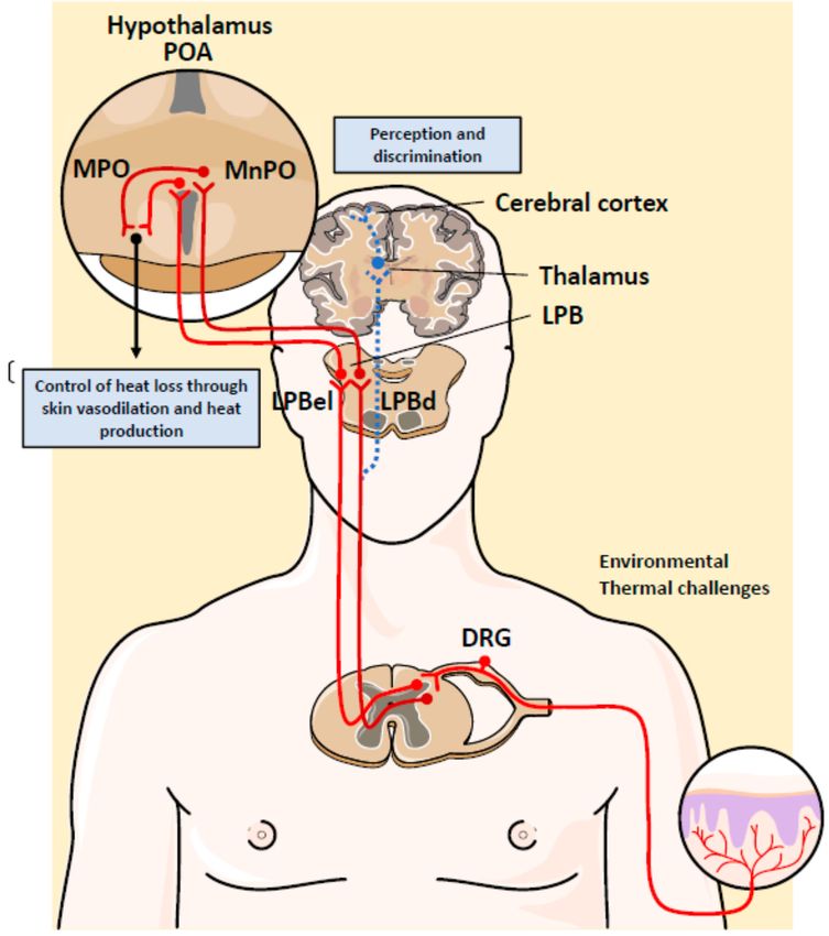
4. Neuronal Control of Body Temperature
Homeostatic control of the Tb is essential for the survival of mammals. It is well-established that
Tb is regulated partly by specific neuronal populations located in the hypothalamus [61]. This part ofCells 2020, 9, 316 7 of 20
the brain works as a thermostat to maintain the Tb within a narrow range [62]. The most important
regions of the hypothalamus involved in Tb regulation are the preoptic area (POA) and the posterior
hypothalamic area (Figure 3) [63]. They contain temperature-sensitive neurons that initiate neuronal
responses for heat generation or heat dissipation. This means that the brain itself is an input to regulate
homeostatic responses. These conclusions are based on results obtained from electrophysiological
recordings of the POA and revealed that local or environmental heat activate a subset of neurons
referred to as “warm-sensitive” [8,64,65].
Figure 3. Hypothalamic thermoregulatory network that controls body temperature in mammals.
The hypothalamic neurons of the preoptic area (POA) and the posterior hypothalamic area function as
a body thermostat to maintain a stable body temperature. The ascending temperature information ends
in two anatomically distinct areas of the lateral parabrachial nucleus (LPB): the lateral and dorsal external
LPB (LPBel and LPBd, respectively). Heat and cold have been shown to activate cFOS expression in
LPBd and LPBel, respectively. Altogether, this sophisticated thermoregulatory network highlights the
importance of maintaining body temperature during an environmental temperature challenge. DRG,
dorsal root ganglia; MPO, medial preoptic subnucleus; MnPO, median preoptic subnucleus.
In addition to sensing local brain temperature, POA neurons receive thermal information from
the periphery. It has been reported that three tissues provide an important input: the skin, spinal
cord, and abdominal viscera [66]. The thermosensitivity of these tissues is due to sensory neurons
that measure the temperature. Most of the neurons have cell bodies located in dorsal root ganglia
(DRG) and their axons extend out to the target tissues [67]. Considerable progress has been made to
elucidate the molecular basis of peripheral cold and warmth sensing. These studies have led to the
identification of a number of ion channels activated by a wide spectrum of physical and chemical stimuli.
Those activated by temperature belong to a superfamily of ion channels called transient receptor
potential (TRP) channels [68,69]. Four TRP subtypes are activated by an increase in temperature and
two TRP channels are activated by decreases in temperature [68]. For example, TRPM8 is an ion
channel that admits Ca2+ and Na+ in response to moderate cold (10–25 ◦ C), while several transient
receptor potential cation channels (TRV) have been proposed to sense warmth including TRVP1,
TRVP3, TRCP4 and TRVP2 [70]. The mechanism by which temperature modulates TRP channels
remains to be elucidated.Cells 2020, 9, 316 8 of 20
Temperature information is sensed by these thermoreceptors in DRG neurons and is then
transmitted to the dorsal horn of the spinal cord, where it is further processed before being sent
to the brain (Figure 3). Elegant experiments have been carried out to elucidate the role of these
thermosensitive neurons. Transgenic mice lacking TRVP1 in temperature-sensitive DRG neurons have
reduced spinal neuron responses to heat [71]. Similarly, ablation of TRPM8+ DRG neurons reduced
the number of spinal neurons activated by mild cold, but not by lower temperatures [72]. These results
support the idea that spinal neurons synthesize information from many types of DRG neurons.
Dorsal horn neurons send glutamatergic projection to the brain that synapse with the lateral
parabrachial nucleus (LPB) and the thalamus. Thermal information received in the thalamus is relayed
upward to the somatosensory cortex and other cortex areas, where it mediates the discrimination
of temperature (spinothalamocortical pathway) [73]. The ablation of this thermosensory pathway
does not affect the autonomic response of Tb regulation. However, injuring or silencing of the
LPB abolishes autonomic responses to skin cooling and warming and the temperature preference
in behavioral assays [74]. This result suggests that the spinothalamocortical pathway does not
play a role in the thermal afferent pathway that evokes involuntary thermoregulatory responses to
environmental challenges.
Ascending temperature information terminates in two anatomically distinct areas of LPB: the
external lateral and dorsal LPB (LPBel and LPBd). It has been demonstrated that warm and cold activate
cFOS expression in LPBd and LPBel, respectively [75]. LPB neurons send glutamatergic projections to
the midline POA, where GABAergic and glutamatergic interneurons in the median preoptic (MnPO)
subnucleus are activated [76]. LPBel neurons activate GABAergic MnPO interneurons that inhibit the
distinct population of warm-sensitive neurons in the medial preoptic (MPO) subnucleus that control
cutaneous vasoconstriction, BAT and shivering. Thus, inhibition of neurons in the MPO increases core
body temperature, shivering, metabolism and heart rate. In contrast, glutamatergic interneurons in the
MnPO, which may be excited by glutamatergic inputs from warm-activated neurons in LPDd, excite
warm sensitive neurons in MPO [61,77]. Altogether, this thermoregulatory network is a sophisticated
reflex that is necessary to maintain Tb during an environmental temperature challenge (Figure 3).
5. Adaptive Thermogenesis in Brown Adipose Tissue
Small mammals have a tissue dedicated to heat generation, the BAT [78]. For a long time, it was
known that BAT was present in small mammals such as rodents and neonatal humans. However,
in the last decade it was discovered that active BAT is also found in adult humans [79].
In rodents, BAT is located mainly in the interscapular zone. In adult humans, it is found in the
supraclavicular region, and in the cervical, axillary, paravertebral and perirenal areas [80]. BAT is
called “classic” to distinguish it from inducible or beige adipose tissue, which has unique molecular
and developmental characteristics [81]. Beige adipocytes have the appearance of white adipose
tissue (WAT) until the animal needs to generate more heat. After exposure to cold or other stimuli,
this beige adipose tissue or inducible BAT is enriched in cells with the appearance and functional
characteristics of classic BAT in a process called browning. Although beige and BAT adipose tissue have
different developmental origins and gene expression profiles [82], both are thermogenic. Thermogenic
adipocytes can increase energy expenditure and generate heat by uncoupling the oxidative metabolism
from ATP production. This function is carried out by the uncoupling protein (UCP)1, a proton
transporter located in the internal mitochondrial membrane that uncouples energy generation from
fuel oxidation from ATP production to produce heat. Thus, the electrochemical gradient generated
through the electron transport chain (ETC) is dissipated [83–85]. In brown adipocytes, the high
content of mitochondria and their vascular and nervous supply facilitates thermogenesis activated
by the sympathetic nervous system. The nerve terminals act on α-adrenergic receptors to promote
thermogenesis in BAT. It has been shown that cold enhances sympathetic signaling and that chronic
exposure to cold triggers the expansion and activation of BAT [86], resulting in adaptive thermogenesis.Cells 2020, 9, 316 9 of 20
Adaptive thermogenesis is a mechanism of metabolic heat production that involves stimulation
of the sympathetic nervous system to release norepinephrine (NE) and epinephrine, resulting in the
increased metabolic activity necessary for heat generation in BAT [10,79]. Previous studies have shown
that heat production by adaptive thermogenesis in mice can triple that of basal metabolism, and it is
what increases the most in other animal models [87,88].
Obesity is an important risk factor for type 2 diabetes and cardiovascular disease. Importantly,
BAT has been shown to promote HDL turnover and reverse cholesterol transport [89]. The high
metabolic activity of thermogenic adipocytes confers atheroprotective properties through increased
systemic cholesterol flow through the HDL compartment.
The thermogenic function of BAT requires an adaptive increase in proteasomal activity to ensure
the quality control of cellular proteins. It has been shown that ER-localized transcription factor nuclear
factor erythroid-2, like-1 (Nfe2l1 protein, also known as Nrf1) is an important mediator of brown
adipocyte function, providing a greater proteometabolic quality control to adapt to cold or obesity [90].
It has been described that obesity might affect BAT’s proteasomal activity [90]. A recent epigenomic
study associated an altered methylation pattern of the human NFE2L1 locus with BMI [91]. However,
the molecular mechanism implicated in how this epigenetic variant could affect Nrf1 and proteasome
activity is still unknown.
6. Therapeutic Efficacy of Adaptive Thermogenesis in Obesity
In recent decades, BAT has been extensively investigated for its potential therapeutic role in obesity
and T2D. Previous studies showed that excessive caloric intake could stimulate the expansion of BAT
and the increase in thermogenesis as an adaptive measure to maintain body weight. This mechanism
of diet-induced thermogenesis is mediated by BAT and UCP1 [92]. In fact, in the absence of UCP1,
mice are prone to obesity. Initial studies, where brown adipocytes were genetically ablated with
a toxin driven by the UCP1 promoter [93], demonstrated for the first time the protective effect of BAT
against obesity and T2D. Importantly, these protective effects were observed in mice raised at room
temperature (thermal stress with a Ta of 20–22 ◦ C). Subsequent investigations under Ta conditions
showed that Ucp1−/− mice were very susceptible to hypothermia, due to recurrent tremors, but did not
demonstrate the role of UCP1 in thermogenesis, nor a propensity to develop obesity [94,95]. However,
when mice remained at thermoneutrality, they showed greater metabolic efficiency, which resulted
in an increase in adiposity and obesity development [96]. This is explained because UCP1 knockout
mice are more susceptible to hypothermia, which directly affects most of the systemic effects of energy
metabolism [93].
The preferable fuel source in BAT is lipids, but glucose is also used. Therefore, approaches to
activate BAT and reduce glucose and lipid content through adaptive thermogenesis could be potential
therapies to fight against obesity [83]. The best way to model human energy physiology with mice
is under thermoneutral (30 ◦ C) conditions. Under this situation, cold-induced thermogenesis would
be minimal and will not influence total energy expenditure [97]. Other potential variables that may
account in similar proportions for total energy expenditure compared with a sedentary human would be
BMR (70%), food thermogenesis (10%) and energy expenditure by physical activity (20%) [7]. Because
energy expenditure decreases by approximately 50% in mice at thermoneutrality [98], this implies
that the metabolic phenotype in obesity including adiposity would be highly dependent on the Ta .
An example of this implication is shown in a study about thyroid hormone metabolism [99]. In humans,
hyperthyroidism is associated with a hypermetabolic state, characterized by heat intolerance and fat
loss, while hypothyroidism decreases energy expenditure and promotes cold intolerance and obesity.
Interestingly, the authors have shown that unlike in humans with hypothyroidism, mice that lack
type 2 deiodinase, a key enzyme in the conversion of thyroid hormone, did not develop metabolic
dysfunction when housed at 22 ◦ C. However, when these animals were maintained at thermoneutrality
(30 ◦ C), there was an increase in adiposity, hepatic steatosis and glucose intolerance [99]. Therefore,
they concluded that the accommodation of mice at a Ta resulted in increased adrenergic activity in BAT,Cells 2020, 9, 316 10 of 20
which compensated for the loss of activity of deiodinase type 2 and the production of T3. These findings
suggest that chronic housing of mice under conditions of thermal stress can mask the genetic functions
involved in energy balance and metabolic homeostasis triggering a change in the metabolic phenotype.
6.1. Activating BAT to Treat Obesity
Obesity is a chronic metabolic disorder characterized by ectopic fat deposition and a state of
chronic low-grade inflammation. It is associated with higher free fatty acids, glucose and insulin
levels. Adipose tissues, both WAT and BAT, are highly affected during obesity. These alterations
include adipocyte hyperplasia and hypertrophy [100], endoplasmic reticulum stress [101], oxidative
stress [102], fibrosis [100] and mitochondrial dysfunction [103], among others. Obesity is associated
with severe disorders such as cardiovascular disease, dyslipidemia, T2D or even some forms of cancer.
BAT was initially recognized for its ability to protect animals from hypothermia [104]. However in
the last decade, the discovery that BAT is active in adult humans and that it is reduced in several
conditions such as obesity, T2D and aging has triggered leading research in the BAT field to improve
lipid and glucose homeostasis in the fight against obesity [105–109].
Overfeeding activates BAT’s diet-induced thermogenesis [92]. Thus, several studies have
focused on natural or chemical drugs to enhance thermogenesis such as ginger [110], tea seed
oil [111], berberine [112], butein [113], capsaicin [114], flavonoids such as quercetin [115], calcium
supplement [116] or fluvastatin sodium [117], among others.
Exercise is another inductor of BAT thermogenesis [118]. Interestingly, a positive correlation has
been demonstrated between exercise and increased browning in subcutaneous WAT [119]. The lactate
produced in muscles after exercise or after cold exposure leads to an increase in UCP1 levels in the
adipose tissue [120]. Recently, Takahashi et al. demonstrated that subcutaneous WAT-derived TGF-β2
secreted after exercise or its administration improved glucose homeostasis and insulin sensitivity by
increasing fatty acid oxidation [121]. Furthermore, some myokines related to BAT activation, such as
irisin [122] or β-aminoisobutyric acid, have been shown to decrease weight gain and improve glucose
tolerance in mice [123].
As mentioned above, BAT is innervated by the sympathetic nervous system and controlled
by adrenergic inductors such as norepinephrine. Thus, huge efforts have been made to find novel
β-adrenergic agonists that can potentiate BAT activity and enhance thermogenesis. Some examples are
Cl-316,243 [124] or mirabegron [125]. The latter was initially clinically used for overactive bladder but
was also found to activate BAT in rats and humans [126]. Many other studies have focused on increasing
the BAT mass, i.e., the differentiation of brown adipocytes or WAT browning. One study centered on
fibroblast growth factor-21 (FGF21) [127], which is mainly secreted by the liver and associated with
BAT activity. In humans higher FGF21 levels have been found in serum after cold exposure [128].
Another important thermogenic coactivator is PGC1-α, which is involved in mitochondrial biogenesis
and thermogenesis [129]. Its activity is related to an increase in other transcriptional factors involved
in brown differentiation such as PRDM16 or the PPARs family [130]. Other factors have also been
studied to enhance thermogenesis in obesity such as the bone morphogenetic proteins (BMPs) family,
for example BMP7 [131] or BMP8b [132]. Interestingly, although BMP4 improves the obese phenotype,
it has a tissue-dependent dual effect: it increases browning in subcutaneous WAT; and it increases
the number of lipid droplets and decreases BAT UCP1 expression [133]. Another approach to
activate thermogenesis has been based on PPAR-γ agonists. Some studies showed increased UCP1
levels after treatment with rosiglitazone [134,135]. Other secreted peptides or hormones have been
reported to activate BAT: norepinephrine [136], natriuretic peptides [137], meteorin-like [138], bile acids,
adenosine [139] or activin E [140,141].
In recent years, BAT-derived adipokines, commonly called batokines, have generated considerable
interest among the scientific community for their anti-obesity potential [142]. In 2018, Deshmukh et al.
found a batokine called EPDR1 involved in BAT activation [143]. Another recent and elegant study hasCells 2020, 9, 316 11 of 20
discovered a new chemokine called CXCL14 that is secreted by BAT and induces browning of WAT via
immune cell activation [144].
Finally, alternative therapies are being studied to increase BAT mass and thermogenesis.
These include the direct transplantation of this tissue or differentiated beige cells from preadipocytes,
mesenchymal stem cells (MSCs) or induced pluripotent stem cells (iPSC) [145]. In the future,
more personalized therapies could focus on the intrinsic study of the genome to identify other BAT
activators such as miRNAs to combat obesity [146].
6.2. Role of Thermoneutrality in Obesity and Metabolic Studies: Chronic Cold vs. Thermoneutrality
It is known that an increase in metabolic heat production has physiological effects. In fact, for every
1 ◦ C that the Ta drops, approximately 46.3 kcal/m2 /24 h are required to maintain the core temperature of
the mouse [13]. This increase in metabolic heat production has many physiological effects. For example,
energy expenditure is approximately 50% lower in mice living at thermoneutrality than in mice living
in chronic cold conditions [98]. Therefore, we can deduce that the metabolic phenotypes of obesity and
adiposity are highly dependent on the Ta at which mice are housed (Figure 4).
Figure 4. Metabolic and physiological differences in mice under chronic cold or at thermoneutrality.
Mouse models used to study metabolic diseases are influenced by environmental temperature.
Mice show differences in the metabolic phenotype when housed at a standard temperature (21 ◦ C) vs.
thermoneutrality (30 ◦ C). Mice at standard temperatures are subjected to chronic cold. This triggers
controlled hypothermia where energy expenditure is affected by changes in physiology (food intake,
BAT and WAT physiology and an increase in adaptive thermogenesis) and in metabolism (basal
metabolism, adaptive thermogenesis, diet efficiency, insulin secretion, adipose tissue physiology,
inflammation at adipocyte and vascular levels, and the effect of drugs and therapies against obesity).
Some studies have shown that the lack of efficient induction of the obese phenotype by high fat
diet (HFD) is due to the fact that mice were housed in conditions below their thermoneutrality [57–60].
This effect of low temperatures on obesity has been observed in other animal models [14,78,81],
demonstrating the correlation between low temperature and low effectiveness of the diet, as well
as low levels of insulin and glucose and an altered response in glucose tolerance tests and energy
homeostasis [10,44].Cells 2020, 9, 316 12 of 20
Other researchers found differences in metabolic inflammation [84]. Mice housed at
thermoneutrality vs. chronic cold had greater metabolic inflammation. This was correlated with higher
inflammation in the WAT and at vascular level, promoting atherosclerosis. The authors thus claim that
thermal stress could limit our ability to faithfully model human diseases in mice [84].
The exposure to chronic cold vs. thermoneutrality has also been studied at the pharmacological
level in obesity. The chemical protonophore, 2,4-dinitrophenol (DNP) was used for weight loss in
humans. However, its consumption was discontinued due to toxicity [45,46]. DNP generates heat by
uncoupling mitochondria. Thus, its anti-obesity mechanism is at least due to an increase in energy
expenditure, without a direct effect on food intake. At 22 ◦ C, chronic treatment in mice with a low
dose of DNP had no effect on the phenotype and the only change observed was that BAT became less
active [47]. This indicates that adaptive cold-induced thermogenesis was potentially reduced by the
amount of heat that was generated by DNP-mediated uncoupling [147]. When the same experiment
was performed at thermoneutrality, the same dose of DNP increased energy expenditure, reduced
body weight, reduced adiposity and improved glucose tolerance [47]. Although DNPs have been
discontinued for the treatment of obesity [46], they could serve as a model for the effects of systemic
uncoupling agents, which demonstrate better efficacy at thermoneutrality than at colder temperatures.
Another comparison of the pharmacological effect at chronic cold vs. thermoneutrality is
the β3 adrenergic agonist, Cl316,243. The main effects of β3 agonists are direct stimulation of
BAT thermogenesis and WAT lipolysis [52]. These experiments were performed at 22 ◦ C, and the
pharmacological treatment showed no effect on body weight or adiposity. However, there was greater
energy expenditure compensated with the increase in food intake [52]. At thermoneutrality, Cl316,243
increased energy expenditure, but it also reduced body weight and adiposity.
Altogether, it is clear that the Ta used in experiments with mice is critical and might directly
influence the efficiency and clinical translatability of pharmacological, metabolic and energy
homeostasis studies.
7. Conclusions and Future Perspectives
The mouse is the predominant model for studying human diseases. However, many studies
fail to deliver mechanistic information about human physiology. This failure comes from translating
preclinical studies in mice to therapy in humans. Although we are aware that mice and humans
are two different species, we do not always consider all the external variables of the environment,
which could influence the physiological adaptation of the study model and hinder the reproducibility
of preclinical investigations.
The thermoregulatory network triggered by the hypothalamus is a necessary reflex to maintain
Tb during variations in Ta . Therefore, given the influence that Ta exerts on the physiological and
pathophysiological responses of the mouse, this study variable should be considered for more correct,
efficient translation into human therapy.
The epidemic of obesity and metabolic diseases is increasing exponentially, and current therapies
remain inefficient. BAT has become an attractive potential target to treat obesity due to its thermogenic
capacity. However, environmental variables such as temperature could directly influence both BAT
activity and the dynamics of energy expenditure in the model under study.
For future metabolic studies, it would be important to consider all the variables that may influence
the experimental outcome regarding obesity and insulin resistance. For example, an important point is
the selective breeding of mouse animal models. Relevant specific differences in metabolic activity have
been found in strain-dependent genetic conditions [148–150]. However, the correlation between strain
variables and thermo-neutrality in studies of obesity and associated diseases is still unknown.
Currently, most metabolic studies are carried out at a room temperature of 21 ◦ C, which is
considered a thermoneutral zone for adult humans. However, mice subjected to the same temperatures
experience chronic cold. The cold triggers-controlled hypothermia in which energy expenditure is
affected by the increase in adaptive thermogenesis, by the activation of BAT and tremors due toCells 2020, 9, 316 13 of 20
involuntary muscle contractions (Figure 4). Further studies in mice at thermoneutrality will deepen
our understanding of physiological mechanisms, which could increase the success of translation into
human treatments for obesity and metabolic diseases.
Author Contributions: All authors contributed equally to this work. All authors have read and agreed to the
published version of the manuscript.
Funding: This study was supported by the Ministry of Spain (MINECO) (SAF2017-83813-C3-1-R to DS and LH,
cofunded by the ERDF), the Centro de Investigación Biomédica en Red de Fisiopatología de la Obesidad y la
Nutrición (CIBEROBN) (Grant CB06/03/0001 to DS), the Government of Catalonia (2017SGR278 to DS), and the
Fundació La Marató de TV3 (201627-30 to DS). MB-P is a recipient of a CONICYT doctoral fellowship.
Conflicts of Interest: The authors declare no conflict of interest.
References
1. Lovegrove, B.G. The evolution of endothermy in Cenozoic mammals: A plesiomorphic-apomorphic
continuum. Biol. Rev. 2011, 87, 128–162. [CrossRef] [PubMed]
2. Bennett, A.; Ruben, J. Endothermy and activity in vertebrates. Science 1979, 206, 649–654. [CrossRef]
[PubMed]
3. Pörtner, H.O. Climate Variability and the Energetic Pathways of Evolution: The Origin of Endothermy in
Mammals and Birds. Physiol. Biochem. Zoöl. 2004, 7, 959–981.
4. Wu, C.-W.; Storey, K.B. Life in the cold: Links between mammalian hibernation and longevity. Biomol.
Concepts 2016, 7, 41–52. [CrossRef] [PubMed]
5. Bouma, H.R.; Verhaag, E.M.; Otis, J.P.; Heldmaier, G.; Swoap, S.J.; Strijkstra, A.M.; Henning, R.H.; Carey, H.V.
Induction of torpor: Mimicking natural metabolic suppression for biomedical applications. J. Cell. Physiol.
2012, 227, 1285–1290. [CrossRef] [PubMed]
6. Maloney, S.K.; Fuller, A.; Mitchell, D.; Gordon, C.; Overton, J.M. Translating Animal Model Research: Does It
Matter That Our Rodents Are Cold? Physiology 2014, 29, 413–420. [CrossRef] [PubMed]
7. Karp, C.L. Unstressing intemperate models: How cold stress undermines mouse modeling. J. Exp. Med.
2012, 209, 1069–1074. [CrossRef]
8. Gordon, C. Thermal physiology of laboratory mice: Defining thermoneutrality. J. Therm. Biol. 2012, 37,
654–685. [CrossRef]
9. Gordon, C.J. Temperature Regulation in Laboratory Rodents; Cambridge University Press (CUP): Cambridge,
UK, 1993.
10. Cannon, B.; Nedergaard, J. Nonshivering thermogenesis and its adequate measurement in metabolic studies.
J. Exp. Biol. 2011, 214, 242–253. [CrossRef]
11. Nedergaard, J.; Cannon, B. The Browning of White Adipose Tissue: Some Burning Issues. Cell Metab. 2014,
20, 396–407. [CrossRef]
12. David, J.M.; Chatziioannou, A.F.; Taschereau, R.; Wang, H.; Stout, D.B. The Hidden Cost of Housing Practices:
Using Noninvasive Imaging to Quantify the Metabolic Demands of Chronic Cold Stress of Laboratory Mice.
Comp. Med. 2013, 63, 386–391.
13. David, J.M.; Knowles, S.; Lamkin, D.M.; Stout, D.B. Individually Ventilated Cages Impose Cold Stress on
Laboratory Mice: A Source of Systemic Experimental Variability. J. Am. Assoc. Lab. Anim. Sci. 2013, 52,
738–744. [PubMed]
14. Gouveia, K.; Hurst, J.L. Reducing Mouse Anxiety during Handling: Effect of Experience with Handling
Tunnels. PLoS ONE 2013, 8, 1–8. [CrossRef]
15. Greenberg, G.D.; Howerton, C.L.; Trainor, B.C. Fighting in the home cage: Agonistic encounters and effects
on neurobiological markers within the social decision-making network of house mice (Mus musculus).
Neurosci. Lett. 2014, 566, 151–155. [CrossRef] [PubMed]
16. Haemisch, A.; Gärtner, K. Effects of cage enrichment on territorial aggression and stress physiology in male
laboratory mice. Acta Physiol. Scand. Suppl. 1997, 640, 73–76. [PubMed]
17. Tabarés Seisdedos, R. Health effects of overweight and obesity in 195 countries over 25 years. New Engl. J.
Med. 2017, 377, 13–27.
18. Pörtner, H.O.; Storch, D.; Heilmayer, O. Constraints and trade-offs in climate-dependent adaptation: Energy
budgets and growth in a latitudinal cline. Sci. Mar. 2005, 69, 271–285. [CrossRef]Cells 2020, 9, 316 14 of 20
19. Flouris, A.D.; Piantoni, C. Links between thermoregulation and aging in endotherms and ectotherms.
Temperature 2015, 2, 73–85. [CrossRef]
20. Terrien, J. Behavioral thermoregulation in mammals: A review. Front. Biosci. 2011, 16, 1428. [CrossRef]
21. Zhang, Y.; Proença, R.; Maffei, M.; Barone, M.; Leopold, L.; Friedman, J.M. Positional cloning of the mouse
obese gene and its human homologue. Nature 1994, 372, 425–432. [CrossRef]
22. Tartaglia, L.A.; Dembski, M.; Weng, X.; Deng, N.; Culpepper, J.; Devos, R.; Richards, G.J.; Campfield, L.;
Clark, F.T.; Deeds, J.; et al. Identification and expression cloning of a leptin receptor, OB-R. Cell 1995, 83,
1263–1271. [CrossRef]
23. Shutter, J.R.; Kinsey, A.C.; Scully, S.; Stark, K.L.; Graham, M.; Lüthy, R. Hypothalamic expression of ART,
a novel gene related to agouti, is up-regulated in obese and diabetic mutant mice. Genes Dev. 1997, 11,
593–602. [CrossRef]
24. Ollmann, M.M.; Wilson, B.D.; Yang, Y.-K.; Kerns, J.A.; Chen, Y.; Gantz, I.; Barsh, G.S. Antagonism of central
melanocortin receptors in vitro and in vivo by agouti-related protein. Science 1997, 278, 135–138. [CrossRef]
[PubMed]
25. Fan, W.; Boston, B.A.; Kesterson, R.A.; Hruby, V.J.; Cone, R.D. Role of melanocortinergic neurons in feeding
and the agouti obesity syndrome. Nature 1997, 385, 165–168. [CrossRef] [PubMed]
26. Huszar, D.; Lynch, C.A.; Fairchild-Huntress, V.; Dunmore, J.H.; Fang, Q.; Berkemeier, L.R.; Gu, W.;
Kesterson, R.A.; Boston, B.A.; Cone, R.D.; et al. Targeted Disruption of the Melanocortin-4 Receptor Results
in Obesity in Mice. Cell 1997, 88, 131–141. [CrossRef]
27. Farooqi, I.S.; O’Rahilly, S. Mutations in ligands and receptors of the leptin–melanocortin pathway that lead
to obesity. Nat. Clin. Pr. Endocrinol. Metab. 2008, 4, 569–577. [CrossRef]
28. Kleiber, M. The Fire of Life. An Introduction to Animal Energetics; John Wiley & Sons, Inc.: London, UK, 1961;
p. 454.
29. Schmidt-Nielsen, K.; Knut, S.N. Scaling: Why Is Animal Size so Important? Cambridge University Press:
Cambridge, UK, 1984.
30. Refinetti, R. The circadian rhythm of body temperature. Front. Biosci. 2010, 15, 564–594. [CrossRef]
31. Scholander, P.F.; Hock, R.; Walters, V.; Johnson, F.; Irving, L. Heat Regulation in Some Arctic and Tropical
Mammals and Birds. Biol. Bull. 1950, 99, 237–258. [CrossRef]
32. Abreu-Vieira, G.; Xiao, C.; Gavrilova, O.; Reitman, M.L. Integration of body temperature into the analysis of
energy expenditure in the mouse. Mol. Metab. 2015, 4, 461–470. [CrossRef]
33. Brychta, R.J.; Chen, K.Y. Cold-induced thermogenesis in humans. Eur. J. Clin. Nutr. 2017, 71, 345–352.
[CrossRef]
34. Hill, R.W.; Muhich, T.E.; Humphries, M.M. City-Scale Expansion of Human Thermoregulatory Costs. PLoS
ONE 2013, 8, e76238. [CrossRef] [PubMed]
35. Scholander, P.F.; Hammel, H.T.; Andersen, K.L.; Andersen, K.L.; L yning, Y. Metabolic Acclimation to Cold
in Man. J. Appl. Physiol. 1958, 12, 1–8. [CrossRef] [PubMed]
36. E Wilkerson, J.; Raven, P.B.; Horvath, S.M. Critical temperature of unacclimatized male Caucasians. J. Appl.
Physiol. 1972, 33, 451–455. [CrossRef] [PubMed]
37. Qiu, Y.; Nguyen, K.D.; Odegaard, J.I.; Cui, X.; Tian, X.Y.; Locksley, R.M.; Palmiter, R.D.; Chawla, A. Eosinophils
and type 2 cytokine signaling in macrophages orchestrate development of functional beige fat. Cell 2014, 157,
1292–1308. [CrossRef]
38. Sampath, H.; Flowers, M.T.; Liu, X.; Paton, C.M.; Sullivan, R.; Chu, K.; Zhao, M.; Ntambi, J.M. Skin-specific
Deletion of Stearoyl-CoA Desaturase-1 Alters Skin Lipid Composition and Protects Mice from High Fat
Diet-induced Obesity. J. Biol. Chem. 2009, 284, 19961–19973. [CrossRef]
39. Hirata, M.; Suzuki, M.; Ishii, R.; Satow, R.; Uchida, T.; Kitazumi, T.; Sasaki, T.; Kitamura, T.; Yamaguchi, H.;
Nakamura, Y.; et al. Genetic Defect in Phospholipase Cδ1 Protects Mice From Obesity by Regulating
Thermogenesis and Adipogenesis. Diabetes 2011, 60, 1926–1937. [CrossRef]
40. Nakamura, Y.; Fukami, K.; Yu, H.; Takenaka, K.; Kataoka, Y.; Shirakata, Y.; Nishikawa, S.; Hashimoto, K.;
Yoshida, N.; Takenawa, T. Phospholipase Cδ1 is required for skin stem cell lineage commitment. EMBO J.
2003, 22, 2981–2991. [CrossRef]
41. Narvaez, C.J.; Matthews, D.; Broun, E.; Chan, M.; Welsh, J. Lean phenotype and resistance to diet-induced
obesity in vitamin D receptor knockout mice correlates with induction of uncoupling protein-1 in white
adipose tissue. Endocrinology 2009, 150, 651–661. [CrossRef]Cells 2020, 9, 316 15 of 20
42. Li, Y.C.; Pirro, A.E.; Amling, M.; Delling, G.; Baron, R.; Bronson, R.; DeMay, M.B. Targeted ablation of the
vitamin D receptor: An animal model of vitamin D-dependent rickets type II with alopecia. Proc. Natl. Acad.
Sci. USA 1997, 94, 9831–9835. [CrossRef]
43. Martin, B.; Ji, S.; Maudsley, S.; Mattson, M.P. “Control” laboratory rodents are metabolically morbid: Why it
matters. Proc. Natl. Acad. Sci. USA 2010, 107, 6127–6133. [CrossRef]
44. Overton, J.M. Phenotyping small animals as models for the human metabolic syndrome: Thermoneutrality
matters. Int. J. Obes. 2010, 34, S53–S58. [CrossRef] [PubMed]
45. Dauchy, R.T.; Dauchy, E.M.; Hanifin, J.P.; Gauthreaux, S.L.; Mao, L.; Belancio, V.P.; Ooms, T.G.; Dupepe, L.M.;
Jablonski, M.R.; Warfield, B.; et al. Effects of Spectral Transmittance through Standard Laboratory Cages
on Circadian Metabolism and Physiology in Nude Rats. J. Am. Assoc. Lab. Anim. Sci. 2013, 52, 146–156.
[PubMed]
46. A Wren, M.; Dauchy, R.T.; Hanifin, J.P.; Jablonski, M.R.; Warfield, B.; Brainard, G.C.; E Blask, D.; Hill, S.M.;
Ooms, T.G.; Bohm, R.P. Effect of Different Spectral Transmittances through Tinted Animal Cages on Circadian
Metabolism and Physiology in Sprague–Dawley Rats. J. Am. Assoc. Lab. Anim. Sci. 2014, 53, 44–51.
[PubMed]
47. Horn, M.J.; Hudson, S.V.; A Bostrom, L.; Cooper, D.M. Effects of Cage Density, Sanitation Frequency, and
Bedding Type on Animal Wellbeing and Health and Cage Environment in Mice and Rats. J. Am. Assoc. Lab.
Anim. Sci. 2012, 51, 781–788. [PubMed]
48. Leys, L.J.; McGaraughty, S.; Radek, R.J. Rats Housed on Corncob Bedding Show Less Slow-Wave Sleep. J.
Am. Assoc. Lab. Anim. Sci. 2012, 51, 764–768. [PubMed]
49. Royals, M.A.; Getzy, D.M.; Vandewoude, S. High Fungal Spore Load in Corncob Bedding Associated with
Fungal-Induced Rhinitis in Two Rats. Contemp. Top. Lab. Anim. Sci. 1999, 38, 64–66. [PubMed]
50. Smith, E.; Stockwell, J.D.; Schweitzer, I.; Langley, S.H.; Smith, A.L. Evaluation of cage micro-environment of
mice housed on various types of bedding materials. Contemp. Top. Lab. Anim. Sci. 2004, 43, 12–17.
51. Whiteside, T.E.; Thigpen, J.E.; Kissling, G.E.; Grant, M.G.; Forsythe, D.B. Endotoxin, Coliform, and Dust
Levels in Various Types of Rodent Bedding. J. Am. Assoc. Lab. Anim. Sci. 2010, 49, 184–189.
52. Kokolus, K.M.; Capitano, M.L.; Lee, C.-T.; Eng, J.W.-L.; Waight, J.D.; Hylander, B.L.; Sexton, S.; Hong, C.-C.;
Gordon, C.J.; Abrams, S.I.; et al. Baseline tumor growth and immune control in laboratory mice are
significantly influenced by subthermoneutral housing temperature. Proc. Natl. Acad. Sci. USA 2013, 110,
20176–20181. [CrossRef]
53. Nagy, T.R.; Krzywanski, D.; Li, J.; Meleth, S.; Desmond, R. Effect of Group vs. Single Housing on Phenotypic
Variance in C57BL/6J Mice. Obes. Res. 2002, 10, 412–415. [CrossRef]
54. Nicholson, A.; Malcolm, R.D.; Russ, P.L.; Cough, K.; Touma, C.; Palme, R.; Wiles, M.V. The Response of
C57BL/6J and BALB/cJ Mice to Increased Housing Density. J. Am. Assoc. Lab. Anim. Sci. 2009, 48, 740–753.
[PubMed]
55. Morgan, J.L.; Svenson, K.L.; Lake, J.P.; Zhang, W.; Stearns, T.M.; Marion, M.A.; Peters, L.L.; Paigen, B.;
Donahue, L.R. Effects of Housing Density in Five Inbred Strains of Mice. PLoS ONE 2014, 9, e90012. [CrossRef]
[PubMed]
56. Paigen, B.; Svenson, K.L.; Von Smith, R.; Marion, M.A.; Stearns, T.; Peters, L.L.; Smith, A.L. Physiological
effects of housing density on C57BL/6J mice over a 9-month period. J. Anim. Sci. 2012, 90, 5182–5192.
[CrossRef] [PubMed]
57. A Toth, L.; A Trammell, R.; Ilsley-Woods, M. Interactions Between Housing Density and Ambient Temperature
in the Cage Environment: Effects on Mouse Physiology and Behavior. J. Am. Assoc. Lab. Anim. Sci. 2015, 54,
708–717. [PubMed]
58. Aghajani, M.; Mahdavi, M.R.V.; Najafabadi, M.K.; Ghazanfari, T.; Azimi, A.; Soleymani, S.A.; Dust, S.M. Effects
of Dominant/Subordinate Social Status on Formalin-Induced Pain and Changes in Serum Proinflammatory
Cytokine Concentrations in Mice. PLoS ONE 2013, 8, e80650. [CrossRef] [PubMed]
59. Arndt, S.S.; Laarakker, M.C.; Van Lith, H.A.; Van Der Staay, F.J.; Gieling, E.; Salomons, A.R.; Klooster, J.V.;
Ohl, F. Individual housing of mice — Impact on behaviour and stress responses. Physiol. Behav. 2009, 97,
385–393. [CrossRef]
60. Febinger, H.Y.; George, A.; Priestley, J.; A Toth, L.; Opp, M.R. Effects of Housing Condition and Cage Change
on Characteristics of Sleep in Mice. J. Am. Assoc. Lab. Anim. Sci. 2014, 53, 29–37.You can also read