Hepatitis E and Pregnancy: An Unholy Alliance Unmasked from Kashmir, India
←
→
Page content transcription
If your browser does not render page correctly, please read the page content below
viruses
Review
Hepatitis E and Pregnancy: An Unholy Alliance Unmasked
from Kashmir, India
Mohammad Sultan Khuroo

                                          Digestive Diseases Centre, Dr. Khuroo’s Medical Clinic, Srinagar, Jammu and Kashmir 190010, India;
                                          khuroo@yahoo.com; Tel.: +91-9906591044

                                          Abstract: The adverse relationship between viral hepatitis and pregnancy in developing countries had
                                          been interpreted as a reflection of retrospectively biased hospital-based data collection by the West.
                                          However, the discovery of hepatitis E virus (HEV) as the etiological agent of an epidemic of non-A,
                                          non-B hepatitis in Kashmir, and the documenting of the increased incidence and severity of hepatitis
                                          E in pregnancy via a house-to-house survey, unmasked this unholy alliance. In the Hepeviridae family,
                                          HEV-genotype (gt)1 from genus Orthohepevirus A has a unique open reading frame (ORF)4-encoded
                                          protein which enhances viral polymerase activity and viral replication. The epidemics caused by
                                          HEV-gt1, but not any other Orthohepevirus A genotype, show an adverse relationship with pregnancy
                                          in humans. The pathogenesis of the association is complex and at present not well understood.
                                          Possibly multiple factors play a role in causing severe liver disease in the pregnant women including
                                          infection and damage to the maternal-fetal interface by HEV-gt1; vertical transmission of HEV to
                                          fetus causing severe fetal/neonatal hepatitis; and combined viral and hormone related immune
                                          dysfunction of diverse nature in the pregnant women, promoting viral replication. Management is
                                          multidisciplinary and needs a close watch for the development and management of acute liver failure.
                                          (ALF). Preliminary data suggest beneficial maternal outcomes by early termination of pregnancy in

                                   patients with lower grades of encephalopathy.

Citation: Khuroo, M.S. Hepatitis E
                                          Keywords: hepatitis E; hepatitis E virus; genotypes; pregnancy; epidemic hepatitis; sporadic hepatitis;
and Pregnancy: An Unholy Alliance
                                          acute liver failure; fetus; neonate; delivery; hepatitis E vaccine
Unmasked from Kashmir, India.
Viruses 2021, 13, 1329. https://
doi.org/10.3390/v13071329

Academic Editor: Alexander Ploss          1. Historical Background
                                               Space-time clustering of events in which people fall acutely ill with jaundice, quickly slip
Received: 4 June 2021                     into a coma and die, is an alarming situation, more so when the victims are mostly or
Accepted: 5 July 2021                     exclusively pregnant women [1].
Published: 9 July 2021                         The association between epidemics of jaundice and pregnancy has long been reported
                                          in scientific literature. The earliest recorded epidemic of jaundice with high mortality in
Publisher’s Note: MDPI stays neutral      pregnant women was reported from the French Caribbean colony Martinique in the year
with regard to jurisdictional claims in   1858 [2]. A strange disease had struck the island which left 24 women dead, and 20 of
published maps and institutional affil-   these were pregnant. All the deceased pregnant women had delivered stillborn babies.
iations.
                                          None of the jaundiced soldiers, all males, in the nearby garrison had died. Another notable
                                          epidemic of jaundice occurred in Paris in 1871. Deaths occurred exclusively in gestating
                                          women and autopsies revealed acute yellow atrophy of the liver as the cause of death [3].
                                          Over the ensuing decades until 1946, many countries in Europe recorded several epidemics
Copyright: © 2021 by the author.          of jaundice with high death rates in pregnant women [1]. These epidemics also reported
Licensee MDPI, Basel, Switzerland.        high rates of fetal and neonatal deaths as a result of abortions, premature deliveries,
This article is an open access article    miscarriages, and stillbirths in both dead and surviving pregnant women. All these
distributed under the terms and           outbreaks were likely to have been related to hepatitis E; however, no serological tests of
conditions of the Creative Commons
                                          sera from such outbreaks are available. With improvement in economic conditions and
Attribution (CC BY) license (https://
                                          clean water supplies, epidemic disease with high mortality in pregnant women is no longer
creativecommons.org/licenses/by/
                                          reported from industrialized countries [4].
4.0/).

Viruses 2021, 13, 1329. https://doi.org/10.3390/v13071329                                                    https://www.mdpi.com/journal/viruses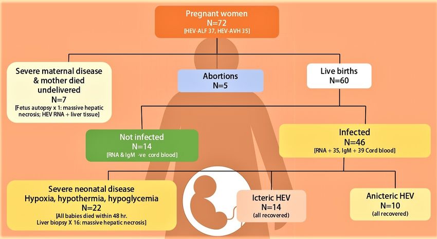
Viruses 2021, 13, x 2 of 24
Viruses 2021, 13, 1329 clean water supplies, epidemic disease with high mortality in pregnant women 2isofno
23
longer reported from industrialized countries [4].
2. Controversy Over Data—West versus East
2. Controversy Over Data—West versus East
In the latter part of the last century, viral hepatitis and pregnancy was a matter of
In the latter part of the last century, viral hepatitis and pregnancy was a matter
investigation and controversy [5]. The data published from various industrialized coun-
of investigation and controversy [5]. The data published from various industrialized
tries of Europe, North America and Australia had indicated that pregnancy does not in-
countries of Europe, North America and Australia had indicated that pregnancy does
crease the severity of disease and /or susceptibility to infection [6–10]. This was expected
not increase the severity of disease and /or susceptibility to infection [6–10]. This was
as hepatitis
expected asEhepatitis
was not endemic
E was not in such regions
endemic of theregions
in such world [11]. However,
of the several
world [11]. reports
However,
from developing countries in Asia especially India, Iran, and the Middle East,
several reports from developing countries in Asia especially India, Iran, and the Middle had shown
that there is increased severity and mortality from viral hepatitis among pregnant
East, had shown that there is increased severity and mortality from viral hepatitis among women
[12–16]. These
pregnant women reports were
[12–16]. based
These on retrospective
reports were based on analysis of hospital
retrospective admissions
analysis and
of hospital
were interpreted in some studies as a reflection of biased hospital-based data
admissions and were interpreted in some studies as a reflection of biased hospital-based collection,
due to
data the fact that
collection, duethese
to thedata
fact predated
that thesethe
datadiscovery
predatedoftheHEV [17]. of HEV [17].
discovery
3. Unmasking the Unholy Alliance
The association between viral hepatitis and pregnancy was delineated with the dis-
covery of HEV from an epidemic of viral hepatitis in Kashmir, India [18] and the reporting
Kashmir, India reporting
disease in
of the true incidence and severity of the disease in pregnancy
pregnancy based
based via
via aa prospective
prospective
door-to-door study [19] (Figure 1).
Figure 1. Gulmarg
Figure 1. Gulmarg Kashmir
Kashmir Epidemic,
Epidemic, 1978–1979.
1978–1979. (A)
(A) Upper
Upper panel.
panel. The
The epidemic
epidemic curve
curve with
with the
the weekly
weekly occurrence
occurrence of of
hepatitis E cases.
hepatitis cases. The region had an open-source water supply from a canal
canal (Ningli Nallah) which originates from
(Ningli Nallah) which originates from the
the
Alpather
Alpather Lake
Lake situated
situated atat the
the foot
foot of
of Apharwat
Apharwat Peaks,
Peaks, Gulmarg.
Gulmarg. After
After passing
passing through
through mountains
mountains as as the
the world-famous
world-famous
Sharanz waterfall,
Sharanz waterfall,the
the stream
streamcrosses
crossesthethevalley
valleyto
tojoin
join the
the Wular
Wularlake.
lake. The
The canal
canal along
along its
its route
route is used for multiple purposes
purposes
including drinking
including drinking water,
water, linen
linen washing,
washing, swimming,
swimming, fishing,
fishing, and
and sewage
sewage andand garbage
garbage disposal,
disposal, and
and thus
thus stays
stays highly
highly
polluted. Lower
polluted. Lowerpanel.
panel.TheThedatadataononincidence
incidence
andand severity
severity of viral
of viral hepatitis
hepatitis in men
in men (15–45
(15–45 years),
years), NPF NPF (nonpregnant
(nonpregnant fe-
females;
males; 15–45 years) and PF (pregnant females) were collected by four door-to-door surveys done at 4 to 6 week intervals
15–45 years) and PF (pregnant females) were collected by four door-to-door surveys done at 4 to 6 week intervals during
during the epidemic. (B) Kashmir strain of HEV (Pinglina epidemic, 1993-94, Kashmir, India [20].). Unrooted phylogenetic
the epidemic. (B) Kashmir strain of HEV (Pinglina epidemic, 1993–94, Kashmir, India [20].). Unrooted phylogenetic tree
tree generated by Maximum Likelihood method using MEGA software (version 10.1.8), on the basis of 326 bp sequences
generated
of HEV ORF1 by Maximum Likelihood
genomic. Reference method using
sequences MEGAHEV
of different software (version
genotypes from10.1.8),
GenBankon the
arebasis of 326
shown bp sequences
in gray of
filled circles,
HEV ORF1 genomic. Reference sequences of different HEV genotypes from GenBank are shown
while country specific HEV sequences are shown in color filled circles. Kashmir strain of HEV was of genotype 1 with in gray filled circles,
while country specific HEV sequences are shown in color filled circles. Kashmir strain of HEV was of genotype 1 with 94.6%
homology with the Burmese isolates of HEV (Courtesy Saleem Kamili & Xia, Guo-Liang, both at CDC, Atlanta, Georgia).
α = 6 pregnant women died.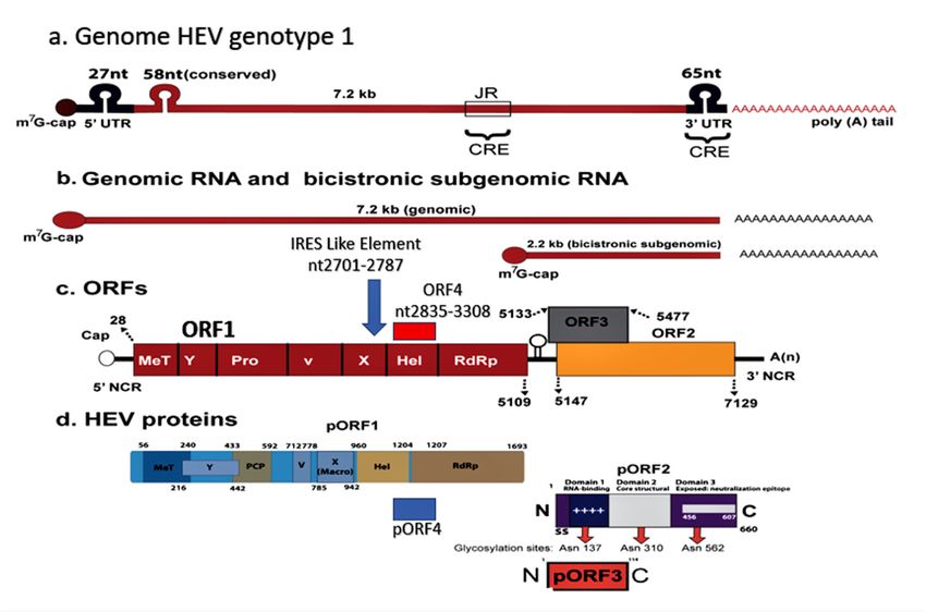
Viruses 2021, 13, 1329                                                                                          3 of 23

                         3.1. Discovery of Hepatitis E
                               The discovery of HEV started with studies carried out on a large-scale epidemic of
                         viral hepatitis in Gulmarg-Kashmir, India in November 1978 [18]. Investigating an epidemic
                         of the size had posed problems of harsh weather, primitive healthcare facilities, a highly
                         compressed time period, lack of funding, and hesitancy of medical manpower to join the
                         team due to fear of personal health risks. To face the challenges, a team of 500 healthcare
                         workers (local inhabitants) opted to reside in the epidemic region, support the primitive
                         healthcare facilities, offer care to the needy at the doorstep, and record every case of
                         hepatitis from the community (Figure 1). Over 9 weeks, 20,083 cases of acute viral hepatitis
                         (AVH) with 600 fatalities were recorded. The epidemic curve was highly compressed with
                         the occurrence of up to 4000 icteric cases per week. The disease selectively affected young
                         adults and presented as acute hepatitis syndrome with cholestatic features in around
                         20% of the affected individuals. Liver histology showed portal and lobular hepatitis
                         with necrosis and ballooning degeneration and Kupffer cell hyperplasia, with a subset of
                         patients showing distinctive features in the form of intracanalicular bile stasis and rosette
                         formation of hepatocytes as a dominant feature. All patients who survived recovered and
                         none developed chronic liver disease [21]. IgG anti-HAV, as a marker of past exposure
                         and immunity to hepatitis A virus (HAV), was reactive in all patients, while none was
                         seropositive for IgM anti-HAV, HBsAg, and IgM anti-HBc. It was postulated that this
                         epidemic of enterically transmitted non-A, non-B hepatitis (ET-NANBH), was caused by an
                         agent different from post-transfusion non-A, non-B hepatitis (PT-NANBH), later identified
                         as hepatitis C virus (HCV) [22].
                               Mikhail Balayan self-infected himself via stool samples from an outbreak of hepatitis
                         that occurred among Soviet troops in Afghanistan. He and his team identified virus-like-
                         particles (VLPs) in his stool samples [23]. Reyes et al. isolated a cDNA from the virus
                         responsible for ET-NANBH in a stool from Burma [24]. Tam et al. cloned and sequenced
                         the full length of HEV [25] and Yarbough et al. developed a serological test for diagnosis of
                         HEV infection [26].
                               After these developments, sera samples (n = 114) collected during the Gulmarg Kash-
                         mir epidemic 1978–79, 71% tested positive for IgG anti-HEV and 75% of these were reactive
                         for IgM anti-HEV, confirming HEV as the causative agent of the epidemic [27]. From 1978
                         to 2013, ten epidemics of viral hepatitis were reported from Kashmir, India [18,19,28–30].
                         Sera from all ten epidemics were tested for IgG anti-HEV, IgM anti-HEV and HEV RNA and
                         the epidemics were confirmed to be caused by HEV [20,30–32]. Partial genomic sequencing
                         of HEV from two such epidemics, namely the Jammu epidemic 1988 and the Pinglina
                         epidemic of 1993–94, were carried out. Both virus strains belonged to HEV-gt1 and had a
                         homology of 94.6%–96.8% to the Burmese strain of HEV (Figure 1) [20,33]

                         3.2. Incidence and Severity of Hepatitis E in Pregnancy
                              During the Gulmarg-Kashmir epidemic of 1978–79, a prospective study was done to
                         define the incidence and severity of hepatitis E in pregnant women compared with non-
                         pregnant women of child-bearing age and men (15–45 years) [19]. The data were collected
                         in Block Sopore, consisting of 15 villages with a population of 16,620. The four door-to-door
                         surveys were conducted at 4-to-6-week intervals to identify every new case of hepatitis
                         (Figure 1). A total of 275 cases of hepatitis E were recorded. Thirty-six (17.3%) of the
                         208 pregnant women were infected with HEV as compared to 71 (2.1%) of 3350 nonpregnant
                         women and 107 (2.8%) of 3350 men. The incidence of disease in the first trimester (3/34;
                         8.8%), second trimester (15/77; 19.2%), and third trimester (18/97; 18.6%) was higher when
                         compared to that in nonpregnant women and men. Acute liver failure (ALF) developed
                         in 22.2% (8/36) of pregnant women with HEV infection, as compared to 2.8% (3/107)
                         of men and no (0/71) nonpregnant women. Nine deaths had occurred, six in pregnant
                         women and three in men. The case fatality rate of HEV infection was 16.6% (6/36) in
                         pregnant women and 2.8% (3/107) in men. None of the nonpregnant women with HEV
                         infection died. None of the 18 pregnant women with HEV infection in their first and second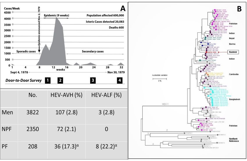
Viruses 2021, 13, 1329                                                                                            4 of 23

                         trimester developed ALF, while 8 (44.4%) of 18 pregnant women with HEV infection in
                         the third trimester developed acute hepatic failure with six deaths. The case fatality rate
                         of HEV infection in the third trimester of pregnancy was 33.3% (6/18). These data were
                         conclusive that, during epidemics, HEV infection showed increased incidence and severity
                         in pregnancy. The incidence was higher in all three trimesters as compared to HEV- infected
                         men and non-pregnant women, while the increased severity of disease was restricted to
                         the third trimester of pregnancy.

                         4. Hepatitis E
                               Hepatitis E is one of the five main forms of viral hepatitis and is caused by infection
                         with HEV [34]. HEV is a group of viruses in the family Hepeviridae [35]. These viruses are
                         quasi-enveloped, have an icosahedral shape with 20 faces, spherical geometry with surface
                         spikes and indentations, and T = 1 symmetry. Genomes are linear, non-segmented, 7.2 kb in
                         length, and have three open reading frames (ORFs), namely ORF1, ORF2 and ORF3 [36].
                         HEV has marked genetic heterogeneity and is divided into two genera, namely Orthohepe-
                         virus and Piscihepevirus [37]. Orthohepevirus has four species A, B, C and D. Orthohepevirus A
                         has eight genotypes (gt). HEV-gt1 and HEV-gt2 infect humans alone. An additional ORF4,
                         spanning nt2835-3308 and overlapping with ORF1, is present in HEV-gt1 alone and its
                         protein expression is regulated via an IRES-like RNA element (nt2701-2787). ORF4 appears
                         to cause endoplasmic reticulum (ER) stress which may be involved in promoting viral
                         replication [38,39] (Figure 2). As C-terminal 19 amino acids are absent in around half of the
                         genomes, only the N-terminal 124 amino acids of pORF4 can interact with other viral and
                         host proteins. This protein functions to enhance viral polymerase activity and promote
                         viral replication and is indispensable for the HEV-gt1 life cycle [40]. HEV-gt3 and HEV-gt4
                         are highly divergent and have been isolated from several animals including pig, wild boar,
                         deer, mongoose, rabbit, goat, horse, bottlenose dolphin, and sheep (HEV-gt3); and pig,
                         wild boar, cattle, cow, sheep, goat, and yak (HEV-gt4) [41,42]. HEV-gt5 and HEV-gt6 infect
                         wild boar in Japan and HEV-gt7 and HEV-gt8 infect dromedary and Bactrian camels, respec-
                         tively [43]. HEV-gt3 and HEV-gt4 from pigs and possibly rabbits are zoonotic, and isolated
                         cases of HEV-gt7 infection in humans have been reported [36,37]. Orthohepevirus B has four
                         genotypes, infects birds, primarily chicken, and causes hepatitis-splenomegaly syndrome
                         and big liver-spleen disease in chicken. Orthohepevirus C causes infection in rats, voles, mice,
                         and ferret and Orthohepevirus D infects bats. Cases of rat HEV infection in humans have
                         been reported [44,45]. Genus Piscihepevirus includes a single species containing cutthroat
                         trout virus [37]. Isolates from moose, fox, and little egret have remained unassigned as
                         yet [35,37]. A recent study showed that people in households with seropositive goats were
                         more likely to be seropositive themselves than persons living in households with seronega-
                         tive goats [46]. Hepatitis E is a global disease and was estimated to have caused 20.1 million
                         incident infections in the year 2005, out of which approximately 3.4 million infections were
                         symptomatic with around 70,000 deaths and 3000 stillbirths [47]. Hepatitis E has distinctive
                         epidemiological and clinical characteristics in developing countries, which contrast sharply
                         with those in the industrialized world [48,49]. Based on disease pattern and prevalence
                         and genotype distribution, four hepatitis E epidemiological patterns are seen [11]. The first
                         is an hyperendemic zone that encompasses many countries in the Indian subcontinent,
                         Southeast Asia, Central Asia, many regions in Africa, and Mexico. Here HEV-gt1 and
                         HEV-gt2 present as an epidemic and an endemic disease. The second is an endemic zone
                         that involves several countries in the Middle East and South America and some regions
                         of Southeast Asia (Singapore). HEV-gt1 causes one-fourth of sporadic hepatitis and ALF
                         in these countries. Epidemics of hepatitis E do not occur. The third epidemiological zone
                         is limited to Egypt wherein HEV-gt1 infections occur at young age similar to hepatitis A.
                         Recent studies have shown that symptomatic HEV infections do occur in elderly people
                         and those with pre-existing liver diseases. [50–52]. Seroprevalence studies have shown
                         high occurrence of asymptomatic infections in the general population as well as pregnant
                         women [53,54]. This could be related to zoonotic food-borne exposure to HEV-gt3 through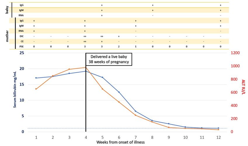
s 2021, 13, x 5 of 24
hepatitis A. Recent studies have shown that symptomatic HEV infections do occur in el-
Viruses 2021, 13, 1329derly
people and those with pre-existing liver diseases. [50–52]. Seroprevalence studies 5 of 23
have shown high occurrence of asymptomatic infections in the general population as well
as pregnant women [53,54]. This could be related to zoonotic food-borne exposure to
HEV-gt3 through cow’s milk, goat milk and goat liver [46,55,56]. It was also observed that
cow’s milk,
naturally acquired goat milk
immunity appearandtogoat liver HEV-exposed
protect [46,55,56]. It was also observed
subjects that naturally acquired
from contracting
immunity appear to protect HEV-exposed subjects from contracting
HEV infection during an epidemic [57]. HEV-gt3 and HEV-gt4 cause sporadic autochtho- HEV infection during
an epidemic [57]. HEV-gt3 and HEV-gt4 cause sporadic autochthonous
nous zoonotic food-borne infection in industrialized countries, which constitute the fourth zoonotic food-
borne infection in industrialized countries, which constitute the fourth zone. HEV-gt3 has
zone. HEV-gt3 has been reported from many European countries, North and South Amer-
been reported from many European countries, North and South America, Russia, Japan,
ica, Russia, Japan, and Australia; while HEV-gt4 is prevalent in many regions of China,
and Australia; while HEV-gt4 is prevalent in many regions of China, several Southeast
several Southeast Asian countries, Japan, and a few countries in Europe.
Asian countries, Japan, and a few countries in Europe.
Figure 2. Hepatitis E virus Genotype 1. Genomic organization. (a) The hepatitis E virus genome; (b) Genomic RNA and
Figure 2. Hepatitis E virus Genotype 1. Genomic organization. (a) The hepatitis E virus genome;
bicistronic sub-genomic RNA; (c) Four Open reading frames (ORFs) and (d) Four encoded proteins (pORF1, pORF2, pORF3,
(b) Genomic RNA and bicistronic sub-genomic RNA; (c) Four Open reading frames (ORFs) and
and pORF4). ORF4, spanning nt2835-3308 and overlapping ORF1 in present in HEV genotype 1 alone, and its protein expression
(d) Four encoded proteins (pORF1, pORF2, pORF3, and pORF4). ORF4, spanning nt2835-3308 and
is regulated via an IRES-like
overlapping ORF1RNA elementin(nt2701-2787).
in present HEV genotype ORF4 encodes
1 alone, a protein
and its protein(pORF4) of 124
expression aa, whichvia
is regulated functions to
enhance viralanpolymerase
IRES-like RNA element (nt2701-2787). ORF4 encodes a protein (pORF4) of 124 aa, which func-cycle.
activity and promote viral replication and is indispensable for the HEV genotype 1 life
tions to enhance viral polymerase activity and promote viral replication and is indispensable for
5. Epidemic
the HEV genotype 1 life cycle.Hepatitis E and Pregnancy
Several large-scale water-borne epidemics of hepatitis have been reported from many
5. Epidemic Hepatitis
regions ofEthe
anddeveloping
Pregnancyworld [1] (Table 1). Initially, these epidemics were designated
empirically as ET-NANBH
Several large-scale water-borne epidemicsbasedofon serological
hepatitis testing
have been for HAV
reported fromand HBV infections.
many
Later, once serological testing for HEV infection was available, most
regions of the developing world [1] (Table 1). Initially, these epidemics were designated such epidemics were
found to be caused by HEV. HEV strains, once characterized,
empirically as ET-NANBH based on serological testing for HAV and HBV infections. were all of HEV-gt1 [58,59].
Epidemictesting
Later, once serological HEV infections had several
for HEV infection wasfeatures in common
available, most such including
epidemicsoccurrence
were in young
adultsby
found to be caused (15–45
HEV.years),
HEV significant cholestatic
strains, once features,
characterized, self-limiting
were disease,
all of HEV-gt1 and high mortality
[58,59].
Epidemic HEV in infections
pregnancy.had several features in common including occurrence in young
adults (15–45 years), significant cholestatic features, self-limiting disease, and high mor-
Table 1. Epidemics of hepatitis E with number of recorded cases, case fatality rate (overall and in pregnant women) and
tality in pregnancy.
relationship with hepatitis E virus genotypes.
Number of
Region Year [References µ] CFR (%) HEV Genotypes
HEV Infections
Overall Pregnancy
Kashmir 1978–2013
55,563 3.19 22.0 HEV-gt-1
[18–20,28–30,32,33]
New Delhi 1956 [58,60,61] 29,300 0.9 10.5 HEV
Kanpur 1991 [62] 79,091 0.06 27.0 HEVViruses 2021, 13, 1329 6 of 23
Table 1. Cont.
Number of
Region Year [References µ] CFR (%) HEV Genotypes
HEV Infections
Overall Pregnancy
Azamgarh 1982 [63] 152 12 39.0 ENANBH
Kolhapur 1981 [58,64,65] 1169 0.25 8.33 HEV-gt1a
Islamabad 1997 [66–68] 3827 0.2 11.4 HEV-gt1b
Rangoon 1985 [25,69] 399 3.5 12.0 HEV-gt1
Kathmandu 1981 [70,71] 12,000 - 21.0 HEV-gt1
Kathmandu 1987 [71,72] 7405 0.41 24.65 HEV-gt1
Bangladesh 2008 [73,74] 4198 0.47 19.0 HEV-gt1
Bangladesh 2010 [74,75] 2162 0.55 25 HEV-gt1
Turkmenistan 1985 [76,77] 16,175 0.12 27.4 HEV-gt1
Uzbekistan 1985 [78,79] 12,000 - 7.1 HEV-gt1
Xinjiang 1986 [80,81] 120,000 0.59 13.3 HEV-gt1
Indonesia 1991 [82] 1688 1.78 26.3 HEV
Algeria 1980 [83,84] 788 1.39 100 HEV-gt1
Sudan 2006 [85,86] 253 13.5 31.1 HEV-gt1
Djibouti 1998 [87,88] 42 9.5 33.3 HEV-gt1
Central African Republic 2002 [87,89] 715 0.55 14.28 HEV-gt1
Somalia1993 [87,90] 11,413 2.9 13.8 HEV-gt1
Kenya 1991 [91,92] 1702 3.70 14.28 HEV-gt1
Sudan 2004 [85,87] 2621 1.71 31.14 HEV-gt1
Uganda 2007 [93] 4789 1.50 6.87 HEV-gt1
Mexico 1986 [94] 223 1.35 0 HEV-gt2
Namibia 1995 [95] >600 0.50 1 death β HEV-gt2
Namibia 1983 [96,97] 201 3.48 85.7 HEV-gt1
Nigeria 2018 [98,99] 146 1.37 8 HEV-gt1 & HEV-gt2
Central African Republic 2008 [100] 222 1.8 20 HEV-gt1
Chad 2004 [101] 989 3.0 - HEV-gt1 & HEV-gt2
Namibia 2017 [102] 7247 0.80 6.00 HEV
Chad 2016 [103] 1293 0.69 3.16 HEV-gt1
CFR = Case fatality rate, gt = genotype, ENANBH = Epidemic non-A, non-B hepatitis, β = number of pregnant women not mentioned in
calculating CFR. µ = The references include reports of the epidemic and further studies on the stored samples to characterize the epidemic
and the HEV genotypes.
From 1978 to 2013, 10 large-scale water-borne epidemics of hepatitis E were recorded
in Kashmir [18–20,28–30,32,33]. A total of 55,563 persons had contracted the disease and
1775 died with a case fatality rate (CFR) of 3.19%. CFR of HEV in pregnant women during
these epidemics was 22%. A retrospective analysis of sera from a large-scale water-borne
epidemic that occurred in Delhi in 1955–56 revealed that the epidemic was caused by
HEV [58,60,61]. This epidemic affected an estimated 29,300 patients with 266 deaths.
Overall, CFR was 0.9% and CFR in pregnancy was 8.5%. A massive epidemic of hep-
atitis E involving an estimated 79,000 cases visited Kanpur, UP, India in 1992, with a
CFR of 27% in pregnant women [62]. Several other outbreaks have been reported from
other parts of India [63,64,104–106], Pakistan [66–68,107], Burma [25,69], Nepal [70–72],
and Bangladesh [73–75,108,109], and all of these showed high CFR in pregnant women [58].
Several regions in Central Asia namely Turkmenistan [76,77], Uzbekistan [78,79], Tajik-
istan [110], and Kirgizstan [111], have been hit by epidemics of viral hepatitis caused
by HEV-gt1. These epidemics affected between 10,000 and 30,000 persons and saw high
mortality in pregnant women with CFR ranging from 7% to 27%. Xinjiang region in the
northwest of China recorded a massive outbreak of viral hepatitis affecting 120,000 peo-
ple (mostly Uighurs) in the autumn of 1987–88. CFR in pregnant women was 13% [80].
The outbreak was later confirmed to be caused by HEV-gt1 [81]. Regions of South-East Asia,
namely Indonesia [82] and Vietnam, [112] have reported several epidemics of hepatitis E
with a high fatality rate of up to 26% in pregnant women. Several outbreaks caused by
HEV-gt1 have been reported from many regions of Africa including Somalia [87,90], Al-Viruses 2021, 13, 1329 7 of 23
geria [83,84,113], Côte d’Ivoire [114], Botswana [115], Djibouti [87,88] and Central African
Republic [87,89], with higher fatality in pregnant women. Of late, outbreaks of hepatitis E
in refugee camps among displaced people in several African countries including Soma-
lia [87,90,116], Kenya [91,92], Sudan [85,86] and Uganda [93,117] have occurred. All these
epidemics have reported higher death rates in pregnant women.
HEV-gt2 was the incriminating agent for two outbreaks of hepatitis that occurred
in two villages 70 km south of Mexico City in 1986 [94]. Of the 223 cases, three women
died with an overall CFR of 1.35%. Higher fatality in pregnant women was not reported.
The epidemic caused by HEV-gt2 from Namibia in 1995 also did not report higher deaths
in pregnant women [92,95,102]. However, a previously reported epidemic from Namibia in
1983 was caused by HEV-gt1 [96,97] and of the 201 cases six of the seven deaths were seen in
pregnant women. Epidemics of hepatitis in Nigeria [98,99], Central African Republic [100],
and Chad [101,103] were caused by both HEV-gt1 and HEV-gt2 and all reported higher
deaths in pregnant women. Few cases of HEV-gt3 and HEV-gt4 infections reported in
pregnant women have not been associated with severe disease or deaths [118,119]. An HEV-
gt3 outbreak on a cruise ship causing 33 infections did not cause higher mortality in
pregnant women [120]. HEV-gt3 and HEV-gt4 are prevalent in industrialized countries
and have not been associated with higher mortality in pregnant women [121]. Thus, higher
mortality of epidemic hepatitis E in pregnancy is genotype-specific and associated with
HEV-gt1 and not with other genotypes causing human infections, namely HEV-gt2, HEV-
gt3, and HEV-gt4. However, adverse pregnancy outcomes including miscarriage and
stillbirths have been reported in pregnant rabbits experimentally infected with HEV-gt3
and HEV-gt4 [122–124].
6. Sporadic Hepatitis E and Pregnancy
In 1983, 293 patients with acute sporadic viral hepatitis, of whom 155 cases were
caused by HEV infections were reported [20,125]. The mode of transmission was enteric,
mostly based on person-to-person contact. The disease occurred in young adults with
relative sparing of children. The clinical profile resembled AVH with cholestasis in a
subset of patients. The disease was self-limiting and none of the patients developed
chronic hepatitis or cirrhosis on follow-up. All these features resembled the epidemic HEV
described from Kashmir [18]. The disease occurred in 19 pregnant women. The overall
case fatality rate (CFR) was 12.3% and CFR in pregnant women was 35.6%. After this,
the etiology, clinical course, and outcome of AVH in 76 pregnant women and 337 non-
pregnant women of childbearing age were studied [126]. The prevalence of HEV in
pregnant women was 85.5% (65/76 patients) as against 41.5% (140/337) in nonpregnant
women. The prevalence of HEV infection was 76.9% (4/13), 88.9% (12/18), 83.8% (23/37),
and 100% (8/8) in first, second, third trimesters, and puerperium, respectively. The CFR
of HEV infection in pregnant women was 69.2% (45/65) as against 10.0% (14/140) in
nonpregnant women. The CFR was 40% (4/10) in the first trimester as against 74.5% (41/55)
in the second trimester and beyond. A north Indian study reported HEV as the cause of
acute sporadic hepatitis in 82%, 49%, and 57% of pregnant women, nonpregnant women,
and men, respectively [127]. Several other studies of acute sporadic viral hepatitis showed
a higher prevalence of HEV infections and higher CFR in pregnant women [128–132]
(Table 2).
Regarding HEV genotypes prevalent in acute sporadic hepatitis in India, Arankalle
et al. [65] studied 17 HEV isolates (both epidemic and sporadic) from India and found all
to be related to various subtypes of HEV-gt1. In another study, Indian swine were found
to be infected by HEV-gt4, while all Indian human isolates studied were HEV-gt1 [133].
Gupta et al. [59] studied sequences of 74 patients with acute sporadic hepatitis E from
North India and found all the isolates were related to HEV-gt1a. Thus, HEV circulating in
India and causing acute sporadic viral hepatitis with higher mortality in pregnancy is also
genotype-specific and associated with HEV-gt1.Viruses 2021, 13, 1329 8 of 23
Table 2. Prevalence of HEV infection among pregnant women with acute sporadic viral hepatitis.
Study
Author, Year. HEV-AVH (%) HEV-ALF (%) HEV Status
Material
[References]
PF Others PF Others PF Others
Khuroo et al., 1983 [125] 27 266α 19 (70.4) 136 (51.1) 6 (31.6) 13 (9.6) HEV
Nayak et al., 1989 [127] 169 70β 138 (81.6) 34 (48.6) 21 (28.5) ETNANBH
Jaiswal et al., 2001 [129] 127 146β 83 (65.4) 129 (88.4) 44 (53.0) 17 (13.2) HEV
Khuroo et al., 2003 [126] 76 337β 65 (85.5) 140 (41.5) 46 (70.8) 14 (10) HEV
Beniwal et al., 2003 [130] 97 - 46 (47.4) - 18 (39.1) - HEV
Patra et al., 2007 [128] 220 - 132 (60) - 73 (55.3) - HEV
PF = Pregnant females, α = all age groups, β = nonpregnant women of childbearing age.
7. HEV-ALF and Pregnancy
Several large series of ALF and its relationship with pregnancy have been reported
from India. In one study, amongst 180 patients with ALF [134] forty-nine of the 111 women
were pregnant. Seventy-nine patients were related to HEV and the remaining 101 pa-
tients were caused by HAV (4 cases), HBV (25 cases), HCV (13 cases), HDV (two cases),
drug (one case), and non-A-E agents (56 cases). HEV was the cause of ALF in 47 of the
49 pregnant women as against 14 of the 34 nonpregnant women of childbearing age.
Acharya et al. [135] from the All India Institute of Medical Sciences (AIIMS), New Delhi, re-
ported 423 patients with ALF. Of the 223 women, 53 were pregnant. The etiology included
HAV (seven cases), HBV (117 cases), HDV (16 cases), NANBH (264 cases), and drugs
(19 cases). Thirty-one of the 50 cases from the NANBH group were caused by HEV. Subse-
quently, 1015 patients with ALF were reported from the same center. 249 of the 647 women
were pregnant. HEV was the etiological cause in 342 patients, while 651 patients had
non-HEV etiology [136]. HEV was the cause of ALF in 145 of the 244 pregnant women,
100 of the 329 nonpregnant women, and 97 of the 420 men. In another study, HEV was
the cause of ALF in 102 of the 139 pregnant women as against 111 of the 181 nonpregnant
women (p < 0.03) [137]. Kar et al. studied 100 patients with ALF, 50 of whom were pregnant
and another 50 nonpregnant women of childbearing age. ALF was caused by HEV in
28 pregnant women against 7 of the 50 nonpregnant women [138]. Sequencing data of
all HEV positive sera detected HEV-gt1. These data point to the fact that a substantial
proportion of ALF in India is seen in pregnant women and HEV is the dominant etiology.
HEV-ALF in pregnant women starts with prodrome followed by other features of
acute viral hepatitis [29]. However, a rapidly evolving devastating illness develops
within a short pre-encephalopathy period (5.8 ± 5.3 days), characterized by encephalopa-
thy, cerebral edema with features of cerebellar coning, coagulopathy, and upper GI
bleed [134,135,139]. In addition, the occurrence of “Disseminated intravascular coagu-
lation (DIC)” is a distinctive feature of HEV-ALF during pregnancy [140], resembling a
Schwartzmann phenomenon.
The prognosis of HEV-ALF in pregnant women has been studied by several investiga-
tors [134,136]. The investigators from AIIMS, New Delhi, questioned the worse prog-
nosis of HEV-ALF during pregnancy [136,141]. The authors compared the mortality
rates in 249 pregnant women, 341 non-pregnant women, and 425 men, 15 to 45 years
of age. The mortality rates in the three groups were 53.8%, 57.2%, and 57.9%, respectively
(p = 0.572). A prospective study on 180 pregnant women with ALF revealed 79 with HEV-
ALF and 101 with non-HEV-ALF [134]. The prognosis in patients with HEV-ALF was better
than those with non-HEV-ALF (CFR 51.9% in HEV-ALF versus 84.2% in non-HEV-ALF).
Factors predictive of poor prognosis included non-HEV etiology, prothrombin time > 30 s,
grade of coma > 2, and age > 40 years and did not include pregnancy per se or duration
of pregnancy. The fact that pregnant women acquired ALF more often did not mean that
such patients will have higher mortality [142].and 101 with non-HEV-ALF [134]. The prognosis in patients with HEV-ALF was better
than those with non-HEV-ALF (CFR 51.9% in HEV-ALF versus 84.2% in non-HEV-ALF).
Factors predictive of poor prognosis included non-HEV etiology, prothrombin time >30
sec, grade of coma > 2, and age > 40 years and did not include pregnancy per se or duration
Viruses 2021, 13, 1329
of pregnancy. The fact that pregnant women acquired ALF more often did not mean9 that of 23
such patients will have higher mortality [142].
8.
8. Proposed
Proposed Hypothesis
Hypothesis on on Pathogenesis
Pathogenesis of of Mortality
Mortality in
in HEV-Infected
HEV-Infected Pregnant
Women
Pregnant Women
The pathogenesisof
The pathogenesis ofhigher
highermorbidity
morbidity andand mortality
mortality duedue to HEV
to HEV infection
infection in preg-
in pregnancy
nancy is complex and remains to be fully understood. The lack
is complex and remains to be fully understood. The lack of a proper animal modelof a proper animal modelto
to study the pathogenesis of HEV-gt1 in pregnancy
study the pathogenesis of HEV-gt1 in pregnancy has been an issue. has been an issue. Rhesus monkey
(Macaca
(Macaca mulata)
mulata) is an established
established animal
animal model
model forfor HEV
HEV infection
infection [32,143],
[32,143], but
but HEV-gt1
HEV-gt1
infection
infection to to pregnant
pregnant monkeys
monkeys does does notnot result
result in
in ALF
ALF [144,145] and thus is not useful to
study the pathogenesis
study pathogenesis of the disease. Pregnant Pregnant rhesus
rhesus monkeys
monkeys can be infected with
HEV-gt4 with
HEV-gt4 withresultant
resultanthigh
highviral
viraltiters,
titers, longer
longer duration
duration of infection,
of infection, obstetric
obstetric events
events and
and vertically transmitted fetal infections [146]. A successful animal
vertically transmitted fetal infections [146]. A successful animal model for HEV-gt4 has model for HEV-gt4
has been
been established
established in pregnant
in pregnant BABL/c BABL/c miceRabbits
mice [147]. [147]. Rabbits
are neware newmodels
animal animal formodels
hep-
for hepatitis
atitis E, but HEV-gt1
E, but HEV-gt1 is not infectious
is not infectious to the[148].
to the animal animal [148].occurrence
Besides Besides occurrence
of ALF in
of ALF inwomen,
pregnant pregnant women, extrahepatic
extrahepatic manifestations manifestations including neurological,
including neurological, renal,
renal, hematologi-
hematological,
cal, and pancreatic andare
pancreatic
associatedarewith
associated with HEVThe
HEV infections. infections. The pathogenesis
pathogenesis of HEV in the of
HEV in the extrahepatic tissues is either due to direct cytopathic effect mediated
extrahepatic tissues is either due to direct cytopathic effect mediated by the virus replica- by the virus
replication
tion or immunological
or immunological mechanisms
mechanisms causedcaused
by an by an uncontrollable
uncontrollable host response
host response [149].[149].
Sev-
Several important facts have recently emerged to explain the complex
eral important facts have recently emerged to explain the complex relationship between relationship between
HEV and
HEV and pregnancy
pregnancy (Figure
(Figure 3).
3).
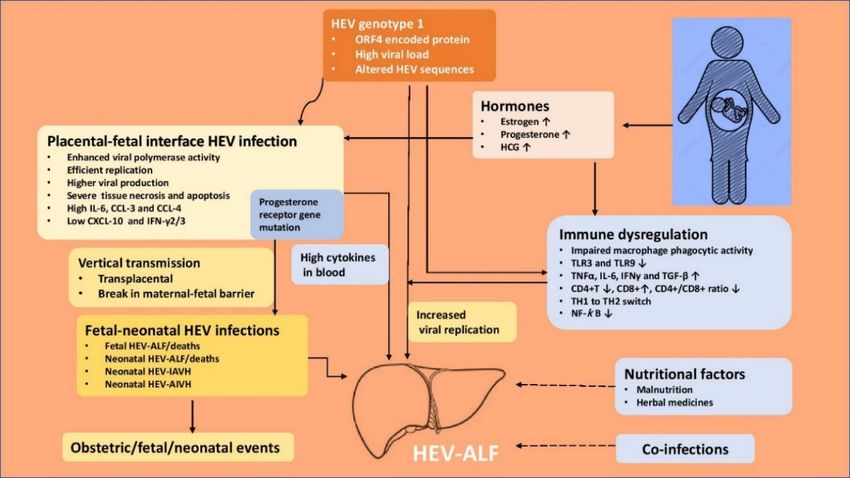
Figure 3. Pathogenesis
Figure 3. Pathogenesis of
of hepatitis
hepatitis E
E virus-related
virus-related acute
acute liver
liver failure
failure in
in pregnancy.
pregnancy. IAVH
IAVH = = icteric
icteric acute
acute viral
viral hepatitis,
hepatitis,
AIAVH = Anicteric acute viral hepatitis, HEV-ALF = Hepatitis E virus related acute liver failure.
AIAVH = Anicteric acute viral hepatitis, HEV-ALF = Hepatitis E virus related acute liver failure.
8.1.
8.1. HEV
HEV Genome,
Genome, Heterogeneity
Heterogeneity and
and Variants
Variants in
in Pregnancy
Pregnancy
The
The data
data available
available from epidemic and sporadic hepatitis E indicate that pregnant
women
women acquire
acquireHEV
HEV[19,126]
[19,126]and
anddevelop
developALF more
ALF often
more than
often others
than [134–136]
others and and
[134–136] this
this phenomenon is limited to infection with HEV-gt1 [59,65]. Other hepatitis viruses
(HAV, HBV and HCV) and other HEV genotypes (HEV-gt2 and HEV-gt3) do not cause
higher deaths in pregnant women [150]. Among the HEV family, HEV-gt1 alone has
ORF4 which encodes a protein, pORF4 of 124 aa [38,39]. The encoded pORF4 by the
HEV-gt1 genome interact with other viral and host proteins, enhance viral polymerase
activity and promote viral replication [123]. HEV-gt1, and not HEV-gt3, causes necrosis
and apoptosis in the maternal–fetal interface possibly caused by mitochondrial damage
and activation of caspase family membranes [151]. This leads to alterations of the placental
barrier architecture and promotes vertical transmission [38]. High levels of HEV RNA
related to HEV-gt1 have been correlated with increased severity of disease in pregnantViruses 2021, 13, 1329 10 of 23
women [138,152]. HEV sequences in patients with HEV-ALF (five patients) were compared
with those of HEV-AVH (five patients) and showed six unique amino acid substitutions in
the ORF1 region of HEV [153], namely F179S, A317T, T735I, L1110F, V1120I, and F1439Y.
Two of these (L1110F and V1120I) which occurred in the helicase domain pointing to its
role in determining the outcome of HEV infections.
8.2. Immune Response in HEV Infected Pregnant Women
The immunological alterations in pregnancy are complex, showing immune toler-
ance to an allogenic fetus and host defense against pathogen. This is accomplished by
the maternal–fetal interface (decidua) consisting of decidual stromal cells, decidual im-
mune cells, and trophoblast cells [154]. The maternal–fetal interface contains natural
killer (NK) cells, macrophages, dendritic, and T cells which interact with invading fetal
extra-villous trophoblast for placentation, fetal growth, and pregnancy outcome [155].
Pregnancy suppresses cell-mediated immunity at the maternal–fetal interface to tolerate
fetal antigens and maintains a normal humoral immune response against pathogens [156].
Pregnancy causes a shift from a Th1 to a Th2 cytokine response, allowing maternal–fetal
tolerance for fetus development [157].
HEV-gt1 infection in pregnant women causes several alterations in innate and adap-
tive immune response, which helps virus replication and increases the severity of disease
in the pregnant women [123]. Pregnant women with HEV-ALF have impaired macrophage
phagocytic activity and downregulation of TLR3 and TLR9 expression, impeding MyD88-
mediated IFN production [158]. This may lead to inadequate triggers for the innate immune
response which in turn could lead to enhanced viral replication and severe liver injury.
HEV-gt1 (but not HEV-gt3) in pregnant women was shown to evade early antiviral re-
sponse and cause adverse consequences due to poor IFN response in placental cells [151].
Pregnant women with HEV-ALF have significantly higher levels of pro-inflammatory
cytokines (TNF-alpha, IL-6, IFN-gamma, and TGF-beta) and this had a significant positive
correlation with viral load, serum bilirubin, and prothrombin time. Increased severity of
disease in pregnant women with HEV-ALF may be mediated through cytokines [159,160].
Pregnant women with HEV-ALF have lower CD4+ T cell counts, higher CD8+ T cell counts,
and lowered CD4/CD8 ratio than women with HEV-negative ALF [161]. Pregnant women
with HEV-ALF have excessive Th2 switching, which dysregulates balance between toler-
ance and immunity [162,163]. Pregnant women with HEV-ALF show higher DNA-binding
activity of NF-κB and absence of p65 expression, leading to deregulated immunity and
severe liver damage [164]. Recently the role of immune cells in the spread of HEV infection
to various body organs has been studied. Immune cells are permissive to HEV and help
the virus enter various body organs, as shown in vivo and in vitro studies [165,166]. It is
possible that this may be one of the ways in which the virus reaches the maternal–fetal
interface and causes vertical transmission.
8.3. Hormones and HEV in Pregnancy
Hormones in pregnancy may enhance HEV viral replication. Women experience a
sudden and marked increase in estrogen and progesterone levels during pregnancy [167].
Estrogen helps the fetus to develop and mature. Progesterone suppresses the maternal
immune response by stimulation of Th2 and reduction of Th1 cytokines, thereby pre-
venting maternal rejection of the trophoblast [168]. Pregnant women with HEV infection
have higher levels of estrogen, progesterone, and HCG than those without HEV infection.
Higher hormone levels are apt to further dampen the immune response and help viral
replication [155]. Estradiol has been shown to facilitate HEV replication in an in vitro ex-
periment in A546 cells [169]. High estrogen in pregnancy causes placental dysfunction and
leads to preterm delivery, low birth weight infants and fetal mortality [170]. Bose et al. [171]
studied deregulation of the progesterone receptor signaling pathway in pregnant women
with HEV-ALF, HEV infection, and healthy controls. Patients with HEV-ALF show proges-Viruses 2021, 13, 1329 11 of 23
terone receptor gene mutations and a high IL-12/IL-10 ratio and are associated with a poor
maternal outcome.
8.4. Nutritional Status and HEV in Pregnancy
Malnutrition had been proposed to explain the reports of higher deaths with viral
hepatitis in pregnancy in developing countries. However, the nutritional assessment
of pregnant women in these reports had not been evaluated [13]. During the Gulmarg-
Kashmir HEV epidemic 1978-79, caloric and protein intake as well as estimation of serum
protein as an index of nutritional status in pregnant women were determined and com-
pared with those in nonpregnant women (15–45 years) [19]. The caloric and protein intake
and serum protein in pregnant women were within normal ranges (3242 + 551 cal/day,
60 + 20 g/day, and 3.3 + 0.4 g/dL respectively) and did not differ from those seen in non-
pregnant women (300 + 450 cal/day, 50 + 15 g/day, and 3.2 + 0.6 g/dL; p > 0.50). The caloric
and protein intake and serum proteins were well in acceptance with those recommended
for Indian women [172]. Of significance was the observation that pregnant women who
developed HEV-ALF had excellent nutritional status. Thus, malnutrition contributing
to severe disease in pregnant women in developing countries was not collaborated by
these data.
8.5. Fetal HEV Infections and Maternal Mortality, Obstetric Events and Neonatal Outcome
Morbidity and mortality among pregnant women and their neonates and obstetric
events may be a reflection of the severity of the HEV infection in the mother [19,126,128].
However, there is growing evidence that vertically transmitted HEV infections causing fetal
HEV infections may directly contribute to maternal mortality, obstetric events, and neonatal
outcome [173]. Several investigators from India and the Middle East [140,174–183] have
reported vertical transmission of HEV and resultant peri-natal morbidity and mortality
(Table 3).
Table 3. Vertical transmission of HEV and maternal and obstetric events and neonatal outcome.
Author Year. HEV- Maternal & HEV-Neonatal Status Pattern of Outcome of
[References] PF Obstetric Events Neonatal HEV HEV-Infected Neonates
Babies HEV-Infections Disease
6 (RNA 5, IgM 3, Died 2 (HEV-ALF, Liver
ALF 6, Died 3, HEV-ALF 2, biopsy 1 MHN),
Khuroo et al., 10 (DUD 2), FTD 7, 8 IgG- I-HEV 1,
1995 [174] PD 1. AI-HEV 3. Recovered 4, RNA in
Seroconversion 1). 2 lasted 1 month.
Died 6 (HEV-ALF, Liver
biopsy 1 MHN with HEV
ALF 15, Died 9 19 (Died 1 due HEV-ALF 6, RNA in liver), Recovered
Khuroo et al., 26 prematurity) + 15 (RNA 10, I-HEV 4, AI-HEV 9. RNA lasted for 4 weeks
2009 [175] (DUD 5), FTD 15, IgM 12). in 4, 8 weeks in 1,
PD 4, Ab 2. 2 aborted. 5.
32 weeks in 1. IgM lasted
for 4 weeks in 3,
for 8 weeks in 2.
ALF 16, DIC 9, 25 (RNA 20, IgM HEV-ALF 14, Died 14 (HEV-ALF,
Khuroo et al., 36 Died 10, FTD 26, 33 + 3 aborted. I-HEV 9, Liver biopsy 14 MHN),
2006 [140] PD 7, Ab 3. 24). AI-HEV 2. Recovered 11.
HEV-ALF 2,
Kumar et al., 28 ALF 6, Died 3 26 I-HEV 21, Died 2 (HEV-ALF),
2001 [177] (DUD 2), PD 2. 26 (RNA 26) Recovered 24.
AI-HEV 3.
Singh et al.,
22 ALF 14, Died 14. NK *, 6 3 (RNA 3). I-HEV 1. -
2003 [176]
Kumar et al., 28 ALF 9, Died 7, NK *, 18 6 (RNA 4, IgM 3). - -
2004 [178] PD 18.
FTD 92 (Vaginal
Chibber et al., 92 delivery 80, 92 4 (RNA 4, IgM 4). I-HEV 4 *** -
2004 [179]
Caesarean in 12)
El Sayed Zaki 9 (RNA 5, IgM 1, RDS with icterus
9 9 ** 9 5, I-HEV 3, -
et al., 2013 [181] IgG 6). Sepsis 1.Viruses 2021, 13, 1329 12 of 23
Table 3. Cont.
Author Year. HEV- Maternal & HEV-Neonatal Status Pattern of Outcome of
[References] PF Obstetric Events Neonatal HEV HEV-Infected Neonates
Babies HEV-Infections Disease
Sharma et al., 144 ALF 41, Died 6 128 59 (RNA 15, - -
2017 [180] (DUD 6). IgM 59).
Bonney et al., ALF 2, Died 2 Recovered, RNA -ve
3 (DUD 1), PD 1, 1 + 1 aborted 1 (RNA 1, IgM 1). I-HEV 1.
2012 [182] Ab 1. 3 weeks., IgM -ve 4 weeks.
Fetal ascites at 15
1 (IgM cord blood, weeks pregnancy, Healthy baby delivered 38
Pradhan et al., 1 Fetal HEV-AVH 1
2012 [183] 15 weeks. amniotic fluid & resolved in weeks., LFT normal,
serum at birth). follow up IgM +ve.
PF = Pregnant females, ALF = acute liver failure, I-HEV = Icteric hepatitis E virus infection, AI-HEV= Anicteric hepatitis E virus infection,
MHN = Massive hepatic necrosis, DIC = disseminated intravascular coagulation, FTD = full term delivery, DUD = mother died with
baby undelivered, Ab = abortion, PD = premature delivery, RNA = HEV RNA +ve, IgM = IgM anti-HEV +ve, IgG = IgG anti-HEV +ve,
RDS = respiratory distress syndrome. LFT = liver function tests, * = total babies born not known, ** = all deliveries had complicated clinical
course, *** = baby developed icterus at 6 weeks of birth.
In a seminal piece of research, mother-to-child transmission of HEV in 10 pregnant
women was studied [174]. Two mothers died with babies undelivered. Six of the eight live-
born babies showed evidence of HEV infection at birth. HEV RNA was detected in cord and
birth blood samples in five, IgM anti-HEV in three and IgG anti-HEV seroconversion in four
babies. Two babies died within 24 h from hypothermia and hypoglycemia. A liver biopsy
in one baby revealed massive hepatic necrosis. Of the remaining six, one baby had icteric
hepatitis and the other three had anicteric hepatitis. In another study, vertical transmission
of HEV in 15 of the 19 babies born to HEV-infected pregnant women was studied [175].
Six of these died within first week of life. The remaining nine babies survived and five
showed HEV RNA for varying intervals lasting up to 32 weeks. All surviving babies
recovered and none developed chronic liver disease. Recently the relationship of severity
of disease in 36 pregnant women (HEV-AVH, 20 and HEV-ALF, 16) with the severity of
HEV infection in fetuses and newborn babies was evaluated [140]. Babies born to HEV-ALF
mothers were more often HEV infected, viremic, and born with severe disease than those
with HEV-AVH. DIC in mothers with HEV-ALF occurred exclusively in pregnant women
who delivered babies with HEV-ALF. All the six mothers who survived had delivered
babies within 4 days (2.3 ± 1.0 days) of onset of encephalopathy. Based on these data, it was
postulated that severe fetal disease is the likely cause of increased severity of HEV infection
in the mother, akin to the mirror syndrome in pregnancy, and delivery of baby performed
early in the course of the disease may reverse the severe maternal disease [184–186].
Vertically transmitted fetal HEV infections and HEV infections in new-born babies
have a wide spectrum of manifestations (Figure 4). Severe fetal disease results in intrauter-
ine fetal death, and often the mother also had severe disease with DIC and died before
delivery. Autopsies of such fetuses have shown massive hepatic necrosis. A significant pro-
portion with severe disease present with hypothermia, hypoxia, and hypoglycemia at birth
and die within 24 to 48 h of birth. Liver biopsies in such patients also show massive hepatic
necrosis. Babies born with HEV infection may be asymptomatic with mild abnormality of
liver tests (anicteric HEV) or present with jaundice with elevated live tests (Icteric HEV).
The disease is self-limiting and serial follow-up reports clinical, serological and virological
recovery in few weeks. A few babies show prolonged viremia lasting for up to 32 weeks,
with eventual recovery.
8.6. Maternal–Fetal Interface HEV Infections
The role of HEV-gt1 is increasingly being recognized in causing infection and dysreg-
ulation at the maternal–fetal interface which leads to vertical transmission and increased
systemic inflammation and consequent severe maternal disease [187]. Based on HEV
transplacental transmission to the fetus, Bose et al. [188] studied placental HEV replication
in 90 pregnant women (HEV-AVH 68 and HEV-ALF 22) and detected replicative HEV
RNA and HEV RNA staining by ORF3, which correlated with fetal and maternal mor-Viruses 2021, 13, x 13 of 24
encephalopathy. Based on these data, it was postulated that severe fetal disease is the
Viruses 2021, 13, 1329 13 of 23
likely cause of increased severity of HEV infection in the mother, akin to the mirror syn-
drome in pregnancy, and delivery of baby performed early in the course of the disease
may reverse the severe maternal disease [184–186].
talityVertically
in HEV-ALF transmitted
patients.fetal HEV infections
El-Mokhtar andfound
et al. [189] HEV that
infections
HEV-gt1in new-born
replicatedbabies
more
have a wide
efficiently spectrum
with a higher of degree
manifestations (Figure 4).
of inflammatory Severe fetal
response disease results inprimary
in non-decidualized intrau-
terine
humanfetal death, and
endometrial often the
stromal cellsmother
than inalso had severe
HEV-gt3. diseasebelieved
The authors with DICthatandthis
died before
infection
delivery. Autopsies
mediates vertical of such fetuses
transmission of HEV have shown
to the fetus.massive
Recently,hepatic
Gouillynecrosis. A infected
et al. [151] significantex
proportion with severe
vivo maternal–fetal disease
interface present
with withand
HEV-gt1 hypothermia,
HEV-gt3 and hypoxia,
showed and hypoglycemia
HEV-gt1 was more at
birth andthan
efficient die within
HEV-gt3 24in
toviral
48 h replication
of birth. Liver
andbiopsies
productionin such
and patients
showed also
moreshow
severe massive
tissue
hepatic
damagenecrosis.
(necrosisBabies born withwith
and apoptosis) HEVupregulated
infection may beCCL-3,
IL-6, asymptomatic
and CCL-4with mild
and abnor-
downreg-
ulated of
mality CXCL-10 and(anicteric
liver tests IFN-γ2/3. The or
HEV) authors
presentconcluded that HEV
with jaundice withplacental
elevated replication
live tests (Ic-is
genotype-specific
teric and HEV-1
HEV). The disease tropism at and
is self-limiting the maternal–fetal
serial follow-up interface
reportsalong withserological
clinical, the extent
of tissue
and damage,
virological pro-inflammatory
recovery in few weeks. cytokines, and chemokines
A few babies show prolongedmightviremia
be responsible
lasting for
the severe maternal disease.
up to 32 weeks, with eventual recovery.
Figure 4.
Figure 4. Fetal-neonatal
Fetal-neonatalhepatitis E virus
hepatitis infection
E virus recorded
infection in fetuses
recorded and and
in fetuses neonates fromfrom
neonates 72 pregnant women
72 pregnant with hepa-
women with
titis E virus infection seen at Sher-I-Kashmir Institute of Medical Sciences, Srinagar Kashmir, India from Dec 1993 onwards
hepatitis E virus infection seen at Sher-I-Kashmir Institute of Medical Sciences, Srinagar Kashmir, India from Dec 1993
[140,174,175]. ALF = acute liver failure, RNA = HEV RNA, IgM = IgM anti-HEV.
onwards [140,174,175]. ALF = acute liver failure, RNA = HEV RNA, IgM = IgM anti-HEV.
8.6. Maternal–Fetal Interface HEV Infections
9. Management
The
HEVrole of HEV-gt1
infection is increasingly
in pregnant being recognized
women requires in causing
a team approach, infectionby
determined and dysreg-
stage and
ulation at the maternal–fetal interface which leads to vertical transmission
severity of the disease in the mother, occurrence of obstetrical complications, and severity and increased
systemic inflammation
of vertically transmitted and consequent
disease severe maternal disease [187]. Based on HEV
in the fetus/neonate.
transplacental transmission to the fetus,
HEV-AVH is usually a self-limiting disease Bose et al. [188]
and studied
needs placental
supportive HEV replication
medical treatment.
in 90 pregnant women (HEV-AVH 68 and HEV-ALF 22) and detected
At the onset, prodromal symptoms are limited to anorexia, fever, nausea, vomiting, and replicative HEVab-
RNA and HEV RNA staining by ORF3, which correlated with fetal and
dominal discomfort and generally subside within a week. A few patients may need a short maternal mortality
in HEV-ALF
hospital stay patients. El-Mokhtar
for intravenous fluidsetgiven
al. [189]
for found
severe that HEV-gt1
vomiting. replicated
Otherwise, more effi-
patients are
ciently with a higher degree of inflammatory response in non-decidualized
advised bed rest at home with bathroom privileges during prodrome and icteric disease. primary hu-
man
Later,endometrial stromal
physical activity cells than
is restricted inwork
and HEV-gt3.
can be The authors
resumed believed
when that
disease this infection
recovery occurs.
mediates
A portablevertical transmission
high caloric diet withofhigh
HEV to the fetus.and
carbohydrate Recently,
low fatGouilly et al.
is usually [151] infected
advised, but has
ex
no vivo maternal–fetal
benefit interface
in disease recovery. with HEV-gt1
Cholestatic and HEV-gt3
symptoms and showed
if intractable HEV-gt1with
can be managed was
more efficient than
antihistamines, HEV-gt3 in viral
cholestyramine, replication
and/or and production
ursodeoxycholic and showed
acid (UDCA). more severe
Corticosteroids
should not be administered unless there is associated autoimmune hepatitis.
Patients are at high risk for ALF and the disease course can be unpredictable. So,
a close watch on impending signs of liver failure (shrinking liver size, high or rising INR,
rapid rise in serum bilirubin, and development of ascites or coagulopathy) must be kept.You can also read