5 Control of Vector-Borne Diseases in Dogs and Cats - ESCCAP Guideline 05 Third Edition - March 2019
←
→
Page content transcription
If your browser does not render page correctly, please read the page content below
Control of Vector-Borne
5     Diseases in Dogs and Cats
ESCCAP Guideline 05 Third Edition – March 2019

                                                 1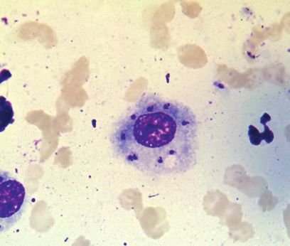
ESCCAP
                   Malvern Hills Science Park, Geraldine Road, Malvern,
                       Worcestershire, WR14 3SZ, United Kingdom

                             First Published by ESCCAP 2012

                                  © ESCCAP 2012–2019

                                     All rights reserved

    This publication is made available subject to the condition that any redistribution or
     reproduction of part or all of the contents in any form or by any means, electronic,
         mechanical, photocopying, recording or otherwise is with the prior written
                                     permission of ESCCAP.

    This publication may only be distributed in the covers in which it is first published
                   unless with the prior written permission of ESCCAP.

        A catalogue record for this publication is available from the British Library.

                                 ISBN: 978-1-907259-69-2

2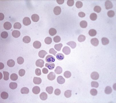
TABLE OF CONTENTS

INTRODUCTION                                            5

1. CONSIDERATION OF PET HEALTH AND LIFESTYLE FACTORS    8

2. PREVENTION AND CONTROL OF VECTOR-BORNE DISEASES      9

2.1. Insect-borne diseases                              9

2.1.1. Canine leishmaniosis                             9

2.1.2. Dirofilariosis and other filarial infections    17

2.1.3. Bartonellosis                                   24

2.1.4. Viral infections                                26

2.2. Tick-borne diseases                               26

2.2.1. Babesiosis (Piroplasmosis)                      26

2.2.2. Ehrlichiosis                                    30

2.2.3. Anaplasmosis                                    32

2.2.4. Borreliosis (Lyme disease)                      35

2.3. Vector-borne viral diseases                       37

APPENDIX 1 – BACKGROUND                                40

             Control of Vector-Borne
  5          Diseases in Dogs and Cats
  ESCCAP Guideline 05 Third Edition – March 2019

                                                            3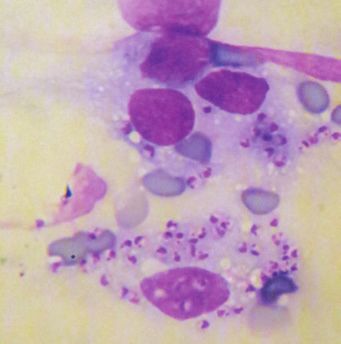
TABLES

    Table 1: Insect-borne infectious agents of dogs and cats in Europe                             6

    Table 2: Tick-borne infectious agents of dogs and cats in Europe                               7

    Table 3: Leishmania species infecting dogs and cats in Europe                                  9

    Table 4: Chemotherapy of canine leishmaniosis                                                 14

    Table 5: Filarial species infecting dogs and cats in Europe                                   17

    Table 6: Morphological features of blood microfilariae from filarial worms of dogs and cats   21

    Table 7: Prevention of dirofilariosis in dogs and cats in Europe                              22

    Table 8: Babesia species of dogs and cats and their vectors in Europe                         26

    Table 9: Distribution of canine Babesia spp. in Europe                                        27

    Table 10: Clinical manifestations of canine babesiosis                                        28

    Table 11: Chemotherapy of babesiosis in dogs                                                  29

    Table 12: Anaplasma spp. affecting dogs and cats in Europe                                    32

    Table 13: Distribution of pathogenic Anaplasma spp. in Europe                                 33

    Table 14: Clinical and laboratory findings of pathogenic Anaplasma infections in dogs         33

    Table 15: Vector-borne viruses which can affect dogs or cats in Europe                        37

    Table 16: Distribution of vector-borne viral infections affecting dogs and cats in Europe     38

    Table 17: Clinical manifestations of vector-borne viral infections in dogs                    38

    FIGURES

    Figure 1: Leishmania life cycle                                                                9

    Figure 2: Approximate distribution of canine leishmaniosis in Europe                          11

    Figure 3: Leishmania amastigotes                                                              12

    Figure 4: Dirofilaria immitis life cycle                                                      18

    Figure 5: Dirofilaria repens life cycle                                                       18

    Figure 6: Approximate distribution of Dirofilaria immitis and Dirofilaria repens in Europe    19

    Figure 7: Flea life cycle                                                                     24

    Figure 8: Babesia life cycle                                                                  27

    Figure 9: Babesia canis piroplasms in red blood cells                                         28

4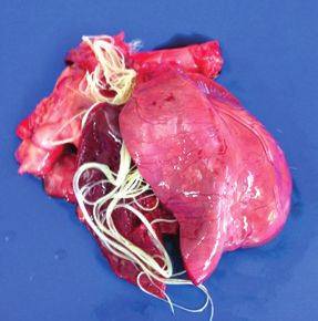
INTRODUCTION
Vector-borne diseases are caused by a wide range of infectious agents including viruses, bacteria and
parasites (protozoa and helminths), which are transmitted by a variety of arthropod vectors such as ticks,
lice, fleas and Diptera (mosquitoes, phlebotomine sand flies1, muscid flies).
Vector-borne pathogens or diseases are important because:
They may be highly pathogenic in dogs and cats.
Their transmission is often unpredictable.
Their diagnosis and control are difficult.
Variable clinical signs can develop after long incubation periods and these are rarely pathognomonic.
Animals may have persistent infections and thus act as reservoirs.
Several are important zoonoses, such as leishmaniosis, borreliosis, rickettsiosis, bartonellosis,
anaplasmosis and dirofilariosis.
Climatic and ecological changes, national regulations on the management of stray dogs and cats together
with an increase in pet travel and translocation of pet animals can influence the epidemiological situation
of vector-borne diseases in Europe. Rare diseases may increase in frequency in certain areas, either due
to the increased importation of infected animals or because the causative agents and their vectors spread
to and establish in previously non-endemic areas. Such an expansion of endemic areas has been recorded
for various parasitic diseases such as dirofilariosis, babesiosis and leishmaniosis. Babesiosis, for example,
has been observed across central Europe in the past few years, emerging from previous endemic regions
in Europe. Another important feature of these diseases is their increasing occurrence in wild animals, which
could act as reservoirs.
The effective control of vector-borne diseases requires a thorough knowledge of the infectious agents, their
vectors and major hosts. Besides giving an overview of the majority of vector-borne diseases of dogs and
cats, this guideline focuses on the following important infections/diseases: leishmaniosis, dirofilariosis,
bartonellosis, babesiosis, ehrlichiosis, anaplasmosis and vector-borne viral diseases.
The following vector-borne diseases are not presented in detail in this guideline, but are mentioned here and
in the tables:
Haemoplasmosis (formerly haemobartonellosis) – small Gram-negative bacteria, mycoplasmas
or haemoplasmas, that attach to the surface of red blood cells, e.g. Mycoplasma haemocanis and
M. haemofelis, in dogs and cats, respectively. Other less pathogenic species have been described mainly
in cats: Candidatus Mycoplasma haemominutum and Candidatus Mycoplasma turicensis, but also in
dogs: Candidatus Mycoplasma haematoparvum. Although their mode of natural transmission is still not
known, ticks and fleas could be implicated and direct transmission may also be possible.
Rickettsiosis (e.g. Rickettsia conorii, R. slovaca, R. felis) – small intracellular Gram-negative bacteria that
typically cause fever in the acute phase in susceptible hosts. They are transmitted by many arthropods.
Hepatozoonosis (e.g. Hepatozoon canis) – protozoal pathogens of dogs transmitted by the ingestion of
an infected tick.
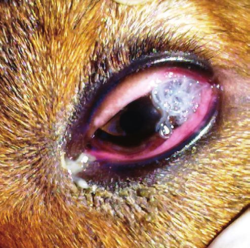
Onchocercosis (Onchocerca lupi) is an ocular disease of varying severity in dogs. This filarioid has also
been recognised as a zoonotic agent, though the information about the biology and epidemiology of this
infection is largely unknown.
Thelaziosis (Thelazia callipaeda) – a nematode located in the conjunctival sac which is transmitted by
drosophilid flies.
1
phlebotomine sand flies – within Europe Psychodid sand flies of the genus Phlebotomus are responsible for the transmission of
Leishmania infantum. These will be referred to throughout the text as phlebotomes.
5Table 1: Arthropod-borne infections of dogs and cats in Europe
Disease or Causative agents Vector1 Host Geographic Severity of
infection distribution clinical signs
DISEASES CAUSED BY PROTOZOA
Leishmaniosis Leishmania Phlebotomes Dogs, cats, foxes, Southern Europe Subclinical–severe
infantum hares, humans,
other mammals
DISEASES CAUSED BY HELMINTHS
Dipylidiosis Dipylidium caninum Fleas, lice Dogs, cats, foxes, Europe Subclinical
Filariosis humans
Dirofilaria immitis Culicidae Dogs, cats, foxes, Southern and parts Subclinical–severe
humans of central Europe
D. repens Culicidae Dogs, cats, foxes, Southern, central Minor–moderate
humans and eastern Europe
Acanthocheilonema Fleas Dogs, foxes Spain, France, Italy, Minor
dracunculoides & (A. reconditum), Portugal, Greece
A. reconditum louse flies,
Culicidae and
Rhipicephalus
sanguineus
(A. dracunculoides)
Thelaziosis Thelazia callipaeda Drosophilid flies Dogs, cats, foxes, Italy, France, Minor–moderate
(Phortica spp.) wolves, humans, Switzerland, Spain,
other mammals Portugal, Romania,
Germany
BACTERIAL INFECTIONS OR DISEASES
Rickettsiosis Rickettsia felis Fleas Dogs, cats, Europe Subclinical–
Other hedgehogs, moderate
humans
Bartonellosis (cat Bartonella henselae Fleas, (ticks) Cats (reservoir Europe Subclinical–minor
scratch disease) host), humans
Bartonellosis (dog Bartonella vinsonii Arthropod vectors Dogs Europe Moderate–severe
endocarditis) and others
Haemoplasmosis Mycoplasma Fleas (ticks) Cats, dogs Europe Cats: minor–severe
haemofelis (cats), suspected Dogs: subclinical
M. haemocanis
(dogs) and others
Tularaemia Francisella Ticks, mosquitoes, Cats (dogs), other Europe Subclinical–severe
tularensis Tabanidae mammals, humans
VIRAL INFECTION
West Nile virus West Nile virus Culex spp. and Horses, humans, Romania, Czech Subclinical–severe
infection (WNV), Flavivirus other mosquitoes (dogs, cats); Republic, Italy,
reservoir: birds France, Portugal,
Greece, Spain
1
non-insect vectors given in parentheses.
6Table 2: Tick-borne infectious agents of dogs and cats in Europe
Disease Causative agents Vectors Hosts Geographic Severity of
distribution clinical signs
DISEASES CAUSED BY PROTOZOA
Babesiosis Babesia canis Dermacentor Dogs, wolves Western, southern Moderate–severe
(piroplasmosis) reticulatus and central
Europe up to the
Baltic following
distribution of
vector
B. vogeli Rhipicephalus Dogs Southern Mild–moderate
sanguineus Europe following
distribution of
vector
B. gibsoni and Haemaphysalis spp., Dogs, wolves Sporadic and rare Moderate–severe
B. gibsoni- like Dermacentor spp. in Europe
Babesia microti- Ixodes hexagonus2 Dogs, foxes Northwestern Moderate–severe
like/Babesia vulpes Spain, Portugal,
Italy, Croatia,
France, Sweden
Hepatozoonosis Hepatozoon canis1 Rhipicephalus Dogs, foxes Mainly southern Mostly mild
sanguineus Europe infection; subclinical
Hepatozoon felis, Unknown Cats Spain, Portugal Subclinical
Hepatozoon spp.
DISEASES CAUSED BY NEMATODES
Filariosis Acanthocheilonema Rhipicephalus Dogs, cats Southern Europe Minor
(Dipetalonema) sanguineus3
dracunculoides,
Acanthocheilonema
(D.) reconditum,
Cercopitiphilaria spp.
DISEASES CAUSED BY BACTERIA
Bartonellosis Bartonella henselae, Many animals, Fleas suspected3, Throughout Europe Commonly
Bartonella vinsoni, dogs, cats, humans organism found in subclinical infection
Bartonella spp. Ixodes spp. ticks
Borreliosis Borrelia burgdorferi Many animals Ixodes ricinus, Throughout Europe Mostly subclinical
(Lyme disease) complex (especially especially rodents, I. hexagonus,
B. garinii and dogs, cats, humans I. trianguliceps,
B. afzelii in Europe) I. persulcatus
Ehrlichiosis Ehrlichia canis Dogs (cats) Rhipicephalus Southern Europe Moderate–severe
(monocytic) sanguineus following distribution
of vector
Neoehrlichiosis Candidatus, Rodents, humans, Ixodes ricinus Europe Unknown
Neoehrlichia dogs
mikurensis
Anaplasmosis Anaplasma Many animals, Ixodes ricinus, Throughout Europe Mild and subclinical
(granulocytic phagocytophilum dogs, cats, humans I. trianguliceps infections common
ehrlichiosis)
Anaplasmosis Anaplasma platys Dogs Rhipicephalus Southern Europe Commonly
(infectious cyclic sanguineus following distribution subclinical
thrombocytopenia) of vector infections
Rickettsial Rickettsia conorii Dogs Rhipicephalus Southern Europe Commonly
infections sanguineus following distribution subclinical infection
(Mediterranean of vector
spotted fever/MSF)
Coxiellosis Coxiella burnetii Ruminants, dogs, Ixodes spp.,3 Throughout Europe Subclinical Infection
(Q Fever) cats, humans Dermacentor spp.
Tularaemia Francisella Lagomorphs, cats Ixodes spp.,3 Southern Europe Subclinical infection,
tularensis Dermacentor spp., occasionally
Haemaphysalis moderate to severe
spp., Rhipicephalus in young cats
sanguineus
DISEASES CAUSED BY VIRUSES
European tick- TBE virus, Many animals, Ixodes ricinus, Central, eastern and Can be moderate–
borne encephalitis (Flavivirus) rodents, dogs I. trianguliceps, northern Europe severe but not
I. persulcatus commonly reported
Louping ill infection Louping-ill virus, Many animals, Ixodes ricinus, UK, Ireland Can be moderate–
(Flavivirus) mainly sheep, dogs I. trianguliceps severe but not
commonly reported
1
Transmission of Hepatozoon spp. is by ingestion of an infected tick and not a tick bite.
2
Potential competent vector but not yet experimentally demonstrated.
3
Ticks are not the sole arthropod vectors for these diseases.
71. CONSIDERATION OF PET HEALTH AND LIFESTYLE FACTORS
Animals require care tailored to their individual needs. Certain factors may dictate more intensive monitoring
and/or treatment, while others may warrant a less aggressive approach.
Animal
The age and health status of the animal are important, including its history and origin. Some breeds or
individuals have a genetically-determined susceptibility to some diseases such as leishmaniosis, while other
concomitant infections may predispose to, or aggravate, vector-borne diseases.
Environment
Dogs and cats housed in kennels and catteries or animals living outdoors may be at greater risk of acquiring
vector-borne diseases compared to individual animals living indoors. Fleas and R. sanguineus however,
frequently live and may establish infestations indoors.
The risk of transmission may also depend on various local conditions such as (micro-) climate and local
topography.
Nutrition
Poor nutrition may increase susceptibility to many diseases including vector-borne diseases.
Location and travel
Dogs and cats living in, or travelling to, specific geographical areas endemic in certain vector-borne diseases
are at a higher risk of infection. The following situations could present a higher risk:
animals travelling with their owners on holiday,
animals being rehomed,
animals going to boarding facilities,
animals attending dog and cat shows,
animals taken into fields for countryside walks
animals taking part in hunting activities.
Guests visiting with their pets can also bring parasites into environments, causing infestations in pets that
don’t travel.
Transmission by blood transfusion
Veterinary surgeons should be aware that most of these infections may be present in the blood of animals
that appear healthy. It is especially important to avoid causing iatrogenic infection from these individuals. In
particular, animals that are to act as blood donors should be screened and demonstrated to be seronegative/
polymerase chain reaction (PCR) negative (depending on the screened pathogen) for relevant infections prior
to donating blood.
82. PREVENTION AND CONTROL OF VECTOR-BORNE DISEASES
2.1. Insect-borne diseases
2.1.1. Canine leishmaniosis
2.1.1.a. Agents and vectors
In Europe, canine leishmaniosis is predominantly caused by Leishmania infantum which comprises various
enzymatic types (zymodemes). Other species (L. tropica, L. major) have rarely been diagnosed (Table 3).
The vectors are several species of blood-sucking flies of the genus Phlebotomus (Phlebotominae subfamily;
phlebotomes = sand flies).
Dogs are considered the main reservoir of L. infantum infection but cats can also be hosts. Many other
mammalian species can become infected including humans and this parasite has been isolated from various
mammals such as rodents (e.g. rats and squirrels), hares and rabbits. Horses, goats, sheep, cats and wild
canids including foxes, wolves and jackals can become infected but the epidemiological role of these hosts
has not yet been clearly established.
sand fly takes a macrophages phagocytize
blood meal and the promastigotes
injects promastigote
into the skin promastigotes
transform into
amastigotes
and multiply in
macrophages
promastigotes
divide in the the cell ruptures and
gut and migrate free amastigotes
to proboscis multiply in new
macrophages and
infect other body cells
amastigotes in the
skin macrophages
amastigotes transform
into the promastigote
stage in sand fly gut sand
sandfly
flytakes
takesaablood
bloodmeal
mealand
andingests
ingest
macrophages infected with amastigotes
Figure 1: Leishmania life cycle
The development of phlebotomes takes place in terrestrial habitats. Eggs are laid in soil rich in organic
matter and the larvae develop through four instars before they pupate and the adults emerge (Figure 1).
The seasonal dynamics of phlebotomes have not been fully explored; however, it is known that some
palaearctic species overwinter as 4th stage larvae. Phlebotomes have a nocturnal circadian activity with
most species seeking their hosts after sunset. Activities vary from species to species and within their habitat.
During the day, adult phlebotomes rest in dark, humid places especially cracks and holes in stone walls, wood
piles, animal stables and basements or dark cellars of houses.
9Phlebotomes are widespread in the Mediterranean region, Africa and the Middle East. They are well adapted,
depending on the species, to tropical or subtropical climates and even to arid habitats. Furthermore, it has
been known for decades that the Phlebotomus perniciosus endemic area extends up to northern France, and
this species was found in localised areas in southern Germany and southern Switzerland.
Table 3: Leishmania species infecting dogs and cats in Europe
Causative agent Vector Hosts
Leishmania infantum Phlebotomus spp. (sand flies) e.g. P. perniciosus, Dogs, foxes, jackals, hares, rabbits, rodents, cats,
(variety of zymodemes) P. ariasi, P. perfiliewi, P. neglectus, P. tobbi, various other mammals, humans
P. langeroni
L. tropica P. sergenti, P. arabicus Dogs, humans, hyraxes
L. major P. papatasi Rodents, dogs, humans
2.1.1.b. Biology and transmission
Leishmania spp. occur and multiply in two well-differentiated forms: intracellular amastigote stages infecting
cells of the vertebrate host and extracellular flagellated promastigote stages in the gut of phlebotomes.
Leishmania spp. are highly vector-specific and are transmitted by the blood-sucking females of several
Phlebotomus species while feeding on their hosts. Vector activity is linked to a minimum temperature of
15°C.
The parasite development in the vector is temperature dependent and lasts around 7–14 days at
temperatures above 18°C.
Other ways of transmitting Leishmania not dependent on phlebotomes have been reported: intra-uterine
from mother to offspring, via infected blood donors or through venereal transmission. However, their
epidemiological significance is still unknown. In non-endemic areas, these alternative transmission routes
might explain infections occurring in the absence of a competent vector. Furthermore, direct transmission
through biting, wounds or transmission via other hematophagous arthropods (e.g. ticks, fleas) have been
postulated, but remain unproven.
There is some evidence of resistance in certain dog breeds (e.g. the Ibizian hound) as well as susceptibility
of other breeds to disease development (e.g. German Shepherds, Rottweilers, Cocker Spaniels and Boxers)
but no sex- or age-dependent risks have been described. Infected but clinically healthy dogs, including
those which have undergone successful chemotherapy, may represent potential parasite carriers.
The incubation period can vary between three months to years and is dependent on the individual immune
response of the infected dogs.
After local multiplication of parasites in dendritic cells and macrophages in the skin, dissemination primarily
occurs via the lymphatic system and blood. Parasites can be found mainly in the skin, lymph nodes,
spleen, liver, bone marrow and many other organs or body fluids (e.g. intestine, saliva, semen, conjunctiva
and urine).
Clinical signs are only observed in a low proportion of infected dogs. Infected but clinically healthy dogs
represent an important reservoir of infection for phlebotomes.
The main risks in endemic areas are related to vector exposure and abundance of reservoir hosts which
include dogs living outdoors, stray dogs, hunting dogs and dogs adopted from animal shelters in endemic
areas being rehomed in non-endemic areas. Different studies suggest that cats might act as alternative
reservoir hosts of L. infantum based on the PCR detection of infections in peripheral blood in up to 20% of
cats in Portugal and 60% in Sicily; however, few of them had shown clinical disease. Further investigations
are required to confirm the possible role of cats in L. infantum transmission.
102.1.1.c. Distribution in Europe
Canine leishmaniosis is endemic in southern Europe with prevalence rates of infection of up to 60% in
exposed populations. Figure 2 shows the approximate northern limit of the endemic area. Outside this area,
many imported cases of canine leishmaniosis and a few cases in cats have been diagnosed and treated.
However, there are a few reports of isolated cases in dogs which did not travel through or reside in endemic
areas. Most probably, focal transmission can occur for a limited period of time if there is sufficient infection
pressure from imported infected dogs and competent vectors.
Endemic countries
Autochtonous cases, non-vectorial transmission
(vertical, transfusion, dog biting) and imported cases
Imported cases
No reported cases/ no data available
Canary Islands
Figure 2: Approximate distribution of canine leishmaniosis in Europe
112.1.1.d. Clinical signs/laboratory abnormalities
In endemic areas, a large number of infected dogs may be clinically healthy.
Clinical signs are highly variable depending on immune responses, disease history and possibly many other,
as yet unknown, factors. Local cutaneous lesions at the site of the initial phlebotome bites are often the
first signs observed before disseminated infection occurs. Typical Phlebotomus bite sites include the ear
pinnae, nose and abdomen. The localised lesions sometimes go unnoticed or are misdiagnosed as tick or
simply insect bites. They consist of single or several specific papular to ulcerative lesions, called chancres or
“chancre d´inoculation”. They last several weeks but are self-limiting. During this period, infected dogs may
remain seronegative but later around 25% seroconvert and the disease becomes generalised. In affected
dogs, enlargement of single or multiple lymph nodes may be evident accompanied by weight loss, anorexia
and weakness.
More severe clinical signs may develop and the disease can be fatal if therapy is not instituted. Severe
clinical signs include skin lesions like alopecia, nodules, ulcers, hyperkeratosis, intense exfoliative
dermatitis, mucocutaneous lesions and onychogryphosis (nail hyperplasia). Generalised cutaneous forms
of the disease are normally non-pruritic and are most often keratoseborrhoeic but may also be ulcerative,
papular or pustular or, less frequently, nodular. General signs might include loss of body weight, asthenia
(weakness), pallor of mucous membranes, muscular atrophy, splenomegaly, epistaxis and haematuria.
Other clinical signs may include gastrointestinal disorders (vomiting, diarrhoea and chronic colitis), lameness
(polyarthritis, osteomyelitis, polymyositis), vascular disorders (systemic vasculitis, arterial thromboembolism),
glomerulonephritis (polyuria and polydipsia), ocular lesions (blepharitis, conjunctivitis, keratoconjunctivitis,
anterior uveitis). Even uncommon but already reported cardiorespiratory and neurological disorders may
occur in some cases.
Although the clinicopathological abnormalities may be variable, there are many common findings such
as a normocytic normochromic non-regenerative anaemia and, less frequently, thrombocytopenia and
leucopenia. Plasma protein changes with hyperglobulinaemia and hypoalbuminaemia are particularly
common. Proteinuria and variable azotaemia with an increase in the urine protein/creatinine ratio, due
essentially to immune-mediated glomerulonephritis, are present in some sick dogs and are considered an
indicator of poor clinical prognosis.
2.1.1.e. Diagnosis
To reduce the potential for transmission of
Leishmania from dogs to vectors, the diagnosis
should be confirmed and treatment instituted as early
as possible. Clinical signs, together with relevant
epidemiological information and various laboratory
test abnormalities strongly indicate a tentative
diagnosis. CBC, biochemical profile and urinalysis
including a urine protein/creatinine ratio (only in
proteinuric dogs) should always be performed.
Direct diagnosis is possible by detecting the
amastigote stages in Giemsa or Diff-Quick
stained smears obtained from superficial lymph
nodes or bone marrow aspirates or by observing
the promastigotes after in vitro culturing of samples
(Figure 3). The sensitivity of parasite detection is Figure 3: Leishmania infantum amastigotes in a
macrophage from a lymph node aspirate
lower with skin biopsies and is generally reduced in
clinically healthy, infected dogs, but can be increased
by molecular or immunohistochemical techniques.
12Polymerase chain reactions (PCRs), mostly targeting repetitive sequences, have proven to be highly sensitive
compared with laborious in vitro cultivation, combined with the fact that they are not impaired by microbial
contamination. However, the diagnostic sensitivity is dependent on the quality of the clinical samples. Lymph
node aspirates, especially from animals with lymphadenopathy, are most convenient while bone marrow
sampling is more invasive but may be indicated for special cases such as suspect but clinically unremarkable
animals. Blood samples and conjunctival swabs can be used in clinical cases but the diagnostic sensitivity
is low while skin biopsies have been shown to be a useful alternative for sensitive molecular diagnosis.
Quantitative PCR allows the parasitic load to be estimated in comparable tissues, which could be useful for
follow-up during treatment although this approach needs to be thoroughly evaluated.
Serology is the most commonly used first step allowing the detection of a specific antibody response
in dogs around 12 weeks after initial infection. In subclinical infections, this period may extend to years.
Different laboratory-based methods have been used to detect anti-Leishmania antibodies such as the
indirect fluorescent antibody test (IFAT), enzyme linked immunosorbent assays (ELISA) and Western blot
(WB). Both the sensitivity and specificity of these tests vary according to the defined cut-off values in different
laboratories. Point-of-care tests (“rapid tests”) based on immunochromatographic methods have been
developed and many commercial in-house kits are now available for practitioners for a qualitative diagnosis
in the clinic or for use in epidemiological field studies. These tests have a reasonable sensitivity for the initial
detection of seropositive dogs. However, for the confirmation of clinical cases and for clinical management
post chemotherapy, especially in animals with low specific antibody reactions, methods allowing quantitative
estimations (e.g. IFAT, ELISA) are required. IFAT, ELISA and rapid test results based on whole antigens have
to be carefully interpreted in non-DIVA vaccinated dogs (differentiating infected from vaccinated animals)
because recently-immunised dogs may remain positive for up to 6 months.
2.1.1.f. Control
Treatment
Before initiating chemotherapy, animal owners should be informed about the prognosis, treatment cost and
the fact that the dog remains infected even when a clinical cure is achieved. Furthermore, there are certain
country-specific veterinary public health regulations that have to be respected. Although euthanasia of infected
dogs is not mandatory in any European country, there is an obligation for practitioners to communicate all
new cases to the appropriate authorities in some countries such as Portugal, Italy and Greece.
Indications for treatment
Indication for treatment includes dogs with clinical signs and clinicopathological abnormalities associated
with a positive serology and/or the evidence of the parasite in target organs. The drugs which are mostly
used for the treatment of canine leishmaniosis are listed in Table 4 (see www.esccap.org for links to approved
products for specific countries). Generally, in non-endemic areas, single drug treatments with allopurinol or
meglumine antimoniate or, more recently, with miltefosine, have been used successfully. In European endemic
areas with a high seasonal infection pressure, combined therapy is recommended (see Table 4).
Besides specific therapy, supportive treatment together with an appropriate diet is recommended.
A commercially available diet for clinically affected dogs without signs of renal disease is available containing
moderate protein levels supplemented with omega acids, zinc sulphate and antioxidants.
An improvement may be observed within a few weeks after beginning chemotherapy but clinical cure is only
achieved after several months. As the Leishmania infection is not eliminated by treatment with currently available
compounds, relapses are common. First indicators of relapse are clinical signs and/or clinicopathological
abnormalities associated with the disease, together with a significant rise in specific antibody reactions by
ELISA or by IFAT (a 2–4 fold increase in titre) when examined by the same laboratory.
If there is no clinical improvement following a course of treatment, an alternative drug or a different dosage
should be considered. Alternatively, the diagnosis should be queried or the animal should be examined
for the presence of concomitant vector-borne diseases such as ehrlichiosis, anaplasmosis, babesiosis,
hepatozoonosis, or other diseases such as endocrinopathies, neoplasias, or immune-mediated diseases, all
of which may affect the response to treatment.
13Table 4: Chemotherapy of canine leishmaniosis
Drugs* Dosage Route of administration
Meglumine antimoniate 50 mg/kg (BID) for 4–6 weeks Subcutaneous injection
Allopurinol** 10 mg/kg (BID) for 6–18 months Oral
Miltefosine 2 mg/kg once daily for 4 weeks (with food) Oral
Meglumine antimoniate + allopurinol** see above for both compounds Subcutaneous injection + oral
Miltefosine + allopurinol** see above for both compounds Both oral
* Most cytotoxic drugs are teratogenic in humans therefore gloves must be worn when administering.
** Not registered for veterinary use in Europe.
Numerous pharmacokinetic studies have shown that administration of meglumine antimoniate by
intramuscular or subcutaneous injection is more effective in maintaining sustained drug plasma concentrations
than intravenous injections. After intravenous administration, plasma concentrations fall within two hours
whereas they fall within four hours after intramuscular administration. When injected subcutaneously, plasma
concentrations rise after five hours and remain at therapeutic levels for at least 12 hours. It must be stressed
that repeated intramuscular injections frequently lead to the development of painful oedematous reactions
and myositis and are therefore not recommended; subcutaneous injections, being safer and painless, are
preferred, although some side effects may be presented (potential nephrotoxicity and panniculitis at the
injection site). Different dosages of meglumine antimoniate have been tested but the most widely accepted
regimen is indicated in Table 4.
Allopurinol is commonly administered twice daily in a whole dose of 10 mg/kg bodyweight orally for 6–18
months with generally satisfactory results, a clinical cure being observed in most dogs within 6–12 months
of treatment. After a clinical cure has been achieved, it is advisable to stop treatment and monitor the dog
for possible relapses after three months and subsequently at six-monthly intervals. As with all other drugs,
relapses are relatively common but animals can generally be re-treated with the same compound. Some side
effects have been reported including the development of xanthine nephrolithiasis (few reports) and dogs on
long-term therapy with allopurinol should be checked using urinalysis and/or abdominal ultrasonography.
Usually, xanthinuria has a good prognosis and this side effect disappears shortly after reducing the dosage
or the cessation of treatment (if this is deemed necessary).
Moreover, miltefosine, an alkylphospholipid molecule, has shown therapeutic effectiveness comparable with
that of antimonial compounds. Side effects including vomiting, diarrhoea and anorexia of varying severity
have been reported but these are quick to resolve if the drug is administered with food.
Several clinical trials, combining two compounds, e.g. antimonials or miltefosine plus allopurinol, have shown
that they induce a clinical remission in most cases and partial reduction of the parasitic burden (see Table 4).
The use of non-specific immune modulatory drugs has been reported as potentiating the immune system
of sick dogs to control the infection and to prevent development of clinical disease in uninfected dogs.
Recently, domperidone, which has been launched in several European countries, has shown to manage the
early stages of the disease or to prevent development of clinical disease as part of an integrated control
programme. Further results, however, are needed in order to better evaluate these therapies.
The curative effects of some other drugs, either used alone or in combination, have been reported for the
treatment of canine leishmaniosis, with varying efficacy (aminosidine, furazolidone, marbofloxacin, perifosine,
oleylphosphocholine). None are currently recommended for use as first-line therapy but some would be
helpful as complementary drugs in some clinical cases (e.g. antibiotics). Amphotericin B, once recommended
for cases refractory to antimonials, is not well accepted due to its nephrotoxicity and the invasive intravenous
route of administration. Furthermore, it is a first-line drug for visceral leishmaniosis in humans and the
World Health Organization (WHO) and several public health committees have argued for restricted use of
Amphotericin B (liposomal formulations) in dogs to avoid selection for resistance.
14Resistance to drugs used for the chemotherapy of Leishmania infantum in dogs To date, resistance has been observed against meglumine antimoniate in vitro. Moreover, disease relapse in dogs with leishmaniosis during allopurinol treatment has recently been described and associated with allopurinol resistance of L. infantum isolated from relapsed animals. Further studies are needed to better evaluate this fact. Control strategies Some control strategies used in the past, such as the culling of seropositive dogs in endemic areas, have been shown to be ineffective in reducing Leishmania transmission. Prevention of phlebotome bites by the application of repellents/insecticides in the form of impregnated collars, spot-on and spray formulations is currently the most promising strategy; spray preparations have a short duration of effect. The basic objective is to interrupt parasite transmission and thus control the disease. The phlebotome season in endemic areas may vary from year to year and from region to region. As a general rule, in endemic areas, the season starts in April and continues until November. Numerous studies have assessed the efficacy of pyrethroids in preventing phlebotome bites. For example, it has been observed that dog collars impregnated with 4% deltamethrin possess a repellent effect against phlebotomes lasting from one week after application to over six months, thereby resulting in a significant decrease in the incidence of disease in endemic areas such as Italy or Spain over a period of 2–3 years. Applications of permethrin alone, or in combination with other insecticides as a spot-on, have also been shown to protect dogs against phlebotome bites within hours (i.e. after 24 hours) and for up to 3–4 weeks thus decreasing the incidence of canine leishmaniosis in endemic areas. Data of two clinical field studies performed in Leishmania infantum endemic areas using a slow-release imidacloprid (10%)/flumethrin (4.5%) collar indicate a significant reduction in the risk of Leishmania infection in treated dogs compared to non-treated dogs, while the efficacy of the product in the prevention of sand fly bites has not been established. These studies show that the interruption of Leishmania transmission through the external application of pyrethroids to dogs could be a major tool if incorporated into future disease control programmes in regions where pet dogs are the main reservoir of L. infantum. Finally, other control measures to reduce disease transmission include keeping dogs indoors during dusk and dawn throughout the whole risk season, the use of insecticidal room sprays, protective nets at windows and doors (mesh size
Moreover, in 2016 the European Commission approved the marketing of a new vaccine composed of Protein
Q, a recombinant protein constructed from the union of five antigenic fragments of four proteins of the parasite
Leishmania infantum. This vaccine can be given to seronegative dogs over six months of age as an initial
single injection followed by annual revaccination. The vaccine is indicated to reduce the risk of developing
an active infection, clinical disease or both, following exposure to L. infantum. This new vaccine does not
interfere with the detection of anti-L. infantum antibodies and thus allows the discrimination of vaccinated
from naturally infected dogs.
Resistance to repellents and insecticides: There are no reports of resistance of phlebotomes to pyrethroids.
2.1.1.g. Public health considerations
Human visceral leishmaniosis caused by L. infantum is an important vector-borne zoonotic disease in southern
Europe. Clinical cases of human leishmaniosis generally prove fatal without therapy, especially in children and
immunocompromised patients.
Evidence points to the spread and (re-)emergence of this disease in humans in some endemic areas. In
Madrid, there were more than 700 new clinical cases (visceral and cutaneous) reported between 2009 and
2016. The epidemiological and molecular typing-based studies of this outbreak suggest different genotypes
circulating among sand flies, dogs and other alternative hosts (mainly hares and rabbits). These scientific
studies have contributed to a better understanding of this important zoonosis.
The responsibility of veterinary practitioners must be to adequately manage the disease in dogs to reduce
parasite transmission since dogs are the main reservoir of infection.
The following principles must be stressed:
A thorough diagnostic procedure should be established to identify infected and/or sick dogs.
The best treatment for sick dogs should be chosen, bearing in mind the potential risks for the development
of resistance to “first line” drugs used in humans.
The use of topical insecticides should be recommended for all dogs at risk and especially for infected
clinically healthy or sick dogs even after successful chemotherapy; these should be applied throughout
the risk season which depends on climatic conditions. In the southern European endemic areas, the
risk season is between April and November. In warmer areas all year round use of repellents is strongly
recommended.
In endemic areas, kennels housing stray, hunting or breeding dogs should maintain a strict vector-borne
disease monitoring program; this should be combined with measures designed to prevent disease
transmission by phlebotomes and thus avoid the risk of focal, highly endemic transmission.
To avoid an extension of endemic areas, Leishmania-infected dogs should not be translocated to non-
endemic areas where phlebotomes may be present.
162.1.1.h. Feline leishmaniosis
Despite the high prevalence of canine leishmaniosis in endemic areas, feline leishmaniosis is frequently
subclinical. Over 80 clinical cases in cats have been described in literature in Europe, South America and
Texas, USA. These cases were characterised by cutaneous lesions (nodular and ulcerative lesions), enlarged
lymph nodes, weight loss and ocular lesions. Weight loss, reduced appetite, dehydration and lethargy are the
most frequent non-specific signs. The pathogenesis of feline leishmaniosis is not currently known and the
pattern of immune response to L. infantum infection has never been investigated in cats.
Xenodiagnoses studies performed in two chronically infected cats have shown that a cat can be infectious
to Phlebotomus, the competent L. infantum vector. However, the role of the cat in the transmission cycle of
leishmaniosis is not clear.
The diagnosis protocol is exactly the same as for the dog including parasitological, molecular and serological
techniques. The diagnostic evaluation should always be completed with CBC, a biochemical profile, urinalysis
with a quantitative evaluation of proteinuria and any other tests recommended for excluding other compatible
or concurrent diagnoses. Regarding therapy, a few leishmanicide drugs have been tested on cats but there
is no specific information on pharmacokinetic and pharmacodynamic studies of these compounds in cats.
Allopurinol is the most frequently used oral drug at a dosage of 10–15 mg/kg bodyweight twice a day for
several months.
Finally, in terms of prevention, there are currently no drugs available for cats to avoid phlebotomine bites and
to prevent Leishmania transmission. Permethrin cannot be used as a preventative in cats due to its toxicity
for this species. Flumethrin collars have low sand fly repellency properties and may be an off-label option.
Further information about canine and feline leishmaniosis can be found on the LeishVet website at
www.leishvet.org
2.1.2. Dirofilariosis and other filarial infections
2.1.2.a. Agents and vectors
Filarial worms are nematodes infecting the connective tissues and vascular system of dogs and cats.
Mosquitoes, but also fleas and ticks, act as vectors for the different species (Table 5). Dirofilaria immitis, the
canine and feline heartworm, is the most pathogenic species, while D. repens, which causes subcutaneous
dirofilariosis, is the most important species responsible for zoonotic infections in Europe.
Table 5: Filarial species infecting dogs and cats in Europe (see Table 6 for morphology of microfilariae)
Filarial parasite Vectors Prepatent period Length of adult worms Location of adult
worms
Dirofilaria immitis Mosquitoes (Culicidae) 120–180 days M: 12–18 cm Pulmonary arteries/
F: 25–30 cm right heart
Dirofilaria repens Mosquitoes (Culicidae) 189–259 days M: 5–7 cm Subcutaneous tissue/
F: 10–17 cm muscular fasciae
Acanthocheilonema Fleas and ticks 427–476 days M: 9–17 mm Subcutaneous tissue/
(formerly Dipetalonema) F: 21–25 mm muscular fasciae,
reconditum peritoneal cavity, kidney
Acanthocheilonema Fleas and ticks 120 days M: 15–31 mm Peritoneal cavity
(formerly Dipetalonema) (R. sanguineus) F: 33–55 mm
dracunculoides
Cercopithifilaria spp. Ticks (R. sanguineus) Unknown M: unknown Subcutaneous tissue/
F: 23–24 mm muscular fasciae
M: male; F: female
172.1.2.b. Biology and transmission mosquito feeds
and infective larvae
enter the wound
Filarial nematodes are parasites of domestic and
wild carnivores, mainly canids. However, due to humans can also
become infected
the low host specificity of their arthropod vectors, larvae migrate
to proboscis
many mammalian hosts can be infected, including of mosquito
adult parasites
humans. In such hosts, the parasites generally do sexually reproduce in the
not develop to the adult stage. vertebrate host
Dirofilaria immitis and D. repens microfilariae
are released by female worms into the blood
stream where they become available to blood- larvae develop
inside the mosquito
sucking mosquitoes. Microfilariae develop to
the infective stage (L3) and are transmitted via
saliva during feeding. Dirofilaria immitis larvae
mosquito feeds
undertake an extensive migration to reach the and microfilariae
pulmonary arteries and the right heart where are transferred
they develop into the adult stages and mate.
In dogs, adult worms have a lifespan of up to Figure 4: Dirofilaria immitis life cycle
seven years (although survival in cats is shorter),
and microfilariae survive 2–18 months in the
mosquito feeds
bloodstream. The infective larvae of D. repens and infective larvae
migrate into the subcutaneous connective tissues enter the wound
where they reach maturity. Adult worms are found
between subcutaneous and deep connective humans can also
larvae migrate become infected
tissue layers in most parts of the body. Adults can to proboscis
live for several years (Figures 4 and 5). of mosquito
adult worms
mature in subcutaneous
Acanthocheilonema (syn. Dipetalonema) connective tissues
reconditum is found in subcutaneous tissues and
fasciae, the peritoneal cavity and the kidneys of
canids, Cercopithifilaria grassii is a parasite of the
subcutaneous tissues and fasciae of canids and larvae develop
A. dracunculoides is a parasite of the peritoneal inside the mosquito
cavity. For treatment and prevention purposes,
circulating microfilariae of these species must
be differentiated from those of D. immitis and mosquito feeds
and microfilariae
D. repens. are transferred
Figure 5: Dirofilaria repens life cycle
182.1.2.c. Distribution in Europe
The frequency of transmission and the spread of Dirofilaria spp. infections depend upon environmental factors
such as temperature, the density of vector populations and the presence of microfilaraemic dogs, which are
the main reservoirs of infections. Due to tourism and animal adoption, infected dogs are increasingly being
moved from endemic areas such as Portugal, Spain, Italy and Greece to non-endemic areas.
Dirofilaria immitis
Dirofilaria repens
Canary Islands
Figure 6: Approximate distribution of Dirofilaria immitis and Dirofilaria repens in Europe (© ESCCAP)
Dirofilaria immitis is endemic across southern Europe and in the Czech Republic, Slovenia, Romania and
Bulgaria (Figure 6). The endemic areas of D. immitis and D. repens overlap in many regions. Recently,
D. repens infections in dogs that had never left Germany, Austria or Poland have been documented.
Feline Dirofilaria infections occur in areas where canine infections are highly prevalent; the prevalence in cats,
however, is generally only a tenth of that in dogs.
Acanthocheilonema dracunculoides infections have prevalence rates of up to 14% in hunting dogs and dogs
living outdoors in some countries and regions of Europe such as Spain and southern Italy (Sicily).
192.1.2.d. Clinical signs
Infections with D. immitis may cause severe and potentially fatal disease in dogs and cats. Despite its name,
heartworm disease is essentially a pulmonary disease because the worms are predominantly located in the
pulmonary arteries and the right heart is involved only in the later stages.
DOG
Clinical signs of the disease caused by Dirofilaria immitis develop gradually and may begin with a chronic
cough which may be followed by moderate to severe dyspnoea, weakness and sometimes syncope after
exercise or excitement. At this stage, auscultation may reveal abnormal pulmonary sounds (crackles) over
the caudal lung lobes and a split second heart sound can often be heard. Later, when right congestive heart
failure is developing, ascites, and less often oedema in the limbs, may be observed together with anorexia,
weight loss and dehydration. Arterial damage is usually more severe in dogs that perform intensive physical
exercise. Sudden death is rare and usually occurs following respiratory distress or progressive emaciation.
During the chronic stages of the disease, there may be a sudden onset of acute signs. For example, after
severe spontaneous thromboembolism following the natural death of many heartworms, dogs may show
acute life-threatening dyspnoea and haemoptysis.
In small dogs, the displacement of adult worms from the pulmonary arteries to the right heart, due to pulmonary
hypertension and a sudden decrease in right cardiac output, is a common event. In such cases, affected
dogs present the so-called “caval syndrome”. Dyspnoea, a tricuspid cardiac murmur and haemoglobinuria, due to
mechanical haemolysis in the right cardiac chambers, are the most typical signs and the outcome is usually fatal.
CAT
Most cats show no clinical signs of Dirofilaria immitis for a long time after initial infection and may spontaneously
self-cure. Others may suddenly show a dramatic acute syndrome usually with respiratory signs such as
coughing, dyspnoea and haemoptysis; vomiting also frequently occurs. Sudden death in apparently healthy
cats is not an infrequent consequence of infection. In most cases, the onset of clinical signs seems to be
related to the natural death of parasites or to the arrival of pre-adult heartworms (L5) in the pulmonary arteries.
Feline heartworm disease is now recognised as a significant pulmonary syndrome defined as “Heartworm
Associated Respiratory Disease” (HARD). Clinical signs associated with HARD are anorexia, lethargy, weight
loss, coughing, rapid heart rate, vomiting, diarrhoea, blindness, convulsions, collapse and sudden death.
Dirofilaria repens is the most frequent species associated with subcutaneous filariosis of dogs and cats. In
some cases, subcutaneous, non-inflammatory nodules containing adult parasites or immature stages can
be observed. These “cold” nodules are not painful and appear free within the skin. The parasite may also be
observed during surgery in the perimuscular fasciae, perirenal fat or abdominal cavity or may migrate to the
ocular conjunctiva. Rarely, in cases of heavy infection and sensitised patients, pruritus, pustules, ulcerative
lesions and exfoliative dermatitis may be seen, associated with microfilariae in the skin.
Infections with A. reconditum, A. dracunculoides and C. grassii are mostly subclinical. Differentiation of all
species that produce microfilariae in the bloodstream is necessary for treatment and prevention purposes.
2.1.2.e. Wolbachia species and filarial worm symbiosis
Gram-negative bacteria of the genus Wolbachia are obligate endosymbionts of both D. immitis and D. repens.
These bacteria play an important role in the pathogenesis and immunology of heartworm infection. Wolbachia
can be eliminated from filarial worms through antibiotic therapy of the infected host (e.g. doxycycline). Such
depletion of Wolbachia is often followed by clear anti-inflammatory effects, thus, antibiotic treatment may be
used concomitantly with the use of adulticidal therapeutic agents.
2.1.2.f. Diagnosis
DOG
Heartworm infection in dogs can be detected with blood tests which demonstrate the presence of circulating
microfilariae or adult antigens in serum or plasma samples. Further diagnostic procedures are required to
determine the severity of disease and possible treatment options.
20Blood examination for microfilariae
Blood samples should be examined after concentration by the Knott or the filtration test and morphological
identification carried out (Table 6). NB Wet blood smears do not allow species identification and have a very
low sensitivity. Furthermore, negative test results for microfilariae cannot rule out infection as they are not
always present (occult infection). Molecular testing (PCR) is available in some laboratories to differentiate
species of filarial worms.
Table 6: Morphological features of blood microfilariae1 from filarial worms of dogs and cats
Species Length (µ) Width (µ) Features
Dirofilaria immitis 301.77 ± 6.29 5–7 No sheath, cephalic end pointed, tail straight with the end pointed.
290–330 APh-S: two activity spots located around the anal and the excretory pores.
D. repens 369.44 ± 10.76 6–8 No sheath, cephalic end obtuse, tail sharp and filiform often ending like an
300–370 umbrella handle. APh-S: one spot around the anal pore.
Acanthocheilonema 264.83 ± 5.47 4 No sheath, cephalic end obtuse with a prominent cephalic hook, tail button
reconditum 260–283 hooked and curved. APh-S: activity throughout the body.
A. dracunculoides 259.43 ± 6.69 4–6.5 Sheath, cephalic end obtuse, caudal end sharp and extended.
190–247 APh-S: three spots which include an additional spot in the medium body.
1
Microfilariae (n=10) measured after concentration by the Knott test; when using the Difil® test, lengths are shorter. APh-S: acid
phosphatase stain.
Blood/serological tests for adult female antigens
Antigens from adult female heartworms are present approximately 6–8 months post infection. Commercial
tests based on ELISA or immunochromatographic methods designed to detect these antigens are considered
highly specific and some of them can be used in-house for a quick diagnosis.
Radiographs, electrocardiography and echocardiography
Auxiliary evaluations are often useful in determining the extent of pulmonary and cardiac pathological changes
in infected dogs; mainly thorax X-Ray and echocardiography.
CAT
Blood examination for microfilariae
Detection of microfilariae in the blood of infected cats is unlikely to be successful, therefore the sensitivity
is very low.
Blood/serological tests for adult female antigens
Tests detecting adult female heartworm antigens have a very high specificity and can thus provide definitive
proof of infection. In many cases, however, these tests yield false-negative results because of low worm
burdens or the presence of only male or immature worms. A negative test therefore does not rule out infection.
Heat treatment of serum prior to antigen testing may increase the sensitivity of antigen testing in cats.
Thorax radiographic alterations
Thorax radiographic alterations compatible with echocardiography are very useful for the diagnosis of the
disease in cats. Cardiac ultrasound allows direct visualisation of the parasites in the right atrium and ventricle,
in the main pulmonary artery and in the origin of both its main branches.
The specificity is virtually 100%, and the sensitivity in cats is very high as only a short portion of the caudal
pulmonary arteries cannot be examined. Cardiac ultrasonography should always be performed when feline
heartworm infection is suspected.
212.1.2.g. Control
Treatment
Adulticidal therapy (D. immitis) in dogs:
I. The organic arsenical compound melarsomine dihydrochloride is the only effective drug available for treating
adult heartworm infections in dogs. The currently-accepted regimen is a two-step treatment to reduce the risk of
pulmonary thromboembolism: after one initial treatment of 2.5 mg/kg bodyweight, given by deep intramuscular
injection in the lumbar muscles, the recommended follow-up treatment is administered 30–60 days later (2.5
mg/kg bodyweight twice at an interval of 24 hours). Complications, due to pulmonary thromboembolism,
should be reduced by the restriction of exercise following treatment and by the administration of heparin and
a corticosteroid (e.g. prednisone at 0.5 mg/kg BID 1st week, 0.5 mg/kg SID 2nd week, 0.5 mg/kg EOD 3rd
and 4th weeks) after melarsomine dihydrochloride injections.
Wolbachia (obligate, intracellular, gram-negative, endo-symbiotic bacteria) has been implicated as crucial in
the pathogenesis of filarial diseases. Doxycycline reduces the Wolbachia burden in all stages of heartworms.
Thus, administration of doxycycline at 10 mg/kg daily for 4 weeks before the administration of melarsomine
dihydrochloride is strongly recommended.
II. Surgical intervention is advised when multiple worms have been displaced into the right cardiac chambers
producing sudden onset of caval syndrome.
Adulticidal therapy (D. immitis) in cats:
There is no registered adulticide drug for cats. Decreasing doses of prednisolone are advised in cats in order
to relieve respiratory distress with an initial dose of 2 mg/kg bodyweight per day. If a cat presents with severe
signs of HARD, high doses of oral prednisolone (1–2 mg/kg bodyweight 3 times a day) are recommended.
Adulticidal therapy in canine and feline D. repens infection:
Spot-on moxidectin is licenced in some European countries as an effective adulticide therapy for D. repens
infection in dogs. Because of the zoonotic potential of D. repens, microfilaraemic dogs should be treated
monthly for 12 months with preventative drugs able to kill microfilariae (see below).
Control strategies for dogs
Topical or oral macrocyclic lactones administered monthly throughout the transmission season are
effective against D. immitis third stage larvae (L3) and L4 which have developed within the previous 30
days thus preventing disease caused by adult worms. Several compounds alone, or in combination with
other parasiticides, are available for oral administration or topical application (Table 7); for links to approved
compounds in individual countries see www.esccap.org. An injectable, sustained-release macrocyclic lactone
has been approved in some European countries for use only in dogs older than six months and is registered
to give protection for one year.
Prevention, through monthly administration of macrocyclic lactones, should start before the mosquito season
in spring and continue until late autumn. Recently, topical administration of permethrin with dinotefuran has
shown repellent efficacy against mosquitoes on dogs for at least 4 weeks. In southern Europe, protection
against heartworm should be carried out from May until the end of November. In hyperendemic areas, a year-
round preventive therapy is recommended.
Table 7: Prevention of dirofilariosis in dogs and cats in Europe: minimal and maximal dosages of macrocyclic lactones
Compound Presentation Dog (min.–max. dosage) Cat (min.–max. dosage)
Ivermectin Tablets/chewable tablets 6–12 µg/kg 24–71 µg/kg
Milbemycin oxime Flavoured tablets 0.5–2.5 mg/kg 2–4 mg/kg
Moxidectin Tablets 3–6 µg/kg 1–2 mg/kg
Injectable 0.17 mg/kg
Topical 2.5–6.25 mg/kg
Selamectin Topical 6–12 mg/kg 6–12 mg/kg
22You can also read