Review: An update on clinical, genetic and pathological aspects of frontotemporal lobar degenerations - FTD Talk
←
→
Page content transcription
If your browser does not render page correctly, please read the page content below
Neuropathology and Applied Neurobiology (2015), 41, 858–881                                         doi: 10.1111/nan.12250

Review: An update on clinical, genetic and
pathological aspects of frontotemporal
lobar degenerations
T. Lashley*, J. D. Rohrer†, S. Mead‡ and T. Revesz*
*Queen Square Brain Bank for Neurological Disorders, Department of Molecular Neuroscience, †Dementia Research
Centre, and ‡Department of Neurodegenerative Disease, UCL Institute of Neurology, London, UK

T. Lashley, J. D. Rohrer, S. Mead and T. Revesz (2015) Neuropathology and Applied Neurobiology
An update on clinical, genetic and pathological aspects of frontotemporal lobar degenerations

The development of our understanding of frontotemporal              the clinical concept of FTD evolved and show that FTLD,
dementia (FTD) has gathered pace over the last 10 years.            once thought of as a single disorder, represents a hetero-
After taking a back seat to Alzheimer’s disease for many            geneous group of diseases with overlapping clinical symp-
years FTD has emerged as a significant group of heteroge-           toms, multiple causative genes and varying underlying
neous diseases often affecting people under the age of 65.          pathology. We also provide a brief summary of the clinical
FTD has also been brought into the spotlight as the major           manifestations, summarize the major genetic aspects and
disease entities of the group have clinical, genetic and            describe the main pathological features seen in the differ-
pathological links to motor neuron disease/amyotrophic              ent subtypes of FTLD. We also summarize the correlations
lateral sclerosis, indicating that they form a disease spec-        that exist between clinical presentations and pathological
trum. In this review, we overview how the pathological              variants. An overview of the main pathogenic mecha-
concept of frontotemporal lobar degeneration (FTLD) and             nisms is also provided.

Keywords: classification, frontotemporal dementia, frontotemporal lobar degeneration, FUS, pathology, tau, TDP-43

                                                                    The estimated prevalence of FTD is 15–22/100 000 and
Introduction
                                                                    population studies indicate an equal gender distribution
Frontotemporal lobar degeneration (FTLD) is a pathologi-            [3]. Clinically FTD patients can present with one of three
cal term used for the description of a clinically, pathologi-       canonical clinical syndromes: behavioural variant FTD
cally and genetically heterogeneous group of disorders, in          (bvFTD) and two language variants, semantic dementia
which relatively selective degeneration of the frontal and          and progressive nonfluent aphasia (PNFA) (see later). FTD
temporal lobes is a prominent and common feature [1].               can overlap with motor neuron disease/amyotrophic
The clinical term frontotemporal dementia (FTD) is used             lateral sclerosis (MND/ALS) (FTD-MND), corticobasal syn-
for the description of a group of early onset dementias,            drome (CBS) and progressive supranuclear palsy (PSP)
which is the second most common dementing disorder                  syndrome [4]. FTD is a highly heritable disorder with
among individuals under the age of 65 years [2].                    approximately 30–50% of cases reporting a positive
However, in 25% of the cases FTD presents in old age [3].           family history [5]. Mutations in three genes microtubule-
                                                                    associated protein tau (MAPT), progranulin (GRN) and
                                                                    chromosome 9 open reading frame 72 (C9orf72) genes
                                                                    being responsible for most of the familial cases, and about
Correspondence: Tamas Revesz, Queen Square Brain Bank, UCL Insti-
tute of Neurology, Queen Square, London WC1N 3BG, UK. Tel: +44
                                                                    10–20% of all cases with FTD [5,6]. The current
207 8378370; Fax: +44 207 2784993; E-mail: t.revesz@ucl.ac.uk       neuropathological classification of FTLDs recognizes five
858                                                                                      © 2015 British Neuropathological Society
Frontotemporal lobar degeneration, a review   859

major subgroups, three of which are characterized by spe-
                                                                      Evolution of the pathological concept of FTLDs
cific proteinaceous inclusions: tau in FTLD-tau, 43 kDa
transactive response DNA-binding protein (TDP-43) in                  The concept of circumscribed cerebral cortical atrophy
FTLD-TDP and fused in sarcoma (FUS) in FTLD-FUS. The                  is over 120 years old (Figure 1). Between 1892 and
protein nature of the ubiquitin-positive inclusions has not           1906, Arnold Pick, the eminent Czech-Austrian
been identified in the fourth group and is currently classi-          neuropsychiatrist and head of the Department of Psy-
fied as FTLD-UPS, represented mostly by cases of affected             chiatry at the Charles University in Prague, in a series of
individuals of a Danish pedigree due to mutation in the               pioneering papers described a number of patients with
charged multivesicular body protein 2B (CHMP2B) gene                  such type of cortical atrophy. Pick, trained with Meynert
while no inclusions are found in a small minority of the              in Vienna and Westphal in Berlin, is one of the forefathers
cases designated as FTLD-ni. In this review, we also                  of modern cognitive neurology, whose scientific ideas were
provide a summary of the evolution of the pathological                greatly influenced by the work of John Hughlings Jackson
and clinical concepts of FTD, and discuss the genetics and            in London [7–9]. The first of Pick’s studies on circum-
the main neuropathological findings of the FTLD sub-                  scribed cerebral atrophy is his seminal paper, published in
groups and summarize issues related to the pathogenesis               the Praguer Medizinische Wochenschrift in 1892, in
of the relevant disorders. The most recent classification is          which he described the case of August H, a 71-year-old
also provided in this review.                                         male with progressive cognitive decline and prominent

                           Major mile stone s of th e e volu tionof th e FTLD conce pt

Figure 1. Major milestones of the development of the pathological concept of frontotemporal lobar degeneration (FTLD).

© 2015 British Neuropathological Society                                                                       NAN 2015; 41: 858–881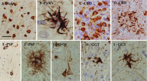
860     T. Lashley et al.

Figure 2. Illustration of argyrophilic Pick bodies by Alois Alzheimer. (Z Gesamte Neurol Psychiatr 1911; 4: 356–85).

speech disorder consisting of poor understanding of                     on this notion of clinical and morphological heterogeneity
speech and written commands, paraphasias and partial                    of a disease for which the umbrella term PiD was still used.
preservation of repetition, for which Pick used Wernicke’s              These authors distinguished three major categories of PiD
term, ‘transcortical sensory aphasia’ [10]. Post mortem                 on the basis of the distribution of the pathology and the
examination, performed by Pick’s pathologist colleague                  presence or absence of swollen, achromatic neurons (Pick
Chiari, confirmed cerebral atrophy, which was more                      cells) and the argyrophilic Pick bodies. Cases with both
prominent in the left than in the right hemisphere and                  Pick bodies and Pick cells were classified as group A, cases
Pick postulated that the particularly severe atrophy of the             that only possessed swollen neurons belonged to group B,
left superior temporal gyrus (‘atrophia cerebri praecipue               while both Pick cells and Pick bodies were absent in group
haemisphaerii sin. in regione gyri primi lobi sphenoidalis’) was        C. The striking absence of either Pick bodies or Alzheimer-
responsible for his patient’s aphasia [10]. Pick’s original             type pathological changes in the majority of cases with
paper provided no detailed microscopic description and                  FTD was also emphasized in the subsequent pioneering
the argyrophilic globular neuronal cytoplasmic inclu-                   studies of the research groups of Lund and Manchester
sions, subsequently named as Pick bodies and are consid-                Universities [16,17]. A pattern of pathological changes
ered pathognomonic of Pick’s disease (PiD) today, were                  was also identified in such cases, which includes loss of
described and illustrated by Alois Alzheimer in 1911                    neurons accompanied by gliosis and microvacuolation of
(Figure 2) [11]. As a result of clinicopathological observa-            the neuropil in superficial cortical laminae without
tions a new disease entity was gradually emerging, and in               amyloid plaques, neurofibrillary tangles or other ‘silver
1925 the medical eponym PiD was first introduced by A.                  positive’ changes (for a review see [18]). In keeping with
Gans as ‘Ziekte van Pick’ in the Dutch [12], and a year                 the ‘nonspecific’ nature of such pathology, the term
later by Onari and Spatz, who were aware of Gans’s work,                ‘dementia lacking distinctive histological features’ (DLDH)
as ‘Picksche Krankheit’ in the German medical literature                was also coined and subsequently widely used [19].
[13].                                                                   During the 1990s, as a prelude to the molecular under-
   In the following decades, the clinical and neuro-                    standing of FTLDs, ubiquitin-positive inclusions were rec-
pathological heterogeneity of cases with frontotemporal                 ognized in extramotor cerebral structures such as neurons
lobar atrophy became apparent, which was also supported                 of the dentate fascia or the frontal and temporal cortices in
by findings indicating that argyrophilic Pick bodies were               cases with MND/ALS with associated FTD [20,21].
present only in about 20% of larger case series [14]. The               The link between ubiquitin-immunoreactive extramotor
neuropathological classification of PiD, put forward by                 neuronal cytoplasmic inclusions, initially described in
Constantinidis and his colleagues in 1974 [15], was based               MND/ALS, and FTLD was subsequently firmly established
© 2015 British Neuropathological Society                                                                          NAN 2015; 41: 858–881
Frontotemporal lobar degeneration, a review   861

by the recognition that such inclusions also occur in cases   C9orf72 gene is a common genetic cause of ALS/MND and
with progressive FTD, but without clinical evidence of        FTLD (C9FTD/ALS), is a recent addition to the list of genes
either upper or lower motor neuron involvement, which         whose mutations play an important role in FTLD [44,45].
resulted in the introduction of the FTLD with ubiquitin-      This rapid increase in knowledge also allowed that a
positive inclusions (FTLD-U) concept [22], subsequently       molecular classification of FTLDs has become available
underpinned by several clinicopathological studies [23–       (Figures 1 and 3), which is now widely accepted and used
26]. This knowledge gave the impetus for pathological         [32,46].
studies to be performed, which demonstrated that
ubiquitin-positive inclusions are characteristic perhaps in
                                                              Clinical concept of FTD
a majority of clinically well-documented FTD cases
[23,26] and that the neuropathological diagnosis of           After the publication of Pick’s original paper in the late
DLDH is only rarely justified [26–28]. The following          19th century, further publications documented that
two decades witnessed several landmark discoveries. In        patients with circumscribed cerebral atrophy had both
the 1990s, the tau isoform composition of sporadic            personality change and language impairment consistent
tauopathies, such as PiD, PSP and corticobasal degenera-      with the modern day split of FTD into behavioural and
tion (CBD) [29–31], which according to the currently          language variants. However, despite these descriptions the
used classification system are part of the FTLD-tau spec-     early 20th century saw limited reports of such cases in the
trum [32], was identified. Furthermore, in 1998, muta-        Western world, although in Japan the disorder Gogi
tions in the microtubule-associated protein tau gene          aphasia (a progressive language disorder) was described in
(MAPT) were reported, indicating that a significant pro-      the 1940’s [47].
portion of hereditary FTDs, previously linked to chromo-         The first modern accounts of the progressive language
some 17 (17q21–22), is due to mutations in this gene          disorders in the Western literature were by Warrington
[33,34]. Further landmark discoveries include the recog-      [48] and Mesulam [49]. Warrington described the selec-
nition that TDP-43 is the main component of the               tive impairment of semantic memory in a group of
ubiquitin-positive inclusions in the majority of the          patients, although she did not use the term semantic
FTLD-U cases and MND/ALS [35]. Involvement of TDP-43          dementia, which was coined a number of years later
in both FTLD-TDP and MND/ALS also provided a firm             [1,50]. Mesulam independently described a group of
molecular link between FTLD and MND/ALS underpin-             patients with progressive language problems initially
ning the notion that these two large groups of                calling them ‘slowly progressive aphasia without general-
neurodegenerative disorders represent two, often overlap-     ized dementia’ [49], and then later ‘primary progressive
ping ends of a disease spectrum. This is also supported by    aphasia’ (PPA) [51], a term that has stuck to this present
clinical data indicating that about 30% of patients with      day. Around the same time period, in the late 1980s and
FTD develop clinical signs of MND/ALS and that 50% of         early 1990s, a group of researchers came together to start
MND/ALS patients show some evidence of cognitive defi-        to understand better the bvFTD. This led to the first Inter-
cits [36]. In addition to the MAPT gene, a number of          national Conference of Frontal Lobe Degeneration of
further genes associated with different forms of familial     non-Alzheimer type in Lund, Sweden and subsequently a
FTD and/or MND/ALS was identified, including the              set of diagnostic criteria in 1994 [52].
progranulin (GRN) gene, whose mutations are responsible          The late 1990s and early 2000s saw a flowering of FTD
for Frontotemporal dementia and parkinsonism linked to        research. One of the keys to this was the development of
chromosome 17 (FTDP-17). FTDP-17 in families without          criteria for both the behavioural and language variants by
harbouring an MAPT mutation [37,38] or the much rarer         a group of researchers in the field in 1998 [1]. These cri-
mutations in the valosin-containing protein (VCP) gene in     teria coined the term FTLD and defined a behavioural
familial FTD [39] also associated with Paget’s disease and    variant (called FTD) and two variants of progressive
inclusion body myositis [40], TARDP and FUS genes in          aphasia: PNFA and semantic dementia. Arguments over
MND/ALS [41,42], as well as the CHMP2B gene in famil-         the nosology of these language variants has continued
ial FTD linked to chromosome 3 (FTD-3), described in a        over the years and in 2004, a third variant was described
Danish pedigree [43]. The discovery in 2011 that intronic     by Gorno-Tempini et al., called logopenic aphasia (LPA)
hexanucleotide (GGGGCC) repeat expansion in the               [53]. More recently, this variant was incorporated into a
© 2015 British Neuropathological Society                                                          NAN 2015; 41: 858–881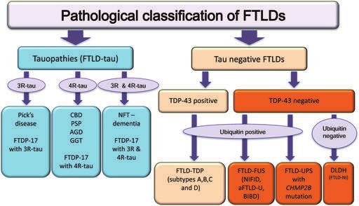
862     T. Lashley et al.

Figure 3. A schematic illustration of the pathological classification of frontotemporal lobar degenerations. Current classification is based on
the molecular features of the disease-associated, inclusion forming proteins, morphological phenotypes and genetic data (for description of
the different disease groups and individual diseases see text). 3R-tau, three-repeat tau; 4R-tau, four-repeat tau; aFTLD-U, atypical
frontotemporal lobar degeneration with ubiquitin immunoreactive neuronal inclusions; AGD, argyrophilic grain disease; BIBD, basophilic
inclusion body disease; CBD, corticobasal degeneration; DLDH, dementia lacking distinctive histology; FTLD, frontotemporal lobar
degeneration; FTDP-17, frontotemporal dementia and parkinsonism linked to chromosome 17; GGT, globular glial tauopathy, NFT-dementia,
neurofibrillary tangle dementia; NIFID, neuronal intermediate filament inclusion disease.

new set of diagnostic criteria for the progressive language                  Neither of the current diagnostic criteria describes the
disorders (PPA), which were renamed, the nonfluent/                       overlap of FTD with MND/ALS or the atypical parkinso-
agrammatic variant, the semantic variant and the                          nian disorders, CBS and PSP. The overlap with these disor-
logopenic variants of PPA [53]. Around the same time, an                  ders has been increasingly recognized in recent years with
international consortium came together to update the                      a substantial proportion of bvFTD patients (and a smaller
diagnostic criteria for the behavioural variant, now                      number of nonfluent aphasia cases) developing MND/ALS
known as bvFTD [54].                                                      or parkinsonism at some point during the disease process.
   bvFTD is characterized by a set of behavioural symp-
toms including disinhibition, apathy, abnormal appetite
                                                                          Genetics of FTD
(commonly a sweet tooth), loss of empathy and obsessive-
compulsive behaviour. However, patients also develop cog-                 A family history of a similar disease is common in FTD,
nitive impairment, usually executive dysfunction initially,               typically in a pattern that suggests dominant inheritance
but other cognitive domains also become involved as the                   [5]. In populations of European ancestry, there are three
disease progresses. Patients with nonfluent variant of PPA                major FTD disease genes (MAPT, GRN and C9orf72) and
develop agrammatism and/or apraxia of speech, while                       many rarer disease genes. It is also recognized that muta-
those with the semantic variant develop anomia and                        tions in genes more commonly associated with related
impaired single word comprehension, later developing                      neurodegenerative diseases such as Alzheimer’s disease,
nonverbal semantic impairment as well; the LPA variant is                 MND/ALS, Parkinson’s disease or prion disease can be
characterized by word-finding pauses and impaired                         found in patients with clinical diagnoses of FTD. The fre-
working memory.                                                           quency of mutations in specific regions or clinical centres
© 2015 British Neuropathological Society                                                                            NAN 2015; 41: 858–881
Frontotemporal lobar degeneration, a review 863
varies according to population factors such as founder modify the clinical phenotype of GRN mutation associated
effects, drift and migration; however, typically, a clinic will FTD [63].
find high single digit percentages of all FTD are explained The C9orf72 gene on chromosome 9 has a
by each of the three major genes individually. A substan- hexanucleotide repeat region located in either in the pro-
tial proportion of familial FTD, depending on how this is moter or intron 1 of the gene (depending on the transcript
defined, remains unexplained [5]. A core clinical and variant). When massively expanded, the mutation causes
pathological phenotype is recognized for each gene, FTD, MND/ALS or the combined syndrome [44,45]. Par-
although clinical distinction is imperfect, particularly in ticularly high frequencies of the C9orf72 expansion muta-
the early stages when gene testing is discussed with tion are found in Finland because of a founder effect [64].
patients and their families. Gene panel technologies are Although the repeat region is variable in length in the
now available to screen all known common and rare FTD healthy population (up to around 30 repeats), repeat
genes simultaneously [55,56]. expansions typically seen in patients are far more massively
In pedigrees with several affected individuals, the loca- expanded, typically >400 repeats [44,65,66]. We still do
tion of the responsible gene can be narrowed by genetic not have certainty about the smallest hexanucleotide
linkage using markers across the genome. The region expansion that confers risk. The core clinical phenotype is
including the MAPT gene on chromosome 17 was first the combination of FTD and MND/ALS; however, the
linked with familial FTD in 1994 [57], and mutations in expansion mutation is commonly associated with pure
this gene were first described in 1998 [33]. MAPT gene FTD, pure MND/ALS and may be reported in patients
mutations are typically associated with bvFTD. Associated labelled with AD, movement disorders, ataxias and nonspe-
syndromes can include parkinsonism, aphasia, early cific neuropsychiatric/neurodegenerative syndromes (for a
amnesia and semantic problems. Over 50 different causal review see [67]). No other mutation types have been
mutations are now known. They fall into two broad cat- reported in the gene and the function of the C9orf72
egories: missense mutations, which alter the amino acid protein is not well understood.
code of the protein usually in or near to the fourth repeat Mutations in several other genes have been uncom-
region of the microtubule-binding domains (see later), monly associated with FTD. These might best be classified
and splice site mutations, which alter the inclusion of as rare gene mutations, which typically cause FTD
exon 10 [58]. Common genetic variation near to the (CHMP2B and VCP, the latter in association with inclu-
MAPT gene is a risk factor in PSP and Parkinson’s disease sion body myopathy and/or Paget’s disease of bone),
[59], but not FTD. or more common gene mutations associated with
Mutations in the GRN gene, coincidentally near to neurodegeneration, but rarely the FTD phenotype (Alzhei-
MAPT on chromosome 17, were identified as causal of mer’s disease genes: PSEN1, APP; prion disease gene:
FTD in 2006 [37,38]. The protein product, progranulin, is PRNP; ALS genes: FUS, TARDBP; others: CSF1R).
a secreted glycoprotein, cleaved into granulin peptides and
found in the brain and serum with roles in inflammatory
Neuropathology of FTLD – an overview of
diseases, diabetes and obesity [60]. Complete loss of
current classification
progranulin protein is associated with neuronal ceroid
lipofuscinosis [61]. Like MAPT mutations, they are most As a result of recent advances in the understanding of
commonly associated with the bvFTD; however, nonfluent the molecular mechanisms associated with FTLDs, this
aphasia, CBS and other Parkinsonian syndromes have heterogeneous group of diseases are now subdivided
been reported. Over 150 variants of GRN have been pathologically on the basis of the deposited abnormal
reported in patients [62]. The large majority of mutations intracellular protein aggregates [32,46,68] (Figure 3). In
are premature stop codons, splice site, exon/gene deletions approximately 40% of all FTLD cases the microtubule-
or frameshift mutations, which result in a hemi-loss of associated protein tau forms inclusions in neurons, and
functional protein. Serum levels of progranulin are in some diseases, in both neurons and glial cells (both
reduced by ~50% in mutation carriers. Most of the astrocytes and oligodendrocytes) (FTLD-tau). With the
missense changes in GRN, aside from those in or near exception of a small minority of the cases, the tau-
to the transcription initiation codon are classified as negative cases have neuronal inclusions, which were
variants of unknown significance. Variants of TMEM106b originally identified by their immunoreactivity for
© 2015 British Neuropathological Society NAN 2015; 41: 858–881864 T. Lashley et al.
ubiquitin (FTLD-U) [22,27,69]. The majority of the also supported by experimental data indicating that tau
FTLD-U cases are associated with accumulation of aggregates can induce neurotoxicity [78] and are associ-
TDP-43 [35], and the term FTLD-TDP is assigned to this ated with nerve cell dysfunction [79].
group [32,46]. In a smaller group of the FTLD-U, the The current molecular classification of FTLD-tau is
inclusions are positive for the FUS protein (FTLD-FUS) based upon genetics and the biochemical composition of
[32,70,71], and such cases are responsible for the major- the inclusions. FTLD-tau includes patients with MAPT
ity of tau and TDP-43-negative FTLDs representing gene mutations (FTD and parkinsonism linked to chromo-
about 5–10% of ubiquitin-positive FTLDs. FTLD-FUS some 17 or FTDP-17 MAPT), PiD, PSP, corticobasal degen-
includes three diseases, neurofilament inclusion body eration (CBD), argyrophilic grain disease (AGD), globular
disease (NIFID), atypical FTLD-U (aFTLD-U) and glial tauopathy (GGT) and NFT-dementia, which was
basophilic inclusions body disease (BIBD) [70–73]. Two recently redefined (somewhat controversially [80]) and
neuropathological subtypes still remain elusive; one pri- designated as primary age-related tauopathy or PART
marily representing familial FTLD with CHMP2B muta- (Figures 3 and 4) [32,81–84]. It should be noted that the
tion (FTD-3) and with ubiquitin-positive inclusions, clinical presentation is not that of FTD in the majority of
which are negative for tau, TDP-43 or FUS and is now AGD and PART cases. Although both PSP and CBD are
termed FTLD-UPS, and a second group, termed FTLD-ni, clinically often referred to as atypical Parkinsonian disor-
which contains rare cases with no discernible pathologi- ders, they are now recognized as disorders of both move-
cal inclusions [32]. ment and cognition [85–89]. It is well documented that
both CBD and PSP patients may present with bvFTD [88–
92] or language impairment (PSP-FTD variants) [89,92–
FTLD-tau
94]. It is of note that the ‘cortical’ PSP variants, which, in
The natively unfolded tau protein with a suggested role in addition to PSP-FTD also include PSP-CBS (PSP with CBS),
microtubule assembly and stabilization as well as regula- are associated with increased neocortical tau load due to a
tion of axonal transport, is the most commonly misfolded shift of the tau burden from the basal ganglia towards the
protein forming intracellular inclusions in human cerebral cortices [95–97]. An intriguing aspect of several
neurodegenerative diseases [74]. Tauopathies are respon- protein folding neurodegenerative disorders is clinical
sible for a major group of FTLDs, designated as FTLD-tau disease progression, which is thought to be due to a
[32], which represents a clinically, biochemically, geneti- stereotypic spatial, time dependent expansion of the
cally and pathologically heterogeneous group of diseases underlying pathology. Data from Alzheimer’s disease indi-
with the overarching feature that all forms possess abun- cate that clinical progression is dependent on such an ana-
dant neuronal, and in some variants, both neuronal tomical progression of the neurofibrillary tau pathology,
and glial filamentous inclusions. These are composed described by the Braak and Braak staging system, which is
of abnormally hyperphosphorylated form of tau in widely used in neuropathological practice [98]. Recent
the absence of amyloid-β deposits [74,75]; hence, they experimental data indicate that tau can show prion-like
are ‘primary’ tauopathies [76]. In addition to tau propagation and spread, which is consistent with patho-
phosphorylation, which is an early event of tau aggrega- logical disease progression in human disease [74].
tion, a number of other post-translational modifications of However, apart from AGD [99], staging of tau pathology
tau are also known. These include ubiquitination, has not been established in other forms of FTLD-tau such
acetylation, glycation, glycosylation with O-linked N- as PiD, CBD and PSP despite significant advancement in
acetylglucosamine (O-GlcNAcylation), nitration, sumoy- recognizing disease subtypes.
lation, and truncation, which are also thought to be asso- FTLD-tau can be divided on the basis of the predominant
ciated with the aggregation process [58]. It is widely tau species making up the inclusions. The six tau isoforms,
accepted that the inclusions contribute to disease patho- which are expressed in the adult human brain, are pro-
genesis in tauopathies as they occur in specific brain duced by alternative mRNA splicing from the MAPT gene
regions whose functions are altered by the tau pathology [100,101]. The isoforms differ from one another by
[77]. In this regard, formation of tau aggregates and fila- whether they possess 0, 1 or 2 29-amino-acid-long
ments, which possess a cross-β structure characteristic of N-terminal inserts, encoded by exons 2 and 3 and by the
amyloid fibrils, is a highly relevant process [58], which is presence or absence of a fourth 31-amino-acid-long repeat
© 2015 British Neuropathological Society NAN 2015; 41: 858–881Frontotemporal lobar degeneration, a review 865 Figure 4. Examples of tau pathology in some of the diseases comprising frontotemporal lobar degeneration (FTLD-TAU). Tau immunohistochemistry demonstrates typical Pick bodies in the granular cell layer of the dentate fascia (A). The glial pathology in Pick’s disease includes ramified astrocytes (B). Pretangles in the granular cell layer of the dentate fascia (C) together with a characteristic astrocytic plaque (D) in corticobasal degeneration (CBD). Four-repeat tau-positive coiled bodies in white matter (E) and tufted astrocytes in grey matter structures (F) are characteristic pathological features of progressive supranuclear palsy (PSP). FTDP-17 due to mutations of the microtubule-associated protein tau (MAPT) gene have a number of pathological variants, determined by the localization of the mutations; cases with an exon 10+16 intronic mutation also show extensive four-repeat tau-positive glial pathology and numerous pretangles and neurofibrillary tangles (G). Globular glial tauopathy (GGT) is characterized by the presence of globular glial inclusions, including globular oligodendroglial inclusions (H) and globular astrocytic inclusions (I). AT8 immunohistochemistry, bar on A represents 40 microns on A and E; 20 microns on C, D E–I and 5 microns on B. in the microtubule-binding domain of tau, encoded by For diagnosis post mortem microscopic investigation of exon 10. Two major groups of the six tau isoforms, each the brain, including demonstration of the characteristic group containing three isoforms, can be differentiated on neuronal and if present glial inclusions, by tau, and the basis of whether the repeat encoded by exon 10 is if necessary, supplemented by 3R-tau and 4R-tau present or absent. Isoforms with four-repeat sequences are immunohistochemistry (Figure 4) together with docu- designated as 4-repeat-tau (4R-tau) while those without mentation of the anatomical distribution of the tau this sequence are called 3-repeat tau (3R-tau) [100]. In PiD lesions, is required. Microscopic studies may need to be [30] and some forms of FTDP-17 MAPT [102] 3R-tau is the supplemented with genetic and biochemical investigations predominant protein species in the tau filaments making in some cases, especially if a familial FTLD-tau variant is up the inclusions, whereas 4R-tau assembles into filaments suspected. Although there is significant overlap between in CBD, PSP, GGT, AGD and some FTDP-17 MAPT variants different forms of FTLD-tau, the presence of characteristic [102–105]. In a third group of diseases, which includes morphological features associated with the different NFT-dementia and some FTDP-17 MAPT variants, both tauopathies allows a specific diagnosis to be made in the 3R-tau and 4R-tau are present in the inclusions (for a majority of the cases. The presence of Pick bodies in PiD or recent review see [102]). The differences in the tau isoform other relevant neuronal and thread pathology in combina- composition of the tau filaments are also reflected by char- tion with characteristic glial inclusions, such as astrocytic acteristic differences in the ultrastructural morphologies of plaques in CBD, tufted astrocytes in PSP, ramified the filaments [106]. astrocytes in PiD or numerous globular oligodendroglial © 2015 British Neuropathological Society NAN 2015; 41: 858–881
866      T. Lashley et al.

Table 1. Correlations between clinical presentation, genetics and pathology in different subtypes of frontotemporal lobar degeneration

Clinical presentation             Brain pathology                                          Gene                         References

bvFTD                             FTLD-tau                 Pick’s disease                                               [89,92,169,170]
                                                           MAPT mutations                  MAPT                         [89,170]
                                                           CBD                                                          [89,92,170]
                                                           GGT                                                          [83,84]
                                  FTLD-TDP                 Type A                          GRN, C9orf72                 [89,170]
                                  FTLD-TDP                 Type B                          C9orf72                      [170]
                                  FTLD-TDP                 Type C                                                       [170]
                                  FTLD-TDP                 Type D                          VCP                          [89]
                                  FTLD-FUS                 NIFID                                                        [89,158]
                                                           aFTLD-U                                                      [89,158]
Semantic dementia                 FTLD-tau                 Pick’s disease                                               [89,169,170]
                                  FTLD-TDP                 Type C                                                       [89,170]
FTLD-MND                          FTLD-tau                 GGT                                                          [83,84]
                                  FTLD-TDP                 Type A                                                       [89,170]
                                  FTLD-TDP                 Type B                          C9orf72                      [89,170]
PNFA                              FTLD-tau                 Pick’s disease                                               [89,92,169,170]
                                                           CBD                                                          [89,92,170]
                                                           PSP                                                          [170]
                                  FTLD-TDP                 Type A                          GRN, C9orf72                 [89,170]
                                  FTLD-TDP                 Type B                          C9orf72                      [170]
PSPS                              FTLD-tau                 MAPT mutations                  MAPT                         [171]
                                                           CBD                                                          [88,172]
                                                           PSP                                                          [89,92,170]
                                                           GGT                                                          [83,84]
                                  FTLD-FUS                 NIFID                                                        [158]
CBS                               FTLD-tau                 MAPT mutations                  MAPT                         [89]
                                                           CBD                                                          [88,89,92,170]
                                                           PSP                                                          [89,92,96,97,170]
                                                           GGT                                                          [83,84]
                                  FTLD-TDP                 Type A                                                       [89]
                                  FTLD-FUS                 NIFID                                                        [156,158]

aFTLD-U, atypical frontotemporal dementia with ubiquitin-positive inclusions; bvFTD, behavioural variant frontotemporal dementia; C9orf72,
chromosome 9 open reading frame 72; CBD, corticobasal degeneration; CBS, corticobasal syndrome; FUS, fused in sarcoma; GGT, globular glial
tauopathy; GRN, progranulin; MAPT, microtubule-associated protein tau; MND, motor neurone disease; NIFID, neuronal intermediate filament
inclusion disease; PNFA, progressive nonfluent aphasia; PSP, progressive supranuclear palsy; PSPS, progressive supranuclear palsy syndrome;
TDP, transactive response DNA-binding protein; VCP, valosin containing protein.

and astrocytic inclusions in GGT are important helpers of               this has also provided further support for a molecular
the morphological diagnosis (Figure 4) (for a review see                classification of FTLDs [35]. TDP-43 is a highly con-
[107]).The FTDP-17 MAPT variants may show similarities                  served and ubiquitously expressed multifunctional, het-
to PiD, PSP, CBD, AGD or GGT and variability within the                 erogeneous nuclear ribonucleoprotein (hnRNP), encoded
same family is also known [58]. For correlations between                by the TARDBP gene located on chromosome 1. TDP-43
clinical presentation, genetics and pathology see Table 1.              possesses nuclear localization and nuclear export signals
                                                                        and it shuttles between the cell nucleus and cytoplasm.
                                                                        TDP-43 also has an N-terminal domain, two RNA-
FTLD-TDP
                                                                        recognition motifs (RRMs) involved in RNA and DNA
The discovery demonstrating the TDP-43 protein as the                   binding and its glycine-rich C-terminal region contains
main component of the ubiquitin-positive inclusions in                  most of the mutations causing familial MND/ALS and
the majority of FTLD-U, designated as FTLD-TDP, and                     rarely familial FTD [108,109]. The C-terminal region of
also in MND/ALS (ALS-TDP) resulted not only in a better                 TDP-43 contains a prion-like protease resistant domain
understanding of the pathogenesis of these diseases, but                [110]. TDP-43 protein expression is tightly controlled by
© 2015 British Neuropathological Society                                                                         NAN 2015; 41: 858–881
Frontotemporal lobar degeneration, a review 867
autoregulatory mechanisms and both over and under and dystrophic neurites (DNs). Neurons containing TDP-
expression results in impaired neuronal function [111]. 43-positive inclusions lose their normal nuclear TDP-43
Cellular functions of TDP-43 include regulation of RNA staining that can be observed in neurons without an
splicing, translation, miRNA processes and mRNA trans- inclusion, which in every day neuropathological practice
port and stability [112] with more than 6000 RNA is a helper of identifying affected neurons (Figure 5).
targets. The aggregation of TDP-43 is associated FTLD-TDP type A is characterized by numerous NCIs,
with several post-translational modifications including DNs and variable numbers of NIIs and the TDP-43 immu-
phosphorylation of serine residues, ubiquitination, oxi- noreactive lesions are most numerous in layer 2 of
dation, lysine acetylation and C-terminal cleavage. affected cortices. FTLD-TDP type B is characterized by
Pathological examination, utilizing antibodies specific for numerous NCIs in both the superficial and deeper cortical
phosphorylated TDP-43 (pTDP-43) confirms that the layers, but this subtype can exhibit the occasional DNs.
cellular TDP-43 aggregates contain phosphorylated FTLD-TDP type C is associated with abundant long DNs
epitopes [113]. In disease TDP-43 aggregates can accu- often with a corkscrew appearance throughout the corti-
mulate in both the cytoplasm and nuclei of affected cal layers. Some TDP-43 type C cases are associated with
neurons and glia, which results in cellular dysfunction. corticospinal tract degeneration [121]. FTLD-TDP type D
There is now evidence to suggest that TDP-43 in inclu- is characterized by numerous NIIs, DNs and infrequent
sions, the majority of which have been shown to show NCIs (Figure 5) [122,123]. It remains unclear whether
amyloid features [114], has cellular prion-like properties, differences in underlying pathophysiology or selective
which could have relevance for the pathomechanism of neuronal vulnerability determine the distinction between
FTLD-TDP [115]. The disease-associated TDP-43 making the FTLD-TDP subtypes via a single pathogenic mecha-
up the inclusions, is thought to exercise its deleterious nism or whether there are multiple mechanisms involved.
effect via toxic gain of function due to overexpression or For correlations between clinical presentation, genetics
mutant forms, but given the number of its functions it is and pathology see Table 1.
also likely that a loss of function effect also has a role in
the pathogenesis of FTLD-TDP. FTLD-TDP has recently
FTLD-TDP in familial FTD
been reported to be associated with chronic traumatic
encephalopathy [116]. TDP43-positive inclusions are TDP-43 pathology is characteristic not only in sporadic,
also found in around 90% of patients with hippocampal but also in some of the familial forms. In cases with muta-
sclerosis, which is also a feature in the majority of FTLD- tions in the GRN gene FTLD-TDP type A is the character-
TDP suggesting a special relationship between these two istic morphology, while FTLD-TDP type D is restricted to
pathologies [117,118]. cases with mutations in the VCP gene. The majority of the
FTLD cases with C9orf72 repeat expansion mutations
show FTLD-TDP type B pathology, but a proportion of
FTLD-TDP subtypes
them have changes consistent with type A and rare cases
Subtypes of FTLD-U were originally established, using with type C pathology have also been reported
findings of ubiquitin immunohistochemistry, and two [121,124,125]. Families with pathogenic mutations in
major classification systems were in existence [69,119]. both the GRN and the C9orf72 genes have been docu-
One of the strengths of these classifications is that they mented [126,127].
have been validated by clinical, genetic and pathological The characteristic aspect of the pathology of C9FTLD/
correlations. In order to eliminate competing classifica- ALS cases is the presence of star-like p62/sequestosome-1-
tions a single harmonized system was proposed in 2011, positive, but TDP-43-negative NCIs in the hippocampus
which has gained wide acceptance [120]. Accordingly, (granule cell layer and CA4 hippocampal subregion) and
the four different subtypes of FTLD-TDP, which are recog- cerebellar cortex (cerebellar granule cells) (Figure 6)
nized, are based on the predominant lesion type(s) and [44,125,128,129], which are also positive for ubiquitin
the distribution of the pathological inclusions. The and the ubiquitin-binding protein ubiquilin-2. The high
TDP-43 immunoreactive inclusion types considered frequency of C9orf72 repeat expansion in both FTD and
include neuronal cytoplasmic inclusions (NCIs), neuronal ALS has generated great interest in the underlying mecha-
intranuclear inclusions (NIIs), oligodendroglial inclusions nisms in these diseases, of which several nonmutually
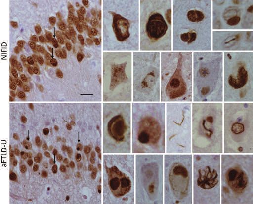
© 2015 British Neuropathological Society NAN 2015; 41: 858–881868 T. Lashley et al. Figure 5. Morphological features of frontotemporal lobar degeneration-transactive response DNA-binding protein (FTLD-TDP) subtypes A, B, C and D. The separation of the different FTLD-TDP subtypes is based on the microscopic features of a number of different TDP-43-positive inclusion types. In FTLD-TDP type A neuronal cytoplasmic inclusions (arrows), short dystrophic neurites and neuronal intranuclear inclusions are characteristic. In FTLD-TDP type B neuronal cytoplasmic inclusions, often with somewhat granular appearances, are seen and the inclusions are present in all cortical layers. FTLD-TDP type C is characterized by unique, long corkscrew-type neurites (arrow). FTLD-TDP type D shows numerous neuronal intranuclear inclusions (arrow) and short neurites while neuronal cytoplasmic inclusions are rare. TDP-43 immunohistochemistry, bar on top panel represents 40 microns on all large panels and FTLD-TDPC inserts; 10 microns on all other inserts. © 2015 British Neuropathological Society NAN 2015; 41: 858–881
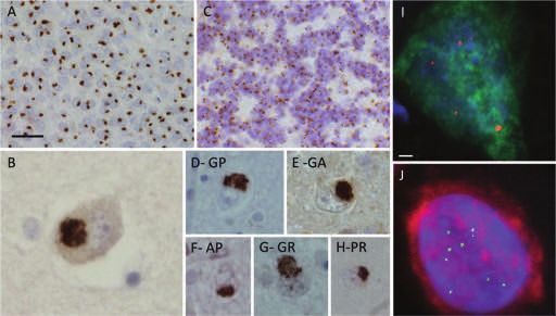
Frontotemporal lobar degeneration, a review        869

Figure 6. Dipeptide repeat pathology in chromosome 9 open reading frame 72 (C9orf72) repeat expansion cases. All cases carrying a
C9orf72 expansion repeat mutation contain additional p62-positive, transactive response DNA-binding protein (TDP)-43-negative neuronal
cytoplasmic inclusions. These inclusions are composed of dipeptide repeat proteins, which are translated from the C9orf72 expansion
repeats. The p62-positive inclusions are numerous in the granule cell layer of the dentate gyrus (A) and are prominent in areas including
the CA4 hippocampal subregion (B) and the cerebellar granule cells (C). Antibodies have also been raised against the 5 dipeptides [antisense:
Pro-Arg (H), Pro-Ala (F); sense: Gly-Ala (E), Gly-Arg (G); antisense or sense Gly-Pro (D)], which show that all dipeptides are present in the
p62 positive inclusions. Representative images of frontal cortex tissue from heterozygous C9orf72 cases with RNA fluorescent in-situ
hybridisation (FISH) for sense foci (red, I) or antisense foci (green, J) with immunostaining for neurons with NeuN (green in I, red in J) and
nuclear DNA staining with 4′,6-diamidino-2-phenylindole (DAPI) (blue). Bar on A represents 40 microns on A and C; 20 microns on D–H;
10 microns on B. Scale bar in I represents 2 μm on I and J.

exclusive possibilities exist (for a review, see [130]). One of          GTPases, suggesting that it may have a role in vesicular
the mechanisms is RNA-related toxicity, based on evidence                trafficking [134]. The possibility that toxicity is, at least
from microsatellite expansion diseases with large repeat                 partly due to a decrease/loss of the function of the C9orf72
expansions. In these disorders, the transcribed repeat RNA               protein is considered and such a hypothesis is supported by
aggregates in the nucleus in discrete structures termed                  findings of decreased levels of GGGGCC repeat-containing
RNA foci, which sequester select RNA-binding proteins                    transcripts in C9FTD brains [44,135]. A third mechanistic
resulting in loss of their function, ultimately leading to               option is that both the sense and antisense transcripts of
disease. RNA foci have also been identified in C9FTD/ALS                 the GGGGCC expansion repeats are translated via the
neurons [44] and, as the GGGGCC repeat expansions are                    mechanism of RAN (repeat-associated non-ATG) transla-
bidirectionally transcribed, the RNA foci are formed of both             tion into C9RANT (RAN-translated) protein aggregates
sense and antisense transcripts, typically found in separate             (antisense: Pro-Arg, Pro-Ala, Pro-Gly; and sense: Gly-Ala,
cells, but they can also co-localize in the same nucleus                 Gly-Arg, Gly-Pro), which form the dipeptide repeat
[131,132]. The possibility between high RNA foci burden                  proteins found in the p62-positive inclusions and
and early age of disease onset has been raised [131,133].                thought to contribute to disease pathogenesis (Figure 6)
Although the precise function of the C9orf72 protein is not              [132,136,137]. The topographical distribution of these
known, it has been shown to be structurally related to a                 dipeptide repeat proteins is similar regardless of the clinical
class of GDP/GTP exchange factors that activate Rab-                     phenotype [138,139]. RAN translation is ‘unconven-
© 2015 British Neuropathological Society                                                                            NAN 2015; 41: 858–881
870    T. Lashley et al.

tional’ mode of translation with unknown mechanism,            the ubiquitin-positive NCIs in motor neurons are positive
which occurs across expanded repeat tracts in the absence      for the 526-amino-acid-long, 53 kDa nucleoprotein FUS
of an initiating codon [140]. This mechanism has been          [41,42] gave the impetus for the investigation of a possible
shown in several microsatellite expansion disorders such       role in FTLDs.
as myotonic dystrophy, type 1 (DM1), spinocerebellar              FUS is a member of the FET family of multifunctional
ataxia, type 8 (SCA8) and fragile X-associated tremor/         DNA/RNA-binding proteins and is encoded by the
ataxia syndrome (FXTAS) and data suggest that RAN              FUS gene located on chromosome 16 [147–149]. FUS is
translation may be a significant mechanism in C9FTD/ALS        ubiquitously expressed multifunctional hnRNP, also
[130,132,141]. Experimental data indicate that both            known as hnRNP P2 [150,151]. Different regions of
arginine-rich proteins and repeat RNA contribute to            the protein are involved in different functions; its
C9orf72-mediated neurotoxicity [131].                          N-terminus is involved in transcription activation [152]
                                                               while the C-terminal region contains multiple domains
                                                               involved in RNA-protein interactions and contains the
Disease staging and progression
                                                               nuclear localization signal, which is targeted by
There have been attempts for establishing stages of disease    transportin1 (TRN1) and is essential for the nuclear
progression in FTLDs, which initially were based on            transport of FUS [153]. The majority of the mutations
progression of clinical signs [142] or macroscopic             causing familial MND/ALS affects the region of the
brain atrophy [143]. A recent study using pTDP-43              nuclear localization signal, which decrease the binding
immunohistochemistry aimed to establish microscopic            affinities of FUS for TRN1 [154]. The subcellular locali-
patterns of disease progression in bvFTD cases [144]. Four     zation of the FUS protein is cell type dependent, for
stereotypical patterns of pTDP-43 pathology suggestive of      example FUS is present in proportionally larger amounts
potentially sequential spreading of the TDP-43 aggregates      in the nucleus than in the cytoplasm of neurons while
along a fronto-occipital gradient could be established;        FUS expression is exclusively nuclear in glia [155]. Under
cases with ‘pattern I’ had widespread pTDP-43 lesions in       normal physiological conditions the FUS protein continu-
the orbital gyri, gyrus rectus and amygdala. In ‘pattern II’   ously shuttles between the cytoplasm and the nucleus
there was evidence for increased pTDP-43 burden with           [150].
lesions emerging in middle frontal and anterior cingulate
gyrus, anteromedial temporal structures, superior and
                                                               Neuropathological features of FTLD-FUS
middle temporal gyri, striatum, red nucleus, thalamus
and precerebellar nuclei. Cases with more advanced             The discovery that FUS mutations are the cause of a
pathology were labelled as showing ‘pattern III’ with          subgroup of familial MND/ALS gave the impetus
involvement of the motor cortex, bulbar somatomotor            for investigating the role of the FUS protein in sporadic,
neurons and the spinal cord anterior horn while cases          tau and TDP-43-negative, ubiquitin-positive FTLD-U
with ‘pattern IV’ pTDP-43 lesions were present in the          cases. These studies showed that, indeed, the FUS pro-
visual cortex [144]. A previous study by the same              tein is a major component of the pathological lesions
research group suggested that in MND/ALS pTDP-43               in such cases comprising about 5–10% of all FTLD-U
aggregates may spread via axonal transport, initially          [70,71,73]. This finding gave the basis for the introduc-
spreading from the motor cortex towards the brainstem          tion of a third smaller FTLD group, appropriately
and spinal cord followed by cortical areas and finally         termed FTLD-FUS, which at present includes three
towards anteromedial temporal lobe structures [145] (for       conditions: NIFID [71,156], aFTLD-U [70] and BIBD
a recent see review [146]).                                    [73].
                                                                  All three diseases classified under the umbrella term
                                                               FTLD-FUS contain FUS-positive inclusions and show
FTLD-FUS
                                                               morphological similarities, but the differences in the
                                                               inclusions types and their locations in the different brain
Introduction and FTLD-FUS subtypes
                                                               regions is sufficient for a specific diagnosis to be made
The recognition that mutations in the FUS gene are asso-       [157,158]. There are considerably more FUS-positive
ciated with familial MND/ALS type 6 (ALS-FUS), and that        inclusions in NIFID compared with aFTLD-U in many
© 2015 British Neuropathological Society                                                           NAN 2015; 41: 858–881
Frontotemporal lobar degeneration, a review 871 Figure 7. Fused in sarcoma (FUS) pathology demonstrated in two diseases of frontotemporal lobar degeneration (FTLD)-FUS, neuronal intermediate inclusion disease (NIFID) and atypical FTLD-U (aFTLD-U). The pathological inclusions found in FTLD-FUS are varied and diverse. Neuronal cytoplasmic inclusions of different morphological types are seen in the granule cell layer of the hippocampus in both the NIFID and aFTLD-U (arrows). FUS immunohistochemistry, bar on A represents 40 microns on granule cell layer panels and between 10 and 20 microns on the smaller panels. anatomical regions including the cerebral cortex, medial ogy of the FUS immunoreactive NCIs is heterogeneous temporal lobe structures, such as subiculum, entorhinal varying from small bean-shaped to larger annular cortex and fusiform gyrus (but not in the granule cell shapes (Figure 7) [158], whereas NII were only occasion- layer of the dentate fascia), subcortical and brainstem ally found in NIFID cases. α-Internexin-positive inclu- nuclei [158]. It is of note that a greater cell loss has sions are a prominent feature of all NIFID cases, been found in these anatomical regions in aFTLD-U, although far less abundant than FUS-positive inclusions, which may account for the lower numbers of inclusions whereas these are absent in both aFTLD-U and in these areas. Hippocampal sclerosis has been described BIBD [157]. The FUS-immunoreactive NCIs in aFTLD-U to be a prominent feature of aFTLD-U, but only occasion- are often compact, round or bean-shaped and vermiform ally seen in NIFID [72,157–159]. Different inclusion NII, described previously [159], and found throughout types have been documented in all three subtypes of the neocortex, granule cells of the dentate gyrus FTLD-FUS [70,71,157,158,160]. In NIFID, the morphol- and striatum [158]. The third FTLD-FUS subtype, BIBD © 2015 British Neuropathological Society NAN 2015; 41: 858–881
872    T. Lashley et al.

shows basophilic NCIs on the haematoxylin and eosin-
                                                               Conclusions and future directions
stained histological sections of cerebral cortices and
FUS immunohistochemistry highlights widespread NCIs            The last 10 years witnessed the discovery of two major
seen not only in cerebral cortices, but also in the basal      FTD genes, the GRN and C9orf72 genes and proteins such
ganglia and brainstem [73,157]. In all three FTLD-FUS          as TDP-43 and FUS together with the recognition of the
subtypes, the cerebellar cortex remain unaffected. FUS-        role of other FET proteins and TRN1 in the FUS inclusions
positive neuronal intranuclear inclusions have also been       in FTLD-FUS. Recognition of C9orf72 as a major gene in
described in polyQ inclusions in Huntington’s disease,         both FTD and MND/ALS trigged major research into the
spinocerebellar ataxia types 1, 2, 3 and dentatorubro-         disease mechanisms of C9FTD-MND/ALS, which has pro-
pallidoluysian atrophy [161,162].                              vided significant results in a relatively short period of time.
   The FUS-positive inclusion of NIFID, aFTLD-U and            This remarkable increase in knowledge has resulted in a
BIBD consistently contain the TATA-binding protein-            better understanding of the pathogenesis of several FTLD
associated factor 15 protein (TAF15) and variably              subgroups, firmly established a molecular link between
contain the Ewing’s sarcoma protein (EWS), which are           FTD and MND/ALS, and also facilitated the introduction
other members of the FET family of proteins [163,164].         of a molecular classification, which has been widely
TRN1, responsible for the transport of FUS from the            accepted and followed in everyday diagnostic practice of
cytoplasm to the cell nucleus, has also been shown to be       neuropathologists. As in other neurodegenerative dis-
present in the NCIs and NIIs in all FTLD-FUS subgroups         eases, the concept of cell-to-cell propagation of disease-
suggesting a role in the pathogenesis of these diseases        associated proteins underlying disease spread and
[165]. However, TAF15 and EWS and TRN1 are not                 progression has been studied in an FTLD-TDP subgroup
present in the inclusions in familial MND/ALS with             with bvFTD, although this is still awaited in all forms of
mutations of the FUS gene [163,166]. For correlations          FTLD-TDP, tauopathies such as PSP and CBD, and in the
between clinical presentation, genetics and pathology see      different disease entities of FTLD-FUS.
Table 1.                                                          Despite the advances several fundamentally important
                                                               questions related to pathogenesis remain unanswered and
                                                               we only enlist here a few. Is there a common pathogenic
FTLD-UPS
                                                               mechanism determining selective neuronal vulnerability
Mutations of the CHMP2B gene, located on chromosome            and linking all forms of FTLDs or, as it seems more likely,
3, are a rare cause of hereditary FTLD. The first mutation,    multiple mechanisms exist? What are the downstream
affecting the splice acceptor site of the sixth (last) exon,   mechanisms driving the TDP-43 pathology to different cell
was described in affected members of a Danish kindred          types and/or different neuronal compartments in the four
with FTD-3 followed by the discovery of a Q165X muta-          pathological subtypes of FTLD-TDP? Detailed pathological
tion in a Belgian FTD family. Both mutations appear to         investigation of larger cohorts with c9orf72 repeat expan-
have a common mechanism; deletion of the C-terminus of         sions is also required. The landmark findings of the past
the CHMP2B protein (for a review, see [167]). The              20 years together with future discoveries, one may trust,
CHMP2B protein is a component of the ESCRT-III complex         will facilitate the translation of this knowledge into
(endosomal sorting complex required for transport-III),        disease biomarkers, allowing precise clinical diagnosis and
which has a role in protein degradation pathways.              ultimately leading to the establishment of effective disease
   Brains of affected individuals show marked fronto-          modifying therapies.
temporal atrophy, but the parietal lobe can also be
affected. There is cortical microvacuolation of the
                                                               Acknowledgements
neuropil accompanied by nerve cell loss and astrogliosis.
Remaining cortical neurons show enlarged vacuoles,             We would like to thank Dr Adrian Isaacs and Dr Sarah
which are thought to represent aberrant large, late            Mizielinska for providing the DPR antibodies and the RNA
endosomes [167]. Ubiquitin and p62-positive NCIs, which        foci images. TL is supported by an Alzheimer’s Research
are tau, TDP-43 and FUS-negative, have been identified in      UK fellowship. TR is supported by a research grant from
the granule cells of the hippocampal dentate gyrus and in      CBD Solutions. JDR is an NIHR Rare Disease Translational
neurons of the frontal cortex [167,168].                       Research Collaboration (TRC) Postdoctoral Fellow. SM is
© 2015 British Neuropathological Society                                                             NAN 2015; 41: 858–881
Frontotemporal lobar degeneration, a review 873
funded by the Medical Research Council. The Queen Tagliavini F, Tiraboschi P, Redaelli V, Prioni S, Grisoli M,
Square Brain Bank is supported by the Reta Lila Weston Borroni B, Padovani A, Galimberti D, Scarpini E, Arighi
Institute of Neurological Studies, UCL Institute of Neurol- A, Fumagalli G, Rowe JB, Coyle-Gilchrist I, Graff C,
Fallstrom M, Jelic V, Stahlbom AK, Andersson C,
ogy. This research was partly supported by the National Thonberg H, Lilius L, Frisoni GB, Pievani M, Bocchetta
Institute for Health Research (NIHR) Queen Square Bio- M, Benussi L, Ghidoni R, Finger E, Sorbi S, Nacmias B,
medical Research Unit in Dementia based at University Lombardi G, Polito C, Warren JD, Ourselin S, Fox NC,
College London Hospitals (UCLH), University College Rossor MN. Presymptomatic cognitive and neuro-
London (UCL). The views expressed are those of the anatomical changes in genetic frontotemporal demen-
tia in the Genetic Frontotemporal dementia Initiative
author(s) and not necessarily those of the NHS, the NIHR
(GENFI) study: a cross-sectional analysis. Lancet Neurol
or the Department of Health. 2015; 14 (3): 291–301
7 Sittig O. Professor Arnold Pick. Arch Psychiatr Nervenkr
Author contributions 1925; 72: 1–20
8 Kertesz A, Kalvach P. Arnold Pick and German
TL, JDR, SM and TR undertook a literature review and neuropsychiatry in Prague. Arch Neurol 1996; 53:
drafted the initial paper and prepared the figures. All 935–8
9 Critchley M, Critchley E. John Hughlings Jackson: Father
authors were involved in editing the paper. TR did the final
of English neurology, New York, Oxford: Oxford Univer-
editing. sity Press., 1998
10 Pick A. Ueber die beziehungen der senilen hirnatro-
References phie zur aphasie. Prag Med Wochenschr 1892; 17:
165–7
1 Neary D, Snowden JS, Gustafson L, Passant U, Stuss D, 11 Alzheimer A. Ueber eigenartige Kranheiltsfalle des
Black S, Freedman M, Kertesz A, Robert PH, Albert M, spateren Alters. Z Gesamte Neurol Psychiatr 1911; 4:
Boone K, Miller BL, Cummings J, Benson DF. 356–85
Frontotemporal lobar degeneration – a consensus on 12 Gans A, De Ziekten van Pick en van Alzheimer.
clinical diagnostic criteria. Neurology 1998; 51: Nederlandsch Tijdschrift v Geneeskunde 1925; 68 (2):
1546–54 1953
2 Rosso SM, Donker Kaat L, Baks T, Joosse M, de Koning I, 13 Onari K, Spatz H. Anatomische beitraige zur lehre
Pijnenburg Y, de Jong D, Dooijes D, Kamphorst W, yon der pickschen umschriebenen grosshirnrinden-
Ravid R, Niermeijer MF, Verheij F, Kremer HP, Scheltens atrophie (‘picksche krankheit’). Ztschr Ges Neurol
P, van Duijn CM, Heutink P, van Swieten JC. Psychiatr 1926; 101: 470–511
Frontotemporal dementia in The Netherlands: patient 14 Escourolle R. La maladie de Pick. Etude D’ensemble et
characteristics and prevalence estimates from a Syntèse Anatomo-Clinique, Paris: Thèse, 1956
population-based study. Brain 2003; 126: 2016–22 15 Constantinidis J, Richard J, Tissot R. Pick’s disease. His-
3 Onyike CU, Diehl-Schmid J. The epidemiology of tological and clinical correlations. Eur Neurol 1974; 11:
frontotemporal dementia. Int Rev Psychiatry 2013; 25: 208–17
130–7 16 Brun A. Frontal-lobe degeneration of non-Alzheimer
4 Neary D, Snowden JS, Gustafson L, Passant U, Stuss D, type. 1. Neuropathology. Arch Gerontol Geriatr 1987; 6:
Black S, Freedman M, Kertesz A, Robert PH, Albert M, 193–208
Boone K, Miller BL, Cummings J, Benson DF. 17 Mann DMA, South PW, Snowden JS, Neary D.
Frontotemporal lobar degeneration: a consensus on Dementia of frontal-lobe type – neuropathology and
clinical diagnostic criteria. Neurology 1998; 51: immunohistochemistry. J Neurol Neurosurg Psychiatry
1546–54 1993; 56: 605–14
5 Rohrer JD, Guerreiro R, Vandrovcova J, Uphill J, Reiman 18 Brun A, Gustafson L. The birth and early evolution of
D, Beck J, Isaacs AM, Authier A, Ferrari R, Fox NC, the frontotemporal dementia (FTD) concept. J Mol
Mackenzie IR, Warren JD, de Silva R, Holton J, Revesz T, Neurosci 2011; 45: 324–9
Hardy J, Mead S, Rossor MN. The heritability and genet- 19 Knopman DS, Mastri AR, Frey WH, Sung JH, Rustan T.
ics of frontotemporal lobar degeneration. Neurology Dementia lacking distinctive histologic features: a
2009; 73: 1451–6 common non-Alzheimer degenerative dementia. Neu-
6 Rohrer JD, Nicholas JM, Cash DM, van Swieten J, Dopper rology 1990; 40: 251–6
E, Jiskoot L, van Minkelen R, Rombouts SA, Cardoso MJ, 20 Okamoto K, Hirai S, Yamazaki T, Sun XY, Nakazato Y.
Clegg S, Espak M, Mead S, Thomas DL, De Vita E, New ubiquitin-positive intraneuronal inclusions in the
Masellis M, Black SE, Freedman M, Keren R, MacIntosh extra-motor cortices in patients with amyotrophic
BJ, Rogaeva E, Tang-Wai D, Tartaglia MC, Laforce R Jr, lateral sclerosis. Neurosci Lett 1991; 129: 233–6
© 2015 British Neuropathological Society NAN 2015; 41: 858–881You can also read