From dinosaurs to birds: a tail of evolution
←
→
Page content transcription
If your browser does not render page correctly, please read the page content below
Rashid et al. EvoDevo 2014, 5:25
http://www.evodevojournal.com/content/5/1/25

 REVIEW                                                                                                                                          Open Access

From dinosaurs to birds: a tail of evolution
Dana J Rashid1*, Susan C Chapman2, Hans CE Larsson3, Chris L Organ1,4, Anne-Gaelle Bebin1,5, Christa S Merzdorf6,
Roger Bradley6 and John R Horner1

  Abstract
  A particularly critical event in avian evolution was the transition from long- to short-tailed birds. Primitive bird tails
  underwent significant alteration, most notably reduction of the number of caudal vertebrae and fusion of the distal
  caudal vertebrae into an ossified pygostyle. These changes, among others, occurred over a very short evolutionary
  interval, which brings into focus the underlying mechanisms behind those changes. Despite the wealth of studies
  delving into avian evolution, virtually nothing is understood about the genetic and developmental events
  responsible for the emergence of short, fused tails. In this review, we summarize the current understanding of the
  signaling pathways and morphological events that contribute to tail extension and termination and examine how
  mutations affecting the genes that control these pathways might influence the evolution of the avian tail. To
  generate a list of candidate genes that may have been modulated in the transition to short-tailed birds, we
  analyzed a comprehensive set of mouse mutants. Interestingly, a prevalent pleiotropic effect of mutations that
  cause fused caudal vertebral bodies (as in the pygostyles of birds) is tail truncation. We identified 23 mutations in
  this class, and these were primarily restricted to genes involved in axial extension. At least half of the mutations
  that cause short, fused tails lie in the Notch/Wnt pathway of somite boundary formation or differentiation, leading
  to changes in somite number or size. Several of the mutations also cause additional bone fusions in the trunk
  skeleton, reminiscent of those observed in primitive and modern birds. All of our findings were correlated to the
  fossil record. An open question is whether the relatively sudden appearance of short-tailed birds in the fossil
  record could be accounted for, at least in part, by the pleiotropic effects generated by a relatively small number
  of mutational events.
  Keywords: Archaeopteryx, Avian, Bird evolution, Confuciusornis, Dinosaur, Jeholornis, Sapeornis, Somitogenesis, Tail

Review                                                                                transformation from the ancestral long 'reptilian' tail
Introduction                                                                          to the short, distally fused tail [1,2]. Yet, the specific
Tails of extant birds are an evolutionary novelty. They are                           developmental mechanisms that facilitated this dramatic
critical for powered flight, ensure reproductive success by                           anatomical change are unknown. This gap in knowledge
attracting mates, and safeguard relatives by communicating                            constitutes a rich vein for future research, one that would
warning signals. Extant bird tails consist proximally of a                            benefit from our current understanding of axial develop-
small series of unfused caudal vertebrae with a high range                            mental mechanisms.
of motion. These articulate to a distal rod-like pygostyle,                             The phenotypic changes that arose in the transition
composed of several fused caudal vertebrae, which sup-                                from ancestral long-tailed to short-tailed birds were
ports the retricial bulb and associated muscles and feathers                          manifested from changes during embryonic develop-
for controlling tail fan and contour shape. This specialized                          ment, indicating that the study of mutations in embry-
tail is present throughout the entire diversity of living birds,                      onic models is essential to elucidating the mechanism of
albeit with many modifications for clade-specific behaviors.                          tail shortening. Mutations in key developmental genes
A well-sampled fossil record documents the evolutionary                               and/or their regulation can cause multiple changes in
                                                                                      morphology. Indeed, pleiotropic effects are observed in
                                                                                      the vertebrate axial skeleton for a number of mutations
* Correspondence: danarashid5@gmail.com
1
 Museum of the Rockies, Montana State University, 600 West Kagy Blvd,                 [3]. Given the multiple phenotypes that can arise with
Bozeman, MT 59717, USA                                                                single mutations, can the perceived sudden appearance
Full list of author information is available at the end of the article

                                        © 2014 Rashid et al.; licensee BioMed Central Ltd. This is an Open Access article distributed under the terms of the Creative
                                        Commons Attribution License (http://creativecommons.org/licenses/by/4.0), which permits unrestricted use, distribution, and
                                        reproduction in any medium, provided the original work is properly credited. The Creative Commons Public Domain
                                        Dedication waiver (http://creativecommons.org/publicdomain/zero/1.0/) applies to the data made available in this article,
                                        unless otherwise stated.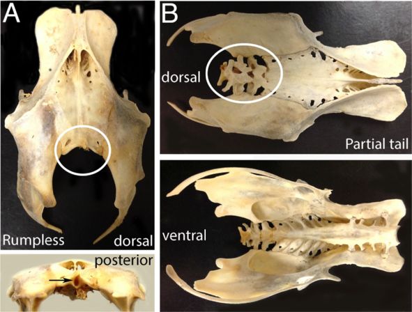
Rashid et al. EvoDevo 2014, 5:25                                                                                Page 2 of 20
http://www.evodevojournal.com/content/5/1/25

of short-tailed birds be due to a lack of intermediate         with the distal ten tightly articulated to form a stiffened
specimens in the fossil record, or from a very limited         rod supporting four unique, ribbon-like, tail feathers.
number of mutations that caused significant alterations           A marked decrease in the number of caudal vertebrae
to the primitive bird skeleton in a relatively short period    becomes very evident in the bird lineage from the Avialae
of time? With reference to the fossil record, we first         subgroup of maniraptoran dinosaurs onward (Figure 2).
review the evolutionary history and early skeletal devel-      Archaeopteryx retained an ancestral caudal vertebral
opment of the bird tail, and discuss the developmental         count of between 20 and 23 [18]. The next most basal
mechanisms involved in axial extension and termination.        bird, Jeholornis, from the Jiufotang Formation of China
We then present a comprehensive survey of mouse tail           and dated at approximately 120 million years old [19], was
mutants with the purpose of examining conserved pat-           also long-tailed, and had 22 caudal vertebrae that are
terns of mutation and likely candidates within the tail        nearly identical to those of Archaeopteryx [12]. The same
gene regulatory network that may have played major             formation preserves the next most basal bird, Sapeornis
roles in reducing the bird tail. These paleontological,        [20,21]. This taxon is the first bird to express a shortened
genetic, and developmental analyses are applied to the         tail with only six to seven free caudal vertebrae and
critical juncture in bird evolution at which tails were        an elongated pygostyle. All taxa derived from the last
truncated and flight was greatly enhanced, in the late         common ancestor of Sapeornis and extant birds retain
Jurassic to Early Cretaceous transition of primitive long-     a reduced tail ending in a pygostyle, suggesting that
tailed to short-tailed birds.                                  this evolutionary transformation is highly adaptive and ne-
                                                               cessary for bird physiology. A single specimen, Zhongornis,
Background                                                     may potentially lie phylogenetically between the long-
The origin of the derived bird tail occurred over a            tailed and short-tailed birds. Zhongornis is known from a
remarkably short evolutionary interval, as evidenced by        single specimen representing a juvenile that was either
the short-lived co-occurrence of both long- and short-         fledged or near fledgling [22]. The juvenile nature of this
tailed birds in equivalent spatio-temporal fossil forma-       taxon makes any phylogenetic placement uncertain, but it
tions (Figure 1). Nearly all non-avian theropod dinosaurs      does have a unique tail morphology that may be either a
sported long, 'reptilian' tails. These taxa were bipedal, so   phylogenetic or ontogenetic signal. Its tail consisted of 13
the tail likely had a counterbalance function. It also had     caudal vertebrae, with the last four partially fused and
robust transverse processes on the proximal caudal ver-        forming what may be a partial pygostyle.
tebrae that would have served as attachment sites for             Despite these variations, there is consensus that short-
the large caudofemoralis muscles that were the primary         tailed primitive birds appear in the fossil record relatively
hind limb retractors [4]. The oldest known bird, Archae-       suddenly, with fewer caudal vertebrae terminating in a
opteryx, dated to 150 million years ago, defines the clade     fused distal pygostyle, with abrupt rather than gradual loss
Aves [5-7] or Avialae [8]. Its fully formed flight feathers,   of tails [2]. These short-tailed birds, the confuciusor-
elongated wings, and evidence of capable powered flight,       nithids, enantiornithines and early ornithurines, had
all ally Archaeopteryx with birds [9,10]. Yet, the presence    acquired a number of other more modern bird-like
of teeth, clawed and unfused fingers, and an elongated,        traits that differed from their long-tailed primitive
bony tail are characteristics shared with non-avian            bird predecessors. These traits included more exten-
theropod dinosaurs. Paravians, including Archaeopteryx,        sive synsacral, sternum, and digit fusion (Figure 1), as well
are characterized by long tails [11,12], some fusion of syn-   as uncinate processes fused to adjacent ribs [23,24].
sacral vertebrae, and varying flight capability (Figure 1).    Osteological modifications were coupled to changes in
Most deinonychosaurians had between 20 and 30 cau-             musculature and behavior. With tail truncation and
dal vertebrae. Oviraptorosaurs, probably the immedi-           multiple bone fusions came advances in flight mechanics.
ate outgroup to Paraves, had relatively shorter tails.         Some of those flight advances can be attributed to the
These shorter tails were due not just to a modest              pygostyle, partly through its contributions to tail feather
decrease in the number of caudal vertebrae relative to         control [25]. Because Jeholornis had a long tail with a
other non-avian theropods, but more generally to a             proximal feather fan, there is some debate about whether
reduction in individual lengths of the more distal cau-        the pygostyle co-evolved with mobile fan-shaped feather
dals [13]. Interestingly, several oviraptorosaurs have         arrays [26]. Whatever their origin, the pygostyle-associated
been documented to have the distal caudal vertebrae            feather fans differed from the frond-type arrays of
co-ossified into a pygostyle-like structure that braced        more primitive long-tailed ancestors [25]. Fan-shaped
a fan-like arrangement of retrices [13-16]. Another            feather arrays play significant roles in sexual selection
more prominent independent reduction of tail length            in modern birds, and likely played analogous func-
occurred in Epidexipteryx, a Mid- or Late-Jurassic manir-      tions in their more primitive short-tailed ancestors
aptoran dinosaur [17]. Its tail had only 16 caudal vertebrae   (Figure 3) [27].
Rashid et al. EvoDevo 2014, 5:25                                                                                                         Page 3 of 20
http://www.evodevojournal.com/content/5/1/25

 Figure 1 Evolutionary tree of Paraves showing important evolutionary changes. Although several other groups of dinosaurs evolved a
 pygostyle (fused posterior tail vertebrae) independently, note that the first birds had long tails and that the fossil record documents a short
 temporal duration of both long- and short-tailed birds followed thereafter exclusively by birds with truncated, distally fused tails.

  Truncation of the bird tail was also concurrent with                       processes and hemal arches (chevrons). In modern birds,
reduction and shortening of the large caudofemoralis                         the CML is absent or reduced, and where present, its
muscle (CML). Reduction of this muscle is not exclusive                      origination site is on the pygostyle [4]. One exception is
to birds and is evident among all maniraptoran subgroups,                    the rumpless Araucana chicken; in this case, the CML
as hypothesized from the lack of a clearly distinguishable                   originates on the pelvis [28]. It is interesting to note that
fourth trochanter, the CML insertion site. More profound                     in Sapeornis, the most basal short-tailed bird, the caudal
CML reductions, however, are predicted in early birds                        vertebrae retained hemal arches [21], but the Confuciusor-
with truncated tails [4]. In theropods and in modern                         nis tail was more derived, and no hemal arches are
reptiles, the CML originates on the proximal caudal                          observed [29] (Figure 2). The presence of hemal arches in
vertebrae, with attachment points on the ventral transverse                  Sapeornis indicate its CML was more substantial than in
Rashid et al. EvoDevo 2014, 5:25                                                                                                      Page 4 of 20
http://www.evodevojournal.com/content/5/1/25

                                                                                    Archaeopteryx

                                                                     Sapeornis

                                                                         Confuciusornis

                                                               Gallus

 Figure 2 Comparison of tail skeletons between Archaeopteryx, Sapeornis, Confuciusornis, and chicken (Gallus gallus). The Archaeopteryx
 tail was modeled after Gatesy and Dial (1996), as well as the Bavarian, Solnhofen, #11, and Thermopolis specimens. For Sapeornis, the tail was
 reconstructed from specimens IVPP V13276, STM 15-15, and DNHM-D3078. The Confuciusornis tail was modeled after Chiappe (2007) and
 specimens GMV-2131, GMV-2132, and GMV-2133. Pygostyles are indicated by arrows. Scale bars equal 2 cm.

 Figure 3 Evolutionary correlation between the pygostyle, tail length, and possible display behavior. This mirror tree, constructed in
 Mesquite [49], shows the correspondence between tail adaptations in theropod dinosaurs, with presence (black) or absence (white) of a
 pygostyle mapped onto the left tree, and presence (black) or absence (white) of evidence that the tail may have been used in display on the
 right tree. Note that the tails of species in bold have shortened tails relative to basal theropods.
Rashid et al. EvoDevo 2014, 5:25 Page 5 of 20
http://www.evodevojournal.com/content/5/1/25
Confuciusornis, suggesting that formation of the pygostyle pairs form is established, at least in part, by the intersec-
alone is not sufficient to cause the degree of CML reduc- tion of two opposing extracellular gradients: the Wnt/Fgf
tion seen in Confuciusornis and in modern birds. gradient and the retinoic acid (RA) gradient. Wnt and Fgf
CML modifications, and others within the tail, may proteins are secreted from the posterior of the embryo,
have facilitated the abrupt transition to short-tailed birds whereas the RA gradient arises from the most recently
due to function decoupling. Decoupling of locomotor formed somites. These soluble factors interact at a critical
structures from each other is a hallmark of the origin of threshold, termed the determination front, where new so-
birds and powered flight and was most focused in the mite formation is initiated (Figure 5) [50-55]. The addition
forelimb and tail [30,31]. The tail of extant birds, for of somite pairs is controlled by an oscillating 'segmen-
example, functions to provide lift, braking, and turning tation clock' signaling cascade, which repeats for each
surfaces for controlled flight [32-36], but is decoupled somite pair. The mechanisms guiding the oscillating clock
from the hind limb and has lost its ancestral contribu- are not completely understood; however, a number of
tions to terrestrial (as opposed to aerial) locomotion. clock participants and their roles have been described
Therefore, the complex functional repertoire of extant [52,56-60]. Among clock genes with time-dependent oscil-
bird tails is achieved by a primary decoupling of the tail lating expression patterns are members of the Wnt, Fgf,
from the hind limbs followed by additional flight adapta- and Notch pathways. The cooperative action of the mo-
tions within the tail. lecular pathways functions to synchronize the oscillation
of the clock, such that a wave front of clock-gene expres-
Tail development sion moves anterior to posterior along the embryonic axis.
Tail structures Negative feedback regulation of clock genes by their
The mechanisms directing tail growth are similar among targets within activated cells as well as RNA instability
vertebrates, and have been evolutionarily conserved since are mechanisms employed to generate oscillating gene ex-
before the dinosaurs. Vertebrate embryo tails are con- pression [61-63]. The boundaries of newly formed somites
structed of the same basic elements, arranged in the same are established by positional expression of Notch pathway
basic pattern (Figure 4). Along the dorsal midline lies the genes; these genes also establish the anterior/posterior axis
neural tube, which gives rise to the brain and spinal cord. of each somite [64-68]. As somites are sequentially added,
Ventral to the floor plate of the neural tube is the noto- ingression through the primitive streak and cell division in
chord, a structure that is the precursor to the nucleus the PSM and CNH feeds into and maintains the PSM for
pulposus in spinal discs [37], and is necessary for proper continued somitogenesis [42]. Krol and colleagues con-
formation of the neural tube and somites [38-40]. Ventral ducted a particularly interesting study comparing the tran-
to the notochord in the posterior part of the embryo is the scriptomes of mouse, chicken and zebrafish during one
hindgut, the most caudal part of which is known as the somite extension. They discovered that despite a high level
tailgut. Somites, discrete paired segments of paraxial of conservation of the major pathways and events of somi-
mesoderm, flank the neural tube and are the developmen- togenesis, the genes that show oscillating expression can
tal precursors of the axial vertebrae, skeletal muscle and differ. Only two Notch pathway proteins, Her1 and Her5,
dermis. In addition, neural crest cells, integral to early were shown to oscillate in all three vertebrates, but
development, overlay the dorsal neural tube and sub- all other identified oscillating proteins, primarily members
sequently migrate ventrally to form the majority of the of the Fgf, Notch, and Wnt cascades, were specific to each
peripheral nervous system, including the sensory ganglia vertebrate. This suggests an unexpected evolutionary
of the tail [41]. A pool of undifferentiated progenitor plasticity in a critical developmental process. Specifically,
mesenchyme cells in the tail bud, the chordoneural hinge members of the Fgf, Notch, and Wnt pathways were likely
(CNH), is the primary source of cells from which tail targets of evolution in axial extension [69].
elongation proceeds [42,43]. Located ventral to the tail tip
and adjacent to the forming tailgut is the ventral ectoder- Regional specification
mal ridge (VER), the remnant of Hensen's node, through The regional identity of the somites, that is, cervical,
which the final gastrulating cells migrate [44-46]. thoracic, lumbar, sacral or caudal, is determined by Hox
gene expression [70]. The Hox genes were first discov-
Axial extension ered in Drosophila, where Hox gene mutations changed
Early in vertebrate embryo development a body plan is the positional identity of segments along the Drosophila
established, whereby somites are added sequentially along body axis [71]. Drosophila and other non-vertebrates
the axis. Somitogenesis has been recently reviewed else- have up to 14 genes contained within one Hox cluster.
where [47,48], but in brief, begins with the formation of Due to tandem genomic duplications, vertebrate Hox
the presomitic mesoderm (PSM) during gastrulation [49]. genes usually appear in four paralogous DNA clusters, A
Following gastrulation, the region of PSM where somite through D. Hox genes within those clusters, numbered 1Rashid et al. EvoDevo 2014, 5:25 Page 6 of 20
http://www.evodevojournal.com/content/5/1/25
A
B C
S
CNH
TG
M
Figure 4 Structures in the embryonic vertebrate tail. (A) Three-dimensional (3-D) reconstruction of an extending vertebrate embryo tail. Axial
structures include the NT and Nc; lateral to these are the paraxial somites and PSM. Somites are the embryonic precursors to skeletal muscle, ribs,
and bony vertebrae; motor and interneurons are derived from the NT; the CNH is the remnant of Hensen's node and contains pluripotent cells;
the PSM is the source of cells from which somites arise; and mesenchyme cells (M) at the distal tip of the tail feed into the CNH. Not shown:
neural crest and ventral structures. Axis indicates Anterior, A; Posterior, P; Dorsal, D; and Ventral, V. (B) Lateral schematic of tail structures. The axial
NT and Nc and paraxial somites and PSM lie dorsal to the TG, which in turn is dorsal to the VER. The VER is the remnant of the Hensen's node
and a source of growth-promoting signals. Not shown: neural crest and PSM. (C) Chick embryo tail stage HH23 stained for somites with FITC-phalloidin.
Abbreviations: CNH, chordoneural hinge; M, mesenchyme, Nc, notochord; NT, neural tube; PSM, presomitic mesoderm; S, somite; TG, tailgut; VER, ventral
ectodermal ridge.
through 13, are collinearly expressed along the body axis transformation, in which vertebrae take on characteris-
sequentially, with Hox1 most rostral and Hox13 most tics that are more anterior or posterior to their position.
caudal [72-77]. In any given vertebrate or non-vertebrate Concurrent disruptions in all three mouse Hox10 genes,
organism, not all 13 or 14 Hox genes within each paralo- for example, cause the lumbar vertebrae to transform
gous cluster are present [78]. Teleost fish sustained an into thoracic-like vertebrae with ribs [82]. Conversely,
additional genome duplication, and therefore, possess loss-of-function of the more posteriorly expressed three
another set of Hox clusters. While four more Hox Hox11 genes in mice results in a failure to form sacral
clusters would be expected, three have been identified, vertebrae, being replaced by vertebrae with lumbar morph-
bringing the total number of clusters in teleosts to seven ology. While these mutations generally preserve the overall
[79]. In vertebrates, Hox genes perform analogous body number of vertebral elements, some Hox gene disruptions
patterning functions to Drosophila and are most evident can increase or (more commonly) decrease total vertebrae
in defining the rostral to caudal identities of vertebrae. numbers (reviewed in [78]).
Most Hox genes are thought to specify regional axial There are additional factors that contribute to regional
identity by initially conferring anteroposterior patterning specification of the tail. Gdf11, for example, which encodes
during gastrulation [80], followed by fine-tuning within a Bmp (Bone morphogenetic protein)-related growth fac-
maturing mesoderm and neuroectoderm (reviewed in tor, acts to establish the trunk-to-tail transition in verte-
[81]). Mutations in Hox genes typically cause homeotic brates [83]. Also involved in caudal axial patterning andRashid et al. EvoDevo 2014, 5:25 Page 7 of 20
http://www.evodevojournal.com/content/5/1/25
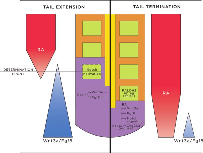
Figure 5 Tail extension and axial termination signaling schematic. During tail extension (depicted on left), somitogenesis is actively
proceeding, with new somites forming from PSM at the determination front. Activities from Cdx proteins, Wnts, and Fgfs establish a posterior
Wnt3a/Fgf8 gradient, which opposes an anterior RA gradient. These opposing gradients allow the creation of the determination front, and
activation of the Notch pathway. Cycling expression patterns of Wnt, Fgf, and Notch pathway genes follow a clock wave-front model, promoting
somite induction, segmentation and differentiation in successive waves, to add somites sequentially, rostral to caudal, down the vertebrate axis.
During tail termination (right), the RA gradient is unopposed, due to progressively decreasing concentrations of Wnts and Fgfs. Contributions
from RA (increased in chick via RALDH2), Hox genes, decreased concentrations of Cyp26a1 (mouse), Wnts and Fgfs, inhibition of the Notch
pathway, apoptosis, and loss of cell division and cell recruitment in the CNH act to terminate the tail. Abbreviations: CNH, chordoneural hinge;
RA, retinoic acid.
posterior expansion are the Cdx genes, which are disruption in the mouse causes limb abnormalities [89].
expressed primarily in the posterior end of the vertebrate In the mouse, loss-of-function studies have shown that
embryo. Cdx and Hox genes are related, and are thought caudal vertebrae are also specified by Hoxb13 [90]. Adding
to have derived from the same ProtoHox cluster [84]; both further complexity to the system, Hoxb8 in the mouse is
have been highly conserved throughout evolution and play expressed in mesoderm in the tail (and also more anteri-
roles in body patterning from cnidarians to higher verte- orly). Hoxb8 knockout does not alter caudal vertebrae
brates. Three separate Cdx genes are generally expressed identity [91], but it can rescue tail vertebrae in Cdx2/4
in vertebrates, and if two of these are knocked out in the mouse mutants [81], indicating that it may contribute to
mouse (Cdx2 and Cdx4), axis extension is prevented and caudal somite extension. Further genetic analyses will help
some trunk and tail structures do not form [85]. Control to determine whether the functions of these Hox genes in
of Cdx and Hox genes is mediated through the Fgf, Notch, the tail are universal in vertebrates.
RA and Wnt signaling pathways, underlining the crucial
contributions of these pathways to extending and shaping Tail termination
the spinal column [78,86,87]. Whether a tail is long, like that of a mouse, or short, as
in a chicken, a number of converging developmental
Caudal positional identity events ensure that the tail stops elongating at a charac-
The correlations between Hox gene expression pattern teristic point in each species and does not grow indefin-
and vertebral specification are not simple, and functional itely. At the termination of axial extension, secondary
redundancies are prevalent. In both chick and mouse, body formation is completed, with somites, neural tube,
the anterior expression boundary of Hoxd12 marks the and notochord development concluded. At this point,
transition between sacral and caudal vertebrae [88]. the pool of progenitor mesenchyme cells within the
Hoxd12 contributes to caudal specification, but its CNH is depleted by a combination of events: epiblastRashid et al. EvoDevo 2014, 5:25 Page 8 of 20 http://www.evodevojournal.com/content/5/1/25 migration through the VER is complete, the non-prolif- also influences the expression of Fgf8 [103], downreg- erating mesenchyme that feeds into the CNH population ulation of Wnt3a at the end of somitogenesis inhibits is also depleted and the neuroblast cells originating in tail growth by influencing both RA effects as well as the CNH population have exited the cell cycle. Since through inhibition of Notch-directed somite formation most of these structures and cell populations are required and maturation. for axial extension, the loss of nearly any one of them will Changes in the VER occur as tail growth comes to an halt progression of the tail. The vertebrate embryo hedges end, terminating notochord elongation, inhibiting somi- its bets, however, and has distinct concurrent mecha- togenesis [105], and also terminating caudal gastrulation nisms for terminating some if not all of these elements [106]. The thickened ventral epithelium that characterizes (Figure 5). the VER reaches its peak at approximately the 45-somite When somitogenesis is nearing completion, the rate of stage in the chick; subsequently, the VER gradually dimin- cell addition to and somite separation from the PSM ishes until it disappears altogether [105]. The dissipation begin to slow down. In the chick, the first 45 pairs of of the VER is concomitant with loss of its signaling. Before somites take 90 minutes each to form; thereafter, the the VER declines, it expresses several secreted proteins, segmentation clock slows such that the final 5 to 8 caudal including Sonic Hedgehog (Shh) [107], Fgf17, Bmp2, and somites (predecessors of the pygostyle) take 150 minutes Wnt5a [105,108], and functions to control the expression to form [42]. One likely candidate for this deceleration is of Noggin in overlying ventral mesoderm [106]. Noggin, WNT3a. While somitogenesis is robustly proceeding, up in turn, is a powerful modulator of Bmps, and hence plays to HH (Hamburger Hamilton [92]) stage 21, WNT3a is significant roles in Shh/Bmp signaling cascades. As the highly expressed in the tail mesenchyme, but as somite VER recedes, it can no longer maintain its signaling, and addition and the need for PSM nears its close, WNT3a is Noggin expression in ventral mesoderm is downregulated. gradually downregulated between HH22 and HH25 [42]. When the VER is ablated, it prevents the ingression of RALDH2, an enzyme involved in the synthesis of RA from epiblast cells that are needed for feeding cells into the vitamin A, is correspondingly upregulated in the tail mes- non-proliferating mesenchyme of the tail and thus, the enchyme in the chick and is thought to be responsible for CNH. The neural tube can temporarily extend without WNT3a and FGF8 downregulation. This creates an imbal- the presence of the notochord (albeit with ventral pat- ance of signaling factors, and thereby promotes the effects terning abnormalities) [38,39], but somite formation and of RA. Exposing mouse embryos to increasing levels of patterning are disrupted [40], and the tail prematurely RA induces more severe axial truncation (Figure 6A) [93]. truncates. Thus, the indirect effects of VER disruption In addition to contributing to the loss of the growth factor affect the tail progenitor population and lead to termin- Fgf8, RA causes differentiation of somitic cells and con- ation of tail elongation. comitant reduction in cell division; without Wnt3a, these Ablation studies, in which different axial structures are effects act as barriers to further somite addition. In removed in a living embryo, have shed light on the inter- the mouse, RA is also promoted by downregulation dependence of a number of these structures for axial of Cyp26a1 [42], an enzyme that normally metabolizes extension. As mentioned above, VER or notochord abla- RA [94]. When Cyp26a1 is downregulated, RA concentra- tion results in the failure to form the complete secondary tion effectively increases, further inhibiting tail growth neural tube and caudal somites, and leads to premature [95,96]. Interestingly, a critical level of RA signaling is axial truncation. In studies where the neural tube was required, as either augmenting or decreasing the amount ablated, the dermamyotome region of differentiated of RA causes premature termination of somitogenesis somites was absent, likely due to disruptions in Bmp [93,97,98]. In this finely tuned system, RA is required for and Wnt signaling from both the dorsal neural tube maturation of recently made somites before the next pair and overlying dorsal ectoderm [109]. Removal of the of somites can form, but prolonged exposure to RA CNH population causes subsequent loss of neural tube, prevents further somite addition. Wnt3a expression also notochord, somite formation inhibition and shortened affects somitogenesis via its cross-talk with the Notch path- tails [39,43]. If the somites themselves are removed, neural way. Specifically, Wnt3a and Notch pathway genes regulate crest cell delamination and subsequent migration come each others' expression levels and patterns [57,99-103], and to a halt; an imbalance between Noggin and Bmp4 is Notch pathway genes are intrinsically tied to the segmenta- thought to be responsible [110]. Reminiscent of the tion clock and somite boundary formation [47,48,104]. As balance between Wnt3a, Fgf8 and RA, there is also a an indication of the coordination between these various balance between Noggin, Shh and Bmps between pathways, loss of Fgf4 and Fgf8 in the mouse tail PSM different structures in the tail. The opposing functions results in loss of Wnt3a, downregulation of Notch sig- of these proteins help to pattern the neural tube naling, and inhibition of Cyp26a1 [99], all of which act [111] and are also involved in somite segmentation together to prematurely truncate the tail. Since Wnt3a and differentiation [112]. Disruption of this balance
Rashid et al. EvoDevo 2014, 5:25                                                                                                          Page 9 of 20
http://www.evodevojournal.com/content/5/1/25

                               A                                        RA

                                  s1

                                  c1

                                          WT

                               B                                 C

                                       WT

                                        Hoxb13KO
                                                        **                    WT               Hoxb13 Ectopic
                                                                                               Overexpression

 Figure 6 Experimental manipulations affecting the length of the vertebrate tail. (A) Increasing RA exposure in mouse embryos leads to
 progressive loss of caudal and sacral vertebrae. s1 indicates first sacral vertebrae and c1 indicates first caudal vertebrae. Data adapted from Shum
 et al. 1999 [93]. (B) Hoxb13 knockout (Hoxb13KO) in the mouse increases caudal vertebrae number by 2 and causes more barrel-shaped as
 opposed to hourglass-shaped vertebrae. Bars indicate experimental marking of equivalent numbered vertebrae; arrowheads indicate caudal
 vertebra #30 in both wildtype (WT) and Hoxb13KO; asterisks indicate two additional caudal vertebrae. Data adapted from Economides et al. 2003
 [90]. (C) Precocious ectopic overexpression of Hoxb13 in the mouse causes prematurely truncated tails. Data adapted from Young et al. 2009 [81].
 RA, retinoic acid.

likely plays multiple roles in terminating elongation of                     employed by embryos to help sculpt morphological fea-
the tail.                                                                    tures and cull extraneous cells, and is often considered
   Applying information from all of these ablation studies                   to be a default pathway for those cells that find them-
to the normal process of tail termination has helped us                      selves within inappropriate environments. During axial
to understand why the disruption of one structure leads                      extension, apoptosis is kept at bay at least in part by sur-
to the termination of others, and helps to explain why                       vival promoting signals through the Shh/Noggin/Bmp
peripheral and motor nerves are missing at the end of                        cascade [39,117], but this cascade is largely disrupted as
the tail. Just as Noggin is upregulated with somite                          tail growth slows. At these later stages, there is consider-
ablation, it is also upregulated in the dorsal neural tube                   able cell death in the tail bud [118], which depletes the
at the end of the naturally waning tail. This enhanced                       available pool needed for somite production. Signaling
expression is thought to inhibit neurogenic neural crest                     from Wnt3a and Fgfs is necessary for maintaining the
derivatives, causing loss of peripheral nerves [113]. Lack                   pool of undifferentiated PSM cells which give rise to
of motor neurons just anterior to the end of the tail is                     somites [55,119], and downregulation of these pathways
likely due to termination of the neural tube (from which                     in the waning tail contributes to apoptosis. In higher
motor neurons derive [114]), the loss of Shh signaling                       vertebrates, this severe reduction in PSM, however, is
from the notochord and neural tube [115], which is                           caused only in part by apoptosis. Due to RA effects,
required to pattern the motor neurons in the ventral                         there is reduced cell division [42,120], and neuroblast
neural tube, and/or the presence or absence of an as-yet                     cells exit the cell cycle, further depleting the progenitor
unidentified diffusible signal intrinsic to the tail [116].                  population. However, PSM has not been completely
   Another process involved in tail cessation is apoptosis,                  eradicated before the segmentation oscillator comes to a
or programmed cell death. Apoptosis is a mechanism                           halt, and the remaining PSM becomes unresponsive to
Rashid et al. EvoDevo 2014, 5:25 Page 10 of 20
http://www.evodevojournal.com/content/5/1/25
signals that promote axial extension [42]. The concept thoracic (1), lumbar (6), sacral (2) and sacro-caudal (5)
that apoptosis plays a contributing but not solitary role vertebrae (Figure 7) [126] (in chick, there is no evidence
in tail cessation is further substantiated by the fact that that caudal vertebrae are incorporated into the syn-
significant mesenchyme cell death occurs even while the sacrum [88]), the chick tail achieves its maximum num-
tail is robustly extending [121], and apoptosis has been ber of somites by E5. Although the synsacral somites
inhibited in different vertebrates, but longer tails have form as separate blocks of tissue, the chondrified
not been documented [122]. synsacral cartilages fuse together to form a continuous
Finally, the prescribed species-specific number of somites structure, devoid of intervertebral discs. Distinct ossifica-
that are formed is controlled by Hox genes [123,124], and tion centers for each of the vertebrae are retained, with
the most caudally-expressed Hox genes act to terminate the onset of ossification observed in a rostral to caudal
tail elongation. Precocious over-expression of Hox13 para- sequence from E15 onward (Figure 7). In addition to the
logs at the posterior end of mouse [81] and chick [125] centrum of the vertebrae, the free sternal ribs have ossi-
embryos leads to prematurely truncated tails with loss of fication centers. The lumbar vertebrae that follow have
caudal vertebrae (Figure 6C). Conversely, targeted knock- transverse processes, but these do not have independent
out of Hoxb13 in the mouse leads to expansion of tail ossification centers, rather ossifying from the pedicle
structures, including neural tube, PSM, and two extra situated between the centrum proximally and the trans-
caudal vertebrae (Figure 6B). A reduction in the level of verse processes distally. The ventral processes abut and
apoptosis in PSM was also observed in these knockout become fused to the ilium. Notably, the transverse dorsal
mice, which ties Hox13 genes to depletion of the mesen- processes and dorsal ligament uniting the lumbar verte-
chyme cells needed for somitogenesis. In this same brae ossifies postnatally forming a continuous plate of
study, Hoxb13 was also shown to inhibit neuronal bone, or sacral shield (Figure 7, adult). This is a common
proliferation, which, combined with the normal loss of feature of birds from neornithines to modern birds,
caudal neural crest-derived neurogenic cells, doubly helping to strengthen the fused synsacrum [1]. The rigid
ensures the lack of spinal ganglia at the end of the tail synsacrum and ilium fuse to form an immovable struc-
[90]. Another potential mechanism Hox13 genes employ ture with osteoblasts visible in the ilium from E14. The
to terminate the tail is to intersect the Wnt/Fgf/RA gradi- transverse and ventral processes of the two sacral verte-
ent by downregulating Cyp26a1 [81], providing yet an- brae abut and fuse to the medial posterior curve of the
other example of the level of involvement of this gradient ilium. These processes are sometimes referred to as sa-
on tail cessation. cral ribs, having their own ossification centers, similar to
To summarize, perturbations in virtually any of the tail sternal ribs [127]. Beyond the synsacrum, the free caudal
elongation processes described above lead to termination vertebrae develop ossification centers at E18, and finally,
of extension, and numerous perturbations are built into by E19 the fused cartilaginous elements of the pygostyle
the axial extension system to ensure proper tail length. follow suit (not shown). Ossification of the axial verte-
Imbalances in the Wnt/Fgf/RA and Noggin/Shh/Bmp brae and pelvic girdle is complete by hatching [126].
gradients are largely responsible for stopping tail growth. Extending beyond the synsacrum, the mature tail in the
Once disrupted, the signaling cascades generated from chick consists of 5 to 6 free caudal vertebrae (there are 5
the gradients no longer properly coordinate with other to 8 free caudal vertebrae among birds in general) and
cascades such as Notch, thereby disabling the elongation the pygostyle (a fusion of the final 5 to 6 somites).
machinery. Hox13 paralog genes further inhibit tail
elongation, likely through their interactions with the Mutations that cause tail truncation
regulatory factors that control these gradients [81]. Fi- Relating the developmental events of axial extension and
nally, increased apoptosis at the termination of somito- termination back to the process of evolution, one needs
genesis removes all remaining progenitor cells. All of to consider birds as organisms that sustained one or
these coordinating pathways are orchestrated through more mutations that converted long theropod tails to
the different tail structures, and signaling between the short avian tails terminating in a fused, distal pygostyle.
structures maintains their inter-dependence so that if Considering the many redundancies in the process of tail
even one structure fails, the rest eventually follow suit. cessation, it follows that just one mutation could have
Species-specific differences in the way the orthologous truncated the posterior axis. Alternatively, the short,
pathways are modulated likely account for the varying fused tails of early birds could have been the result of a
tail lengths observed among vertebrates [123,124]. suite of mutations that occurred over a longer period of
time, and the fossil record is incomplete. Complicating
Skeletal development of the bird synsacrum and tail the genetics behind the transition to short-tailed birds is
Following the formation of somites that will contribute the nature of the mutations that could have occurred.
to the synsacrum, an axial structure with 14 fused Mutations can occur within gene coding sequence, in cisRashid et al. EvoDevo 2014, 5:25 Page 11 of 20 http://www.evodevojournal.com/content/5/1/25 Figure 7 Embryonic events during the termination of the chick embryo tail. Embryonic day, E12 to E17 chondrified skeletons (blue) of chick embryos, with ossified cells (red) detectable from E14 to E17. Compare the E17 chondrified skeleton and the adult skeleton showing the fused synsacrum and bony plate in the latter; the 5 free caudal vertebrae and the pygostyle already patterned during somitogenesis. regulatory regions (CREs) outside coding sequence that mechanisms were possible. It remains to be asked, given control gene expression, by DNA deletion, or by gene the lack of dinosaur DNA, how can we parcel out those duplication [128-130]. The prevailing theory is that most mutations that affect morphological changes in the tail phenotypic changes in evolution are due to changes in and may have converted theropod tails to bird tails? CREs [128]. Alterations in the regulation of gene expres- One way to study the ancestral ties between organisms sion would allow for fewer pleiotropic and potentially is to proceed with an evolutionary developmental biol- deleterious effects of critical genes, by affecting some ogy or 'evo-devo' approach. This approach is particularly but not all expression patterns. Despite the potentially appealing when studying theropod-to-bird evolution, higher chance that changes in CREs were responsible because despite the lack of dinosaur DNA, we can for short fused tails, any of the other above-mentioned still examine gene pathways that potentially generated
Rashid et al. EvoDevo 2014, 5:25                                                                              Page 12 of 20
http://www.evodevojournal.com/content/5/1/25

dinosaur traits. In terms of tail morphology, the gene        of the caudal vertebral bodies, and fusion of the most
pathways that are involved in tail elongation and termin-     distal caudal vertebrae into the pygostyle [25]. Bone
ation in different organisms can be studied side-by-side,     fusion is indeed highly evident in the modern bird
and modulations of those pathways that generate long ver-     skeleton. Fusions are observed not just in the pygostyle,
sus short tails can be compared. In considering the many      but also in the synsacrum and in the dorsal vertebrae
pathways involved in tail elongation and cessation, how do    anterior to the synsacrum, between the ribs as cross
we narrow down the list of candidate genes that may have      bridges called uncinate processes, and in the distal limbs.
been modulated by mutation? For this particular study, we     Between 150 and 120 million years ago, long before the
looked to the mouse, the vertebrate organism with the         Cretaceous Tertiary Extinction, these modern bird traits
greatest accumulated data on mutations. Most mouse            were evolving in primitive birds and other maniraptoran
mutational data has been generated by targeted gene           dinosaurs, and a number of variations of these traits
disruption, which causes phenotypes that are likely more      have been observed in fossil specimens from this time-
extreme than mutations that would occur in, say, CREs.        frame [2]. From the accumulated list of short-tailed
Despite the preponderance of targeted transgenesis, sub-      mouse mutants, it is evident that most mutations affec-
stantial mutational information has also been contributed     ted more than just the tail, and a whole host of other
by chemical, radiological, or transposon induction of         pleiotropic defects were also observed, including, among
random mutations, as well as by studies of spontaneous        others, more anterior bone fusions [see Additional file 1].
mutations. However the mutations occurred, the mouse is       The question then becomes, are there any morphological
a reasonable place to begin the examination of those genes    traits that co-segregate with reduced numbers of caudal
whose modulation affects tail morphology.                     vertebrae for single mutations, and do any of these traits
                                                              co-segregate in the fossil record? Fusions between various
Morphological analysis of mouse mutants                       vertebral surfaces are observed in the bird skeleton, so
A list of mouse tail mutants was generated from the           different types of fusions were considered. Among the
MGI Jackson Laboratories database [131] and the litera-       mouse mutants with decreased numbers of caudal verte-
ture [see Additional files 1 and 2]. From this list, a num-   brae (n = 105), it was interesting to note that 34% (36/105)
ber of interesting and surprising correlations surfaced.      also displayed vertebral fusions (including fusions of
Immediately obvious was the observation that of the           neural arches, articular surfaces/zygopophyses, transverse
159 mutants with affected tails, only two, the Hoxb13         processes, spinous processes, or vertebral bodies). Of the
(Figure 6B) and Slx4 knockout mice, have increased num-       36 with vertebral fusions, 53% had fused ribs. Of all
bers of caudal vertebrae, and these mutations cause only      mutants with decreased numbers of caudal vertebrae, only
modest increases. Indeed, the tail suffers from a particu-    three also had digit fusion, which does not constitute a
lar developmental precariousness, as seen in the prepon-      significant degree of co-segregation but indicates that digit
derance of mutations causing short tails, suggesting that     fusion is also possible with truncated tail mutations.
tail growth is relatively easily disrupted. While this re-      Because modern and primitive short-tailed birds exhibit
mains to be studied across vertebrates, in this particular    both truncated tails and fused vertebrae [1,2], we asked
case, one could propose the argument that the early           the opposing question: If a mouse mutation transpired
decoupling of the tail from hind limb locomotion in man-      which caused caudal vertebral body fusion (the predomin-
iraptoran theropods may have facilitated tail reduction       ant type of vertebral fusion observed in the pygostyle of
through a process of relaxed purifying selection. Relaxed     modern and short-tailed primitive birds), what was the
purifying selection has been demonstrated to promote          chance that caudal vertebrae number was also decreased?
phenotype plasticity [132], and thus, may also facilitate     Seventeen of 23 caudal vertebral body fusion mutants, or
rapid evolutionary change. The distal portion of the tail,    74%, also had truncated tails (Table 1). A high percentage
once completely decoupled from hind limb function, may        of caudal vertebral body fusion mutants (48%) also dis-
have been relatively free to accumulate mutations without     played fused ribs. Thus, in the mouse, if a caudal vertebral
deleterious effects and thereby facilitate the evolution      body fusion event occurred, there was a nearly even
of novel morphologies, namely a radically shortened tail      chance the mouse also had fused ribs and a significantly
and pygostyle.                                                better than even chance that it also had a truncated tail
  To correlate the mouse mutants with specific skeletal       (for a complete list of mouse posterior vertebral fusion
differences observed between theropods, primitive birds       mutants and additional information on the caudal verte-
and modern birds, several parameters were taken into          bral fusion mutants, see Additional files 2 and 3). Since
consideration. When modern bird tails are compared            there is a fairly high correlation of vertebral fusion,
with those of their more primitive bird or non-avian          rib fusion, and truncated tails with mouse mutants,
theropod ancestors, there are three primary differences:      we next asked whether these traits also co-segregate
reduction in the number of caudal vertebrae, shortening       in the fossil record in the transition from non-avian
Rashid et al. EvoDevo 2014, 5:25 Page 13 of 20
http://www.evodevojournal.com/content/5/1/25
Table 1 Caudal vertebral body fusion mouse mutants
Mutant Affected vertebraea cdl v #b Fused ribs Fused digits Structure affected Relevant
pathways
C T L S cdl So NT NC VER
Ankrd13a ✓ Nd
Cenpj ✓ ✓ Nd
CREB ✓ ✓ ✓ ✓ ✓ ✓ Notch/Wnt
Dkk1 doubleridge/null ✓ ✓ ✓ ✓ ✓ Wnt
Dll3 ✓ ✓ ✓ ✓ ✓ ++ ✓ ✓ Notch/Wnt
f flexed tail; Sfxn1 mutation ✓ ✓ ✓ +/− ✓ ✓ ✓ Bmp/Shh
Fgf3 ✓ ++ ✓ ✓ Notch/Wnt
Hes7 ✓ ✓ ✓ ✓ ✓ ++ ✓ ✓ Notch/Wnt
Ikkα ✓ ✓ ✓ + ✓ Notch; FGF
Jsr jumbled spine and ribs ✓ ✓ ++ ✓ ✓ Notch/Wnt
Knk kinked tail ✓ + ✓ Notch; Wnt
Lrp6 crooked tail ✓ ++ ✓ ✓ ✓ Notch/Wnt
mea meander tail ✓ ✓ ✓* ✓ ++ Wnt
Meox1/Meox2 ✓ ++ ✓ ✓ Retinoic Acid
Noto ✓ ✓ ✓ ++ ✓ ✓ ✓ Wnt
Nrarp ✓ ✓ ✓ ✓ + ✓ ✓ Notch/Wnt
Ppp5c ✓ Wnt
Ror2 ✓ + ✓ ✓ ✓ Notch/Wnt; Wnt
Rpl38 ✓ ✓ ✓ ++ ✓ ✓ Hox
Rps7 ✓* ✓ + nd
Sulf1/Sulf2 ✓ ✓ ✓ ✓ ✓ Bmp/Shh
Vangl2 ✓ ✓ ✓ ✓ ++ ✓ ✓ Wnt
Wnt5a ✓ ✓ +++ ✓ ✓ ✓ Wnt
a b
Selected phenotypes and relevant genetic pathways for each mutant are indicated. Asterisks indicate fusion other than vertebral body fusion. For the decreased
number of caudal vertebrae (Cdl vertebrae #) column, +: less than half of the tail is absent; ++: half or more of the tail is absent; and +++: tail is severely
truncated. Check marks indicate the presence of a particular trait. For a more comprehensive list of posterior vertebral body fusion mouse mutants (those which
have fusions posterior to the cervical vertebrae, as seen in modern birds), and for more information on the caudal vertebral body mutants, see Additional files 2 and 3.
Abbreviations: C, cervical; cdl, caudal; L, lumbar; NT, neural tube; NC, notochord; nd, not determined; S, sacral; So, somite; v, vertebrae; VER, ventral ectodermal ridge.
maniraptorans and primitive long-tailed birds to short- In the mouse mutants analyzed, vertebral fusions were
tailed birds. correlated not just with short tails, but also with fused
Correlations between certain maniraptoran traits have ribs. Rib fusions are likely observed with these mutations
been noted before, especially the co-incidence of short because somitogenesis is the most common developmen-
tails and the presence of a pygostyle [133]. The co-segre- tal event affected, and vertebral ribs arise from somites
gation of short tails and a pygostyle applies to a wide [135-138]. If rib fusions were coincident with a vertebral
range of feathered non-avian maniraptorans and primitive fusion mutation, they could have altered the axial skeleton
birds, lending credence to the possibility that the traits are in a few different ways. Very proximal rib fusions could
pleiotropic and linked by a single mutational event, irre- have helped establish the wide dimensions of the ilium
spective of adaptive advantage. This is not to suggest that (synsacrum) by increasing bone mass at the point ribs
the same mutation occurred in these different groups; the attach to the axial skeleton. Very distal rib fusions could
number of different mouse mutants with these traits also have increased the breadth of the sternum, as
indicates that a number of different genes were likely seen in the Gnai3 mouse mutant [139]. Branching of
causative. It should be noted that not all dinosaurs ribs is occasionally observed in some mutants, such as
with a pygostyle had short tails (for example, Beipiaosaurus Tbx6 [140,141] [see Additional file 3], which could have
[134]). These long-tailed dinosaurs with a pygostyle, been the mechanism behind the formation of uncinate
however, were in the minority, just as are the mouse processes. This possibility seems less likely, however,
mutants with fused distal vertebrae and unaffected tail considering that quite a number of non-avian thero-
length. pods exhibited uncinate processes with long tails andRashid et al. EvoDevo 2014, 5:25 Page 14 of 20
http://www.evodevojournal.com/content/5/1/25
no pygostyle, such as Oviraptor philoceratops, Veloci- In addition to posterior truncation, stretches of accessory
raptor mongoliensis, and Diononychus antirrhopus. Also, neural tube and notochord occur when RA is injected into
Sapeornis had a short tail with a rod-like pygostyle and no the tailbud, indicative of enhanced neural differenti-
uncinate processes [21]. Even if uncinate processes had ation [143]. Premature neural differentiation is evident
already evolved through separate means, however, we in a number of the mouse mutants with truncated tails
hypothesize their fusion to adjacent ribs (seen only in Aves [see Additional file 1] and is also evident in the rumpless
[23]) could have been facilitated through a vertebral fusion Araucana chicken [144]. To our knowledge, no manipu-
mutation. lations in the bird have resulted in longer tails with
increased numbers of caudal somites/vertebrae. One study
Genetic analysis of mouse mutants attempted to extend neural crest in chick embryo tails
Investigation into the genetic pathways that are modulated by inhibiting Noggin [113]. While certain neural crest
by these caudal vertebral body mutations in the mouse markers were indeed upregulated at the end of the
also proved insightful (Table 1 and [see Additional file 3]). tail, additional somites were not added and tail length
Of the 20 (of 23) mutations whose affected pathways were remained unchanged. Just as in the mouse, manipulations
previously studied, 10 involve Notch or Notch/Wnt sig- in the tail are far more likely to reduce or otherwise fail to
naling. The remaining mutations have the following path- alter length as opposed to increase tail length.
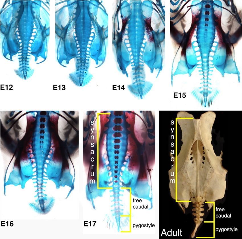
way associations: seven involve Wnt signaling (possibly The only known spontaneous mutation that trun-
independent of Notch), two are associated with BMP/Shh cates the avian axial skeleton, namely in the rumpless
cascades, and one each is involved in Hox or RA signaling. Araucana chicken (Figure 8A), was identified as a gain-
The genes that were mutated appear to have developmen- of-function mutation of the proneural (Iroquois) Irx1 and
tal roles that would be expected, which include functions Irx2 genes [145]. Iroquois genes are tied to Notch, Wnt,
in somitogenesis (most prevalent), neural tube and noto- and Bmp/Shh signaling [146-148], and in addition to their
chord biogenesis and patterning, mesoderm establishment proneural role, they establish tissue borders during devel-
and maintenance, neurogenesis, angiogenesis, and VER opment. Interestingly, heterozygotes of the rumpless locus
signaling. Of those associated with the Notch pathway, the retain 2 to 4 caudal vertebrae, and these are irregularly
majority (6/10) were involved in somite segmentation or fused (Figure 8B) [149,150], adding this mutation to those
differentiation; these include CREB, Dll3, Fgf3, Hes7, Lrp6, that cause both short tails and fused vertebrae. The most
and Nrarp. It is intriguing to note that in the chick, the caudal somites are never generated and the pygostyle,
Notch pathway members Lnfg, Nrarp, and Meso (the chick therefore, never forms. While there is no equivalent gain-
homolog of Mesp2), are all downregulated as somitogen- of-function mouse mutant, loss-of-function mutations in
esis slows [42], at an equivalent point at which the mouse either Irx1 or Irx2 in the mouse do not cause posterior
tail would still be actively extending. Interestingly, several truncation or fused vertebrae, emphasizing an important
mutations among members of this particular pathway, caveat with this study that mutation of the same genes
including Dll3, Hes7, Lnfg, Lrp6 [142], Mesp2, and Tbx6, can be manifested differently depending on the nature of
are reasonably well tolerated and cause spondylocostal the mutation.
dystosis (SCD) disease in humans [3]. Individuals suffering It was previously estimated that among a variety of
from this disease display fused ribs and vertebrae with un- normal-tailed chicken breeds, a tailless phenotype was
affected reproductive capacity, as in the mouse mutants. consistently observed in approximately one out of every
thousand chicks hatched, making further tail trunca-
Experimental manipulations and one spontaneous mutation tion a relatively common chicken mutant phenotype
that affect chick tail morphology [151]. Well-tolerated, relatively common tail truncat-
As in other vertebrate species, the chick tail is often ing mutations (especially if dominant and germ-line)
neglected as a focus of research. There are, however, that conferred certain advantages would theoretically
additional studies that deserve mention here apart from promulgate the evolutionary transition from long- to
the RA and Hox13a manipulations already cited. To short-tailed birds.
date, targeted transgenesis in the bird is largely unreported,
and even transgenic overexpression has been restricted to Additional considerations
a small handful of genes. Genes or proteins can be modu- If a single mutation occurred that shortened the tail and
lated in other ways, however, and the chick embryo is fused the distal caudal vertebrae in short-tailed primitive
amenable to studies such as microinjection, electropor- birds, it would seem logical that occasionally, that gene
ation of DNA or RNA, viral transfection, and insertion of modulation would spontaneously reverse resulting in more
matrices soaked with diffusible proteins or other factors. recent long-tailed birds. Several lines of evidence contradict
The specific morphological changes upon ectopically this possibility, however. First, while not impossible, direct
applied RA in the tailbud are particularly interesting. reversals of mutations are extremely unlikely. IndirectYou can also read