The Molecular 'Myc-anisms' behind Myc-Driven Tumorigenesis and the Relevant Myc-Directed Therapeutics - MDPI
←
→
Page content transcription
If your browser does not render page correctly, please read the page content below
International Journal of
            Molecular Sciences

Review
The Molecular ‘Myc-anisms’ behind
Myc-Driven Tumorigenesis and the Relevant
Myc-Directed Therapeutics
Jessica McAnulty            and Analisa DiFeo *
 Department of Pathology, University of Michigan, Ann Arbor, MI 48109, USA; jlmc@umich.edu
 * Correspondence: adifeo@med.umich.edu; Tel.: +1-734-936-5685

 Received: 16 November 2020; Accepted: 11 December 2020; Published: 13 December 2020

 Abstract: MYC, a well-studied proto-oncogene that is overexpressed in >20% of tumors across all
 cancers, is classically known as “undruggable” due to its crucial roles in cell processes and its lack
 of a drug binding pocket. Four decades of research and creativity led to the discovery of a myriad
 of indirect (and now some direct!) therapeutic strategies targeting Myc. This review explores the
 various mechanisms in which Myc promotes cancer and highlights five key therapeutic approaches
 to disrupt Myc, including transcription, Myc-Max dimerization, protein stability, cell cycle regulation,
 and metabolism, in order to develop more specific Myc-directed therapies.

 Keywords: myc; cancer; inhibitors; transcription; stability; max; cell cycle; metabolism; synthetic lethality

1. Myc’s Role as a Transcription Factor
      Think about any key cellular process, and the Myc family most likely has a role in it: proliferation,
metabolism, differentiation, and apoptosis. The Myc transcription factor family consists of c-Myc,
N-Myc, and L-Myc. Discovery of c-Myc led to finding N-Myc, primarily expressed during development
or in neuroblastoma, and L-Myc, expressed in lung tissue and small-cell lung cancer [1,2]. Although the
family shares stretches of homologous regions and some related targets, N-Myc and L-Myc are
less characterized. c-Myc (hereon referred to as Myc), is the most well-studied family member
as it is an influential protooncogenic transcription factor that binds to about 15% of genes [3–5].
In order to regulate gene expression, Myc recruits or interacts with many different cofactors, including
histone acetyl transferases (CBP, p300, GCN5/TRAPP), P-TEFb and polymerases, and chromatin
remodelers (BRD4, the SWI/SNF complex, SIRT1) [5,6]. It is important to note that Myc can also repress
gene expression by binding to the promoter region and interacting with MIZ1 and SP1 to displace
co-activators or by recruiting DNA methyltransferases [5]. Furthermore, there are two structural
components of the MYC gene that are essential to drive its role as a transcription factor: the E box and
the basic helix-loop-helix leucine zipper domain.
      The canonical Myc E Box DNA binding motif (50 -CACGTG-30 ) is one of the most frequent
regulatory motifs in the human genome [7]. Although Myc is not the only transcription factor that
can occupy this motif, elevated levels of Myc will replace the other bound transcription factors [8],
demonstrating how Myc can influence the transcription of many genes and diverse processes in
proliferating cells. A Myc core signature of 50 common target genes across four human cancer cell
types and human embryonic stem cells revealed Myc’s influence in RNA processing and ribosome
biogenesis [9]. Other diverse functions of Myc target genes include cell cycle regulation, metabolism,
cell adhesion, and signal transduction [5,10,11].
      However, Myc does not exclusively bind to the E-box to modulate transcription. In repression of
gene transcription, cofactors recruit Myc to the promoters lacking the E-box and interfere with active

Int. J. Mol. Sci. 2020, 21, 9486; doi:10.3390/ijms21249486                          www.mdpi.com/journal/ijms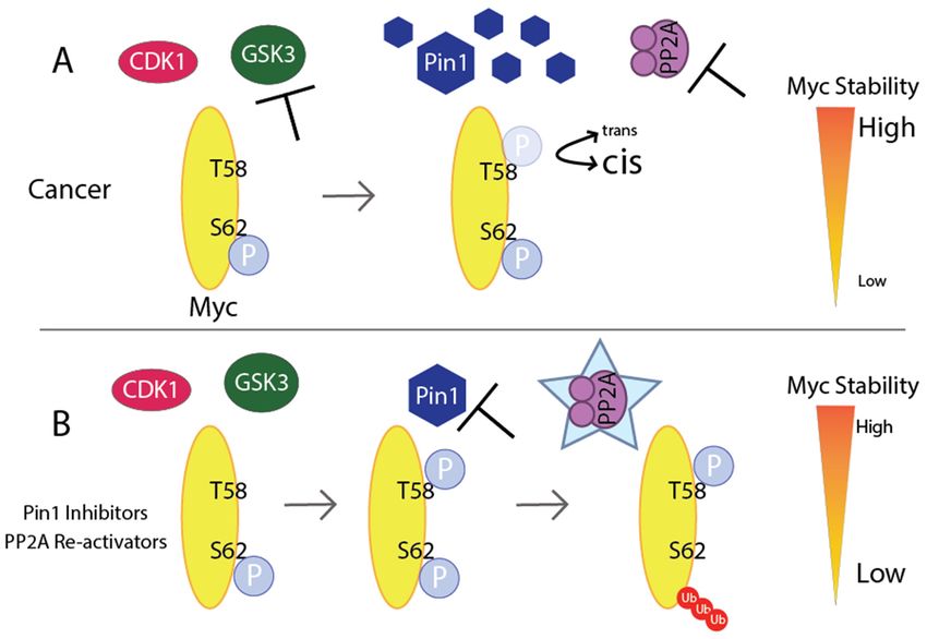
Int. J. Mol. Sci. 2020, 21, 9486 2 of 28
transcription factors [12–14]. Furthermore, Myc can amplify transcriptional signals by accumulating at
the promoters of active genes, even in those with low-affinity E-box-like sequences [15,16]. There is
still debate of whether Myc drives global amplification of transcription [15,16] or if global amplification
is an indirect consequence of Myc’s selective regulation of gene targets [17–19].
In addition to the E box binding motif, the basic helix-loop-helix leucine zipper (bHLHZip) domain
is crucial for Myc’s activity. To take on its role as a transcription factor, Myc must heterodimerize with
Myc-associated factor
Int. J. Mol. Sci. 2020,X21,(Max);
x FOR PEER Myc
REVIEWis incapable of homodimerizing and is inactive2as of 29a monomer.
Max binds accumulating
to Myc at the at the bHLHZip
promoters ofdomain [20,21],
active genes, and
even in this
those heterodimerization
with is required to bind
low-affinity E-box-like sequences
to the E box[15,16].
consensusThere issequence and
still debate of activate
whether Myc transcription [22,23]. However,
drives global amplification overexpression
of transcription [15,16] or if of Max
global amplification
leads to transcriptional is an indirect
repression consequence
as the of Myc’s selective
Max homodimers regulationMyc/Max
antagonize of gene targets [17–19].
heterodimers [22,24].
In addition to the E box binding motif, the basic helix-loop-helix leucine zipper (bHLHZip)
Mad, a transcriptional repressor, can also reduce Myc-driven transcription by dimerizing with Max [5].
domain is crucial for Myc’s activity. To take on its role as a transcription factor, Myc must
heterodimerize with Myc-associated factor X (Max); Myc is incapable of homodimerizing and is
2. Dysregulation
inactive ofas Myc Leads toMax
a monomer. Cancer
binds to Myc at the bHLHZip domain [20,21], and this
heterodimerization is required to bind to the E box consensus sequence and activate transcription
Normally, Myc expression is tightly controlled at each molecular level (transcriptionally,
[22,23]. However, overexpression of Max leads to transcriptional repression as the Max homodimers
post-transcriptionally, translationally,
antagonize Myc/Max heterodimersand post-translationally
[22,24]. Mad, a transcriptional via protein
repressor, can stability,
also reduceand
Myc-via protein
driven
interactions), and transcription
has a shortby dimerizing
half-lifewith
of Max
20–30[5]. min [25–29]. Given that there are many levels of
regulation,2.asDysregulation
a consequence, there are many opportunities for which control of MYC can go awry.
of Myc Leads to Cancer
For instance, point mutations, chromosomal translocations, and gene amplification, or other factors
Normally, Myc expression is tightly controlled at each molecular level (transcriptionally, post-
that activate transcriptiontranslationally,
transcriptionally, or stabilizeand Myc, have been found
post-translationally in a wide
via protein range
stability, andofvia cancers,
protein which are
further described by Meyer
interactions), and hasand Penn
a short and Kalkat
half-life of 20–30 et al.[25–29].
min [30,31]. Thisthat
Given oncogenic activation,
there are many levels ofwhich leads
to sustainedregulation,
levels ofasMyc,
a consequence,
contributes theretoare many opportunities
tumorigenesis andfor which control
evasion of MYC can go awry.checkpoints
of tumor-suppressive
For instance, point mutations, chromosomal translocations, and gene amplification, or other factors
leading to uncontrolled cell growth. MYC expressing tumors thus become addicted to and depend on
that activate transcription or stabilize Myc, have been found in a wide range of cancers, which are
the oncogene, as described
further shown in cancer
by Meyer andmodels
Penn and with conditional
Kalkat activation
et al. [30,31]. This oncogenicof MYC [32].
activation, whichOn the contrary,
leads
MYC leads
inactivationtoofsustained levelstooftumor
Myc, regression
contributes to in transgenic
tumorigenesismouse models,
and evasion of displaying Myc’s vital role
tumor-suppressive
checkpoints
in tumor initiation andleading to uncontrolled
maintenance cell growth. MYC expressing tumors thus become addicted to
[33–35].
and depend on the oncogene, as shown in cancer models with conditional activation of MYC [32]. On
MYC amplification is found
the contrary, inactivation of MYC in leads
21% tooftumor
patients across
regression 33 different
in transgenic cancers
mouse models, [36], particularly
displaying
breast cancer, lung
Myc’s vitalsquamous
role in tumorcell carcinoma,
initiation uterine[33–35].
and maintenance carcinoma, esophageal carcinoma, and ovarian
MYC 1).
cancer [25] (Figure amplification
The highest is foundratesin 21%
of of patients across 33
amplification different
are seen in cancers [36], particularly
high-grade serousbreast
ovarian cancer
cancer, lung squamous cell carcinoma, uterine carcinoma, esophageal carcinoma, and ovarian cancer
wherein greater than 50% of tumors harbor this genomic alteration. MYC translocation affects several
[25] (Figure 1). The highest rates of amplification are seen in high-grade serous ovarian cancer
hematological malignancies,
wherein greater than 50%including
of tumors harbormultiple myeloma,
this genomic Burkitt’s
alteration. lymphoma,
MYC translocation affectsdiffuse
several large cell
lymphoma,hematological
and T-cellmalignancies,
acute leukemia including [37]. Alternatively,
multiple some lymphoma,
myeloma, Burkitt’s tumors that dolarge
diffuse display MYC
not cell
lymphoma,
amplification and T-cellphosphorylation
show extreme acute leukemia [37].levels Alternatively,
whichsome aid in tumors
Mycthat do not [38–41].
stability display MYC
amplification show extreme phosphorylation levels which aid in Myc stability [38–41].
MYC Amplification Across Cancers
40
% MYC Amplification
30
20
10
0
no C
C ph ma
St no s
Pa ma a
te s
d Li e
A ung Pr Ne r
no Sq sta k
ca ua te
in s
M ore er
de rc a
l a
M rv a
l
en h m ot LB a
al o b o r a
C e me
a
si Es arc rian
nc ch
or o ul lio L
C lad ma
G S no al
an a
& ve
a l C io m
U rea
o c
rc mo
ci u
o m
rin
ra a m
G m
dr C to s D om
tic ph tif m
m
oc rom a M he C
ci C
el ic
a t
l d
ar a g
el c
ar R
ve o o
B o
o
s a
no v
e
ci O
o
ea
e
H
as M
ve
ar
de
ng L
er
U
C
va
w
e
In
Lo
rin
bl
Lu
st
lio
te
in
a
U
G
ra
re
A
B
B
Figure 1. MYC amplification across cancers. Percentage represents number of patients with MYC
amplification for that cancer type. Red bars represent cancers in which >10% of patients harbor MYC
mutations. Data from The Cancer Genome Atlas Pan Cancer 2018 Dataset, cancer.gov/TCGA.
Int. J. Mol. Sci. 2020, 21, 9486                                                                                               3 of 28
              Int. J. Mol. Sci. 2020, 21, x FOR PEER REVIEW                                                        3 of 29

     With Myc’s Figure  1. MYC amplification
                    prominent       role across across
                                                   manycancers.  Percentage
                                                            cancers,    therepresents
                                                                             idea of Mycnumber asofapatients
                                                                                                     clinicalwith  MYC is too good to
                                                                                                                target
                amplification for that cancer type. Red bars represent cancers in which >10% of patients harbor MYC
be true. Although      targeted inhibition of MYC via siRNA reduces tumor burden in mice with very
                mutations. Data from The Cancer Genome Atlas Pan Cancer 2018 Dataset, cancer.gov/TCGA.
few toxicities despite Myc’s influence on global transcription [33,35,42,43], global MYC knockout is
embryonic lethalWithinMyc’s
                         mice. prominent   role across measures
                                  Thus, cautious        many cancers,  inthe idea of Myc
                                                                          observing         as aeffects
                                                                                         side    clinical target is too goodMyc need
                                                                                                          of disrupting
           to be true. Although targeted inhibition of MYC via siRNA reduces tumor burden in mice with very
to be addressed     [44]. The less expected problem is that direct inhibition of Myc is not possible with
           few toxicities despite Myc’s influence on global transcription [33,35,42,43], global MYC knockout is
current therapeutic      approaches—Myc
           embryonic lethal                          lacks
                               in mice. Thus, cautious       both enzymatic
                                                          measures                   activity
                                                                      in observing side    effectsand   an active
                                                                                                   of disrupting   Myc site for a small
                                                                                                                         need
molecule to    disrupt
           to be  addressedprotein-protein        interactions
                              [44]. The less expected    problem is[45].    Myc’s
                                                                      that direct      primary
                                                                                  inhibition        nuclear
                                                                                               of Myc           localization
                                                                                                       is not possible   with   further
           current  therapeutic   approaches—Myc        lacks both   enzymatic    activity  and  an
escalates the problem. Nonetheless, scientific discoveries led to creative ways to downregulate Myc. active site for  a small
           molecule to disrupt protein-protein interactions [45]. Myc’s primary nuclear localization further
This review   focuses on how Myc’s oncogenic activation leads to tumorigenesis through initiating
           escalates the problem. Nonetheless, scientific discoveries led to creative ways to downregulate Myc.
transcription,
           Thisincreasing
                 review focusesstability,
                                   on howand Myc’sinfluencing      cell cycleleads
                                                     oncogenic activation      andtometabolism,
                                                                                        tumorigenesiscoupled        with descriptions
                                                                                                         through initiating
of the indirect   inhibitors
           transcription,        of Mycstability,
                            increasing      that target     each mechanism
                                                     and influencing      cell cycle(Figure       2). The molecular
                                                                                       and metabolism,       coupled with   changes in
which MYC  descriptions
              becomesofan    theoncogene
                                 indirect inhibitors   of Myc translocations,
                                             (mutations,        that target each mechanism       (Figure 2). The
                                                                                     and amplification)         is molecular
                                                                                                                   beyond the scope
           changes in which MYC becomes an oncogene (mutations, translocations, and amplification) is
of this review  [30–32].
              beyond the scope of this review [30–32].

                   Figure 2. Schematic presenting the various cellular processes to target through inhibition or
      Figure 2. Schematic presenting the various cellular processes to target through inhibition or reactivation
                  reactivation in the nucleus (left) or cytoplasm (right) upon Myc-induced tumorigenesis. All will be
      in the nucleus    (left)
                  described  in or cytoplasm
                                detail             (right) upon Myc-induced tumorigenesis. All will be described in
                                       in this review.
      detail in this review.
              3. Disrupting Myc Stability to Inhibit Its Actions as a Transcription Factor
3. Disrupting Myc Stability to Inhibit Its Actions as a Transcription Factor
                   In cancer, Myc’s aberrant function as a transcription factor leads to increased cell proliferation,
            cell differentiation,
      In cancer,  Myc’s aberrant  cell function
                                        adhesion, as
                                                  anda transcription
                                                       angiogenesis [10]. Hereleads
                                                                       factor   we will focus on inhibiting
                                                                                    to increased  cell proliferation,
cell differentiation, cell adhesion, and angiogenesis [10]. Here we will focus on inhibiting transcription,
disrupting Myc/Max dimerization, and enhancing protein degradation as strategies to disrupt Myc
gene and protein stability and therefore Myc-driven tumorigenesis.

3.1. Myc Drives Aberrant Transcription
     As discussed, MYC amplification is common among many cancer types. This amplification of MYC
results in increased binding of Myc to promoters and enhancers of active genes, which magnifies the
transcriptional signal [15,16] and as a consequence, increases global transcription. During transcription,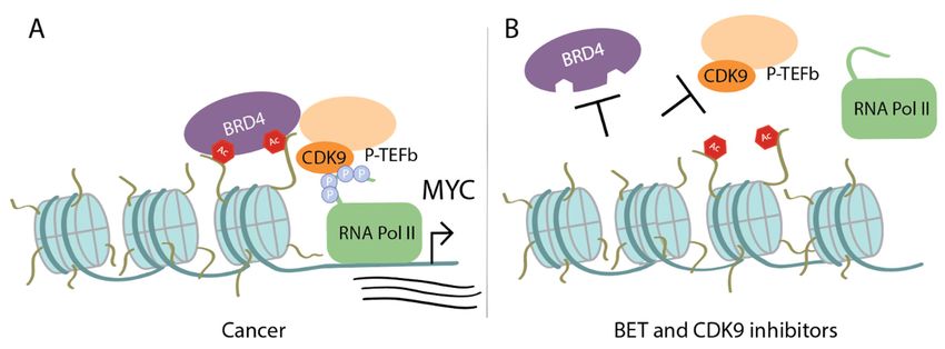
transcription, disrupting Myc/Max dimerization, and enhancing protein degradation as strategies to
disrupt Myc gene and protein stability and therefore Myc-driven tumorigenesis.
3.1. Myc Drives Aberrant Transcription
Int. J. Mol. Sci. 2020, 21, 9486 4 of 28
As discussed, MYC amplification is common among many cancer types. This amplification of
MYC results in increased binding of Myc to promoters and enhancers of active genes, which
Myc magnifies
recruits the
thetranscriptional pause-release
transcriptional signal [15,16] andcomplex P-TEFb (a heterodimer
as a consequence, increases globalof cyclin-dependent
transcription.
During
kinase transcription,
9 (CDK9) and cyclinMyc recruits
T1, T2,the or transcriptional
K) [6,16,46]. pause-release
P-TEFb leads complex P-TEFb (aofheterodimer
to activation transcriptional
of cyclin-dependent kinase 9 (CDK9) and cyclin T1, T2, or K) [6,16,46]. P-TEFb
elongation by phosphorylating RNA polymerase II (Pol II) via CDK9, stimulating pause release [47–49]. leads to activation of
transcriptional elongation by phosphorylating RNA polymerase II (Pol II) via
Furthermore, BRD4, part of the bromodomain and extra-terminal motif (BET) protein family, also recruits CDK9, stimulating
pause release [47–49]. Furthermore, BRD4, part of the bromodomain and extra-terminal motif (BET)
P-TEFb to promoters to initiate transcription elongation [50]. The overlapping roles of BET proteins
protein family, also recruits P-TEFb to promoters to initiate transcription elongation [50]. The
and Myc in recruiting P-TEFb suggests BET proteins or CDK9 as therapeutic targets. First, BET proteins
overlapping roles of BET proteins and Myc in recruiting P-TEFb suggests BET proteins or CDK9 as
are known to regulate MYC transcription [51]. A recent study demonstrated in normal cells that
therapeutic targets. First, BET proteins are known to regulate MYC transcription [51]. A recent study
BRD4demonstrated
has even more in control
normal overcellsMyc
that by binding
BRD4 has andevenphosphorylating
more control over Threonine
Myc by58binding
on Myc,and leading
to degradation [52]. Threonine
phosphorylating However,58Myc is also
on Myc, capable
leading of regulating
to degradation BRD40 s histone
[52]. However, acetyl
Myc is also transferase
capable of
activity [52]. Additional
regulating BRD4′s histone studies aretransferase
acetyl needed toactivity
better [52].
understand
Additional how this circular
studies are neededbalance may be
to better
understand
affected how
in cancer. this circular
CDK9 balance target
is a potential may beas affected in cancer.
it is part CDK9is
of P-TEFb, is necessary
a potential target as it is part and
for proliferation
of P-TEFb,
maintenance of isMYC-overexpressing
necessary for proliferation and maintenance
hepatocellular carcinomaof MYC-overexpressing
[53], and is required hepatocellular
for maintenance
carcinoma
of gene silencing[53],
inand is required
several cancerforcellmaintenance of gene silencing
lines [54]. Another in several
tumorigenic cancer
feature cell lines
of Myc [54].
is looping to
Another tumorigenic feature of Myc is looping to tumor-specific super-enhancers (sites defined by
tumor-specific super-enhancers (sites defined by multiple enhancers abnormally bound by a plethora of
multiple enhancers abnormally bound by a plethora of transcription factors, such as BRD4 and
transcription factors, such as BRD4 and CDK9) [55]. Therefore, inhibiting MYC transcription indirectly
CDK9) [55]. Therefore, inhibiting MYC transcription indirectly via BET inhibitors or affecting
via BET inhibitorsoforMyc
transcription affecting transcription
target genes of Myc
by inhibiting target
CDK9 aregenes by inhibiting
promising strategies CDK9
that haveareshown
promising
strategies that have shown efficacy in
efficacy in Myc-driven cancers (Figure 3). Myc-driven cancers (Figure 3).
Figure
Figure 3. Upon
3. Upon MYCMYC amplificationinincancer,
amplification cancer, Myc
Myc recruits
recruitsadditional
additional transcriptional cofactors
transcriptional to drive
cofactors to drive
transcription:
transcription: (A) BRD4
(A) BRD4 binds
binds to acetylated
to acetylated lysines
lysines on histone
on histone tails tails
and and recruits
recruits P-TEFb
P-TEFb (which
(which includes
includes CDK9), that phosphorylates the carboxy terminal domain of RNA Pol II. Myc can also
CDK9), that phosphorylates the carboxy terminal domain of RNA Pol II. Myc can also individually
individually recruit P-TEFb. (B) Treatment with bromodomain and extra-terminal motif (BET)/BRD4
recruit P-TEFb. (B) Treatment with bromodomain and extra-terminal motif (BET)/BRD4 inhibitors
inhibitors prevents BRD4 from binding to histone tails and treatment with CDK9 inhibitors disrupts
prevents BRD4 from binding to histone tails and treatment with CDK9 inhibitors disrupts CDK90 s
CDK9′s kinase activity. Thus, both result in failure of activating transcription of MYC or Myc target
kinase activity. Thus, both result in failure of activating transcription of MYC or Myc target genes.
genes.
Targeting MYC Transcription—BET Inhibitors, BRD4 Degraders, CDK9 Inhibitors
Targeting MYC Transcription—BET Inhibitors, BRD4 Degraders, CDK9 Inhibitors
The The
BETBET proteins, BRD2,
proteins, BRD2,BRD3,
BRD3,BRD4,
BRD4, and testis-specificBRDT,
and testis-specific BRDT, areare epigenetic
epigenetic readers
readers and and
histone acetyl
histone transferases
acetyl that
transferases activate
that activatetranscription viabinding
transcription via bindingtoto specific
specific acetylated
acetylated lysine
lysine residues
residues
on histone tails.
on histone The
tails. Thebound
bound BET proteinsregulate
BET proteins regulate chromatin
chromatin remodeling
remodeling via H3K122
via H3K122 acetylation
acetylation and
act as
and act as scaffolds
scaffoldstotoform
form transcription complexes
transcription by recruiting
complexes transcriptional
by recruiting activatorsactivators
transcriptional such as P- such
TEFb [50,56,57]. Furthermore, BRD4 influences mitotic progression by binding
as P-TEFb [50,56,57]. Furthermore, BRD4 influences mitotic progression by binding selectively selectively to to
transcriptional start sites of M/G1 genes [58]. Oncogenes, such as MYC, have a transcriptional
dependency on BRD4 and recent findings suggest additional non-transcriptional functions of BRD4 in
cancer [59]. Bromodomains have a mostly hydrophobic pocket with aromatic rings and is an ideal
size for protein–protein interactions, making bromodomains attractive and obtainable therapeutic
targets, unlike Myc [56]. BET protein inhibitors compete for access to the bromodomain and upon
binding, disrupt chromatin remodeling and prohibit expression of target genes, including MYC.
Filippakopoulos et al. and Nicodeme et al. independently designed some of the first bromodomain
inhibitors, known as JQ1 and iBET respectively, that are highly specific towards the BET protein
family [60,61]. Initial studies showed efficacy of JQ1 downregulating both MYC expression and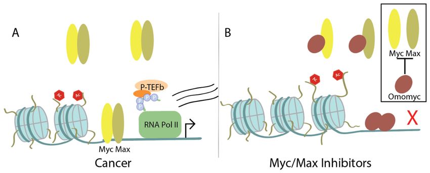
Int. J. Mol. Sci. 2020, 21, 9486                                                                     5 of 28

Myc’s transcriptome genome-wide in Myc-addicted hematological malignancies [62–64], and solid
cancers [65–68]. iBET’s proof-of-concept in preventing BET proteins from binding to acetylated histones
was demonstrated in an inflammation context [61], although a follow-up study exhibited that iBET
was capable of downregulating MYC expression, but to a lesser extent than JQ1 [51]. It is important to
note that both JQ1 and iBET lack specificity for a particular BET protein family member, which limits
their therapeutic availability [69–71]. Therefore, these BET inhibitors serve best as tools to improve our
understanding of targeting bromodomains and the effects on MYC. The discovery of JQ1 and iBET
inspired the development of additional BET inhibitors, with 10 inhibitors being assessed in clinical trials,
including MK-8628/OTX015. Phase Ib trials included six solid tumors such as NUT midline carcinoma
(NMC), which harbors an oncogenic form of BRD4, known as BRD4-NUT. The trial (NCT02259114)
completed with a recommended dose for Phase II studies, although the NMC patients that initially
responded, relapsed several months after treatment [72]. BET inhibitors as a whole currently appear to
have limited therapeutic response and dose-limiting toxicities. More preclinical research will increase
the biological knowledge on mechanisms of action and resistance of BET inhibitors.
      In addition to BET inhibitors, there are also BET degraders that utilize a concept designed in
2000: PROteolysis TArgeting Chimeric (PROTAC) [73]. PROTAC protein degraders link the protein of
interest to an E3 ligase in order to ubiquitinate the protein of interest for degradation. This approach
has been adapted to a variety of targets, including the androgen receptor, estrogen receptor, BCL2,
CDK9, and BET proteins to name a few [74]. PROTAC technology has entered clinical trials, including
Arvinas’s ARV-110 for patients with metastatic castration-resistant prostate cancer (NCT03888612)
which has shown efficacy and a promising safety profile in Phase I [75]. The first PROTAC BET
degraders, including MZ1, a BRD4-specific degrader, were designed in 2015 and demonstrated
increased apoptotic response compared to nonspecific BET inhibitors, but a modest decrease in MYC
expression [76,77].
      It appears the antitumor efficacy of both BET inhibitors and BET degraders is most likely due to
global transcription downregulation, rather than downregulation in MYC transcripts specifically [78].
Devaiah et al. recently discovered crucial molecular differences in Myc stability between BET
inhibitors and BET degraders. Since endogenous BRD4 destabilizes Myc, treatment with a BRD4
degrader, such as MZ1, enhances Myc stability, but treatment with a BET inhibitor, such as JQ1,
does not affect BRD40 s phosphorylation of Myc and therefore Myc’s half-life is unaffected while MYC
transcription is downregulated [52]. Several PROTAC BRD4 degraders demonstrate robust decreases
of MYC expression throughout 3–24 h [79,80], though there are no current clinical trials on BET/BRD4
degraders. Perhaps later timepoints and investigation of phophorylated-S62-c-Myc expression will aid
in understanding long-term effects of BET degraders on Myc stability. For further reading, detailed
reviews on bromodomains and their inhibitors are cited [56,81–84].
      CDK9 is another potential therapeutic target, given its kinase activity in the P-TEFb complex
which releases paused RNA Pol II to initiate transcription. CDK9 inhibitors demonstrate efficacy in
downregulating MYC transcripts and Myc stability across hepatocellular carcinoma [53], mixed-lineage
leukemia [85], diffuse large B-cell lymphoma [86], acute myeloid leukemia [87], and pancreatic
cancer [88]. Although preclinical studies have shown efficacy in targeting CDK9, the sequence
similarity to other cyclin-dependent kinases made specificity difficult. However, several groups
succeeded in creating CDK9-specific inhibitors and PROTAC degraders [38,54,89]. A recent study
demonstrated that CDK9-specific inhibitor, MC180295, downregulates MYC and leads to reactivation
of epigenetically silenced tumor suppressor genes [54]. Thus, downregulation of MYC is not due to
off-target effects of nonspecific CDK inhibition. Initial nonselective CDK inhibitors did not succeed
in clinical trials, most likely due to toxicities from off-target effects. These trials included patients of
many cancer types and were not selective to MYC-amplified patients [90]. However, CDK9-specific
inhibitors, such as BAY 1143572 (NCT01938638), are beginning to enter clinical trials in patients with
advanced cancer and will evaluate MYC expression as a biomarker [91,92].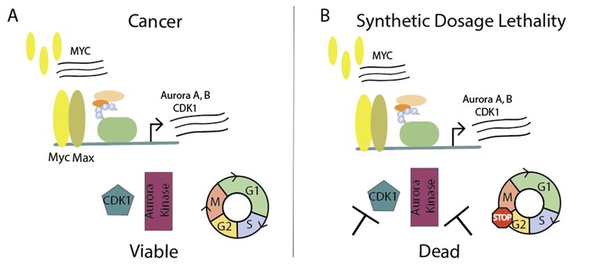
Int. J. Mol. Sci. 2020, 21, x FOR PEER REVIEW                                                               6 of 29

        study demonstrated that CDK9-specific inhibitor, MC180295, downregulates MYC and leads to
        reactivation of epigenetically silenced tumor suppressor genes [54]. Thus, downregulation of MYC is
        not due to off-target effects of nonspecific CDK inhibition. Initial nonselective CDK inhibitors did not
Int. J. succeed    in clinical
        Mol. Sci. 2020, 21, 9486trials, most likely due to toxicities from off-target effects. These trials included 6 of 28
        patients of many cancer types and were not selective to MYC-amplified patients [90]. However,
        CDK9-specific inhibitors, such as BAY 1,143,572 (NCT01938638), are beginning to enter clinical trials
        Additionally,
        in patients with combining        CDK9
                            advanced cancer     andand
                                                     willBET   inhibitors
                                                          evaluate           synergistically
                                                                     MYC expression            improves
                                                                                       as a biomarker       anti-proliferative
                                                                                                        [91,92].
activity inAdditionally,
               several cancers,       with no
                               combining       hematological
                                            CDK9                  toxicity synergistically
                                                    and BET inhibitors      or weight lossimproves
                                                                                             shown inanti-proliferative
                                                                                                         vivo [93–95]. Of the
sameactivity
         note, BETin several   cancers,
                       inhibitors    werewith  noefficacious
                                            also  hematological whentoxicity
                                                                       paired or weight loss showninhibitors,
                                                                                 with additional     in vivo [93–95].  Of PI3K,
                                                                                                                 such as
        the same    note, BET    inhibitors were   also efficacious  when    paired with  additional
ERK, or BCL2 inhibitors [81]. Readers are referred to reviews further discussing targetable           inhibitors, such as Myc
        PI3K, ERK,    or BCL2    inhibitors [81]. Readers   are referred  to reviews further  discussing
cofactors that aid in tumorigenesis [13], such as G quadraplex stabilizers [96,97]. In all, BET and       targetable Myc
        cofactors that aid in tumorigenesis [13], such as G quadraplex stabilizers [96,97]. In all, BET and CDK9
CDK9 inhibitors vastly affect transcription and as a result, downregulate MYC expression indirectly;
        inhibitors vastly affect transcription and as a result, downregulate MYC expression indirectly;
improving       their specificity is expected to increase their therapeutic benefit.
        improving their specificity is expected to increase their therapeutic benefit.

3.2. Myc/Max   Dimerization
     3.2. Myc/Max Dimerization
      Another  wayway
          Another    of affecting   Myc
                         of affecting    transcriptional
                                       Myc   transcriptional stability
                                                               stabilityisisbybypreventing
                                                                                 preventingMycMycfrom
                                                                                                   from interacting
                                                                                                          interacting with
DNA. with DNA. Myc must dimerize with Max in order to drive gene expression, though a recent structuralstudy
       Myc  must   dimerize    with  Max   in order  to  drive  gene   expression,    though   a recent  structural
demonstrates   that against
     study demonstrates       previous
                           that          belief, Myc
                                against previous        is stabile
                                                    belief, Myc isin   the absence
                                                                    stabile           of binding
                                                                              in the absence       DNA [45].
                                                                                              of binding   DNA Although,
                                                                                                                 [45].
Max Although,   Max heterodimerization
      heterodimerization     with Myc is with    Myc isfor
                                            required      required
                                                             Myc’s for   Myc’s oncogenic
                                                                     oncogenic     activityactivity  [98]. Therefore,
                                                                                             [98]. Therefore,   inhibiting
Myc inhibiting
      and Max Myc    and Max dimerization
                dimerization    prevents Myc   prevents   Myc from gene
                                                  from initiating    initiating   gene transcription.
                                                                             transcription.   There areThere
                                                                                                          twoare  two
                                                                                                               immediate
     immediate   challenges:  (a) targeting  the bHLHZip      domain    is nonspecific   to Myc/Max
challenges: (a) targeting the bHLHZip domain is nonspecific to Myc/Max and therefore could present     and  therefore
     could present off-target effects and (b) there are no apparent pockets for which a small molecule can
off-target effects and (b) there are no apparent pockets for which a small molecule can bind [99,100].
     bind [99,100]. Despite this, there has been success in disrupting the Myc/Max interaction with several
Despite this, there has been success in disrupting the Myc/Max interaction with several mini-proteins
     mini-proteins or molecules, including Omomyc, 10058-FA, 10074-G5, KJ-Pyr-9, MYCMI-6, and KI-
or molecules,  including
     MS2-008 (Figure   4). Omomyc, 10058-FA, 10074-G5, KJ-Pyr-9, MYCMI-6, and KI-MS2-008 (Figure 4).

           Figure
      Figure       4. Heterodimerization with
               4. Heterodimerization         with Max
                                                   Max is is required
                                                             requiredforforMyc’s
                                                                            Myc’soncogenic
                                                                                   oncogenic activity: (A) Upon
                                                                                                 activity: (A) Upon
           heterodimerization, Myc/Max binds to the E-box and initiates transcription. This is a normal cellular
      heterodimerization, Myc/Max binds to the E-box and initiates transcription. This is a normal cellular
           process, but in cancer, Myc amplification further increases Myc activity; (B) Treating with Omomyc,
      process, but in cancer, Myc amplification further increases Myc activity; (B) Treating with Omomyc,
           a dimerization inhibitor that preferentially binds to Max or homodimerizes, displaces Myc at E-boxes
      a dimerization inhibitor that preferentially binds to Max or homodimerizes, displaces Myc at E-boxes
           and decreases Myc transcription. Other discussed Myc/Max inhibitors either disrupt Myc/Max dimers
      and decreases   Myc transcription. Other discussed Myc/Max inhibitors either disrupt Myc/Max dimers
           or block Myc’s interaction with DNA, but not both.
      or block Myc’s interaction with DNA, but not both.
     Disrupting Myc/Max Dimerization
Disrupting Myc/Max Dimerization
         The most well-known, and perhaps the first, Myc/Max dimerization inhibitor is Omomyc, a
    The mostnegative
    dominant    well-known,
                        mutant and  perhaps
                               of Myc’s       the first,
                                        bHLHZip   domain Myc/Max     dimerization
                                                           with 4 amino            inhibitor
                                                                        acid mutations  in the is Omomyc,
                                                                                                leucine
a dominant  negative
    zipper that        mutant
                prevents      of Myc’s
                         Myc/Max        bHLHZip domain
                                  heterodimerization          with 4 amino
                                                      [101]. Omomyc   was a acid mutations
                                                                            laboratory       in the leucine
                                                                                       tool developed
zipper that prevents
    to bind and inhibitMyc/Max
                        Myc. Overheterodimerization    [101].
                                   the past two decades,      Omomyc
                                                         research       wasaabetter
                                                                  produced     laboratory  tool developed
                                                                                    understanding    of
to bind and inhibit Myc. Over the past two decades, research produced a better understanding of
how the molecular tool functions: Omomyc reduces the amount of Myc that can bind to promoters by
either heterodimerizing with Myc in the cytoplasm, heterodimerizing with Max, or homodimerizing.
Recent data show Omomyc preferentially binds to Max or homodimerizes [102]. The Omomyc
homodimers or Max/Omomyc heterodimers are transcriptionally inactive complexes that bind
specifically to E-box sequences and displace Myc/Max heterodimers resulting in decreased Myc-driven
transcription [102–105]. Importantly, Omomyc is specific towards Myc’s function and does not suppress
gene expression of other E-box-binding transcription factors [103].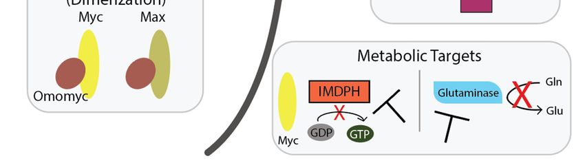
Int. J. Mol. Sci. 2020, 21, 9486                                                                     7 of 28

      Omomyc has shown efficacy in several tumor studies when it is conditionally or transiently
expressed in the cell or linked with a cell penetrating Phylomer [43,106–108]. However, Omomyc is
indeed capable of penetrating cells, including non-small cell lung cancer, neuroblastoma, glioblastoma,
and melanoma cell lines, due to its basic region [103]. Until recently, in vivo proof-of-concept was
lacking. Beaulieu et al. show Omomyc downregulates Myc target gene expression and prevents tumor
progression in lung adenocarcinoma in vivo models via intranasal administration (2.37 mg/kg) over
four weeks [103]. Similarly, in a lung adenocarcinoma xenograft model, paclitaxel combined with
Omomyc administered intravenously diminished tumor growth over 30 days [103]. Both models
showed no significant changes nor toxicities in blood counts or pathology reports of all major organs.
In non-tumor-bearing mice, Demma et al. show Omomyc injected intravenously (5.22 mg/kg) primarily
distributes to the liver and kidneys and has a short half-life in plasma [102]. Although this study
used a higher dosage of Omomyc than the cancer study, toxicities of Omomyc in normal cells must be
considered in future preclinical studies. Dr. Soucek, who created and studied Omomyc over the past
20 years, created the company Peptomyc to develop and sponsor Omomyc-derived clinical candidates;
the first clinical trial is anticipated to start in 2021.
      While Omomyc is capable of disrupting Myc/Max dimerization and preventing Myc from
interacting with DNA, other Myc/Max inhibitors are typically characterized by one of those two actions.
In 2002, Berg et al. demonstrated the proof-of-concept of using combinatorial chemical libraries to
find small molecule inhibitors of protein–protein interactions, including Myc/Max [109]. Shortly after,
Yin et al. identified specific Myc/Max inhibitors from a combinatorial library including 10058-FA and
10074-G5, that result in G0/G1 cell cycle arrest and apoptosis in vitro [110]. However, in vivo studies
show both 10058-FA and 10074-G5 are rapidly metabolized and lack anti-tumor activity [111,112].
Therefore, these compounds best serve as molecular tools and a starting point for new compound
development. More recent Myc/Max inhibitors include KJ-Pyr-9 and MYCMI-6. KJ-Pyr-9 has sufficient
pharmacokinetic properties to penetrate tissue and prevent tumor growth, but cannot reduce existing
tumors; its inability to decrease tumor size may be due to residual Myc activity as an effect of incomplete
Myc inhibition [113]. MYCMI-6 was identified in 2018 as a promising Myc/Max-specific inhibitor
that halts Myc-driven transcription, induces apoptosis, and reduced tumor proliferation in vivo [114].
Interestingly, these different Myc/Max inhibitors all initiate different biological effects.
      Recently, Han et al. identified novel Myc-binding inhibitors, MYCi361 and MYCi975, that appear
to act through disrupting Myc/Max dimers and increasing Threonine (T)58 phosphorylation of Myc,
which leads to Myc degradation. MYCi-induced degradation could be a result of changes in Myc
confirmation as it interacts with MYCi; it is important to note that not all Myc/Max inhibitors lead to Myc
degradation [115]. Treating cells with proteasome inhibitor MG132 or exposing non-phosphorylatable
Myc (T58A mutant) cells to MYCi361 rescues or prevents the MYCi361-induced Myc degradation [116].
In vivo studies utilized a Myc-driven prostate cancer mouse model, MycCaP, in which tumors were
significantly decreased upon MYCi361 treatment. Additional studies are required to determine
its efficacy in other cancers. Given that MYCi treatment modified the tumor microenvironment
through increased expression of PD-L1, Han et al. demonstrated synergistic effects with MYCi361 and
anti-PD1 in the MycCaP model, despite the model’s documented resistance to anti-PD1 therapy [116].
MYCi975 performs similarly to MYCi361 but has a higher therapeutic index and better tolerability
in vivo of up to ten time the anti-tumor efficacious dose. There is promise for future studies on
MYCi975 due to its inhibition of cancer cell growth and reduction of Myc target gene expression
in vitro and decreased tumor growth in vivo with high tolerability. These compounds represent a new
class of directly targeting Myc and inhibiting Myc/Max dimers, which led to Myc degradation.
      Agents that inhibit Myc/Max from binding DNA have also been pursued, although they lack
in vivo data and specificity towards Myc/Max [117]. One approach to prevent Myc binding to DNA
is by targeting one of the many cofactors that recruits Myc to its target genes. WDR5 is an adapter
protein that interacts with histone methyltransferase and serves as a scaffold for chromatin; it recruits
Myc to chromatin and the Myc-WDR5 interaction is required for Myc-driven tumorigenesis [118].
Int. J. Mol. Sci. 2020, 21, 9486 8 of 28
Thomas et al. recently discovered that WDR5 stabilizes the Myc/Max interaction with DNA and a
mutant Myc that cannot bind to WDR5 leads to tumor regression in a Burkitt lymphoma in vivo
model [118]. However, the mutant Myc was capable of binding to chromatin, suggesting that targeting
WDR5 does not affect Myc’s ability to interact with DNA. Given the antitumor effect and the druggable
pockets within WDR5, it is a viable anti-Myc contender to pursue; additional recent advances with
WDR5 are described in the Metabolism section of this review.
Alternative approaches that are not widely explored include stabilizing Max. In 2019, Struntz et al.
discovered KI-MS2-008, which stabilizes Max homodimers while decreasing both Myc binding at
promoters and Myc protein levels [119]. KI-MS2-008 proved efficacious in T cell acute lymphoblastic
leukemia and hepatocellular carcinoma in vivo models with a reduction in tumor burden and no
toxicities in liver or kidney [119]. Further studies are needed to determine the mechanism of action to
optimize for in vivo use, but for now, KI-MS2-008 serves as an instrument to investigate the importance
of Max dimerization in cancer.
Exploring the Myc/Max interaction has been a popular avenue for disrupting Myc-driven
transcription. Sammak et al.’s high resolution crystal structure of the Myc and Max heterodimer in the
absence of DNA will aid in development of future Myc-targeting therapeutics [45]. Pursing additional
compound libraries, such as Carabet and colleagues’ computational screen to discover inhibitors
of Myc-max in silico, can further broaden our understanding of inhibiting Myc/Max dimers [99].
Future Myc/Max dimerization inhibitors must overcome challenges faced by current therapeutics such
as fast metabolism, poor penetrability, and nonspecific targets.
3.3. Myc Protein Stability
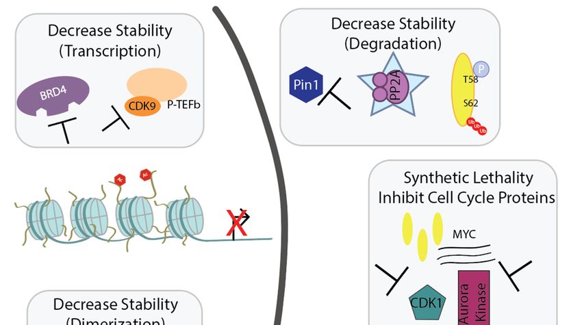
The short half-life of Myc is evidence for Myc’s highly controlled turnover. Myc’s stability is
regulated by phosphorylation on serine 62 (S62) and threonine 58 (T58) by several proteins through
the Raf-MEK-ERK kinase and phosphatidylinositol-3 kinase (PI3K)-Akt pathways [28,120]. First,
extracellular signal-regulated kinase (ERK), CDK1, or growth signals stabilize Myc by phosphorylating
S62. Glycogen synthase kinase 3 (GSK3) is recruited to phosphorylate T58, which is required for
Myc degradation. In brief, Pin1 isomerizes proline 63 on Myc, in which protein phosphatase 2A
(PP2A), a serine/threonine phosphatase, can now dephosphorylate Myc at S62 [121]. The unstable
Myc, with only T58 phosphorylation remaining, becomes ubiquitinated by Fbw7 and is sent for
degradation [122]. Again, these many levels of regulation provide multiple opportunities for cancer
hijacking. In cancers that lack MYC amplification, there are increases in the stabilizing pS62-Myc
and decreases in the degrading pT58-Myc, therefore promoting Myc’s stability and activity [38–41].
Studies show that mutating T58 to alanine, a non-phosphorylatable residue, results in stable Myc
expression and tumorigenic properties, suggesting Myc stability has a role in transformation [123–125].
In cancer, we see faulty regulation of these proteins that modify the phosphorylation on Myc
and promote stabilization. We will describe three scenarios—activation of PI3K/AKT signaling
(which inhibits GSK3B), overexpression of Pin1, and suppression of PP2A activity—that stabilize Myc.
PI3K, PTEN, and upstream components of the PI3K/AKT pathway are commonly mutated in cancer
to promote pathway activation [126]. Activated AKT phosphorylates (and therefore inhibits) GSK3,
which in turn enhances Myc stability [127,128] as GSK3 cannot phosphorylate T58-Myc. This is just
one example of how an upstream signaling pathway (MAPK, Wnt, Notch) can quickly trickle down to
promoting cancer through Myc.
Peptidyl-prolyl cis-trans isomerase NIMA-interacting 1 (Pin1), an isomerase that specifically
recognizes the serine/threonine-proline motif, is overexpressed in several cancers including pancreatic,
breast, and prostate and its expression correlates with poor clinical outcomes [129–131]. Furthermore,
Pin1 promotes several hallmarks of cancer through inactivating 26 tumor suppressors and activating
56 oncogenes [132,133]. By catalyzing the cis/trans conformational change of the target protein, such as
Myc, isomerases like Pin1 gain control of the target protein’s stability, activity, and localization [134].
As mentioned, Myc’s stability is regulated through phosphorylation on S62 and T58 and these sites areInt. J. Mol. Sci. 2020, 21, x FOR PEER REVIEW 9 of 29
Peptidyl-prolyl cis-trans isomerase NIMA-interacting 1 (Pin1), an isomerase that specifically
recognizes the serine/threonine-proline motif, is overexpressed in several cancers including
Int. J. Mol. Sci. 2020, 21, 9486 9 of 28
pancreatic, breast, and prostate and its expression correlates with poor clinical outcomes [129–131].
Furthermore, Pin1 promotes several hallmarks of cancer through inactivating 26 tumor suppressors
and activating 56 oncogenes [132,133]. By catalyzing the cis/trans conformational change of the target
recognized by trans-specific
protein, phosphatases;
such as Myc, isomerases therefore,
like Pin1 Pin1ofcan
gain control stabilize
the target Myc in
protein’s the cis-confirmation
stability, activity, and and
preventlocalization
degradation [135]. On the contrary, Pin1 can revert Myc back to the trans-confirmation
[134]. As mentioned, Myc’s stability is regulated through phosphorylation on S62 and after
phosphorylation
T58 and these of sites
T58,are
which allowsbyPP2A
recognized to remove
trans-specific phosphorylation
phosphatases; from
therefore, Pin1 canS62 to promote
stabilize Myc Myc
in the cis-confirmation
degradation [28]. However,and preventconsideration
another degradation [135]. On the
is that contrary,
PP2A Pin1 can revert
is commonly Myc backin
inactivated tocancers
(describedthe trans-confirmation
below), and so even afterifphosphorylation
Pin1 reverts Myc of T58, which
back allows
to the PP2A to remove phosphorylation
trans-confirmation, Myc would unlikely
from S62 to promote Myc degradation [28]. However, another consideration is that PP2A is
get degraded in the absence of PP2A activity. More research is needed to better understand how T58
commonly inactivated in cancers (described below), and so even if Pin1 reverts Myc back to the trans-
phosphorylation
confirmation, affects Pin1 activity
Myc would unlikely getanddegraded
S62 dephosphorylation.
in the absence of PP2A Additionally,
activity. MorePin1 can is
research promote
self-ubiquitination
needed to of Fbw7,
better the E3 ubiquitin
understand how T58 ligase that ultimatelyaffects
phosphorylation degrades Pin1Myc [136]. and
activity Pin1’sS62influence
on Myc’s dephosphorylation. Additionally,
transcriptional activity Pin1 can potentiates
and stability promote self-ubiquitination
tumorigenesisofand Fbw7,is athe E3 ubiquitin
potential therapeutic
target forligase that ultimately degrades
MYC-overexpressing cellsMyc [136]. Pin1′s influence on Myc’s transcriptional activity and
[132,135–137].
stability potentiates tumorigenesis and is a potential therapeutic target for MYC-overexpressing cells
Lastly, PP2A is a ubiquitously expressed tumor suppressor that accounts for a majority of the
[132,135–137].
phosphataseLastly, activity in cells and dephosphorylates a range of substrates such as Akt, p53, β-catenin,
PP2A is a ubiquitously expressed tumor suppressor that accounts for a majority of the
and Mycphosphatase
[138]. The activity
holoenzyme
in cellscan
andcontain a variety of
dephosphorylates different
a range scaffoldsuch
of substrates (A) asand regulatory
Akt, (B) subunits
p53, β-catenin,
with a common
and Myc [138].catalytic (C) subunit,
The holoenzyme canwith multiple
contain a varietyisoforms for each
of different subunit
scaffold (A) and[139]. Inactivation
regulatory (B) of
subunitsPP2A
PP2A through with ainhibitor
commonokadaic
catalytic acid
(C) subunit,
results in with multiple isoforms
tumorigenesis for each transformation
and cellular subunit [139]. [140].
PP2A isInactivation
commonlyofinactivated
PP2A through PP2A inhibitor
in cancer, including okadaic
lung,acid results
colon, in tumorigenesis
breast, skin, cervix, and andcellular
ovarian [139].
transformation [140]. PP2A is commonly inactivated in cancer, including lung, colon, breast, skin,
This PP2A inactivation occurs through phosphorylation, somatic mutation, or increased expression
cervix, and ovarian [139]. This PP2A inactivation occurs through phosphorylation, somatic mutation,
of endogenous
or increasedinhibitors such
expression as SET andinhibitors
of endogenous CIP2A such [141–144].
as SET andIn the case[141–144].
CIP2A of Myc,InPP2A inactivation
the case of
preventsMyc, dephosphorylation of S62, dephosphorylation
PP2A inactivation prevents therefore stabilizing of S62,Myc and promoting
therefore transformation
stabilizing Myc and promoting[28,121].
In sum,transformation
inhibition of GSK3[28,121].through
In sum,PI3K, overexpression
inhibition of GSK3 through of Pin1,
PI3K,and inactivationofofPin1,
overexpression PP2A andpromote
inactivation of PP2A promote stability of Myc (Figure 5). Although there are
stability of Myc (Figure 5). Although there are many opportunities to increase Myc stability, many of many opportunities to
increase Myc stability, many of these proteins are also potential therapeutic targets to promote Myc’s
these proteins are also potential therapeutic targets to promote Myc’s degradation.
degradation.
Figure 5. Disrupting Myc stability: (A) In cancer, PI3K signaling inactivates GSK3, preventing
phosphorylation of T58 Myc. Pin1 overexpression keeps Myc in the cis-confirmation, preventing PP2A
trans-specific enzyme from binding to Myc. Furthermore, PP2A is inactivated in several cancers,
and therefore S62 remains phosphorylated. All of this leads to high Myc stability. (B) Inhibition of PI3K
allows for GSK3 to phosphorylate T58 on Myc, which is required for degradation. Pin1 inhibitors and
PP2A activators allow for PP2A to recognize and remove the phosphorylation of S62, leading to low
stability and Myc’s degradation.Int. J. Mol. Sci. 2020, 21, 9486 10 of 28
Enhancing Degradation of Myc
Given the various levels regulating Myc degradation, numerous compounds have been developed
to enhance Myc degradation through inhibition of PI3K or Pin1 and re-activation of PP2A. Becker and
collogues demonstrated efficacy in combining a PI3K inhibitor with a microtubule destabilizer in
high-Myc expressing cells. First, they eloquently demonstrated that unphosphorylated S62-Myc binds
to mitotic tubules and is protected from degradation [145]. Given this interaction, treatment with a
microtubule destabilizer, vincristine, drastically reduced Myc protein and P493-6 B-cell lymphoma
cells with ectopic Myc expression were more sensitive to colony forming unit inhibition than Myc
low-expressing cell lines. Since PI3K/AKT inhibits GSK3B activity and therefore stabilizes Myc,
Becker and collogues investigated the addition of PI3K inhibitor idelalisib following the G2-M arrest
induced by vincristine. Treating first with vincristine followed by idelalisib led to higher cell death and
decreased clonogenic growth than either compound alone across 16 Burkitt lymphoma and DLBCL
cell lines [145]. Furthermore, this combination lead to reduction of Myc and tumor viability in two
lymphoma in vivo models, in which the compounds as single agents were not effective. These results
suggest a novel avenue of disrupting Myc stability via microtubule destabilizers followed by PI3K
inhibition to further decrease Myc protein levels. Another targetable signaling pathway that influences
Myc degradation is the MEK/ERK pathway. As mentioned, ERK maintains S62 phosphorylation of
Myc, which promotes Myc’s stability [120]. Therefore, inhibition of the MEK/ERK pathway through
MEK inhibitor U0126 reduced Myc expression and growth in rhabdomyosarcoma cell lines [146].
Furthermore, inhibition of the MEK/ERK pathway or the consequent decrease in Myc expression,
a known driver of radioresistance, sensitizes cancer cells to radiation therapy [147,148].
Aside from Pin10 s influence over Myc’s stability, there are several other mechanisms in which
Pin1 can promote tumorigenesis such as sustaining proliferative signaling and downregulating tumor
suppressors [132]. More than ten Pin1 inhibitors have been developed that demonstrate anticancer
activity, including sensitizing various cancer cells to chemotherapy [132]. We will discuss two Pin1
inhibitors—All-trans retinoic acid (ATRA) and KPT-6566, that have more favorable specificity and
safety profiles than other Pin1 inhibitors. ATRA is clinically used for acute promyelocytic leukemia
(APL), although its drug target was unknown. Through a mechanism-based high throughput screen,
Wei and collogues discovered ATRA directly binds and degrades Pin1 [149]. ATRA was capable of
decreasing Pin1 and tumor growth in APL mouse models and APL human patients’ bone marrow,
along with in vivo models of triple negative breast cancer [149] and acute myeloid leukemia [150];
both cancers overexpress Pin1. However, ATRA has a short half-life of 45 minutes and moderate
anti-cancer activity. Yang and collogues developed an improved, controlled-release formulation of
ATRA (ATRA-PLLA microparticles) that demonstrated selectivity for Pin1 inhibition and improved
anti-cancer efficacy in xenografts of hepatocellular carcinoma, a cancer that is enhanced by Pin1 [151].
Several other liposomal ATRA delivery methods have been developed and performed well in clinical
trials for APL patients [152], although it appears trials for solid tumors utilizing the improved ATRA
formulation are lacking. Additionally, these studies did not specifically investigate the effects of
ATRA and Myc. Several older studies across small cell lung cancer, breast cancer, and colon cancer
demonstrated treatment with ATRA decreased Myc expression at the gene or protein level [153–155].
Selective Pin1 inhibitor KPT-6566, which was also identified through a mechanism-based screen, sets
Pin1 for degradation. When KPT-6566 binds to the catalytic site of Pin1, reactive oxygen species are
produced and DNA damage occurs, leading to cell death particularly in Pin1-overexpressing cancer
cells [156]. There are no data on KPT-6566 decreasing tumor volume in vivo, but in mice injected with
MDA-MB-231 cells, KPT-6566 daily treatment reduced metastatic spread and showed no toxicities
in vital organs [156]. Again, these studies did not investigate the effects of Pin1 inhibition on Myc.
More development is necessary to improve efficacy and drug-likeness of Pin1 inhibitors, especially in
the context of Myc-driven cancers.
Compounds that target PP2A, which is the main phosphatase the regulates Myc stability,
have shown promise in promoting Myc degradation and cell death. There are several methodsInt. J. Mol. Sci. 2020, 21, 9486 11 of 28
published on indirectly activating PP2A as an anti-cancer treatment, such as antagonizing the
endogenous PP2A inhibitors SET (via OP449 [41,157] or FTY720 [158,159]), and CIP2A (via bortezomib,
erlotinib, or celastrol) or disrupting PP2A post translational modifications [143]. SET-inhibitor OP449
increased PP2A activity dose-dependently and OP449-treated leukemia xenografts had a two-fold
reduction of tumor burden [157]. In breast cancer, OP449 decreased both phosphorylation levels
of S62-Myc and Myc transcriptional activity across several cell lines in vitro. OP449 additionally
induced apoptosis while reducing tumor volume and increasing PP2A activity in vivo [41]. In terms
of disrupting CIP2A, the described inhibitors were primarily discovered as a proteasome inhibitor
(bortezomib), EGFR kinase inhibitor (erlotinib), or anti-cancer (celastrol), but indirectly or independently
reduce CIP2A expression or activity [143,160]. Small molecule activators of PP2A (SMAPs) have
also emerged as a new class of validated compounds that re-activate PP2A through binding to the A
scaffolding subunit of PP2A. As PP2A reactivates, S62-Myc becomes dephosphorylated and Myc is sent
for degradation. Recently, SMAPs demonstrated efficacy through binding to PP2A in in vivo models
of Burkitt’s lymphoma, non-small cell lung cancer, and triple-negative breast cancer—all Myc-driven
cancers, representing Myc amplification, post-translational stabilization, and overexpression [161].
SMAPs also display efficacy in prostate and pancreatic cancer models [162,163]. Dr. Narla, one of the
developer of SMAPs, serves as Chief Scientific Officer for Rappta Therapeutics to further develop these
anti-cancer molecules that reactive PP2A [164,165]. In all, targeting Myc’s protein stability may help
reduce toxicity that is expected with a complete loss of Myc.
4. Taking Advantage of MYC Overexpression to Initiate Synthetic Dosage Lethality in the
Context of Cell Cycle
Since transcription factors pose as difficult drug targets, leveraging synthetic lethality offers an
alternative approach of antitumoral therapy. Synthetic lethality occurs when a mutation or inhibition
of two specific genes leads to cell death, but a mutation or inhibition of just one gene does not affect
viability [166]. Synthetic dosage lethality is when manipulation of expression levels leads to cell death;
for example, overexpression of gene A and presence of gene B is viable, but the combination of gene
A overexpression and loss or lower expression of gene B results in cell death. Therefore, synthetic
lethality, or more specifically synthetic dosage lethality, can be advantageous in cancer as the tumors
already have mutations or oncogenic addiction, such as overexpression of MYC. A synthetic lethal
approach affects the mutated tumor cells and spares the normal cells.
Synthetic lethal targets are identified in an unbiased, high-throughput fashion through RNA
interference (RNAi) or CRISPR screens on isogenic cells—cells that differ by a mutation in a single
gene. Although the idea sounds swift, identifying clinically relevant synthetic lethal interactions have
proven difficult due to validation of lethal mutants by recovery, condition-dependent interactions,
and rarity [166]. However, PARP inhibitors successfully demonstrated this concept clinically when
given to cancer patients with BRCA mutations, such as in breast and ovarian cancer [167,168].
Understanding the biological results of MYC overexpression will help identify second-site targets
that lead to synthetic lethality. Reports show that MYC-overexpressing cancer cells have increased
sensitivity to apoptosis in response to cytotoxic drugs or radiation [169]. However, the opposite appears
to be true in melanoma, in which lower MYC expression improves susceptibility to chemotherapy and
radiation due to reactive oxygen species production and mismatch repair protein inhibition [170,171].
As Myc is a master regulator of cell proliferation and metabolism, genes affiliated with these processes
offer a promising avenue to identify synthetic lethal targets.
Cells overexpressing MYC have more mitotic abnormalities, such as altered spindle morphology
and mitotic timing [172]. During mitotic stress, Myc worsens mitotic dysfunction and enhances
apoptosis, which explains the many cell cycle proteins as targets for synthetic lethality. In normal
conditions, advancing through the cell cycle phases of G1, S, G2, and M requires four heterodimers of
cyclin-dependent serine/threonine kinases (CDK) and cyclins: CDK1, 2, 4, 6, and cyclins A, B, E, D,
all of which are Myc target genes [173]. Cyclin B1 binds to CDK1 at the G2-M transition, activating theYou can also read